Ugur Keklikci
A–F, Birgul Dursun
B, C, Abdullah Kursat Cingu
C, DTopical Cyclosporine A 0.05% Eyedrops
in the Treatment of Vernal Keratoconjunctivitis
– Randomized Placebo-Controlled Trial*
Department of Ophthalmology, Dicle University Faculty of Medicine, Diyarbakir, Turkey
A – research concept and design; B – collection and/or assembly of data; C – data analysis and interpretation; D – writing the article; E – critical revision of the article; F – final approval of article; G – other
Abstract
Background. Vernal keratoconjunctivitis (VKC) is a chronic, bilateral inflammation of the conjunctiva that mostly affects children and young adult males. Management of VKC is primarily aimed at reducing symptoms and pre-venting serious vision threatening sequelae.
Objectives. To assess the efficacy of topical cyclosporine A (CsA) 0.05% on the signs and symtomps in the man-agement of VKC.
Material and Methods. This is a placebo-controlled, randomized prospective study. Sixty-two patients with VKC were included in this study. Patients were randomly assigned (1 : 1) to treatment with topical 0.05% CsA eyedrops or a placebo (artificial tears) for a period of 4 weeks, 4 times daily. Ocular signs and symptoms were in all patients scored at entry and at the end of 4 weeks.
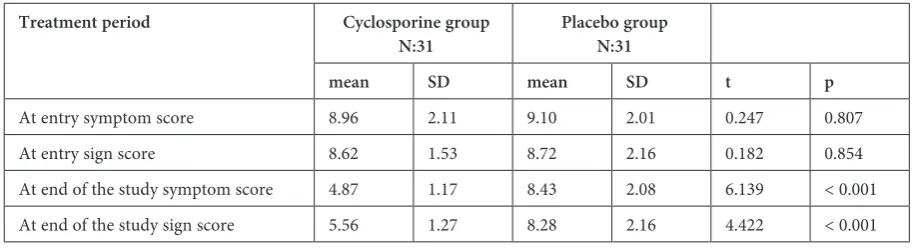
Results. When pre-treatment mean signs and symptoms scores were compared in both groups, there was no sig-nificant difference (p > 0.05). However, mean post-treatment scores as regards signs and symptoms were found to be lower in cyclosporine group than those in placebo group (p < 0.001). No side effects of the treatment with CsA 0.05% eyedrops were observed.
Conclusions. It was found that topical CsA 0.05% eyedrops were safe and effective in the treatment of patients with VKC (Adv Clin Exp Med 2014, 23, 3, 455–461).
Key words: cyclosporine, sign, symptom, topical, vernal keratoconjunctivitis.
Adv Clin Exp Med 2014, 23, 3, 455–461 ISSN 1899–5276
ORIGINAL PAPERS
© Copyright by Wroclaw Medical University
Vernal keratoconjunctivitis (VKC) is a chron-ic, bilateral inflammation of the conjunctiva. VKC mainly affects boys in their first decade of life and the sequelae of the disease may be responsible for permanent visual impairment. Diagnosis is based on symptoms and signs including itching, pho-tophobia, sticky mucous discharge, giant papillae on the upper tarsal conjunctiva or at the limbus, Trantas’ dots, superficial keratopathy, and corneal shield ulcer [1–3]. Recent studies have document-ed various alterations of cornea, conjunctiva and tear film in VKC patients [4–6]. The disease was bilateral in 96.7% of the cases; all unilateral cases involved the tarsal form of VKC [7, 8]. Approx-imately 23% of patients had a perennial form of
VKC from disease onset, and more than 60% had additional recurrences during the winter [1]. In the Mediterranean area and other temperate regions, the intensity of the disease increases in spring and summer and decreases in fall and winter [9].
The pathogenesis of the disease is not com-pletely known. Conventionally, VKC was consid-ered primarily a type 1 hypersensitivity reaction. However, approximately 50% of the patients with VKC have no familial or personal history of ato-py, and a large proportion have negative results on the Standard allergic diagnostic tests, confirming that it is not solely immunoglobulin E (IgE)-me-diated [10]. Recent studies suggest a more com-plex non-IgE-dependent pathogenic mechanism.
Several studies have documented the presence of the Th2 lymphocytes subtype in tears and in con-junctival biopsy samples of patients with VKC. His-topathologically, VKC is characterized by conjunc-tival infiltrations with eosinophils, degranulated mast cells, basophils, plasma cells, lymphocytes, and macrophages. T cell culture from conjunctival scraping of VKC patients yielded mainly Th2-type clones. Th2-derived cytokines such as IL-4, IL-5, IL-13, growth factors and enzymes are found in the conjunctiva of VKC patients [11]. In addition, some researchers have demonstrated the possible involvement of neural factors, sex hormones and conjunctival histaminase deficiency in the patho-genesis of the disease [2, 10, 11].
Several earlier reports indicate that topical an-ti-inflammatory and anti-allergic eye drops are the mainstay of treatment for VKC; however, a gold standard treatment has not yet been established for this disease [3]. Topical anti-allergic and top-ical antihistamines are effective in reducing signs and symptoms of the disease. Non-steroidal an-ti-inflammatory agents also produce a beneficial effect on the course of VKC [1].Steroids can be highly effective, but may cause unwanted elevation of intraocular pressure in steroid responders and increase the risk of corneal infection through lo-cal immunosuppression. In addition, induction of cataract and delayed wound healing can be prob-lematic. Cyclosporine A (CsA) is a non-steroidal immunomodulator that inhibits antigen depen-dent T cell activation. CsA also has a direct inhib-itory effect on eosinophil and mast cell activation and release of mediators, which is likely to be im-portant in allergic inflammation [12–15].
In this placebo-controlled, randomized pro-spective study, we investigated the effects of topi-cal CsA 0.05% on the clinitopi-cal signs and symptoms of patients with VKC.
Material and Methods
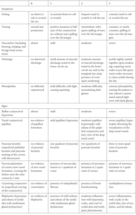
This is a randomized placebo-controlled trial, which comprises 62 patients with moderate or se-vere VKC admitted to the Department of Ophthal-mology, Faculty of Medicine, Dicle University, be-tween January 2010–January 2011. The diagnosis of VKC was made, based on the patients’ history and the presence of typical clinical signs and symp-toms. The clinical forms of the disease was consid-ered to be tarsal in 67.9% of cases, limbal in 19.8% of cases, mixed in 12.3% of cases. Scores of the signs and symtomps were obtained from each pa-tient at the beginning and at the end of the study. A clinical score (0–3 – 0 = absent; 3 = severe) was given, considering the severity of the following eye
symptoms and signs. Symptoms included itching, tearing, discomfort, mucous discharge, and photo-phobia. Signs, however, included bulbar conjunc-tival hyperemia, upper tarsal conjuncconjunc-tival papillae, punctate keratitis, corneal neovascularization, cic-atrizing conjunctivitis, and blepharitis (Table 1). The patients were classified as having severe VKC if the score was 5 or more points for one eye for each scale, and as moderate if the score was be-tween 3 and 5 points.
Patients were randomly selected according to their application to our clinic to receive either topical CsA 0.05% (Restasis, Allergan, Waco, TX, USA) or preservative-free artificial tears (Refresh Tears, Allergan, Waco, TX, USA) for a period of 4 weeks, 4 times daily.The two preparations, which were applied to the patients, were closed with the same cover by different number to be prevented from bias by the researcher. All patients had been treated with a variety of topical medications be-fore enrollment. If patients were using topical cor-ticosteroid, anti-allergic or anti-histaminergic eye drops, this medication was discontinued 1 week before the start of the trial. None of the patients had earlier taken systemic steroids and other anti-inflammatory or immunosuppressive drugs. The study was performed according to the guidelines of theDeclaration of Helsinki; furthermore, we ob-tained approval for the study from the local Ethics Committee.
Statistical Analysis
In the study, Chi-square (χ2) test was used to
compare sex and age rates between the two groups. In addition, Independent Student’st test was used to analyse the matched sample. Data was analysed by using SPSS 15.0 statistical package for Windows (SPSS Inc., Chicago, IL, USA) program. Two-sid-ed p values were considerTwo-sid-ed statistically significant at p ≤ 0.05.
Results
Table 1. Grading of symptoms and signs of vernal keratoconjunctivitis
0 1 2 3
Symptoms
Itching no desire to rub or scratch the eye
occasional desire to rub
or scratch frequent need to scratch or rub the eye constant need to rub or scratch the eye
Tearing normal tear
production positive sensation of full-ness of the conjunctival sac without tears spilling over the lid margin
intermittent, infre-quent spilling of tears over the lid margin
constant, or nearly constant, spilling of tears over the lid mar-gins
Discomfort (including burning, stinging, and foreign body sensa-tions)
absent mild moderate severe
Discharge no abnormal
discharge small amount of mucoid discharge noted in the lower cul-de-sac
moderate amount of mucoid discharge noted in the lower cul-de-sac and in the marginal tear strip; presence of crust upon awakening
eyelids tightly matted together upon awaken-ing, requiring warm soaks to pry lids apart; warm soaks necessary to clean eyelids during the day
Photophobia no difficulty
experienced mild difficulty with light causing squinting moderate difficulty, necessitating dark glasses
extreme photophobia, causing the patient to stay indoors; cannot stand natural light even with dark glasses Signs
Bulbar conjunctival
hyperemia absent mild moderate severe Tarsal conjunctival
papillary no evidence of papillary formation
mild papillary hyperemia moderate papillary hypertrophy with edema of the palpe-bral conjunctiva and hazy view of the deep tarsal vessel
severe papillary hyper-trophy obscuring the visualization of the deep tarsal vessels
Punctate keratitis (superficial epithelial keratitis and punctate staining of the cornea with fluorescein)
no evidence of punctate keratitis
one quadrant of punctate
keratitis two quadrants of punctate keratitis three or more quad-rants of punctate keratitis
Neovascularization of cornea (new vessel formation, crossing the limbus onto the clear cornea by 2 mm
no evidence of new vessel formation
presence of neovascular-ization in 1 quadrant of corne
presence of neovascu-larization in 2 quad-rants of cornea
presence of neovascu-larization in 3 quad-rants of cornea
Cicatrizing conjunctivi-tis (superficial scarring of the conjunctiva)
no evidence of
cicatrization presence of subepithelial fibrosis presence of fornix foreshortening symblepharon forma-tion
Blepharitis (hyperemia and edema of eyelid skin with meibomian gland dysfunction)
no evidence of
blepharitis presence of mild redness and edema of the eyelid with meibomian gland dysfunction
moderate inflamma-tion with hyperemia, scales, and scurf of eyelid skin and tooth-paste phenomenon
pre-treatment mean signs and symptoms scores were compared in both groups, there was no sig-nificant difference (p > 0.05). However, mean post-treatment scores as regards signs and symp-toms were found to be lower in cyclosporine group than those in placebo group (p < 0.001).
Discussion
VKC is a chronic, seasonally exacerbated aller-gic inflammatory disease of the ocular surface, af-fecting mainly children and adolescents. Males are typically more affected than females. In the pres-ent group of VKC patipres-ents, the male: female ratio was 2.2 : 1. This tendency is confirmed in the liter-ature. In the studies reported earlier, at the time of diagnosis, 83% of patients were under 10 years of age and 4% were over 20 years of age [7, 16]. In the present study, however, we determined the mean age of the patients as 9.8.
Management of VKC is primarily aimed at re-ducing symptoms and preventing serious vision threatening sequelae. Steroids can be highly ef-fective, but there are risks of glaucoma, superin-fection with viruses and bacteria due to local im-munosuppression, delayed wound healing, and cataract induction warrant cautious use of topical steroids [17–24]. Therefore, non-steroidal medi-cations are desirable for the treatment of VKC as alternative, but new topical agents with dual anti-allergic activity (mast cell stabilizers and antihista-mine) may also be used for long-term treatment of
allergic inflammation to alleviate signs and symp-toms of the disease [21, 25, 26]. A recent study has reported that topical interferon alpha 2b treatment seems to offer a safe and effective alternative for the treatment of refractory vernal keratoconjunc-tivitis for a brief period [27].
CsA, a macrolide antibiotic, has been isolat-ed from the soil fungus Tolypocladium inflatum. CsA is effective in controlling ocular inflamma-tion, blocking Th2 lymphocyte proliferainflamma-tion, and IL-2 production. It also inhibits histamine release from mast cells and basophils, and through a duction of IL-5 production, it may reduce the re-cruitment and the effects of eosinophils on the conjunctiva [1, 15, 28, 29].Moreover, the thera-peutic efficacy of CsA in VKC, a conjunctival hy-perproliferative disorder, seems to be related to the drug’s efficacy in reducing conjunctival fibroblast proliferation rate and IL-1b production. Multiple studies have reported a beneficial effect of topi-cal cyclosporine to relieve symptoms of VKC in patients with different severity grades of the dis-ease [2, 30–33]. In a recent study, both topical treatment of 0.1% FK-506 ophthalmic ointment and 2% CsA eye drops have been reported to be effective for VKC. They also reported that FK-506 ophthalmic ointment twice daily brought about an improvement of symptoms of VKC similar to that of CsA eye drop four times daily [32].
In the field of ophthalmology, topically applied CsA in various oil-based solvents was first used to inhibit experimental corneal allograft reaction in the early 1980s [34, 35]. Later, the drug was found
Table 2. The characteristics of the patients and the outcomes of the analysis
Demographic
characteristic Cyclosporine groupN:31 Placebo groupN:31 Difference
mean %/SD mean %/SD X2/t p
Gender Female
Male 922 29.0370.97 1021 32.2667.74 0.147 0.713 Mean age 9.9 3.13 9.7 2.63 –0.238 0.812
Table 3. Mean scores of symptoms and signs of patients before and after treatment period
Treatment period Cyclosporine group
N:31 Placebo groupN:31
mean SD mean SD t p
useful in patients with various inflammatory oc-ular surface disorders [2, 22, 36–38].Topical CsA 2% dissolved in maize oil has been shown to be beneficial for many years in the management of se-vere allergic eye disease [39].However, side effects, whether related to the oil solvent or the CsA itself, were common. Lid skin maceration, allergic reac-tion, and marked blurring of vision after drop in-stillation were attributed to the vehicle; however, intense stinging was a side effect of the CsA. In-deed, this is a well-known side effect that limits its clinical use [22]. Topical CsA has been effec-tive in treating patients with severe resistant VKC since 1986 [40]. Several studies have reported the effectiveness of CsA 2% eyedrops in improving signs and symptoms of VKC [30, 41, 42]. In re-cent years, several studies have reported a good response to low-concentrated topical cyclospo-rine to relieve symptoms of VKC [2, 22, 33, 43]. The mechanism(s) of the beneficial effect of top-ical CsA in VKC patients, however, is not clear. In vitro studies have demonstrated that CsA in-hibits activation and proliferation of T lympho-cytes via blockage of IL-2 gene expression. Topical CsA treatment has also been shown to reduce the T lymphocyte population in conjunctival biopsy specimens of VKC and atopic keratoconjunctivitis (AKC) patients. Both studies concluded that topi-cal CsA treatment either in VKC or AKC patients significantly reduced the number of total lympho-cytes infiltrating the conjunctiva and reduced the CD4+ subset only slightly and insignificantly [28].
Akpek et al., [22] in their placebo-controlled study, reported that CsA 0.05% had a steroid spar-ing and beneficial effect on the scores of symptoms and signs during treatment, without any side ef-fects in patients with AKC. However, Daniell et al., [15] in their placebo-controlled trial, failed to show any beneficial effect in terms of final clini-cal score with the addition of topiclini-cal CsA 0.05% in patients with steroid-dependent allergic eye
disease. Nevertheless, they found that patients in the CsA treated group showed significantly greater improvement over time in lid margin thickening, inferior conjunctiva papillae, inferior and superi-or conjunctival hyperemia, and csuperi-orneal tearfilm deficiency. In our study, there was a significant improvement for post-treatment scores of signs and symptoms in CsA group with respect to pl-ecebo group.We verified that VKC was success-fully treated with cyclosporine 0.05% for 4 weeks, 4 times daily. No side effects attributed to topical CsA 0.05% treatment were encountered, suggest-ing that low-concentrated topical CsA is of benefit in the treatment of patients with VKC. Spadavec-chiaet al., [2] in their placebo-controlled study, re-ported that a lower concentration of CsA (1.25% and 1%) was safe and well tolerated as well as sig-nificantly improved the ocular signs and symptoms of VKC. This also suggests that lower concentra-tion of tested cyclosporine was effective for almost complete recovery of severe forms of VKC. This is an important finding, considering that cyclospo-rine is an immunosuppressive drug and that the use of low concentrated eyedrops might avoid sys-temic effects. Similarly, Ozcan et al. [43]showed that topical CsA 0.05% had a significant benefi-cial effect on patients with VKC and AKC. In our study, topical low concentrated cyclosporine was found to be effective in treatment of VKC, and no systemic effects were observed. The results of ear-lier studies are compatible with our findings [2, 30, 43, 45]. For instance, in a recent randomized con-trolled study, it was shown that topical 0.05 % cy-closporine was safe and effective for the long-term prevention of VKC relapses [44].
In conclusion, we used cyclosporine A 0.05% eyedrops in the treatment of patients with VKC. Topical CsA 0.05% was found to be effective in the treatment of patients with VKC. No side ef-fects that can be attributed to topical CsA 0.05% treatment were encountered.
References
[1] Bonini S, Coassin M, Aronni S, Lambiase A: Vernal keratoconjunctivitis. Eye 2004, 18, 345–351.
[2] Spadavecchia L, Fanelli P, Tesse R, Brunetti L, Cardinale F, Bellizzi M, Rizzo G, Procoli U, Bellizzi G, Armenio L: Efficacy of 1.25% and 1% topical cyclosporine in the treatment of severe vernal keratoconjunctivitis in childhood. Pediatr Allergy Immunol 2006, 17, 527–532.
[3] Mantelli F, Santos MS, Petitti T, Sgrulletta R, Cortes M, Lambiase A, Bonini S: Systematic review and meta-analysis of randomised clinical trials on topical treatments for vernal keratoconjunctivitis. Br J Ophthalmol 2007, 91, 1656–1661.
[4] Emre S, Başer E, Oztürk B, Zorlu S, Uzun O, Gülhan C: Corneal biochemical features of patients with vernal keratoconjunctivitis. Graefes Arch Clin Exp Ophthalmol 2013, 251, 555–558.
[5] Sangwan VS, Jain V, Vemuganti GK, Murthy SI: Vernal keratoconjunctivitis with limbal stem cell deficiency. Cornea 2011, 30, 491–496.
[6] Le Q, Wang Y, Xu J:In vivo confocal microscopy of long-standing mixed-form vernal keratoconjunctivitis. Ocul Immunol Inflamm 2010, 18, 349–351.
[8] Keklikci U, Soker SI, Soker Cakmak S, Ozkul S, Sakalar YB, Unlu K: Unilateral vernal keratoconjunctivitis: A case report. Eur J Ophthalmol 2007, 17, 973–975.
[9] Pucci N, Massai C, Bernardini R, Caputo R, Mori F, De Libero C, Novembre E, De Martino M, Vierucci A: Eyelash length in children with vernal keratoconjunctivitis: effect of treatment with cyclosporine eye drops. Int J Immunopathol Pharmacol 2007, 20, 595–599.
[10] Bozkurt B, Artac H, Arslan N, Gokturk B, Bozkurt MK, Reisli I, Irkec M: Systemic atopy and immunoglobulin deficiency in Turkish patients with vernal keratoconjunctivitis. Ocul Immunol Inflamm 2013, 21, 28–33.
[11] Labcharoenwongs P, Jirapongsananuruk O, Visitsunthorn N, Kosrirukvongs P, Saengin P, Vichyanond P: A double-masked comparison of 0.1% tacrolimus ointment and 2% cyclosporine eye drops in the treatment of vernal keratoconjunctivitis in children. Asian Pac J Allergy Immunol 2012, 30, 177–184.
[12] Nussenblatt RB, Palestine AG: Cyclosporine A: immunology, pharmacology and therapeutic uses. Surv Ophthalmol 1986, 31, 159–169.
[13] Borel JF, Baumann G, Chapman I, Donatsch P, Fahr A, Mueller EA, Vigouret JM: In vivo pharmacological effects of cyclosporin and some analogues. Adv Pharmacol 1996, 35, 115–246.
[14] Whitcup SM, Chan CC, Luyo DA, Bo P, Li Q: Topical cyclosporine inhibits mast cell-mediated conjunctivitis. Invest Ophthalmol Vis Sci 1996, 37, 2686–2693.
[15] Daniell M, Constantinou M, Vu HT, Taylor HR: Randomised controlled trial of topical ciclosporin A in steroid dependent allergic conjunctivitis. Br J Ophthalmol 2006, 90, 461–464.
[16] Bonini S, Bonini S, Lambiase A, Marchi S, Pasqualetti P, Zuccaro O, Rama P, Magrini L, Juhas T, Bucci MG: Vernal keratoconjunctivitis revisited: a case series of 195 patients with long-term followup. Ophthalmology 2000, 107, 1157–1163.
[17] De Smedt S, Nkurikiye J, Fonteyne Y, Tuft S, De Bacquer D, Gilbert C, Kestelyn P: Topical ciclosporin in the treatment of vernal keratoconjunctivitis in Rwanda, Central Africa: a prospective, randomised, double-masked, controlled clinical trial. Br J Ophthalmol 2012, 96, 323–328.
[18] Ang M, Ti SE, Loh R, Farzavandi S, Zhang R, Tan D, Chan C: Steroid-induced ocular hypertension in Asian children with severe vernal keratoconjunctivitis. Clin Ophthalmol 2012, 6, 1253–1258.
[19] Oner V, Türkcü FM, Taş M, Alakuş MF, Işcan Y: Topical loteprednol etabonate 0.5 % for treatment of vernal keratoconjunctivitis: efficacy and safety. Jpn J Ophthalmol 2012, 56, 312–318.
[20] Tabbara KF: Ocular complications of vernal keratoconjunctivitis. Can J Ophthalmol 1999, 34, 88–92. [21] Leonardi A: Vernal keratoconjunctivitis: pathogenesis and treatment. Prog Retin Eye Res 2002, 21, 319–339. [22] Akpek EK, Dart JK, Watson S, Christen W, Dursun D, Yoo S, O’Brien TP, Schein OD, Gottsch JD: A
random-ized trial of topical cyclosporin 0.05% in topical steroid-resistant atopic keratoconjunctivitis. Ophthalmology 2004, 111, 476–482.
[23] Bonini S, Sacchetti M, Mantelli F, Lambiase A: Clinical grading of vernal keratoconjunctivitis. Curr Opin Allergy Clin Immunol 2007, 7, 436–441.
[24] Dada T, Konkal V, Tandon R, Singh R, Sihota R: Corneal topographic response to intraocular pressure reduction in patients with vernal keratoconjunctivitis and steroid-induced glaucoma. Eye 2007, 21,158–163.
[25] Verin P, Allewaert R, Joyaux JC, Piozzi E, Koliopoulos J, Bloch-Michel E: Lodoxamide Study Group Comparison of lodoxamide 0.1% ophthalmic solution and levocabastine 0.05% ophthalmic suspension in vernal keratoconjunc-tivitis. Eur J Ophthalmol 2001, 11, 120–125.
[26] Abelson MB: A review of olopatadine for the treatment of ocular allergy. Expert Opin Pharmacother 2004, 5, 1979–1994.
[27] Turan-Vural E, Acar BT, Acar S: The efficacy of topical interferon alpha 2b treatment in refractory vernal kera-toconjunctivitis. Ocul Immunol Inflamm 2012, 20, 125–129.
[28] Avunduk AM, Avunduk MC, Erdol H, Kapicioglu Z, Akyol N: Cyclosporine effects on clinical findings and impression cytology specimens in severe vernal keratoconjunctivitis. Ophthalmologica 2001, 215, 290–293. [29] Bielory L, Bonini S, Bonini S: Allergic eye disorders. Clin Allergy Immunol 2002, 16, 311–323.
[30] Pucci N, Novembre E, Cianferoni A, Lombardi E, Bernardini R, Caputo R, Campa L, Vierucci A: Efficacy and safety of cyclosporine eyedrops in vernal keratoconjunctivitis. Ann Allergy Asthma Immunol 2002, 89, 298–303. [31] Keklikci U, Soker SI, Sakalar YB, Unlu K, Ozekinci S, Tunik S: Efficacy of topical cyclosporin A 0.05% in
con-junctival impression cytology specimens and clinical findings of severe vernal keratoconjunctivitis in children. Jpn J Ophthalmol 2008, 52, 357–362.
[32] Labcharoenwongs P, Jirapongsananuruk O, Visitsunthorn N, Kosrirukvongs P, Saengin P, Vichyanond P: A double-masked comparison of 0.1% tacrolimus ointment and 2% cyclosporine eye drops in the treatment of vernal keratoconjunctivitis in children. Asian Pac J Allergy Immunol 2012, 30, 177–184.
[33] Tesse R, Spadavecchia L, Fanelli P, Rizzo G, Procoli U, Brunetti L, Cardinale F, Miniello VL, Bellizzi M, Armenio L: Treatment of severe vernal keratoconjunctivitis with 1% topical cyclosporine in an Italian cohort of 197 children. Pediatr Allergy Immunol 2010, 21, 330–335.
[34] Hunter PA, Wilhelmus KR, Rice NS, Jones BR: Cyclosporin A applied topically to the recipient eye inhibits cor-neal graft rejection. Clin Exp Immunol 1981, 45, 173–177.
[35] Kana JS, Hoffmann F, Buchen R, Krolik A, Wiederholt M: Rabbit corneal allograft survival following topical administration of cyclosporin A. Invest Ophthalmol Vis Sci 1982, 22, 686–690.
[37] Gunduz K, Ozdemir O: Topical cyclosporin as an adjunct to topical acyclovir treatment in herpetic stromal kera-titis. Ophthalmic Res 1997, 29, 405–408.
[38] Sall K, Stevenson OD, Mundorf TK, Reis BL: Two multicenter, randomized studies of the efficacy and safety of cyclosporine ophthalmic emulsion in moderate to severe dry eye disease. Ophthalmology 2000, 107, 631–639. [39] Hingorani M, Moodaley L, Calder VL, Buckley RJ, Lightman S: A randomized, placebocontrolled trial of topical
cyclosporin A in steroid-dependent atopic keratoconjunctivitis. Ophthalmology 1998, 105, 1715–1720.
[40] BenEzra D, Pe’er J, Brodsky M, Cohen E: Cyclosporine eyedrops for the treatment of severe vernal keratocon-junctivitis.Am J Ophthalmol 1986, 101, 278–282.
[41] Bleik JH, Tabbara KF: Topical cyclosporine in vernal keratoconjunctivitis. Ophthalmology 1991, 98, 1679–1684. [42] Gupta V, Sahu PK: Topical cyclosporin A in the management of vernal keratoconjunctivitis. Eye 2001, 15,
39–41.
[43] Ozcan AA, Ersoz TR, Dulger E: Management of severe allergic conjunctivitis with topical cyclosporin a 0.05% eyedrops. Cornea 2007, 26, 1035–1038.
[44] Lambiase A, Leonardi A, Sacchetti M, Deligianni V, Sposato S, Bonini S: Topical cyclosporine prevents seasonal recurrences of vernal keratoconjunctivitisin a randomized, double-masked, controlled 2-year study. J Allergy Clin Immunol 2011, 128, 896–897.
[45] Pucci N, Caputo R, Mori F, De Libero C, Di Grande L, Massai C, Bernardini R, Novembre E: Long-term safety and efficacy of topical cyclosporine in 156 children with vernal keratoconjunctivitis. Int J Immunopathol Pharmacol 2010, 23, 865–871.
Address for correspondence:
Ugur Keklikci
Department of Ophthalmology Dicle University Faculty of Medicine TR-21280, Diyarbakir
Turkey
Tel.: + 90 412 248 80 01 E-mail: [email protected]
Conflict of interest: None declared