Angel A. Noda
A–d, f, Islay Rodríguez
A–c, e, f, Brian Mondeja
e, f, carman fernández
e, fDesign, Optimization and Evaluation of a Polymerase
Chain Reaction for Detection of
Borrelia
spp
.
Projektowanie, optymalizacja i ocena reakcji łańcuchowej polimerazy
w celu wykrywania
Borrelia
spp
.
Bacteriology and Mycology department, “Pedro Kourí” Tropical Medicine Institute, Havana, cuba
A – research concept and design; B – collection and/or assembly of data; C – data analysis and interpretation;
D – writing the article; E – critical revision of the article; F – final approval of article; G – other
Abstract
Background. Lyme borreliosis and relapsing fever are important zoonotic diseases worldwide and the improve-ment of diagnostic strategies is a prioritized task considering the morbidity of these diseases in some areas. PcR based methods appear to be of utmost importance because of the high sensitivity and specificity of these assays.
Objectives. To obtain a molecular method based on PcR for the detection of the genus Borrelia infection in dif-ferent specimens.
Results. Sets of reportedprimers were evaluated “in silico” and they did not fulfill the proposal parameters. On the other hand, the two new, designed sets of primers were theoretically efficient for Borrelia dNA amplification. PcR procedures with these primers were standardized with borrelial dNA and optimum annealing temperatures, prim-er concentrations and reaction cycle numbprim-ers wprim-ere detprim-ermined. The PcR analytical sensitivity was 10 genomes pprim-er reaction for each technique. Both PcR were highly specific to different Borrelia species dNA and to samples (sera, cerebrospinal liquids and hard ticks) infected artificially with a Borrelia strain, visualizing the amplification of the expected dNA fragment. No amplification was obtained when other microorganisms were used. 36 human clinical samples were negatives in a preliminary study.
Conclusions. Both sets of primers with their respective PcR protocols showed similar results, which suggest that each one can be used indistinctly in detecting Borrelia spp., mainly in countries where the situation of these diseases are unknown (Adv Clin Exp Med 2013, 22, 5, 639–653).
Key words:Borrelia spp., dNA, polymerase chain reaction, optimization.
Słowa kluczowe:Borrelia spp., dNA, reakcja łańcuchowa polimerazy, optymalizacja.
Adv clin exp Med 2013, 22, 5, 639–653 ISSN 1899–5276
ORIgINAL PAPeRS
© copyright by Wroclaw Medical University
The genus Borrelia is currently constituted of more than 40 spirochetes species, many of which cause diseases in humans and domestic animals. All known species are transmitted by ticks except Borre-lia recurrentis, the causative agent of relapsing fever (Rf) which is transmitted by human body louse [1].
Two groups of spirochetes are important among these tick-borne species as human patho-gens: Lyme disease (Ld) spirochetes, transmitted by the relatively slow-feeding ixodid (hard ticks) of the genus Ixodes; and Rf spirochetes transmit-ted by the fast-feeding argasid (soft ticks) of the ge-nus Ornithodoros [2].
To date, 18 species have been named with-in the group of Ld spirochetes. Of these, B. afzelii,
B. burgdorferi sensu stricto and B. garinii have been confirmed as agents of localized, disseminated and chronic manifestations of Lyme borreliosis. B. spiel-manii has been detected in early skin disease, and
B. bissettii and B. valaisiana in specimens from a sin-gle case of Ld [3]. Within the Rf spirochete group, there are also approximately 18 species [4, 5].
techniques [6, 7]. Although the culture of Borre-lia has been the gold standard method for sever-al years, different authors have reported PcR as more sensitive than both culture and serological diagnosis [8, 9]. A culture, in relation to PcR, is more time-consuming and it may not support the growth of particular Borrelia species [10].
The development of PcR has offered a new dimension in the diagnosis of infectious diseases. This molecular technique is now used more fre-quently and offers the most reliable way of identi-fying species of Borrelia [11]. PcR specificity and efficiency can be greatly affected by the way prim-ers are designed and used, hence the importance of an excellent primer design [1].
In the countries where epidemiological, clini-cal or serologiclini-cal evidence of Ld and Rf exist, it is necessary to have specific molecular tools for the detection and identification of Borrelia from clinical samples and ticks. for example, in Mexi-co, patients who had received tick bites were con-firmed positives for Lyme disease which suggests the presence of Borrelia burgdorferi infection [12]. Other studies in Brazil demonstrated the presence of a Brazilian Lyme disease-like illness known as Baggio-Yoshinari Syndrome (BYS), a new and in-teresting emerging tick-borne disease caused by
Borrelia burgdorferi sensu lato spirochetes, only during their cystic forms [13].
Patients with positive Igg Western Blot for
Borrelia burgdorferi were found in colombia [14] and in cuba, and serological evidence suggests the circulation of Borrelia [15, 16].
All the above information indicates the impor-tance of establishing molecular techniques capable of detecting Borrelia spp.
In this work, different primer sets for the am-plification of Borrelia spp. and reported in the sci-entific literature were evaluated, and others were designed, evaluated and applied for the diagnosis of borreliosis in human clinical samples.
Material and Methods
Reference Strains
cultures of B. burgdorferi sensu stricto B31,
B. garinii Ne83 and B. afzelii Ne17 were used. Other strains employed in this study were: My-coplasma genitalium ATcc 33530, Haemophi-lus influenzae type b ATcc 49629, Streptococcus pneumoniae ATcc 49619, Streptococcus agalacti-ae ATcc 13813, Pseudomonas aeuruginosa ATcc 27853, Escherichia coli ATcc 35218, Escherichia coli ATcc 25922 and Leptospira interrogans ca-nicola caca-nicola (Hond Utrech IV), generously do-nated by the National Reference Laboratories of the Bacteriology-Mycology department.
Reference DNA
Quantified dNA extracts from B. hermsii HS1 and B. japonica HO14, kindly sent by Pr. Pedro Anda, were used.
“In silico” Evaluation
of Reported Primers
The genes that code for the 16S rRNA (16S rRNA) subunit and flagellin B (flaB) were select-ed as amplification targets. The evaluatselect-ed primer sets were Bf1-Br1, 16S1A-16S1B [17] and 16S2A-16S2B [18], belonging to 16S rRNA, beyond Bor1-Bor2 [19] introduced in flaB.
during the evaluation, partial sequences of
rRNA 16S and flaB gene were obtained from gen-Bank (http://www.ncbi.nlm.nih.gov/) and aligned using clustal software (www.ebi.ac.uk/clustalw/). The specificity of primers was verified by the com-puter program BLAST (http://blast.ncbi.nlm.gov/ Blast.cgi). The dimer formation was obtained us-ing OPeRON (http://www.operon.com/techni-cal/toolkit.aspx). Other parameters like the melt-ing and annealmelt-ing temperature, primer length, gc content and 3´-extreme stability were evaluated using Bioedit software (www.bioedit.com).
Design of New Primer Sets
for sequence analysis, those highly conserved and those for which numerous sequences report-ed existreport-ed for the different species of Borrelia were used. The sets of primers were designed and eval-uated following the requisites mentioned in the literature. As a result, the Bioedit computer pro-gram (www.bioedit.com) was used to align all the sequences of Borrelia species obtained from gen-Bank. This permitted the search for the most con-served region of the sequences selected.
The sets of primers were named B1/B2 and B3/B4, and were synthetized in the genetic engi-neering and Biotechnology center, Havana, cuba using polyacrylamide gel electrophoresis as meth-od of purification.
B. Burgdorferi s.l.
DNA Extraction
Having sub-cultured the strains in a BSK-H medium, we proceeded to extract the dNA us-ing a High Pure PcR Template Preparation Kit (ROcHe) for genomic dNA.
calculating the concentration. Additionally, an an-alytic electrophoresis in 0.8% agarose gel was car-ried out [20].
PCR
PCR reaction mixture:The PcR was performed in a 50 µI solution containing 0.5 µM of each prim-er, 0.125 U of Taq polymerase (QIAgeN, germany), PcR buffer 1X (dNTP 0.02 mM, MgcI2 0.25 mM,
KcI 0.025M, Tris HcI 0.025 M, Bovine Serum Al-bumin 0.1 mg/mI and sterile bi-distilled water) and dNA template 0.14 µg.
Optimization of thermal cycling parameters: The annealing temperature was the 1st condition to be optimized. The sets of primers B1/B2 were subjected to temperatures of 53, 55, 57 and 59°c and B3/B4 to 55, 57, 59 and 61°c. The numbers of cycles were varied from 40, 35, 30 and 25. The am-plification was done in a thermal cycler (Mastercy-cler personal, eppendorf).
Optimization of reaction components: The primer concentrations were varied from 0.1 µM; 0.2 µM to 0.5 µM for each set of the designed primers.
PCR analytic sensitivity: Serial and decimal di-lutions of B. burgdorferi s.s. (B31) dNA were made from 20 ng/µL up to 2 fg/µL; and finally serial and double dilutions were done from the last one with amplification for estimating the detection limit. The assay was carried out 3 times.
Having determined the PcR analytical sen-sitivity, the effectiveness of the method was eval-uated with 25, 30, 35 and 40 cycles for the PcR reaction.
Assay Specificity: The dNA extracted from strains of bacteria mentioned as Reference strains were utilized in addition to the extracted dNA of
B. hermsii (HS1) and B. japonica (HO14).
Gel electrophoresis: Amplified PcR products were analyzed on 2% agarose gel by electrophore-sis in a 0.5X TBe buffer (Tris-Borato-edTA buf-fer; containing: Tris Base 54 g, Boric acid 27.5 g, edTA (0.5M) 20 mI and sterile bi-distilled wa-ter). To prepare samples for electrophoresis, 2 µl of BLUe/ORANge 6X (PROMegA, MAdISON, WI, U.S.A) for every 10 µl of dNA solution was mixed well and loaded on the well of the gel along with a standard marker named Smart Ladder 100 bp (PROMegA, United States of America). The electrophoresis was run at 110 volts for 45 min.
PCR Evaluation in Ticks
Artificially Infected
Artificial Infection of Ticks
with
B. Burgdorferi
s.s. (B31)
Ticks: for the study, 7 ticks of the Rhipicepha-lus (BoophiRhipicepha-lus) micropRhipicepha-lus cayo coco strain cultured and donated by the National center of Parasitology (Institute of Veterinary Medicine) were used.Artificial infection: It was carried out accord-ing to the report by gern and collaborators [21] using capillaries.
dNA extraction from infected ticks: This was done using Ammonium Hydroxide (NH4OH) and
the heat method described by guy and Stanek, 1991 [22]; Rijpkema et al., 1996 [23]; and with some modifications from Humair et al., 2007 [24].
evaluation of the effectiveness of dNA extrac-tion: The efficiency of tick dNA extraction was tested by amplifying the tick mitochondrial 16S rRNA gene (ribosomal dNA [rdNA]) using-tick specific primers: TQ16S + 1f 5’cTg cTc AAT gAT TTT TTA AAT Tgc TgT gg 3’ and TQ16S-2R 5’ Acg cTg TTA Tcc cTA gAg 3’ [25].
dNA electrophoresis was carried out in 2% agarose gel containing ethidium bromide and dNA fragments visualized under ultraviolet light.
Application of standardized PcRs for amplifi-cation of dNA from ticks infected artificially: The same method was followed as for pure culture, ex-cept that here the extracted dNA from the ticks was added to the reaction mixture.
PCR Evaluation in Clinical
Samples Infected Artificially
clinical samples:A mixture of sera from blood donors and cerebrospinal fluid (cSf) from patients suspected of neurosyphilis, negatives by PcR for B. burgdorferi sensu lato [26] and conserved as part of the collection of sera of the National Laboratory of Spirochetes, IPK, were used.Strains of Borrelia spp.: 0.5 mL of B. burgdor-feri s.s. (B31) culture was subjected to cell count in a Petroff-Hausser chamber to determine the con-centration. Samples with higher counts were dilut-ed with fresh mdilut-edium until there were 5000 and 500 cells for each sample.
Artificial infection of samples: The strain di-lutions were centrifuged and the obtained precip-itates were mixed with 0.5 mL of serum and cfS separately.
Application of the standardized PcRs in arti-ficially infected clinical samples: Preceded in the same way as for the ticks.
Application of PCR to Samples
of Sera and CSF Obtained from
Patients with Clinical Suspicions
of Lyme Disease
clinical samples:28 sera and 8 cSf from 33 pa-tients, conserved as part of the collection of sera of the National Laboratory of Spirochetes, IPK.
dNA extraction: A High Pure PcR Template Preparation Kit (ROcHe) for genomic dNA was used, leaving 200 µl (remaining from the applica-tion of the serological test) of each clinical sample. PcR: The previously standardized PcR with the set of primers B3/B4 was applied.
Statistical Analysis
The results were analyzed using measures of de-scriptive statistic (absolute and relative frequencies).
Results
“In silico”
Evaluation
of
Reported and Designed Primers
Tables 1 and 2 show the results of parameters evaluated for the sets of selected primers and the designed primers are shown in Tables 3 and 4.PCR Standardization
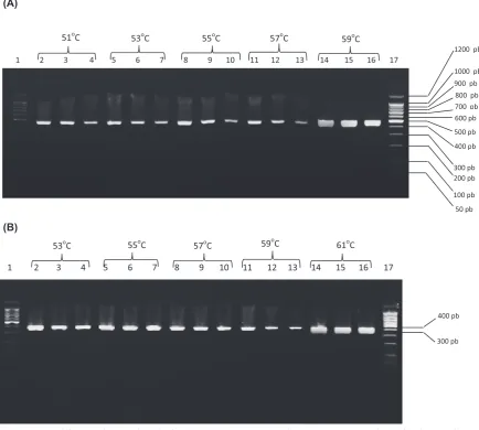
The results of varied hybridization tempera-tures for the set of primers B1/B2 and B3/B4 were visualized for each assayed temperature as shown
in fig. 1. 59ºc and 61ºc were the chosen optimal hybridization temperatures, respectively. The re-sults of PcR with different primer concentra-tions and the elected hybridization temperatures are shown in fig. 2. A decrease in the thickness of the band was evidenced with decreasing centration for primers B1/B2 using a selected con-centration of 0.5 µM. for primers B3/B4, these dif-ferences were not observed for concentrations of 0.2 and 0.5 µM. The concentration of usefulness was 0.2 µM.
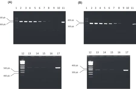
Analysis of PcR sensitivity: The analytic sen-sitivity assay permitted the observation of ampli-fication for both sets of primers up to 2.5 fg/µL of dNA, translated in mass 25 fg (fig. 3), correspond-ing to ten genomes of the bacteria. This result was the same 3 times the assay was done.
When evaluating the number of cycles for the reaction, the smallest dilutions were amplified with 40 cycles. An appreciable decrease in the thickness and intensity of bands was observed as the cycles diminish. for that reason 40 cycles were selected for amplification.
In the specificity assay of each PcR, the am-plification and non-amam-plification of dNA of the related and unrelated strains of Borrelia spp., re-spectively, were verified. Amplification of the pro-spective dNA fragment was demonstrated for the strains of Borrelia.
PCR Evaluation in Ticks
Aartificially Infected
Artificial infection of ticks with subculture of
Borrelia spp.: Most of the ticks observed under the stereoscope showed ingurgitation indicative of feeding with the culture at the end of the period of artificial infection.
evaluation of the effectiveness of dNA extrac-tion:PcR amplification of the dNA fragment that
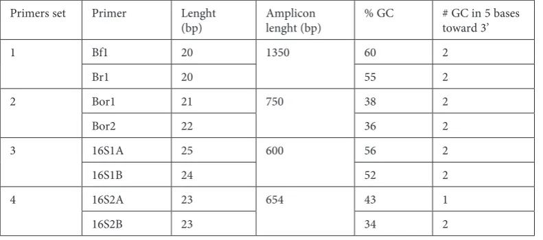
Table 1. characteristics of the evaluated primers reported in the literature Primers set Primer Lenght
(bp) Amplicon lenght (bp) % gc # gc in 5 bases toward 3’
1 Bf1 20 1350 60 2
Br1 20 55 2
2 Bor1 21 750 38 2
Bor2 22 36 2
3 16S1A 25 600 56 2
16S1B 24 52 2
4 16S2A 23 654 43 1
Table 2. characteristics of the evaluated primers reported in the literature Primer Mismatches
(# of sequences) [Stability]
Similitude
(% similitude – # of strains) # of dimers (# of bases) # of nucleotides (dinu-cleotides) repeated 3 or more times
Melting temperature (ºc) Bf1 T:g (1)
[More stables] B. sinicaB. hermsii (100% – 1) (100% – 2)
B. duttonii (100% – 4)
B. hispanica (100% – 8)
B. turcica (100% – 1)
B. sp. (100% – 35)
B. japonica (100% – 1)
B. burgdorferi (100% – 10)
B. recurrentis (100% – 1)
B. valaisiana (100% – 8)
B. garinii (100% – 5)
B. lonestari (100% – 1)
B. turicatae (100% – 1)
B. afzelii (100% – 4)
B. californiensis (100% – 2)
B. andersonii (100% – 3)
B. miyamotoi (100% – 5)
B. spielmanii (100% – 4)
Candidatus B. texasensis
(100% – 1)
1 (6) 0 (0) 64
Br1 g:T (31) [More stables]
A:c (31) [Less stables]
B. sinica (100% – 1)
B. americana (100% – 8)
B. sp. (100% – 10)
B. hermsii (100% – 1)
B. crocidurae (100% – 20)
B. duttonii (100% – 2)
B. hispanica (100% – 11)
B. turcica (100% – 1)
B. japonica (100% – 1)
B. burgdorferi (100% – 43) 0
Bf1 X Br1 1 (4)
0 (0) 62
Bor1 A:c (3) [Less stables]
T:g (19) [More stables]
T:T (5) [Less stables]
B. miyamotoi (100% – 3)
B. persica (100% – 3)
B. hermsii (95% – 40)
B. sp. (95% – 3)
B. recurrentis (95% – 2)
B. duttonii (95% – 3) Uncultured B. sp. (95% – 1)
B. turicatae (95% – 9)
B. lonestari (100% – 2) (95% – 1)
B. parkeri (95% – 8)
No Borrelia (100% – 24) (95% – 1)
1 (4) 1 (0) 58
Bor2 g:T (11) [More stables]
g:g (12) [More stables]
g:T (2) [More stables]
B. duttonii (100% – 11)
B. persica (100% – 3)
B. recurrentis (100% – 21) Uncultured B. sp. (100% – 1) (95% – 5)
B. burgdorferi (95% – 10)
B. afzelii (95% – 12)
B. garinii (95% – 21)
B. crocidurae (95% – 1)
B. hispanica (95% – 6)
B. hermsii (95% – 1)
B. lonestari (95% – 4)
B. lusitaniae (90% – 1)
3 (4 and 6)
Bor1 X Bor2 2 (4)
Table 2. characteristics of the evaluated primers reported in the literature (continuation) Primer Mismatches
(# of sequences) [Stability]
Similitude
(% similitude – # of strains) # of dimers (# of bases) # of nucleotides (dinu-cleotides) repeated 3 or more times
Melting temperature (ºc) 16S1A T:g (1)
[More stables] B. turcicaB. sp. (100% – 26) (100% – 1) Uncultured B. burgdorferi
(100% – 7)
B. japonica (100% – 1)
B. hispanica (100% – 1)
B. burgdorferi (100% – 41)
B. recurrentis (100% – 1)
B. duttonii (100% – 1)
B. valaisiana (100% – 8)
B. lonestari (100% – 1)
B. turicatae (100% – 1)
B. hermsii (100% – 1)
B. afzelii (100% – 3)
B. garinii (100% – 4)
B. valaisiana (100% – 8)
B. californiensis (100% – 2)
B. andersonii (100% – 3)
B. spielmanii (100% – 4)
Candidatus B. texasensis
(100% – 1)
3 (4 and 6) 0 (0) 78
16S1B g:T (11)
[More stables] B. sinica B. americana(100% – 1) (100% – 9)
B. sp. (100% – 20)
B. garinii (100% – 6)
B. turcica (100% – 1)
B. burgdorferi (100% – 55)
B. japonica (100% – 1) Uncultured B. sp. (100% – 1)
B. hermsii (100% – 1)
B. afzelii (100% – 2)
B. spielmanii (100% – 4)
2 (4)
16S1A X 16S1B 0
0 (0) 76
16S2A A:c (8) [Less stables]
g:T (6) [More stables]
B. duttonii (100% – 3)
B. hispanica (100% – 6)
B. turcica (100% – 1)
B. sp. (100% – 26) Uncultured B. burgdorferi
(100% – 6)
B. burgdorferi (100% – 29)
B. garinii (100% – 18)
B. recurrentis (100% – 2)
B. valaisiana (100% – 5) Uncultured B. sp. (100% – 1)
B. lusitaniae (100% – 2)
B. spielmanii (100% – 2)
0 1 (0) 66
16S2B g:T (23)
[More stables]
B. sinica (100% – 1)
B. garinii (100% – 22)
B. burgdorferi (100% – 54)
B. carolinensis (100% – 16)
B. sp. (100% – 5)
1 (8) 16S2A X 16S2B 1 (5)
Table 3. characteristics of the designed primers
Primer Sequence Length
(bp) Amplicon length(bp) % gc # gc in 5 bases toward 3’ B1 5′-TAg ATg AgT cTg cgT cTT ATT A-3′ 22 465 36,36 0
B2 5′-cTT AcA ccA ggA ATT cTA AcT T-3′ 22 36,36 1 B3 5′-TcA cAc Tgg AAc TgA gAT Acg-3′ 21 394 47,62 3 B4 5′-cAA cAg ATT ccA ccc TTA cAc-3′ 21 47,62 2
Table 4. characteristics of the designed primers (continuation) Primer Mismatches
(# of sequences) [Stability]
Similitude
(% similitude – # of strains) # of dimers (# of bases) # of nucleotides (dinu-cleotides) repeated 3 or more times
Melting temperature (ºc) B1 0 B. sinica (100% – 1)
Uncultured B. sp. (100% – 8)
B. americana (100% – 2)
B. sp. (100% – 6)
B. hermsii (100% – 2)
B. crocidurae (100% – 21)
B. garinii (100% – 2)
B. afzelii (100% – 1)
B. burgdorferi (100% – 49)
B. duttonii (100% – 3)
B. hispanica (100% – 14)
B. persica (100% – 4)
B. miyamotoi (100% – 1)
B. recurrentis (100% – 1)
B. valaisiana (100% – 6)
B. microti (100% – 1)
B. turicatae (100% – 1)
B. spielmanii (100% – 6)
B. lonestari (100% – 2)
B. japonica (100% – 3)
B. bissettii (100% – 4)
B. carolinensis (100% – 1)
B. parkeri (100% – 2)
0 0 (0) 60
B2 T:g (10)
[More stables] Uncultured B. hermsii (100% – 11)B. sp. (100% – 5)
B. crocidurae (100% – 21)
B. duttonii (100% – 3)
B. hispanica (100% – 17)
B. turcica (100% – 1)
B. sp. (100% – 9)
B. persica (100% – 5)
B. miyamotoi (100% – 1)
B. recurrentis (100% – 1)
B. valaisiana (100% – 6)
B. microti (100% – 1)
B. theileri (100% – 2)
B. afzelii (100% – 8)
B. turicatae (100% – 1)
B. spielmanii (100% – 6)
B. lonestari (100% – 2)
B. parkeri (100% – 2)
B. japonica (100% – 3)
B. bissettii (100% – 4)
B. carolinensis (100% – 1)
B. burgdorferi (100% – 18)
B. americana (100% – 2)
B. sinica (100% – 1)
1 (6)
B1 X B2 1 (4)
codes for the RNA mitochondrial 16S subunit of ticks allowed the confirmation of nucleic acids ex-tracted from the ticks, except in one case.
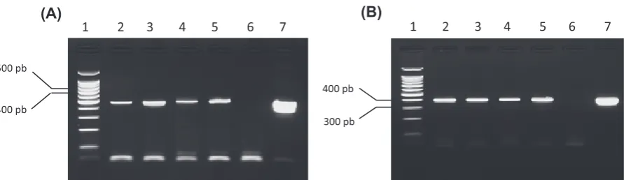
Application of PcRs: Having analyzed the quality of nucleic acids extracted, the PcR tech-nique was applied to detect Borrelia spp. in the ex-tracts and amplification was achieved. With the set of primers B1/B2, amplified dNA were visualized except for the ticks that corresponds to lane 3 and 7 of gel electrophoresis (fig. 4A), while with B3/B4 only the tick belonging to lane 3 did not show am-plification (fig. 4B).
PCR Evaluation of Clinical
Samples Infected Artificially
fig. 5 shows amplification of expected dNA fragments from serum and cfS samples contain-ing Borrelia culture.
Table 4. characteristics of the designed primers (continuation) Primer Mismatches
(# of sequences) [Stability]
Similitude
(% similitude – # of strains) # of dimers (# of bases) # of nucleotides (dinu-cleotides) repeated 3 or more times
Melting temperature (ºc) B3 c:A (2)
[Less stables] B. sinicaUncultured (100% – 1)B. sp. (100% – 8)
B. americana (100% – 2)
B. sp. (100% – 5)
B. hermsii (100% – 2)
B. crocidurae (100% – 16)
B. duttonii (100% – 2)
B. hispanica (100% – 6)
B. garinii (100% – 1)
B. burgdorferi (100% – 27)
B. afzelii (100% – 6)
B. bissettii (100% – 3)
B. carolinensis (100% – 1)
B. valaisiana (100% – 4)
B. lonestari (100% – 2)
B. spielmanii (100% – 6)
B. parkeri (100% – 2)
B. turicatae (100% – 1)
B. miyamotoi (100% – 1)
B. recurrentis (100% – 1)
B. japonica (100% – 3)
0 0 (0) 62
B4 0 B. sinica (100% – 1) Uncultured B. sp. (100% – 5)
B. americana (100% – 5)
B. sp. (100% – 10)
B. hermsii (100% – 2)
B. crocidurae (100% – 21)
B. duttonii (100% – 2)
B. hispanica (100% – 6)
B. garinii (100% – 1)
B. turcica (100% – 1)
B. theileri (100% – 1)
B. burgdorferi (100% – 47)
B. valaisiana (100% – 6)
B. spielmanii (100% – 5)
B. lonestari (100% – 1)
B. japonica (100% – 3)
B. afzelii (100% – 8)
B. bissettii (100% – 2)
B. recurrentis (100% – 1)
B. miyamotoi (100% – 1)
B. turicatae (100% – 1)
0
B3 X B4 1 (5)
(A)
(B)
1200 pb 1000 pb 900 pb
1 2 3 4 5 6 7 8 9 10 11 12 13 14 15 16 17
50 pb 100 pb 200 pb 300 pb 400 pb 600 pb 500 pb 700 pb 800 pb
400 pb
300 pb
1 2 3 4 5 6 7 8 9 10 11 12 13 14 15 16 17
53oC 55oC 57oC 59oC 61oC
51oC 53oC 55oC 57oC 59oC
500 pb
400 pb
1 2 3 4
300 pb 400 pb
1 2 3 4
(A) (B)
Fig. 1. PcR amplification from B. burgdorferi (B31) dNA using annealing temperature gradients for the set of primers B1/B2 (A) and B3/B4 (B). Lines 1 and 17: Molecular weight pattern; lines 2, 5, 8, 11 and 14: dilutions 1/10 of the dNA extracts; lines 3, 6, 9, 12 and 15: dilutions 1/100 of the dNA extracts; lines 4, 7, 10, 13 and 16: dilutions 1/1000 of the dNA extracts
Application of PCR to Samples
of Sera and CSF Obtained from
Patient with Clinical Suspicions
of Lyme Disease
All the 36 clinical samples used in this prelim-inary study were negatives (100%), not being wit-nesses of visible dNA in any of them (results not shown).
Discussion
genomic dNA is frequently used as a target for amplification by PcR of a fragment of the same. for Borrelia spp., the amplification of the plasmid sequence is a non-reliable infection indicator due to different factors that include the crossed reac-tivity with dNA of other species of Borrelia, the genetic variability of plasmid sequences and the lodging of different strains [27].
400 pb 500 pb (A)
300 pb 400 pb
1 2 3 4 5 6 7 8 9 10 11
1 2 3 4 5 6 7 8 9 10 11
500 pb
400 pb
300 pb 400 pb
12 13 14 15 16 17
12 13 14 15 16 17
(B)
1 2 3 4 5 6 7 8 9
453 pb 517 pb
394 pb
1 2 3 4 5 6 7 8 9
(A) (B)
Fig. 3. PcR amplification from B. japonica dNA dilutions. (A) Primers B1 and B2, (B) Primers B3 and B4. Lines 1 and 12: Molecular weight pattern; line 2: dNA 20 ng/µL; line 3: dNA 2 ng/µL; line 4: dNA 0,2 ng/µL; line 5: dNA 0,02 ng/µL; line 6: dNA 2 pg/µL; line 7: dNA 0,2 pg/µL; line 8: dNA 20 fg/µL; line 9: dNA 2 fg/µL; line 13: dNA 10 fg/µL; line 14: dNA 5 fg/µL; line 15: dNA 2,5 fg/µL; lines 10 y 16: negative control (sterile ultrapure water) and lines 11 and 17: positive control (dNA from B. burgdorferi s.s. B31)
The above-mentioned reason gives way for the selection of the gene that codes for 16S rRNA in
Borrelia as a target gene for amplification. It is al-so known that this gene is one of the more con-served in the Borrelia species [28], which would fa-cilitate the discovery of other conserved nucleotide regions where the PcR primers can be inserted.
for complete dNA amplification, the strict scru-tiny of characteristics of the primers that allow them to work effectively was indispensable [29–31].
Primers Bf1 and Br1 amplified a dNA frag-ment of 1,350 base pairs. This is not advisable, es-pecially with the use of a conventional Taq po-limerase, which does not possess proofreader exonuclease activity 3’–5’ and when amplifying a fragment of considerable size because the proba-bility of introducing errors during the polymeriza-tion is higher. Also, with amplicons of a high num-ber of bases, the non-amplification risk increases as a result of loss of sample integrity during its ma-nipulation [32]. This set of primers also presents some mismatches that cause problems for correct annealing.
Bor1 and Bor2 presented a considerable num-ber of mismatches; this can interfere with the cor-rect recognition and ulterior amplification of the targeted fragment. The sequences of these primers allowed dimer formation which should be avoided, or the initial concentration in the reaction mixture of available primers for the amplification is affect-ed, hence decreasing the sensibility of PcR [33]. This set of primers theoretically did not recognize the sequences reported in the gene bank that cor-responds to most representative species of Borrelia
in the group of Lyme disease and relapsing fever. for 16S1A and 16S1B, the fundamental defi-ciency resides in the formation of three and two autodimers respectively; on the other hand, 16S2A and 16S2B did not show the mismatch formation and the theoretical recognition of diverse species dNA of Borrelia, with the negative consequences
already mentioned previously. Taking into consid-eration these results, it was not recommended to use the primers analyzed for the amplification of a dNA fragment of Borrelia spp.
due to the lack of theoretical efficiency of the primers reported in the literature, we decided to break new ground and design new primers capable of amplifying a fragment of dNA of Borrelia spe-cies reported in the gene bank. The two sets of de-signed primers fulfilled the proposed demands.
PcR constitutes a highly sensitive and specif-ic technique for the amplifspecif-ication of a dNA frag-ment. As for the speed of the reaction, the same can be used in routine diagnosis of infectious dis-eases, but for successful results their standardiza-tion was indispensable. Several experiments were required in order to obtain optimal conditions in the course of PcR development.
The logical selection of the 1st parameter to optimize starts with the hybridization tempera-ture of the primer to the sequence of the target. As was mentioned, a higher hybridization temper-ature was chosen in both cases because the bands were neat and more intense. Thicker bands were obtained with dNA extracted from a pure cul-ture. This suggests that while working with clini-cal samples and ticks, the quantity of Borrelia is much smaller [30] and that the probability of ob-taining a defined band with this temperature is higher than with those others that were evaluat-ed. Also, when using higher temperatures of hy-bridization than the range admitted by the primer and the target sequence, the assay gains specificity. consequently, hybridization should take place at a temperature sufficiently high so as to allow a per-fect union of dNA-dNA [34].
In the determination of optimal primer con-centration in each amplification reaction, differ-ences in the width of the bands were observed in relation to the concentrations of primers B1 and B2, this band increased in width and intensity with 400 pb
500 pb
1 2 3 4 5 6 7 1 2 3 4 5 6 7
300 pb 400 pb
(A) (B)
0.5 µM. This result was expected because the prim-er was completely available for annealing to the target sequence and more concentrated primers increased the annealing probability.
No similar pattern was observed with B3 and B4 where the bands observed with 0.2 µM and 0.5 µM were similar in thickness and intensity. 0.2 µM was selected as the optimal concentration because a smaller concentration of primers in the mixture avoids the unspecific union and formation of primer dimers [35]. The usual primer concen-tration in the reaction mixture oscillates between 0.1 and 1 µM [36].
PcR as a molecular technique of dNA ampli-fication has been characterized by speed and high sensitivity that permits the detection of pathogens with few dNA copies in the initial sample [11].
The analytic sensitivity study of both PcR per-mitted the amplification of 25 fg of B.burgdorferi
s.s. dNA that correspond to ten genomes of this bacteria according to that reported by Marconi and garon [37]. These results showed dNA am-plification of Borrelia, andaccording to the reports in the literature, the spirochetes number in human corporal fluid is low (approximately 50 organisms/ /mL). Some patients with arthritis can have up to 10000 spirochetes/mL of synovial liquid while a pa-tient with chronic migratory erythema and system-ic symptoms can have up to 4000 organisms/mL of plasma [38]. It is also known that the number of spirochetes found in different bodily fluids and tissues strongly depends on the phase of the dis-ease (acute, remission or during relapse) [10], but generally the molecular methods are characterized by a high sensitivity and these could be capable of finding a minimum quantity of Borrelia in ade-quate clinical samples from patients with a clinical and epidemiological suspicion of Lyme disease.
The ideal number of cycles for PcR develop-ment depends on the quantity of template dNA in the reaction mixture and the expected yield of PcR. for less than ten copies of template dNA, 40 reaction cycles should be used. If the initial template quantity is more, 25–35 cycles is usually enough.
When we decreased the number of reaction cycles (40) that was used during the standardiza-tion in order to reduce the time of the technique and improve on its specificity, the results were not satisfactory. Since diminishing the number of cy-cles, the dNA quantity visualized in agarose gel also diminished. It was therefore not convenient to ignore the reaction sensitivity and consider the speed of the technique.
The specificity assay on the other hand, was carried out keeping in mind that the dNA used be-longed to bacteria that are related and not related phylogenetically to Borrelia. By obtaining samples
as heterogeneous as possible, the elected pathogens for this assay could be identified from the clinical samples.
In a PcR optimized by Kaufman for the detec-tion of B. burgdorferis.l., different Leptospira sero-vars, as well as bacteria that can be found in clin-ical samples such as Escherichia coli, Pasteurella multocida, Pseudomonas variabilis and Staphylo-coccusintermedius were used to test the specificity. No amplification of the region of interest (85 bp) was observed, although clear unspecific bands above 400 bp for the serovars Pomona and Hardjo of Leptospira interrogans, as well as for Escherichia coli, Pasteurella multocida and Pseudomonas vari-abilis [39] were noted. The previous results differed from ours in that the designed primers B1, B2, B3 and B4, did not amplify except for the evaluated strains of Borrelia and in gel that corresponds to the size of the amplified fragment expected.
After dNA extraction in ticks, it was neces-sary to validate this procedure and prove that the extracted genetic material existed and was under good conditions to be subjected to PcR. Accord-ing to Halos and collaborators, the application of a PcR that amplifies a dNA fragment correspond-ing to 16S mRNA of ticks proves the effectiveness of dNA extraction [25].
dNA amplification of the expected fragment was observed for all ticks fed except for the 1st one. This could have been due to a remnant of bovine blood in this tick. It is reported that hemoglobin in the blood inhibits Taq polymerase and conse-quently PcR [40].
The set of primers B1/B2 used amplified the expected fragment of the ticks dNA except for two of these arthropods. This could be either because they did not feed on the culture or ingested only a small quantity of Borrelia that could not be de-tected by the technique and therefore not visual-ized in 2% agarose gel. However with primers B3/ B4, no visible amplification was noted in only one tick. Our attention was drawn to the fact that in the 7th tick a band was visualized in the gel electro-phoresis. (A band not observed with the 1st set of primer). This assay suggests that PcR with primer B3/B4 was more sensitive than with B1/B2 in ticks. This was also reinforced by the intensity and clar-ity of all the bands observed than when the 2nd set of primer was used.
In countries where Lyme disease is endemic, the prevalence of Borrelia infection in ticks reached values of 15% in germany [41], 23% in china [42] and up to 96% in Switzerland [43]. However in countries where relapsing fever is endemic, the prevalence varied between 2 and 40% [44].
Borrelia spp. in cuban ticks of medical and veteri-nary importance.
The presence of inhibitors was not verified in the serum and cfS during the amplification reac-tions. We therefore suggest the use of both PcRs for the detectionof Borrelia spp. in these samples.
Bearing in mind that the pathogen Borrelia has not been reported in humans in cuba (Lyme dis-ease); it was important to develop a highly sensi-tive and specific system to obtain reliable results for the detection of these microorganisms. We think that the primers designed in this investiga-tion have a series of desired characteristics neces-sary for their effective use.
Both sets of primers with their respective PcR showed similar results, which suggested that each can be used indiscriminately.
The clinical samples coming from patients with clinical suspicion of Lyme borreliosis did not show
Borrelia dNA presence, which can be explained by the symptomatology development in the illness with relationship to the clinical sample taken in addition to the presence in the sample of a smaller number of microorganisms than necessary for the detection limit of the technique. The remainder of the 200 µl of the dNA extraction sample was used
and, since the spirochetes number reported in hu-man corporal fluids is less than 50 organisms/mL, the fact that approximately 10 spirochetes were found in the final volume of the extraction makes it at exactly the PcR detection limit. Because of the above-mentioned, the use of a Nested-PcR is sug-gested to increase the analytical sensitivity of the method. In this case, it is very important to bear in mind the use of an internal control in order to be able to distinguish the inhibited samples.
This was the 1st time in cuba that PcR was applied as a molecular tool for Borrelia detection in clinical samples. It paves the way for the devel-opment of future investigations in this field.
The molecular methods presented here are ex-cellent diagnostic tools for the detection of Borrelia
spp. Briefly, dNA is extracted from the specimen in question, analyzed by PcR using Borrelia spp.
– specific primers, and then separated in agarose gel. These methods are fairly quick and sensitive provided the instrument, reagents, specific prim-ers and enzymes are accessible. We suggest using PcRs optimized in countries where the situation of borreliosis is ignored, since it will allow the detec-tion of any species of the genus Borrelia.
Acknowledgements We would like to thank dr. Lise gern, Phd, from the Biology Institute of Neucha-tel University, Switzerland, for the molecular biology reactive kindly donated and to the National centre of Parasitology, cuba, for the ticks donated.
References
[1] Oshaghi MA, Rafinejad J, Choubdar N, Piazak N, Vatandoost H, Telmadarraiy Z, Mohtarami F, Ravasan NM:
discrimination of Relapsing fever Borrelia persica and Borrelia microtti by diagnostic species-specific primers and polymerase chain reaction-restriction fragment length polymorphism. Vect Born Zoon dis 2011,11, 3.
[2] Schwan TG, Piesman J: Vector interactions and molecular adaptations of Lyme disease and Relapsing fever spi-rochetes associated with transmission by ticks. emerg Infect dis 2002, 8, 2.
[3] Stanek G, Reiter M: The expanding Lyme Borrelia complex-clinical significance of genomic species? clin Microbiol Infect 2011, 17, 487–493.
[4] Rebaudet S, Parola P: epidemiology of relapsing fever borreliosis in europe. feMS Immunol Med Microbiol 2006, 48, 11–15.
[5] Schwan TG, Raffel SJ, Schrumpf ME, Gill JS, Piesman J: characterization of a Novel Relapsing fever Spirochete in the Midgut, coxal fluid, and Salivary glands of the Bat Tick Carios kelleyi. Vect Born Zoon dis 2009, 9, 643– 647.
[6] Wang G, van Dam AP, Schwartz I, Dankert J: Molecular typing of Borrelia burgdorferi sensu lato: taxonomic, epidemiological, and clinical implications. clin Microbiol Rev 1999, 2, 633–653.
[7] Cyr TL, Jenkins MC, Hall RD, Masters EJ, McDonald GA: Improving the specificity of 16S rdNA-based poly-merase chain reaction for detecting Borreliaburgdorferi sensu lato-causative agents of human Lyme disease. J Appl Microb 2005,98,962–970.
[8] Nowakowski J, Schwartz I, Liveris D, Wang G, Aguero-Rosenfeld ME, Girao G, McKenna D, Nadelman RB, Cavaliere LF, Wormser GP: Laboratory diagnostic techniques for patients with early Lyme disease associated with erythema migrans: a comparison of different techniques. clin Infec dis 2001, 33, 2023–2027.
[9] Lebech AM: Polymerase chain reaction in diagnosis of Borrelia burgdorferi infections and studies on taxonomic classification. http://preview.ncbi.nlm.nih.gov/pubmed/11985118 2002, 105, 1–40.
[10] Mantovani E, Costa IP, Gauditano G, Bonoldi VLN, Higuchi ML, Yoshinari NH: description of Lyme disease-like syndrome in Brazil. Is it a new tick borne disease or Lyme disease variation? Braz J Med Biol 2007, 40, 4.
[11] Cerar T, Ruzic-Sabljic E, Glinsek U, Zore A, Strle F: comparison of PcR methods and culture for the detection of Borrelia spp. in patients with erythema migrans. clin Microbiol Infect 2008, 14, 653–658.
[13] Oliveira A, Fonseca AH, Marquez C, Mantovani E, Yoshinari NH: growth, cysts and kinetics of Borrelia garinii
(Spirochaetales: Spirochaetacea) in different culture media. Mem Inst Oswaldo cruz 2010, 105,717–719.
[14] Palacios R, Osorio LE, Giraldo LE, Torres AJ, Philipp MT, Ochoa MT: Positive Igg Western blot for Borrelia burgdorferi in colombia. Mem Inst Oswaldo cruz 1999, 94, 499–503.
[15] Rodríguez I, Fernández C, Sánchez L, Martínez B, Siegrist HH, Lienhard R: Prevalence of antibodies of Borrelia burgdorferi sensu stricto in humans from a cuban village. BRAZ J INfecT dIS 2012,16, 82–85.
[16] Rodríguez I, Ortega LM, Fernández C, Rodríguez ME, Scheurer C, Lienhard R: Borreliosis de Lyme en cuba. A propósito de nuevos casos. Rev Panam Infect 2009,11, 37–41.
[17] Raoult D, Ndihokubwayo JB, Tissot-Dupont H, Roux V, Faugere B, Abegbinni R: Outbreak of epidemic typhus associated with trench fever in Burundi. Lancet 1998, 352, 353–358.
[18] Ritcher D, Schlee DB, Matuschka FR: Relapsing fever–Like Spirochetes Infecting european Vector Tick of Lyme disease Agent. emerg Infect dis2003,15, 234–242.
[19] Picken RN: Polymerase chain reaction primers and probes derived from flagellin gene sequences for specific detec-tion of the agents of Lyme disease and North American relapsing fever. J clin Microbiol 1992, 30, 99–114.
[20] Brusés BLL, Aguirre H, Gorodner MV, Jorge O: comparación de técnicas de extracción de AdN para la detección de
Tripanozoma cruzi mediante la técnica de PcR. UNIVeRSIdAd NAcIONAL deL NORdeSTe. comunicaciones científicas y Tecnológicas 2000.
[21] Gern L, Raiz O, Capiau C, Hauser P, Lobet Y, Simoen E, Voet P, Petre J: Immunization of mice by recombi-nant OspA preparations and protection against Borrelia burgdorferi infection induced by Ixodes ricinus tick bites. Immunol Lett 1994, 39, 249–258.
[22] Guy EC, Stanek G: detection of Borrelia burgdorferi in patients with Lyme disease by the polymerase chain reac-tion. J clin Pathol 1991, 44, 610–611.
[23] Rijpkema S, Golubic D, Molkenboer M, Verbeek-De Kruif N, Schellekens J: Identification of four genomic groups of Borrelia burgdorferi sensu lato in Ixodes ricinus ticks collected in a Lyme borreliosis endemic region of northern croatia. exp Appl Acarol 1996, 20, 23–30.
[24] Humair PF, Douet V, Moran-Cadenas F, Schouls L, Van de Pol I, Gern L: Molecular identification of blood meal source in Ixodes ricinus ticks using 12S rdNA as a genetic marker. J Med entomol 2007, 44, 869–880.
[25] Halos L, Taoufik J, Vial L, Maillard R, Suau A, Menach AL, Boulouis HJ, Vayssier-Taussat M: determination of an efficient and reliable method for dNA extraction from ticks. Vet Res 2004, 35, 709–713.
[26] Postic D, Assous MV, Grimont PAD, Baranton G: diversity of Borreliaburgdorferi sensu lato evidenced by restriction fragment length polymorphism of rrf(5S)-rrl(23S) intergenic spacer amplicons. Int J Syst Bacteriol 1994, 44, 743–752.
[27] Rosa PA, Hogan D, Schwan TG: Polymerase chain reaction analyses identify two distinct classes of Borrelia burg-dorferi. J clin Microbiol1991, 29, 524–532.
[28] Lee SH, Vigliotti VS, Vigliotti JS, Jones W, Williams J, Walshon J: early Lyme disease with spirochetemia – diag-nosed by dNA sequencing. BMc Res Notes 2010, 3, 273.
[29] Hyndman DL, Mitsuhashi M: PcR primer design. Met Mol Biol 2003, 226, 81–88.
[30] Qiagen: Taq PcR Handbook for standard and specialized PcR applications with minimal optimization.editorial QIAgeN companies 2008, 24 pp.
[31] Wilske B: epidemiology and diagnosis of Lyme borreliosis. An Med 2005 37, 568–579.
[32] Fröhler S, Dieterich C: 3Pd: Rapid design of optimal primers for chromosome conformation capture assays. BMc genomics 2009, 10,635.
[33] Aguero-Rosenfeld ME, Wang G, Schwartz I, Wormser GP: diagnosis of Lyme borreliosis. clin Microbiol Rev 2005, 8, 484–509.
[34] Alka D, Sarin BC, Mittar D, Sehajpal PK: Optimization of 38 kda based PcR assay for detection of Mycobacterium tuberculosis from clinical samples. Ind Jour Tuberc 2003,50, 209.
[35] Roche: PcR applications manual 2006, 34pp.
[36] Grunenwald H: Methods in Molecular Biology. p90. In Bartlett JMS, Stirling d, The PcR Revolution 2006, Humana Press Inc, Totowa, New Jersey.
[37] Marconi RT, Garon CF: development of Polymerase chain Reaction Primer Sets for diagnosis of Lyme disease and for species-specific identification of Lyme disease isolates by 16S rRNA Signature Nucleotide Analysis. J clin Microbiol 1992,25, 2830–2834.
[38] Schmidt BL: PcR in laboratory diagnosis of human Borrelia burgdorferi infections. clin Microbiol Rev 1997, 10, 185–201.
[39] Kaufman AC, Greene EC, McGraw AR: Optimization of polymerase chain reaction for the detection of Borrelia burgdorferi in biologic specimens. J Vet diagn Invest 1993, 5,548–554.
[40] Sparagano OA, Allsopp MT, Mank RA, Rijpkema SG, Figueroa JV, Jongejan F: Molecular detection of pathogen dNA in ticks (Acari: Ixodidae): a review. exp Appl Acarol 1999, 23, 929–960.
[41] Schaarschmidt D, Oehme R, Kimmig P,, Hesch RD, Englisch S: detection and molecular typing of Borrelia burg-dorferi sensu lato in Ixodes ricinus ticks and in different patient samples from southwest germany. eur J epidemiol 2001, 17, 1067–1074.
[43] Morán-Cadenas F, Rais O, Jouda F, Douet V, Humai PF, Moret J, Gern L: Phenology of Ixodes ricinus and infection with Borrelia burgdorferi sensu lato along a North- and South-facing altitudinal gradient on chaumont Mountain, Switzerland. J Med entomol 2007, 44, 683–693.
[44] Assous MV, Wilamowski A: Relapsing fever borreliosis in eurasia forgotten, but certainly not gone! clin Microbiol Infect 2009,15,407–414.
Address for correspondence:
Angel A. Noda
Bacteriology and Mycology department “Pedro Kourí” Tropical Medicine Institute Havana
cuba
Tel.: 537 255 35 30
e-mail: [email protected]
conflict of interest: None declared