Introduction
Escherichia coli represents as the most en-countered etiology for urinary tract in-fections (UTIs) identified in the gastroin-testinal tract of mammals. There is a well established belief that uropathogenic E. coli (UPEC) arises from the distal gut microbi-ota1-3 In addition, E. coli is a diverse species
regarding the genetic content and pheno-typic and pathogenic traits.4 The disease
which generates strains of E. coli contains multiple virulence mediators participating in their pathogenesis (diarrhea, dysentery, septicemia, pneumonia, meningitis, and UTI). Also, commensal strains can lead to a disease in host with compromised immune system.5 Some pathogenic strain of E. coli
persist in gastrointestinal tract of human
and are frequently associated with human diseases.
According to multiple virulence determi-nants, E. coli bacteria can be categorized into 3 major subclasses: commensal, in-testinal pathogenic and extra inin-testinal pathogenic E. coli (ExPEC).6 It has been
shown that intestinal and ExPEC strains may be extended from commensal types through obtaining of virulence mediators.7
ExPEC exhibit significant genetic hetero-genicity and represents a wide spectrum of virulence-related factors such as adhesins, toxins, and iron up taking molecules, lipo-polysaccharides, polysaccharide capsules, and invasions, which can be expressed on mobile genetic segments including plas-mids, bacteriophages, and pathogenici-doi 10.15171/ijbsm.2017.11
Prevalence of Genes Encoding Outer Membrane
Virulence Factors Among Fecal
Escherichia coli
Isolates
Ahmad Rashki1*, Hussein Ali Abdi2, Milad Shookohi3
1Associate Professor in Molecular Genetics and Microbiology, Department of Pathophysiology, Faculty of Vet-Medicine, University of Zabol, Zabol, Iran
2PhD Student in Molecular Genetics, Faculty of Sciences, University of Sistan and Baluchestan, Zahedan, Iran 3MSc in Molecular Genetics, Department of Biology, Faculty of Basic Sciences, University of Zabol, Zabol, Iran
Abstract
Objective:Escherichia coli is commensal bacterium of human intestine. The gut is a common pool of E. coli isolates causing urinary tract infections (UTIs). Some of fecal E. coli (FeEC)by the possession of certain virulence factors is able to cause diseases in human and other mammalian models. To evaluate the health threats coordinated with a given fecal source of E. coli strains, we determined the frequency of genes expressing virulence determinants in fecal E. coli isolates collected from human feces in Zabol, southeast of Iran.
Methods: Escherichia coli isolates (n = 94) were separated from the feces of patients attending teaching hospitals, and screened for various virulence genes: fimH, his, hlyA, ompT, irp2, iucD,
iroN, and cnf1 by using the multiplex polymerase chain reaction (PCR) method.
Results: The prevalence of virulence genes was as follows: adhesins (fimH, 98% and iha, 26%), alpha-hemolysins (hlyA, 10%), outer membrane protease (ompT, 67%), aerobactin (iucD, 67%), iron-repressible protein (irp2, 91%) and salmochelin (iroN, 33%) and cytotoxic necrotizing factor 1 (cnf1). According to the diversity of different virulence genes, the examined isolates exhibited 29 different patterns.
Conclusion: Our results demonstrated that most of the assessed isolates harbored several virulence factors. Our findings propose possibility of human feces serving as a source for pathogenic organisms, supporting the notion that fecal materials of humans play a role in the epidemiological chain of extra-intestinal pathogenic E. coli. This is the first report of the frequency of virulence factors among E. coli isolates collected from human feces in Iran.
Keywords: Fecal Escherichia coli, Major virulence factors, Multiplex PCR
*Correspondence to Ahmad Rashki; Department of Pathophysiology, Faculty of Vet-Medicine, University of Zabol, Zabol, Iran.
Tel: +98-5424822250; Fax: +98-5424822251; Email: [email protected]
Received October 26, 2016
Accepted March 6, 2017
Published online March 18, 2017
http://ijbsm.zbmu.ac.ir
Copyright © 2017 The Author(s); Published by Zabol University of Medical Sciences. This is an open-access article distributed under
Please cite this article as follows:Rashki A, Abdi HA, Shookohi M. Prevalence of genes encoding outer membrane virulence factors among fecal
Escherichia coli
ty-associated islands (PAIs).8 The fecal flora from the
hosts provides the most common source for infecting E. coli strain.9 The UTI-causing strains that are commonly
called UPEC are obtained by faces, the place of their en-trance to the urinary tract via colonization of the vaginal introitus and the periurethral location.10E. coli strains
re-siding within intestinal tract and promoting UTI typically contain virulence genes necessitated for colonization of the urinary tract. On the other hand, it seems that some of FeEC strains carrying many genes that encode virulence factors and can cause serious disease at diverse extraint-estinal sites.
Multiple studies have been done in different parts of Iran in order to investigate the frequency of virulence genes among UPEC.11-13 However, there is no information about
the frequency of virulence mediators in FeEC isolated from human in Iran. Therefore, this study was conduct-ed to examine the distribution of 8 virulence factors, in-cluding type 1 fimbriae (fimH), iron-regulated gene ho-mologue adhesion (iha), alpha-hemolysin (hlyA), outer membrane protease (ompT), aerobactin (iucD), yersinia-bactin (irp2), salmochelin receptor (iroN) and cytotoxic necrotizing factor 1 (cnf1) in E. coli strains isolated from human feces in Zabol, southeast of Iran, by using the mul-tiplex polymerase chain reaction (PCR) method.
Methods
Bacterial Isolates
The sample size was calculated as described by Charan and Biswas.14 Swabs were collected directly from stool
samples of the patients with diarrhea admitted to teaching hospitals in Zabol, Iran during July 2014 through October 2014. Samples were suspended into Cary-Blair transport media (Laboratorios Conda, S.A., Spain) and transported to laboratory on ice where one loop from each sample was streaked directly on MacConkey agar (HiMedia Labora-tories) within 4 hours after collection. Plates were
incu-bated at 37°C for 18–24 hours, and up to 3 colonies with typical appearance of E. coli were selected and subjected to biochemical tests including oxidase, indole, methyl red, Voges-Proskauer, nitrate reduction, urease production, Simmons’ citrate agar (HiMedia Laboratories), and var-ious sugar fermentation.15,16
Extraction of DNA
The bacteria were isolated from 1 mL of the E. coli cul-ture grown for 18 hours at 37°C. The bacterial DNA was extracted by boiling methods.17 Briefly, all E. coli isolates
were cultivated overnight (16 hours) in 5 mL Luria-Ber-tani (LB) broth (HiMedia Laboratories) in a shaking in-cubator (200 rpm) at 37°C. Two milliliters of bacterial iso lates were then pelleted, suspended in 200 μL of sterile double-distilled water and boiled at 95°C for 10 minutes. The mixture was cooled on ice (5 minutes), and the su-pernatant was collected following centrifugation (13 000 rpm 5 minutes). After centrifugation, the supernatants were kept as DNA at -20°C until applied for PCR.
Detection of Putative Virulence Genes Using the Multiplex PCR Method
Prevalence of putative virulence genes in FeEC isolates was determined by multiplex PCR.3 Details of primer
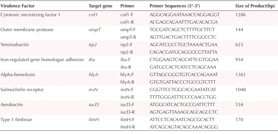
sequences, target genes and products size are shown in Table 1. Amplification of selected genes was performed by setting a net volume of 25 μL. The reaction contained 2 μL of DNA, 12.5 μL of Taq DNA Polymerase Master Mix Red (amplicon, A/S, Denmark), 1 μL of primers (30 pmol concentration for each) (Pishgam, Iran) and 8.5 30 pmol ddH2O. The multiplex PCR was performed considering an initial phase of denaturation (94°C, 5 minutes), fol-lowed by 35 cycles consisting of denaturation (94°C for 30 seconds), annealing (59°C for 50 seconds) and extension (72°C for 70 seconds), and followed by a final extension step at 72°C for 5 minutes. Amplification was performed
Table 1. Primers for the Multiplex PCR Assays
Virulence Factor Target gene Primer Primer Sequences (5'-3') Size of Product(bp)
Cytotoxic necrotizing factor 1 cnf1 cnf1-F AGGCAGGAATAAACCAGGAGGT 1286 cnf1-R ACGAGCAGAATTTGACACACGA
Outer membrane protease ompT ompT-F TGCGATCAGCTCTTTTGCTTCT 144 ompT-R AGTTGACTGACTTTTCGGCCTC
Yersiniabactin irp2 irp2-F AGCATCGCCTGCTAAAACTGAA 623 irp2-R CAGACGATGCAGGGCGTTATTA
Iron-regulated gene homologue adhesion iha iha-F CTGGAAGTCAGCATTCGTGGAA 934 iha-R GATGCCACTCATCCTCAGCAAA
Alpha-hemolysin hlyA hlyA-F GTTAGCGGGTGTCACCAGAAAT 1361 hlyA-R GTGTGATTACCCTGCCGTCTTT
Salmochelin receptor iroN iroN-F CGGTTCCTGGCACGAATATCAT 1048 iroN-R TTTTGGGATTTCCCCAACCTGG
Aerobactin iucD iucD-F ATGGCATCACTGCCGATTCTTT 534 iucD-R AGTGAGTTAAAGCAGCAGCCTC
using a gradient Eppendorf’s Mastercycler® Pro
(Eppen-dorf, Germany). The multiplex PCR products were sep-arated by agarose gel 2% electrophoresis and visualized under UV-induced fluorescence. A 100 bp DNA ladder (Fermentase) was used as size standard (Figure 1). Ampli-fication identities were confirmed by restriction analysis.
Restriction Analysis
Amplified fragments of selected genes were confirmed by restriction analysis. Restriction patterns of 8 sequenc-es were obtained with the Webcutter 2.0, online software (http://rna.lundberg.gu.se/cutter2/). The hlyA, iha, irp2 and iroN gene sequences were restricted with TfiI endonu-clease followed ompT, iucD and fimH sequences restricted with AluI endonuclease. MspI endonuclease was chosen for restriction of cnf1 sequence. Restriction conditions were identical in all cases. Each 30 μL reaction mixture contained 1 μL of restriction endonuclease, 8 μL of PCR product, 3 μL of specific endonuclease enzyme buffer and 18 μL of sterilized dH2O. After overnight incubation, the restriction products were determined by electrophoresis of the digested DNA in 2% agarose gel.
Results
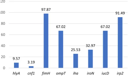
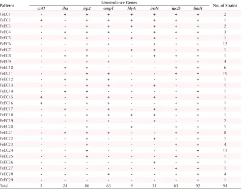
The frequency of virulence genes collected from E. coli isolates of fecal samples is shown in Figure 2. In total, 94/94 (100%) of FeEC isolates represented with at least one of the studied virulence genes, of which 1 (1%), 18 (19%), 10 (11%), and 29 (31%) were detected harboring 1, 2, 3, and 4 virulence factors, respectively. Twenty-nine different virulence combinations were found among fecal E. coli isolates (Table 2). FeEC11 pattern was determined by the presence of the irp2, ompT, iucD and fimH genes which presented the most identified pattern, detected in 19 (20%) isolates. fimH was the frequent virulence gene identified in all 92(98%) FeEC isolates. Among the iron-acquisition genes, irp2 was the most prevalent gene and was identified in 86 (91%) isolates, while iucD and iroN genes were detected in 63 (67%) and 31 (33%) iso-lates respectively. The secretory virulence genes includ-ing hly and cnf were present in 9 (10%) and 3 (3%) FeEC isolates, respectively. The ompT gene was identified in 63 (67%) and the iha was detected in 24 (26%) isolates. The association of 7 genes was recognized in FeEC1 and FeEC2 patterns (3 isolates). One isolate harbored fimH only (FeEC29). The 2 FeEC3 and FeEC4 patterns includ-ed isolates possessing a combination of 6 amplifiinclud-ed genes (Table 2).
The detected virulence genes were confirmed by restric-tion analysis: iha, iroN, irp2 and hlyA amplicons (934, 1084, 623 and 1361 bp respectively) were restricted with TfiI endonuclease enzyme and yielded fragments of 465, 267, 129 and 73 bp; 1004 and 80 bp; 426 and 197 bp; and 1030 and 331bp respectively. The cnf1 amplicon (1286 bp) was restricted with MspI and yielded fragments of 467, 404, 261 and 154 bp. The detected virulence genes were confirmed by restriction analysis: iucD, ompT and fimH amplicons (534, 144 and 170 bp respectively) and were
Figure 1. Electrophoresis of Virulence Selected Genes Among FeEC Isolate Obtained by Multiplex PCR.
Abbreviations: FeEC, fecal Escherichia coli; PCR, polymerase chain reaction.
Each band is indicated by the names of the virulence genes. Lines 1 and “M” denote 100 bp DNA marker.
Figure 2. Prevalence of Virulence Genes Among 94 FeEC Isolates Collected From Human Feces.
Abbreviation: FeEC, fecal Escherichia coli. 9.57
3.19 97.87
67.02
25.53 32.97 67.02
91.49
0 20 40 60 80 100 120
hlyA cnf1 fimH ompT iha iroN iucD irp2
restricted with AluI endonuclease enzyme and yielded fragments of 386 and 148 bp; 135 and 9 bp; and 109 and 61bp respectively (Figure 3).
Discussion
The lower gastrointestinal tract is considered as the rich-est pool of UTI generating organisms. Some strains can colonize the vagina and urinary tract.18 The variety and
heterogeneity of virulence factors, including adhesins, toxins and siderophores emerge to be momentous for E. coli strains and make the development of multiplex PCR method especially significant. In this work, to our knowledge for the first time in Iran, we assessed the prev-alence of virulence gene profile in 94 FeEC isolates Un-derstanding the distribution of virulence determinants in FeEC isolates is important to evaluate their relative con-tribution to the extraintestinal infection. We also assessed multiplex PCR assays to detect virulence factors utilizing a combination of primers previously reported. 3
frequency of the iha gene, as other adhesion, was 26% (24/94). Our laboratory previously showed that the prevalence of iha gene was 29% among UPEC isolates.20
However, no significant differences were observed in re-lation to iha gene. According to our knowledge, to date, no reports have been published about the prevalence of iha gene among FeEC isolates. The findings of our study indicated that the presence of the hlyA and cnf1 genes was 10% and 3% respectively.
In an other study, Usein et al21 demonstrated that the hlyA
and cnf1 genes were found in 35% of E. coli bacteria re-covered from the fecal flora of healthy adult humans. The results showed the variation geographical distribution of these genes. The prevalence of the cnf1 gene was differ-ent in fecal isolates studied by other investigators.22,23 In
accordance with our study, Obiet et al24 also described
that the cnf1 gene was expressed in 3.5% of E. coli isolates collected from diarrheic stool samples in South Africa. In an other study, our group demonstrated that cnf1 gene was more frequently detected in UPEC (28%) in comparison with FeEC isolates (3%).20 Particularly, ExPEC expresses
a richness of apparently excessive iron obtaining systems, including the salmochelin, yersiniabactin, and aerobactin
Table 2. Virulence Gene Patterns Identified Among the Studied Isolates
Patterns Urovirulence Genes No. of Strains
cnf1 iha irp2 ompT hlyA iroN iucD fimH
FeEC1 - + + + + + + + 2
FeEC2 + - + + + + + + 1
FeEC3 - - + + + + + + 2
FeEC4 - + + + - + + + 3
FeEC5 - + + - + + - + 1
FeEC6 - - + + - + + + 12
FeEC7 - - + - + + - + 1
FeEC8 - + + - - + + + 1
FeEC9 - - + + - - - + 4
FeEC10 - + + - - - + + 6
FeEC11 - - + + - - + + 19
FeEC12 - + + + - - - + 1
FeEC13 - - + + - + - - 1
FeEC14 - + + - - - - + 1
FeEC15 + - + + - + - + 1
FeEC16 + - + + - - + + 1
FeEC17 - + - + - + + + 1
FeEC18 - - + + + + - + 1
FeEC19 - - + + - + - + 2
FeEC20 - - + - + - + + 1
FeEC21 - + + + - - + + 8
FeEC22 - - + - - + - + 1
FeEC23 - - + - - - + + 4
FeEC24 - - + - - - - + 11
FeEC25 - - + - - - + - 1
FeEC26 - - - - - + - + 1
FeEC27 - - - - - - + + 1
FeEC28 - - - + - - - + 4
FeEC29 - - - - - - - + 1
Total 3 24 86 63 9 31 63 92 94
Figure 3. Gel Electrophoresis for Digestion Results of Virulence Genes in FeEC Isolates.
L: Lader; Lane 1. Digest of hlyA (1361bp) with TfiI enzyme and production of fragments 331 bp and 1030 bp; Lane2. Digest of
iroN (1084 bp) with TfiI enzyme and production of fragments 80 bp and 1004 bp; Lane 3. Digest of iucD (534 bp) with AluI enzyme and production of fragments 148 bp and 386 bp; Lane 4. Digest of
fimH (170 bp) with AluI enzyme and production of fragments 61 bp and 109 bp; Lane 5. Digest of cnf1 (1286 bp) with MspI enzyme and production of fragments 154 bp, 261 bp, 404 bp and 467 bp;
Lane 6. Digest of iha (934 bp) with TfiI enzyme and production of fragments 73 bp, 129 bp, 267 bp and 465 bp; Lane 7. Digest of irp2
siderophores. In accordance, we observed in our study a very high prevalence of the iron acquisition genes; irp2 (91%), iucD (67%) and iroN (33%). The irp gene cluster is mapped within the high pathogenicity island (HPI) described primarily in Yersinia spp. and horizontal gene transfer has caused it to be present in intestinal and ex-tra intestinal clinical E. coli strains.25 The iroN gene, that
is situated on the iroA gene cluster encodes a receptor which is responsible for iron uptake mediated by the sid-erophores salmochelins, contributes to the virulence of UPEC. This interaction facilitates transport of the com-plex into the bacterial cytosol.26 The virulence genes of
UPEC bacteria such as iroN, iucD and irp2 have been previously recorded from Iran.11,12 However, according to
published data, there is no information on the occurrence of these genes in FeEC isolates.
The distribution of the ompT gene among the studied iso-lates was also similar to that in the previously reported data.27 OmpT rather seems as a conserved protease
ex-ecuting in metabolism of E. coli derivedfrom secretory proteins. This outer membrane protease contributes to the destruction of several proteins interacting with the outer membrane.28 However, there is no report about the
prevalence of ompT gene among FeEC isolates in Iran. The analysis of the association between the presences of different combinations of virulence genes among FeEC isolates, allowed us to divide the tested isolates into 29 vir-ulence patterns noted FeEC1 to 29. Our results revealed the complexity of the properties of virulence markers in fecal E. coli isolates. The pattern FeEC6 included strains simultaneously posi tive for irp2+, ompT+, iucD+, iroN+ and fimH+ (12 isolates). The prevalence of genes coding for
the 2 adhesion pathways (type 1 fimbriae and iha) which confers the ability to colonization among 24 isolates were fit with those published by other investigators.3,29 Among
FeEC isolates, 89 isolates contain genes encoding an iron acquisition protein (yersiniabactin, aerobactin or salmo-chelin receptor). The maximum number of detected am-plicons in 1 isolate was 6. The main novel finding is the high occurrence of genes of outer membrane virulence proteins among FeEC isolates in Zabol, southeast of Iran. Apart from the high occurrence of virulence genes, vari-ous virulence determinants were also revealed in fecal E. coli strains. In our analysis, 74% (70/94) of the bacteria were represented with various virulence genes (4 or more genes). It is comprehensible that the recurrence and prev-alence of virulence features of fecal E. coli strains are ferent in other regions of Iran. Probably, geographical dif-ferences, cultural habitants, dietary features, public and hospital health policies, weather climate of each region and even sampling methods may exert high impacts on detection rate of virulence factors of FeEC isolates. None-theless, further research on the expression of virulence genes and molecular typing methods covering wider geo-graphical areas in Iran is needed to determine the dis-tribution pattern of virulence determinants and develop effective strategies to treat FeEC -induced diseases. This may constitute a limitation of our study.
Conclusion
The FeEC isolates expressing virulence genes may be re-garded as an important organism for extra-intestine in-fections in Iran. The current work is the first report which identified virulence genes among FeEC isolates in Iran. The multiplex PCR designed in the present study suc-cessfully screened FeEC isolates for various E. coli-related virulence genes. Further studies in other parts of Iran are needed to identify virulent factors and to ascertain the pathophysiology of such infectious agents to consider possible prevention interventions.
Ethical Approval
We obtained informed consent form our participants.
Competing Interests
Authors declare that they have no conflict of interest.
Acknowledgments
We wish to thank the staff of the laboratory of microbiol-ogy of the faculty of vet-medicine at university of Zabol.
References
1. Gruneberg RN. Relationship of infecting urinary organism to the faecal flora in patients with symptomatic urinary infection. Lancet. 1969;2(7624):766-768.
2. Johnson JR, Kaster N, Kuskowski MA, Ling GV. Identification of urovirulence traits in Escherichia coli by comparison of urinary and rectal E. coli isolates from dogs with urinary tract infection. J Clin Microbiol. 2003;41(1):337-345.
3. Rashki A. Cervico-vaginopathogenic Escherichia coli
in Iran: serogroup distributions, virulence factors and antimicrobial resistance properties. Microb Pathog. 2014;75:29-34 doi:10.1016/j.micpath.2014.1008.1004. 4. Dobrindt U, Janke B, Piechaczek K, et al. Toxin genes on
pathogenicity islands: impact for microbial evolution. Int J Med Microbiol. 2000;290(4-5):307-311. doi:310.1016/ S1438-4221(1000)80028-80024.
5. Stecher B, Hardt WD. The role of microbiota in infectious disease. Trends Microbiol. 2008;16(3):107-114. doi:110.1016/j.tim.2007.1012.1008.
6. Finlay BB, Falkow S. Common themes in microbial pathogenicity revisited. Microbiol Mol Biol Rev. 1997;61(2):136-169.
7. Russo TA, Johnson JR. Proposal for a new inclusive designation for extraintestinal pathogenic isolates of Escherichia coli: ExPEC. J Infect Dis. 2000;181(5):1753-1754. doi:1710.1086/315418.
8. Rodriguez-Siek KE, Giddings CW, Doetkott C, et al. Comparison of Escherichia coli isolates implicated in human urinary tract infection and avian colibacillosis. Microbiology. 2005;151(Pt 6):2097-2110. doi:2010.1099/ mic.2090.27499-27490.
9. Clermont O, Bonacorsi S, Bingen E. Rapid and simple determination of the Escherichia coli phylogenetic group. Appl Environ Microbiol. 2000;66(10):4555-4558
10. Wilson BA, Salyers AA, Whitt DD, Winkler ME. Bacterial pathogenesis: a molecular approach: American Society for Microbiology (ASM); 2011.
Escherichia coli in Iran: serogroup distributions, virulence factors and antimicrobial resistance properties. Ann Clin Microbiol Antimicrob. 2013;12:8. doi:10.1186/1476-0711-1112-1188.
12. Abdi HA, Rashki A. Comparison of virulence factors distribution in uropathogenic E. coli Isolates from phylogenetic groups B2 and D. Int J Enteric Pathog. 2014;2(4):e21725.
13. Charan J, Biswas T. How to calculate sample size for different study designs in medical research? Indian J Psychol Med. 2013;35(2):121-126. doi:110.4103/0253-7176.116232. 14. Maluta RP, Stella AE, Riccardi K, et al. Phenotypical
characterization and adhesin identification in Escherichia coli strains isolated from dogs with urinary tract infections. Braz J Microbiol. 2012;43(1):375-381. doi:10.1590/S1517-838220120001000045.
15. Chen YM, Wright PJ, Lee CS, Browning GF. Uropathogenic virulence factors in isolates of Escherichia coli from clinical cases of canine pyometra and feces of healthy bitches. Vet Microbiol. 2003;94(1):57-69. doi:10.1016/S0378-1135(1003)00063-00064.
16. Rahdar M, Rashki A, Miri HR, Rashki Ghalehnoo M. Detection of pap, sfa, afa, foc, and fim adhesin-encoding operons in uropathogenic Escherichia coli isolates collected from patients with urinary tract infection. Jundishapur J Microbiol. 2015;8(8):e22647. doi:10.5812/jjm.22647 17. Rashki A. Cervico-vaginopathogenic Escherichia coli
in Iran: Serogroup distributions, virulence factors and antimicrobial resistance properties. Microb Pathog. 2014;75:29-34. doi:10.1016/j.micpath.2014.1008.1004. 18. Moreno E, Andreu A, Pigrau C, et al. Relationship between
Escherichia coli strains causing acute cystitis in women and the fecal E. coli population of the host. J Clin Microbiol. 2008;46(8):2529-2534. doi:2510.1128/JCM.00813-00808. 19. Kaczmarek A, Budzynska A, Gospodarek E. Prevalence
of genes encoding virulence factors among Escherichia coli with K1 antigen and non-K1 E. coli strains. J Med Microbiol. 2012;61(Pt 10):1360-1365. doi:1310.1099/ jmm.1360.044263-044260.
20. Rashki A. Cervico-vaginopathogenic Escherichia coli
in Iran: Serogroup distributions, virulence factors and antimicrobial resistance properties. Microb Pathog. 2014;75:29-34. doi: 10.1016/j.micpath.2014.08.004.
21. Moreno E, Andreu A, Pigrau C, et al. Relationship between
Escherichia coli strains causing acute cystitis in women and the fecal E. coli population of the host. J Clin Microbiol. 2008;46(8):2529-2534. doi:10.1128/JCM.00813-08.
22. Abdi Ha, Rashki A. The phylogenetic study of uropathogenic Escherichia coli strains in Sistan of Iran. J Birjand Univ Med Sci. 2014;21(3):392-400.
23. Usein CR, Damian M, Tatu-Chitoiu D, et al. Comparison of genomic profiles of Escherichia coli isolates from urinary tract infections. Roum Arch Microbiol Immunol. 2003 J;62(3-4):137-154.
24. Yuri K, Nakata K, Katae H, Yamamoto S, Hasegawa A. Distribution of uropathogenic virulence factors among
Escherichia coli strains isolated from dogs and cats. J Vet Med Sci. 1998;60(3):287-290.
25. Caprioli A, Falbo V, Ruggeri FM, et al. Cytotoxic necrotizing factor production by hemolytic strains of Escherichia coli causing extraintestinal infections. J Clin Microbiol. 1987;25(1):146-149.
26. Obi CL, Green E, Bessong PO, et al. Gene encoding virulence markers among Escherichia coli isolates from diarrhoeic stool samples and river sources in rural Venda communities of South Africa. Water SA. 2004;30(1):37-42. doi:10.4314/wsa.v4330i4311.5024
27. Schubert S, Rakin A, Karch H, Carniel E, Heesemann J. Prevalence of the “high-pathogenicity island” of Yersinia species among Escherichia coli strains that are pathogenic to humans. Infect Immun. 1998;66(2):480-485.
28. Hantke K, Nicholson G, Rabsch W, Winkelmann G. Salmochelins, siderophores of Salmonella enterica and uropathogenic Escherichia coli strains, are recognized by the outer membrane receptor Iron. Proc Natl Acad Sci U S A. 2003;100(7):3677-3682. doi:3610.1073/pnas.0737682100. 29. Marrs CF, Zhang L, Tallman P, et al. Variations in 10
putative uropathogen virulence genes among urinary, faecal and peri-urethral Escherichia coli. J Med Microbiol. 2002;51(2):138-142. doi:110.1099/0022-1317-1051-1092-1138.