R E S E A R C H
Open Access
Specific PKC isoforms regulate LPS-stimulated
iNOS induction in murine microglial cells
Jie Wen
1, Rachel Ribeiro
2and Yumin Zhang
1,2*Abstract
Background:Excessive production of nitric oxide (NO) by inducible nitric oxide synthase (iNOS) in reactive microglia is a major contributor to initiation/exacerbation of inflammatory and degenerative neurological diseases. Previous studies have indicated that activation of protein kinase C (PKC) can lead to iNOS induction. Because of the existence of various PKC isoforms and the ambiguous specificity of PKC inhibitors, it is unclear whether all PKC isoforms or a specific subset are involved in the expression of iNOS by reactive microglia. In this study, we employed molecular approaches to characterize the role of each specific PKC isoform in the regulation of iNOS expression in murine microglia.
Methods:Induction of iNOS in response to bacterial endotoxin lipopolysaccharide (LPS) was measured in BV-2 murine microglia treated with class-specific PKC inhibitors, or transfected with siRNA to silence specific PKC isoforms. iNOS expression and MAPK phosphorylation were evaluated by western blot. The role of NF-B in activated microglia was examined by determining NF-B transcriptional response element- (TRE-) driven, promoter-mediated luciferase activity.
Results:Murine microglia expressed high levels of nPKCs, and expressed relatively low levels of cPKCs and aPKCs. All PKC inhibitors attenuated induction of iNOS in LPS-activated microglia. Knockdown of PKCδand PKCb attenuated ERK1/2 and p38 phosphorylation, respectively, and blocked NF-B activation that leads to the expression of iNOS in reactive microglia.
Conclusions:Our results identify PKCδandbas the major PKC isoforms regulating iNOS expression in reactive microglia. The signaling pathways mediated by PKC involve phosphorylation of distinct MAPKs and activation of NF-B. These results may help in the design of novel and selective PKC inhibitors for the treatment of many inflammatory and neurological diseases in which production of NO plays a pathogenic role.
Background
Microglia are distributed throughout the central nervous system (CNS) as resting immunocompetent cells derived from a monocyte/macrophage lineage [1,2]. When acti-vated, microglia protect neurons by clearing toxic cell debris and pathogens, and acting as antigen presenting cells to induce innate immune responses [3]. However, excessive activation of microglia can also release a vari-ety of toxic factors including reactive oxygen species (ROS), reactive nitrogen species (RNS) and proinflam-matory cytokines, which cause toxicity to the
neighboring cells such as neurons and oligodendrocytes (OLs). A pathogenic role for nitric oxide has been impli-cated in many inflammatory and neurodegenerative dis-eases, including multiple sclerosis, stroke and traumatic brain injury [4-7]. Understanding the potential mechan-isms that turn beneficial inflammatory responses into detrimental action is crucial for identifying therapeutic targets to intervene in self-sustained inflammatory cycles.
Nitric oxide (NO), generated from L-arginine by nitric oxide synthase (NOS), has been shown to be both a sig-naling and an effector molecule in diverse biological sys-tems [8-10]. Among the three isoforms of NOS identified, neuronal NOS (nNOS) and endothelial NOS
(eNOS) are Ca2+dependent [8-13], and inducible NOS
(iNOS) functions in a Ca2+-independent manner [10,13].
* Correspondence: [email protected]
1Department of Anatomy, Physiology and Genetics, Uniformed Services University of the Health Sciences, 4301 Jones Bridge Road, Bethesda, MD 20814, USA
Full list of author information is available at the end of the article
Induction of iNOS occurs primarily in astrocytes and microglia in response to endotoxin or to
proinflamma-tory cytokines, such as TNFa, IL-1bor IFNg[14]. Using
pharmacological inhibitors and molecular approaches, studies have shown that NO can react with superoxide to form peroxynitrite in reactive microglia causing toxi-city to neurons and OLs [15,16]. Although it is known that activation of various transcription factors - such as
STAT, NF-B, AP-1, and C/ERP - can contribute to the
production of NO [17-20], the signaling pathways regu-lating expression of iNOS and production of NO in the CNS are still not well understood.
Protein kinase C (PKC) is a family of serine/threonine kinases that regulate cellular responses elicited by hor-mones, neurotransmitters and growth factors [21]. Based on differences in sequence homology between these isozymes and their requirements for cofactors, the
PKC family is divided into conventional PKCs (cPKC:a,
bandg), novel PKCs (nPKC:δ, ε,handθ) and atypical
PKCs (aPKCs: ζ and l/ι) [22,23]. PKC isoforms are
widely expressed in many cell types, including micro-glia/macrophages [24], and studies have shown that PKC activation is an important mediator of microglial activation [25,26]. PKC inhibitors reduce NO synthesis
from IFN-g-treated microglia and PKCδis able to
regu-late NF-B activation and iNOS expression in mouse peritoneal macrophages [27]. Because of the existence of various PKC isoforms and the ambiguity of action of PKC inhibitors, the role of specific PKC isoforms involved in the inflammatory response in microglia has not been elucidated. In this study we used murine microglial cell line BV-2 cells to examine the signaling pathways by which PKC activation leads to iNOS induc-tion in LPS-activated microglia. Our results indicate that all PKC isoforms are expressed in BV-2 cells with a par-ticularly high expression of nPKC. Although several PKC isoforms can mediate lipopolysaccharide- (LPS-)
stimulated increases in iNOS expression, PKCδand b
are likely the major PKC isoforms responsible for PKC function in reactive microglia. Furthermore, we found that distinct mitogen activated protein kinases (MAPKs) are activated in response to specific PKC isoforms and result in iNOS induction. Elucidation of the signaling pathways mediated by the different PKC isoforms in iNOS expression in reactive microglia will facilitate the development of isoform-specific PKC inhibitors with the potential to avoid the side effects of pan-PKC inhibitors.
Methods Materials
Fetal bovine serum (FBS) and Dulbecco’s modified
Eagle’s medium (DMEM) were purchased from
Invitro-gen (Carlsbad, CA). The BV-2 cell line was a Invitro-generous gift from Dr. Feng-Qiao Li, Cognosci Inc., NC. Bacterial
LPS (Escherichia Coli O111:B4) was obtained from
Sigma (St. Louis, MO). 2’,7’-dichlorohydrofluorescein
diacetate (DCF) was purchased from Molecular Probes, Inc. (Eugene, OR). Antibodies against phosphorylated and total p38, extracellular signal regulated kinase 1/2 (ERK1/2) and c-Jun N-terminal kinase (JNK) were pur-chased from Cell Signaling Technology (Danvers, MA). Anti-iNOS antibody was purchased from BD biosciences (San Diego, CA). PKC siRNAs were purchased from Santa Cruz Biotechnology (Santa Cruz, CA). Bisindolyl-maleimide-1 (Bis-1), Rottlerin, GO6976, SB203580, SP600125 and U0126 were purchased from Calbiochem (Gibbstown, NJ). Transfection reagents were from Roche (Basel, Switzerland) and Luciferase assay kit was from Promega (Madison, WI).
Cell culture
Immortalized murine microglial cells (BV-2) were cul-tured in 100 mm dishes in DMEM containing 5% FBS, 1% penicillin/streptomycin at 37°C in an incubator with a humidified atmosphere of 95% air and 5% CO2.
Quantitative real-time PCR and reverse transcriptase PCR analysis
Total RNA was isolated from cultured BV-2 cells using RNeasy Mini Kit (Qiagen, Valencia, CA) and cDNA synthesis from total RNA was performed using a Rever-iAid First Strand cDNA synthesis kit (Fermentas, Glen
Burnie, MD) using 1 µg total RNA and 1μl oligo (dT)18
following the manufacturer’s instructions. Quantitative
real time PCR was conducted with cDNA as a template in a 7500 Real time PCR System using SYBR Green PCR master mix (Applied Biosystems, Foster city, CA). The primers for target genes are shown in table 1. All samples were run in triplicate for PCR amplification. Relative values for mRNA expression were determined from their optimized threshold cycle (CT) normalized
against the CT value of an internal control gene,
GAPDH, by using the comparative CT method (User
Bulletin 7500, Applied Biosystems). To test for downre-gulation of PKC isoforms by specific PKC siRNA, total mRNA isolated from PKC siRNA or RISC-free siRNA-transfected BV-2 cells was used to synthesize cDNA as described above. One microliter of each cDNA, synthe-sized in a reverse transcriptase reaction, was used for
PCR amplification in the presence of 1 U TaqDNA
polymerase in Tag buffer, 0.2 mM each of dNTPs, and 1μM of each primer. Each sample was amplified for dif-ferent cycles according to the expression level of each
gene in the cells. PKCa, bandθwere amplified for 32
cycles, PKC ε and hwere amplified for 28 cycles, and
PKC δwas amplified for 26 cycles. The PCR
was run on 1.5% agarose gels and visualized under UV light.
PKC activity assay
The activity of PKC in BV-2 cells following LPS treat-ment was measured using a PKC activity assay kit from Assay Designs, Inc (Ann Arbor, MI). In brief, BV-2 cells
cultured in 24-well plates were treated with 1 μg/ml
LPS for 30 min and then washed with cold PBS twice and lysed with protein lysis buffer. Whole cell lysates were adjusted to equal protein concentrations with lysis buffer and the same volume of each sample was added to ELISA plates pre-coated with crebtide, a substrate that can be readily phosphorylated by PKC. ATP was added to each well to initiate reaction at 30°C for 90 min. After emptying the contents of each well, phos-phospecific substrate antibody was added and incubated for 1 hr. The phosphorylated crebtide was quantitated
following the manufacturer’s instructions.
Western blot analysis
Whole cell lysates from cultured BV-2 cells were obtained by using ice-cold protein lysis buffer (contain-ing 1 × TBS, 1% Nonidet P-40, 0.5% sodium deoxycho-late, 0.1% SDS, 0.004% sodium azide) with freshly added protease inhibitor cocktail and glycerophosphate and sodium orthovanadate. The lysates were subjected to
centrifugation at 10,000 g for 10 min at 4°C. 5 μg of
whole cell lysates were boiled for 5 min, and separated on Novex 4-12% Bis-Tris gel. Proteins were transferred to PVDF membrane using a Bio-Rad mini-trans-blot cell. Transferred blots were blocked by incubating the membranes with 5% BSA for 1 hr at room temperature to reduce non-specific binding. Blocked membranes were incubated with primary antibodies overnight. These antibodies include rabbit polyclonal anti-phos-phorylated and total ERK1/2, JNK and p38 (all with dilution of 1:1000), mouse anti-iNOS (1:1000), mouse
anti-PKC a, b, δ, ε and g (BD Transduction
Labora-tories, 1:1000) and rabbit anti-PKC h, l, θ and ζ
polyclonal antibodies (Santa Cruz, all with 1:500 dilu-tion). After washing with 1 × TBS-T (Tris-buffered sal-ine containing 1% Tween 20), the membranes were incubated with goat anti-rabbit or goat anti-mouse horseradish peroxidase (HRP) conjugated secondary antibody (1:2000) for 1 hr at room temperature. Finally, the membranes were incubated in Chemiluminescence western blot detection reagents from Pierce (Rockford, IL) for 1 min and protein was visualized with Image Reader LAS-3000 software.
Nitrite measurement
The level of accumulated nitrite in the medium was
determined by the Greiss reaction. Briefly, 50 μl of
Greiss reagent (3.9 mM N-(1-naphthyl)ethylenediamine/ 58 mM sulfanilamide/5% phosphoric acid) was added to
50μl of culture supernatant in a 96-well plate.
Absor-bance was measured at wavelength 550 nm, and nitrite concentration was calculated from a standard curve of sodium nitrite.
siRNA transfection
In order to specify the role of each PKC isoform in iNOS induction by LPS-activated microglia, double-stranded siRNA oligonucleotides for each PKC isoform (purchased from Santa Cruz) were transfected into BV-2 cells with X-treme transfection reagent (60 nmol siRNA/well). The day before the transfection, BV-2 cells were split and plated into 24-well plates at a density of
2 × 10⁵cells/well to assure cells around 80% confluency
at the time of transfection. The transfected cells were continuously incubated at 37°C for 48 hr before use for further experiments. siGLO RISC-free siRNA from Dharmacon was used as a negative control and its fluor-escence was also used for evaluating transfection efficiency.
Plasmid transfection and luciferase assay
The reporter gene with NF-B promoter was transfected into BV-2 cells. In brief, the cells were trypsinized and
Table 1 Primer sequences of mouse PKC isoforms.
PKC isoform Forward Reverse
a 5’-c c c a t t c c a g a a g g a g a t g a-3’ 5’-t t c c t g t c a g c a a g c a t c a c-3’
b 5’-t c c c t g a t c c c a a a a g t g a g-3’ 5’-a a c t t g a a c c a g c c a t c c a c-3’
δ 5’-c a g a c c a a g g a c c a c c t g t t-3’ 5’-g c a t a a a a c g t a g c c c g g t a-3’
g 5’-a c c a g g g c a t c a t c t a c a g g-3’ 5’-c t t c c t c a t c t t c c c c a t c a-3’
ε 5’-g a g g a c t g g a t t g a c c t g g a-3’ 5’-a t c t c t g c a g t g g g a g c a g t-3’
h 5’-c a t c c c a c a c a a g t t c a a c g-3’ 5’-a t a t t t c c g g g t t g g a g a c c-3’
l 5’-t a t g g c t t c a g c g t t g a c t g-3’ 5’-c c t t t g g g t c c t t g t t g a g a-3’
θ 5’-a t g g a c a a c c c c t t c t a c c c-3’ 5’-g c g g a t g t c t c c t c t c a c t c-3’
plated into 96-well plates at a density of 5 × 10⁴ cells/ well. The transfection was performed with FuGene HD transfection reagent. One microgram plasmid containing
NF-B promoter or GFP was mixed with 0.25 μl
FuGene HD in a total volume of 5 μl of serum-free
DMEM for each reaction. At 24 hr after transfection, cells were treated with LPS for 3 hr in the presence of various PKC and MAPK inhibitors. Assessment of luci-ferase activity in transfected cells was carried out with a luciferase reporter assay system from Promega following
the manufacturer’s instructions.
Statistical analysis
Data were analyzed for statistical significance using a two-tailed t test (comparison of two data sets) or with analysis of variance (ANOVA, comparison of multiple data sets). A significant difference was determined as p < 0.05. All experiments were performed in triplicate and have been repeated at least three times.
Results
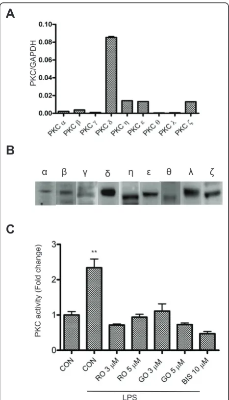
ALL PKC isoforms are present in microglia and activated by LPS
It has been reported that inhibitors of PKC can reduce iNOS induction in reactive microglia [28-30]. However, the specific PKC isoforms that are involved are not known. In order to identify the specific PKC isoforms that are required for iNOS production, we first exam-ined which PKC isoforms are expressed in BV-2 by quantitative real-time PCR. The results indicate that while mRNAs encoding all the PKC isoforms are
detect-able, there are significantly higher levels of nPKC (δ, h,
ε) expression compared to the conventional (a, b, g)
and the atypical (l/ι) isoforms (Figure 1A). Using
iso-form-specific antibodies, we found that each of the PKC isoforms is also expressed in BV-2 cells (Figure 1B). In contrast to a report by Kang and colleagues [31], but
consistent with results from Sun’s group [29], we
detected very low amounts of PKC a and b and very
high levels of PKC δ, suggesting that nPKC isoforms
may account for the major PKC activity in reactive microglia. In order to confirm PKC is activated in LPS-treated microglia, we measured PKC activity in murine BV-2 cells using ELISA. As shown in Figure 1C, PKC activity is elevated after treatment with LPS for 30 min, and suppressed by several PKC inhibitors, which include the pan-PKC inhibitor, Bis-1, the nPKC-selective inhibi-tor, rottlerin, and the cPKC-selective inhibiinhibi-tor, GO6976 [32,33]. These results demonstrate that both cPKC and nPKC might be functionally important in BV-2 cells when activated by LPS.
A
B
**
PKC acti
vi
ty
(Fol
d
change)
LPS
PKC/G
APDH
Į ȕ Ȗ į Ș İ ș Ȝ ȗ
C
Figure 1Expression and activation of various PKC isoforms in BV-2 cells.A, RNA samples isolated from the BV-2 cells were analyzed by quantitative real-time PCR, using primer pairs (see table 1) specific for mouse PKCa,b,g,δ,ε,θ,h,ζandl. GAPDH was used as an internal control. A representative experiment of three that were performed is shown.B, Twenty micrograms of whole cell lysate from BV-2 cells for the detection of each PKC isoform was subjected to western blots with antibodies against PKCa,b,g,δ,ε,θ,h,ζandl. A representative experiment of three that were performed is shown.C, BV-2 cells were treated with LPS (1μg/ml) in the absence or presence of PKC inhibitors for 30 min and then lysed for assaying the PKC activity. CON, RO, GO and BIS stand for control, rottlerin, GO6976 and
PKC inhibitors attenuate iNOS expression in reactive microglia
The discovery of relatively isozyme-specific PKC inhibi-tors has provided important information regarding the function of individual PKC isoforms. It has been
reported that rottlerin specifically inhibits PKCδ while
GO6976 mainly targets conventional PKC, and Bis-1 has inhibitory effects on all PKC isozymes [32,33]. To deter-mine whether iNOS induction is attributable to the acti-vation of PKC, BV-2 cells were treated with LPS in the presence of the aforementioned PKC inhibitors. At 6 hr following LPS treatment, cells were lysed and iNOS pro-duction was determined by western blot. All of the PKC inhibitors were able to suppress iNOS expression to dif-ferent degrees. However, rottlerin seems to have the greatest inhibitory effect (Figure 2A). In comparing
these, rottlerin at 5 μM near completely (94%) blocks
LPS-induced iNOS production, GO6976 at 5μM causes
60% inhibition and Bis-1 at 10 μM inhibits iNOS
pro-duction by 89% (Figure 2B). Consistently, NO produc-tion was also significantly attenuated when cells were treated with PKC inhibitors (Figure 2C). These results confirm that PKC activation is an integral component of LPS-induced iNOS expression and suggest that nPKC isoforms might play a prominent role in iNOS induction in BV-2 cells.
Activation of MAPK occurs downstream PKC, but upstream iNOS induction in reactive microglia
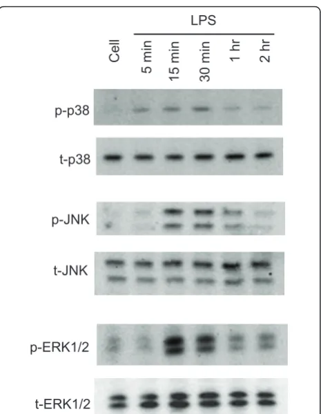
It is well known that MAPK cascades are involved in cytokine- and LPS-mediated iNOS induction in micro-glial cells [34]. However, the involvement of specific MAPKs varies in different cell types and in response to different stimuli. At various times after LPS treatment, all three MAPKs in BV-2 cells are transiently phos-phorylated. p38 phosphorylation occurs at 5 min, reaches maximum at 30 min, and nearly disappears at 1 hr following LPS treatment. The phosphorylation of JNK and ERK1/2 is present after 15 min of LPS treat-ment and remains at the same level until 30 min, fol-lowed by a dramatic reduction at 1 hr (Figure 3).
Using U0126, SB203580 and SP600125, inhibitors of ERK1/2, p38 and JNK, respectively [35], we found that iNOS induction and NO production in reactive micro-glia were significantly inhibited (Figure 4A-C). There was no change in cell viability at 24 hr following drug treatment (data not shown). To investigate the possible relationship between PKCs and MAPKs, we examined activation of MAPKs in the presence of PKC inhibitors. We found that MAPK phosphorylation at 15 min fol-lowing LPS treatment is attenuated by PKC inhibitors, indicating that activation of PKC occurs upstream of MAPKs. The nPKC selective inhibitor rottlerin attenu-ates ERK1/2 phosphorylation by 63%, but has no effect
A
RO
3
ȝ
M
ȕ-actin iNOS
CO
N
LPS
RO
5
ȝ
M
GO 5
ȝ
M
BI
S 10
ȝ
M
CO
N
B
*** *** **
***
C
re
lat
ive int
ens
ity
Nitrite c
onc
entrai
on
(% of
con
trol
)
LPS **
*** ***
on the phosphorylation of p38 and JNK (Figure 4D). GO6976, a cPKC selective inhibitor, not only attenuates the phosphorylation of ERK1/2 by 83%, but also sup-presses the phosphorylation of p38 and JNK by 60% and 47%, respectively (Figure 4D). The general PKC inhibi-tor, Bis-1, inhibits phosphorylation of ERK1/2 by 40% and JNK by 30%. Taken together, these results suggest that although all of the MAPKs are involved in induc-tion of iNOS in LPS-treated microglia, activainduc-tion of spe-cific PKC isoforms may lead to phosphorylation of distinct MAPKs.
Activation of NF-B contributes to PKC-mediated iNOS induction in reactive microglia
NF-B is one of the primary transcription factors that
regulates iNOS expression. The regulation of iNOS mediated by ERK1/2 and p38 MAPK has been shown to require NF-B activation in rat glial cells [34,36]. In this
study, we also investigated whether NF-B is involved in PKC-mediated iNOS production. CAY10470 is a recently developed NF-B inhibitor. It is synthesized from quinazoline derivative 6a, containing
4-phenoxy-phenethyl moiety at the C(4)-position with an IC50of 11
nM to inhibit NF-B activation in human Jurkat cells
[37]. CAY10470 significantly reduces iNOS production
(Figure 5A), implying the involvement of NF-B
activa-tion in iNOS producactiva-tion induced by LPS in BV-2 cells. To further examine the interaction of PKC activation
and NF-B during LPS treatment, we transfected BV-2
cells with an NF-B-responsive luciferase construct con-taining an NF-B response element and luciferase. This construct encodes the firefly luciferase reporter gene under the control of a minimal CMV promoter and tan-dem repeats of the NF-B transcriptional response ele-ment (TRE). The NF-B reporter (Luc) can easily and rapidly monitor NF-B activity in the cells. Our data demonstrate that luciferase activity induced by LPS is significantly inhibited in the presence of the PKC inhibi-tors, rottlerin, GO6976 and Bis-1 (Figure 5B). Similarly,
U0126 and SB203580 also significantly repress NF-B
activity (Figure 5B). Taken together, these results
indi-cate that NF-B acts downstream of PKC and MAPKs
to transcriptionally regulate iNOS production.
The differential role of PKC isoforms in LPS-induced iNOS production and MAPK activation in BV-2 cells
The above results suggest that LPS-induced iNOS pro-duction is mediated by PKC activation and MAPK phos-phorylation. However, because of the lack of specificity and the potential non-target effects of the pharmacologi-cal inhibitors, it is still unclear whether specific PKC isoforms mediate microglial activation by LPS. To test this, we employed RNAi technologies to transfect BV-2 cells with isoform-specific siRNAs to suppress the expression of various PKC isoforms. To test for trans-fection efficiency, we used siGLO RISC-free siRNA as a positive control. siGLO RISC-free siRNA is a stable, fluorescent, and non-targeting control siRNA with RISC-free modification. Following 48 hr of transfection, at least 90% of cells were transfected (Figure 6A). The transfection efficiency was further demonstrated by
downregulation of various PKC isoforms (PKCa,b, θ, δ,
ε and h) using PKC isoform-specific siRNAs by both
conventional and quantitative real-time PCR analysis (Figures 6B and 6C). qRT-PCR data indicated that speci-fic PKC siRNA downregulates relative PKC isoform mRNA level by 3-5 fold.
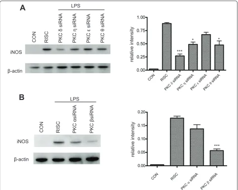
We then examined how downregulation of each speci-fic PKC isoform could affect iNOS induction in BV-2 cells. At 48 hr following PKC siRNA transfection, cells were treated with LPS for 6 hr and iNOS expression was assessed by western blot (Figure 7A, B). Among the p-p38
t-p38
LPS
Cel
l
5 m
in
15 m
in
30 m
in
1 hr 2 hr
p-JNK
t-JNK
p-ERK1/2
t-ERK1/2
nPKC isoforms, knockdown of PKCδ appears to have the greatest inhibitory effect on iNOS expression, with a
more-than-3-fold reduction observed. PKC h and θ
knockdown reduces iNOS by almost 2-fold, and
knock-down of PKCε shows little effect (Figure 7A).
Interest-ingly, downregulation of PKC b, but not PKC a,
significantly attenuates iNOS induction (Figure 7B), even though a very low mRNA expression of both cPKC isoforms is observed in BV-2 cells (Figure 1A, B). There
is a 3-fold reduction in iNOS expression in PKC b
siRNA-transfected cells when compared to RISC-free siRNA-transfected controls (Figure 7B). In summary, these data demonstrate that each PKC isoform has a dif-ferent potency in triggering iNOS induction in
LPS-activated microglia and that selective inhibition of PKC
δ or b may provide more focused anti-inflammatory
effects.
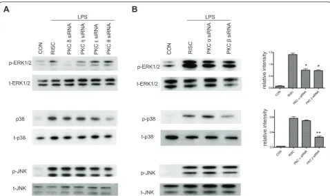
To further identify the specific MAPK pathway through which PKC regulates the expression of iNOS, we examined the effect of PKC siRNAs on phosphoryla-tion of various MAPKs. Similar to the results obtained using PKC inhibitors (Figure 4), downregulation of nPKCs produces various degrees of inhibition of the phosphorylation of ERK1/2 (Figure 8A). Knockdown of
PKC δ almost completely blocks ERK1/2 activation.
PKChsiRNA is shown to inhibit ERK1/2
phosphoryla-tion by 60%, but PKCε and θ siRNAs have no effect.
Interestingly, PKCθ siRNA causes a 75% reduction of
LPS ***
*** ***
B
A
CON U 5
ȝ
M
LPS
iNOS
ȕ-actin
CON
iNOS
ȕ-actin
CON SB 10
M
SP 10
M
LPS
CON
p38
t-p38
p-JNK
t- JNK p-ERK1/2
t-ERK1/2
LPS
CON RO 5
ȝ
M
GO 5
ȝ
M
BIS 10
ȝ
M
CON
Nitrite conce
nt
ratio
n
(% of
control)
C
*** **
*
relativ
e intensity
**
relative intensity
** *
relativ
e intensity
D
Figure 4Involvement of MAPK pathways in LPS-induced iNOS production in BV-2 cells.A-B, BV-2 cells were treated with LPS (1μg/ml) for 6 hr in the presence of SB205380 (SB), SP600125 (SP) or U0126 (U); inhibitors to block phosphorylation of p38, JNK (A) and ERK1/2 (B),
phosphorylation of p38 in LPS-treated microglia (Figure
8A), even though rottlerin doesn’t exhibit any inhibitory
effect (Figure 4C). Compared to the results obtained by using the cPKC inhibitor GO6976 (Figure 3C), we found
that PKC b, but not PKCa siRNA, efficiently blocks
phosphorylation of p38 by 65% based on densitometric analysis of the relative intensity of western blot bands
(Figure 8B). However, both PKCaandbsiRNAs display
nearly 50% inhibitory effects on ERK1/2 phosphorylation (Figure 8B). In addition, the isoform-specific PKC
siRNAs do not affect phosphorylation of JNK (Figure 8A, B), suggesting JNK activation is not involved in iNOS induction downstream of PKC activation. These results not only suggest that various PKC isoforms con-trol diverse downstream MAPKs pathways to affect LPS-induced iNOS production in murine microglia, but also further demonstrate that the commonly used PKC inhibitors are less selective and that the use of individual PKC siRNAs should be more suitable for elucidating sig-naling pathways mediated by the various PKCs.
Discussion
Overproduction of NO by enhanced iNOS induction has been tightly linked to neuroinflammatory and neurode-generative diseases [38-40]. A better understanding of the signaling mechanisms involved in the regulation of microglial iNOS has potential therapeutic implications. Previous studies mostly used PKC activators and inhibi-tors to determine the role of PKC in the regulation of iNOS production in murine microglia [26-30]. However, the absence of selectivity and the potential off-target effects of these pharmacological agents limit the ability to further define isoform-specific functions of the var-ious PKCs. In the present study, we have employed PKC isoform-specific siRNAs to delineate novel molecular signaling pathways linking PKC to iNOS induction in BV-2 cells when exposed to LPS.
Role of the PKC specific isoforms in LPS-induced iNOS production
The PKC family consists of at least 10 serine/threonine protein kinases originally characterized by their depen-dency on lipids for catalytic activity [41,42]. The
cPKCs require DAG and Ca2+, the nPKCs require
DAG but not Ca2+, while the aPKCs require neither.
The different modes of PKC regulation suggest that PKC isoforms may function differently in response to various stimuli. In BV-2 cells, pharmacological inhibi-tion studies suggest that the nPKC and cPKC isoforms are integral to LPS-induced increases in iNOS expres-sion and NO production (Figure 2), and
isoform-speci-fic siRNA knockdown confirms that PKC δand PKCb
are the major nPKC and cPKC isoforms involved in the regulation of LPS-induced iNOS production in murine microglia (Figure 7).
A number of studies have reported that particular PKC isoforms are involved in the production of NO in several different cell types [28,43-45]. Here we
demon-strate a principal role for PKC δ and PKC b in the
response to LPS exposure in murine BV-2 cells. These results are not only consistent with previous studies
showing that PKCδactivation is required for regulating
the production of iNOS in mouse peritoneal macro-phages [27], human leukemia cells [46] and BV-2 cells
A
B
iNOS
CO
N
LPS
10470
ȕ-actin
RLU (10
3)
CO
N
**
0 5 10 15 20
LPS
Figure 5Involvement of the NF-B pathway in LPS-activated BV-2 cells.A, BV-2 cells in 24-well plates were treated with LPS (1
[29], but also for the first time suggest that PKC b might play an important role in LPS-induced iNOS pro-duction in BV-2 cells even with its low levels of expres-sion. It might be concluded that the primary role of
PKCδ results from its high expression relative to other
PKC isoforms (Figures 1A and 1B). However, PKC b
expression is relatively low (Figure 1A) suggesting that induction of iNOS is dependent not only on levels of expression, but also on the activation of distinct PKC
isoforms. Interestingly, PKC a andε have been shown
to be the major PKC isoforms involved in the signaling
pathways by which IFNg induces iNOS expression in
the same cell line [28]. Collectively, these results suggest
that distinct PKC isoforms are activated and implicated in the regulation of iNOS induction in a stimulus-speci-fic manner.
Downstream components of PKC activation in LPS-induced iNOS expression
MAPKs. In the present study we also explored signaling pathways downstream of PKC that increase iNOS expression in response to LPS exposure. In general agreement with the observed effects of the three PKC inhibitors, rottlerin, GO6976, and Bis-1 (Figure 4D),
knockdown of PKC δ, h, a and b expression reduces
LPS-induced phosphorylation of ERK1/2 (Figures 8A
PKC
İ
/GAPDH
***
A
C
***
PKC
ȕ
/GAPDH
PKC
Į
/GAPDH
***
PKC
Ș
/GAPDH
***
PKC
ș
/GAPDH
***
PKC
į
/GAPDH
***
White field TRITC Merge
B
CON PKC
Į
siRNA
CON PKC
ȕ
siRNA
GAPDH
CON PKC
ș
siRNA
PKC
İ
siRNA
PKC
į
siRNA
PKC
Į
siRNA
PKC
ȕ
siRNA
PKC
Ș
siRNA
PKC
Ș
siRNA
CON PKC
į
siRNA
CON CON PKC CON
İ
siRNA
PKC
ș
siRNA
and 8B), whereas downregulation of PKCbsignificantly inhibits LPS-induced phosphorylation of p38 (Figure 8B). No effect on phosphorylation of JNK is observed with individual cPKC or nPKC siRNA (Figures 8A and 8B). Taken together, these results provide strong evi-dence that ERK1/2 and p38 are the main signaling path-ways through which distinct PKC isoforms regulate iNOS induction in response to LPS. Moreover, these results suggest that distinct MAPKs are activated by specific PKC isoforms.
It has been shown that both p38 and ERK1/2 can mediate iNOS expression in glial cells [36]. However, the phosphorylation of ERK1/2 has been found to be
involved in IFNg-, but not in LPS-induced NO
production, although NO production seems to be
coupled to PKC δ activation under both stimulations
[29]. The discrepancy between this report and our cur-rent study is unclear, but may be attributable to differ-ences in the stage of BV-2 cells used in these studies. The same group has recently found that paraquat
toxi-city to microglia is mediated by PKC δ- and
ERK1/2-dependent ROS generation [47]. The fact that neither nPKCs nor cPKCs affect JNK phosphorylation (Figure 6) suggests that JNK is not involved in the signaling path-way of iNOS induction coupling to PKC activation.
Interestingly, PKC θ siRNA significantly blocks p38
phosphorylation (Figure 8A), although the commonly used nPKC inhibitor rottlerin has no inhibitory effect
re
lat
ive int
en
sit
y
re
lat
ive int
ens
ity
iNOS
ȕ-actin
CO
N
PKC
ș
si
RNA
PKC
Ș
si
RNA
PKC
İ
si
RNA
PKC
į
si
RNA
RISC
LPS
A
B
***
* *
PKC
Į
si
RNA
PKC
ȕ
si
RNA
RISC
CO
N
ȕ-actin iNOS
LPS
***
(Figure 4D). Similarly, GO6976 blocks JNK activation (Figure 4D) but the same phenomenon is not observed with the use of cPKC siRNAs (Figure 8B). These results further suggest that it might be misleading to draw con-clusions on the role of specific PKC isoforms in the function of reactive microglia on the basis of pharmaco-logical inhibition.
NF-B. It is known that iNOS expression is
transcrip-tionally regulated. Activation of p38 has been shown to regulate NF-B, C/EBP, and ATF-2 to induce iNOS expression in rat astroglia [48]. However, HIV-1 Tat-induced iNOS expression in human astrocytes is depen-dent on phosphorylation of ERK1/2 and transcriptional activation of C/EBP, but not NF-B [49]. These studies indicate that different transcription factors can be recruited via one or more kinase pathways with respect to different inducers of iNOS. In this study, we find that
activation of NF-B is required for iNOS induction
through the application of CAY10470, an NF-B-specific
inhibitor (Figure 5). The observation that all of the PKC inhibitors - GO6976, rottlerin and Bis-1 - significantly block NF-B activation strongly supports the conclusion
that NF-B activation is required for iNOS induction in
LPS-treated BV-2 cells.
Conclusions
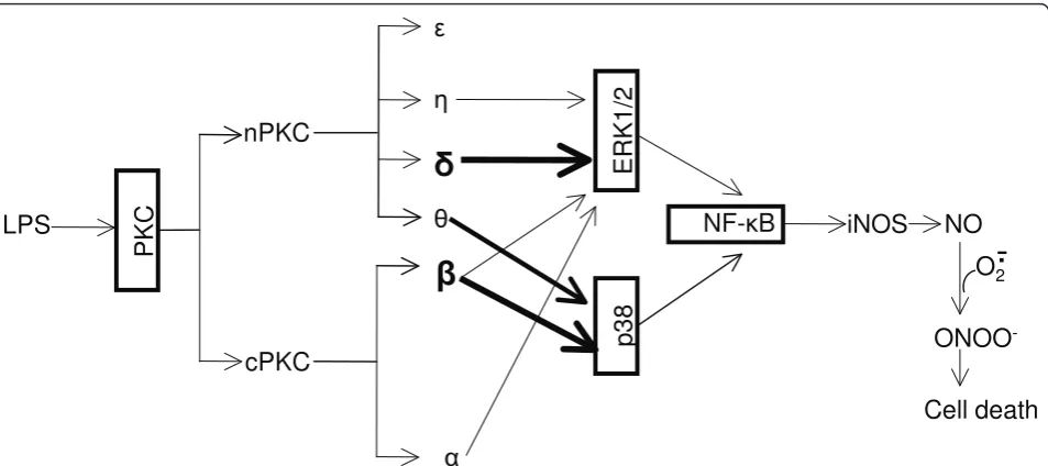
By using pharmacological inhibitors and RNA interfer-ence, we have clearly demonstrated that LPS-induced iNOS expression and NO production in BV-2 is mediated by a signaling pathway involving the sequential activation of PKC, MAPK and NF-B as illustrated in Figure 9. In addition to elucidating the critical role of
PKCδin ERK1/2 phosphorylation and iNOS induction,
our study reveals that PKCbis also a principal PKC
iso-form triggering iNOS induction in reactive microglia, which is coupled through phosphorylation of p38. The
partial inhibitory effects of PKC h and θ on iNOS
induction are due to their attenuation of the phosphory-lation of ERK1/2 and p38, respectively. These data sug-gest that a novel interaction between the distinct PKC isoforms and the various MAPKs promotes iNOS induc-tion. This interaction in different cell types may help to explain the discrepancy in the literature, and may also help guide the design of novel and selective PKC
relativ
e intensity
* *
relativ
e intensity
**
A
p-p38
t-p38
p-JNK
t-JNK p-ERK1/2
t-ERK1/2
PKC
Į
siRNA
PKC
ȕ
siRNA
RISC
CON
LPS
p38
t-p38 t-ERK1/2 p-ERK1/2
CON RISC
LPS
PKC
ș
siRNA
PKC
Ș
siRNA
PKC
İ
siRNA
PKC
į
siRNA
p-JNK
t-JNK
B
Figure 8Effects of downregulation of PKC isoforms on the phosphorylation of MAPK in LPS-treated BV-2 cells.A, At 48 hr following PKCδ,h,εorθsiRNA transfection, cells were treated with LPS (1μg/ml) for 15 min and then lysed for western blot using antibodies against phosphorylated and total ERK1/2, p38 and JNK. A representative experiment of 3 that were performed is shown.B, At 48 hr following PKCaor
inhibitors for the treatment of many inflammatory and neurological diseases in which overproduction of nitric oxide plays a pathogenic role.
Acknowledgements
The authors wish to thank Dr. Thomas Flagg for his careful reading and critical comments and suggestions on the manuscript. This work was supported by grants from the Blast Lethality Injury and Research Program (R600-070-00000-00-106109), the National Multiple Sclerosis Society (RG3741) and start-up fund from the Uniformed Services University (R070 UX).
Author details
1Department of Anatomy, Physiology and Genetics, Uniformed Services University of the Health Sciences, 4301 Jones Bridge Road, Bethesda, MD 20814, USA.2Program in Neuroscience, Uniformed Services University of the Health Sciences, 4301 Jones Bridge Road, Bethesda, MD 20814, USA.
Authors’contributions
JW designed and performed the experiments, analyzed the data and drafted the manuscript. RR performed some experiments and data analysis. YZ conceived of the study, participated in its design and coordination and helped to draft the manuscript. All authors read and approved the final version of the manuscript.
Competing interests
The authors declare that they have no competing interests.
Received: 24 October 2010 Accepted: 21 April 2011 Published: 21 April 2011
References
1. Banati RB,et al:Cytotoxicity of microglia.Glia1993,7(1):111-8. 2. Benveniste EN:Role of macrophages/microglia in multiple sclerosis and
experimental allergic encephalomyelitis.J Mol Med1997,75(3):165-73.
3. Kim SU, de Vellis J:Microglia in health and disease.J Neurosci Res2005,
81(3):302-13.
4. Ono K, Suzuki H, Sawada M:Delayed neural damage is induced by
iNOS-expressing microglia in a brain injury model.Neurosci Lett2010,
473(2):146-50.
5. Beckman JS, Koppenol WH:Nitric oxide, superoxide, and peroxynitrite:
the good, the bad, and ugly.Am J Physiol1996,271(5 Pt 1):C1424-37.
6. Wilms H,et al:Dimethylfumarate inhibits microglial and astrocytic inflammation by suppressing the synthesis of nitric oxide, IL-1beta,
TNF-alpha and IL-6 in an in-vitro model of brain inflammation.J
Neuroinflammation2010,7:30.
7. Block ML, Zecca L, Hong JS:Microglia-mediated neurotoxicity: uncovering
the molecular mechanisms.Nat Rev Neurosci2007,8(1):57-69.
8. Jaffrey SR, Snyder SH:Nitric oxide: a neural messenger.Annu Rev Cell Dev Biol1995,11:417-40.
9. Nathan C, Xie QW:Nitric oxide synthases: roles, tolls, and controls.Cell 1994,78(6):915-8.
10. Bogdan C:Nitric oxide and the immune response.Nat Immunol2001,
2(10):907-16.
11. Popp R, Fleming I, Busse R:Pulsatile stretch in coronary arteries elicits release of endothelium-derived hyperpolarizing factor: a modulator of
arterial compliance.Circ Res1998,82(6):696-703.
12. Ignarro LJ,et al:Nitric oxide as a signaling molecule in the vascular
system: an overview.J Cardiovasc Pharmacol1999,34(6):879-86.
13. Nathan C:Inducible nitric oxide synthase: what difference does it make?
J Clin Invest1997,100(10):2417-23.
14. Pahan K,et al:Lovastatin and phenylacetate inhibit the induction of nitric oxide synthase and cytokines in rat primary astrocytes, microglia,
and macrophages.J Clin Invest1997,100(11):2671-9.
15. Xie Z,et al:Peroxynitrite mediates neurotoxicity of amyloid
beta-peptide1-42- and lipopolysaccharide-activated microglia.J Neurosci2002,
22(9):3484-92.
16. Li J,et al:Peroxynitrite generated by inducible nitric oxide synthase and
NADPH oxidase mediates microglial toxicity to oligodendrocytes.Proc
Natl Acad Sci USA2005,102(28):9936-41.
17. Bolli R, Dawn B, Xuan YT:Emerging role of the JAK-STAT pathway as a
mechanism of protection against ischemia/reperfusion injury.J Mol Cell
Cardiol2001,33(11):1893-6.
18. Janssen-Heininger YM, Macara I, Mossman BT:Cooperativity between oxidants and tumor necrosis factor in the activation of nuclear factor (NF)-kappaB: requirement of Ras/mitogen-activated protein kinases in
ERK
1/2
p
38
cPKC
nPKC
į
Ș
ș
ȕ
Į
İ
iNOS
NF-țB
NO
PKC
O
2Ͳ
.
ONOO
-Cell death
LPS
the activation of NF-kappaB by oxidants.Am J Respir Cell Mol Biol1999,
20(5):942-52.
19. Mattson MP,et al:Roles of nuclear factor kappaB in neuronal survival
and plasticity.J Neurochem2000,74(2):443-56.
20. Saha RN, Pahan K:Regulation of inducible nitric oxide synthase gene in
glial cells.Antioxid Redox Signal2006,8(5-6):929-47.
21. Nishizuka Y:The heterogeneity and differential expression of multiple
species of the protein kinase C family.Biofactors1988,1(1):17-20.
22. Newton AC:Regulation of the ABC kinases by phosphorylation: protein
kinase C as a paradigm.Biochem J2003,370(Pt 2):361-71.
23. Dempsey EC,et al:Protein kinase C isozymes and the regulation of
diverse cell responses.Am J Physiol Lung Cell Mol Physiol2000,279(3):
L429-38.
24. Wadsworth SJ, Goldfine H:Mobilization of protein kinase C in macrophages induced by Listeria monocytogenes affects its
internalization and escape from the phagosome.Infect Immun2002,
70(8):4650-60.
25. Nakai M,et al:PKC and tyrosine kinase involvement in amyloid beta
(25-35)-induced chemotaxis of microglia.Neuroreport1998,9(15):3467-70.
26. Yoon HJ,et al:Phorbol ester synergistically increases interferon-gamma-induced nitric oxide synthesis in murine microglial cells.
Neuroimmunomodulation1994,1(6):377-82.
27. Bhatt KH,et al:Protein kinase Cdelta and protein tyrosine kinase regulate peptidoglycan-induced nuclear factor-kappaB activation and inducible nitric oxide synthase expression in mouse peritoneal macrophages in
vitro.Mol Immunol2010,47(4):861-70.
28. Kang J,et al:Identification of protein kinase C isoforms involved in interferon-gamma-induced expression of inducible nitric oxide synthase
in murine BV2 microglia.Neurosci Lett2001,299(3):205-8.
29. Shen S,et al:Distinct signaling pathways for induction of type II NOS by
IFNgamma and LPS in BV-2 microglial cells.Neurochem Int2005,
47(4):298-307.
30. Han IO,et al:Synergistic expression of inducible nitric oxide synthase by phorbol ester and interferon-gamma is mediated through NF-kappaB
and ERK in microglial cells.J Neurosci Res2003,73(5):659-69.
31. Kang J,et al:Reactive oxygen species mediate A beta(25-35)-induced
activation of BV-2 microglia.Neuroreport2001,12(7):1449-52.
32. Goekjian PG, Jirousek MR:Protein kinase C in the treatment of disease: signal transduction pathways, inhibitors, and agents in development.
Curr Med Chem1999,6(9):877-903.
33. Gschwendt M,et al:Rottlerin, a novel protein kinase inhibitor.Biochem
Biophys Res Commun1994,199(1):93-8.
34. Fiebich BL,et al:Inhibition of LPS-induced p42/44 MAP kinase activation
and iNOS/NO synthesis by parthenolide in rat primary microglial cells.J
Neuroimmunol2002,132(1-2):18-24.
35. Tanel A, Averill-Bates DA:P38 and ERK mitogen-activated protein kinases
mediate acrolein-induced apoptosis in Chinese hamster ovary cells.Cell
Signal2007,19(5):968-77.
36. Bhat NR,et al:Extracellular signal-regulated kinase and p38 subgroups of mitogen-activated protein kinases regulate inducible nitric oxide synthase and tumor necrosis factor-alpha gene expression in
endotoxin-stimulated primary glial cultures.J Neurosci1998,18(5):1633-41.
37. Tobe M,et al:A novel structural class of potent inhibitors of NF-kappa B activation: structure-activity relationships and biological effects of
6-aminoquinazoline derivatives.Bioorg Med Chem2003,11(18):3869-78.
38. Wada K,et al:Role of nitric oxide in traumatic brain injury in the rat.J
Neurosurg1998,89(5):807-18.
39. Endoh M, Maiese K, Wagner J:Expression of the inducible form of nitric oxide synthase by reactive astrocytes after transient global ischemia.
Brain Res1994,651(1-2):92-100.
40. Satake K,et al:Nitric oxide via macrophage iNOS induces apoptosis
following traumatic spinal cord injury.Brain Res Mol Brain Res2000,
85(1-2):114-22.
41. Newton AC:Protein kinase C: structural and spatial regulation by
phosphorylation, cofactors, and macromolecular interactions.Chem Rev
2001,101(8):2353-64.
42. Takai Y,et al:Studies on a cyclic nucleotide-independent protein kinase and its proenzyme in mammalian tissues. I Purification and
characterization of an active enzyme from bovine cerebellum.J Biol
Chem1977,252(21):7603-9.
43. Ginnan R,et al:PKC-delta mediates activation of ERK1/2 and induction of
iNOS by IL-1beta in vascular smooth muscle cells.Am J Physiol Cell
Physiol2006,290(6):C1583-91.
44. Pham TN,et al:Protein kinase C-eta (PKC-eta) is required for the development of inducible nitric oxide synthase (iNOS) positive
phenotype in human monocytic cells.Nitric Oxide2003,9(3):123-34.
45. Nagareddy PR,et al:Selective inhibition of protein kinase C beta(2) attenuates inducible nitric oxide synthase-mediated cardiovascular
abnormalities in streptozotocin-induced diabetic rats.Diabetes2009,
58(10):2355-64.
46. Deb DK,et al:Activation of protein kinase C delta by IFN-gamma.J
Immunol2003,171(1):267-73.
47. Miller RL, Sun GY, Sun AY:Cytotoxicity of paraquat in microglial cells:
Involvement of PKCdelta- and ERK1/2-dependent NADPH oxidase.Brain
Res2007,1167:129-39.
48. Bhat NR,et al:p38 MAPK-mediated transcriptional activation of inducible nitric-oxide synthase in glial cells. Roles of nuclear factors, nuclear factor kappa B, cAMP response element-binding protein,
CCAAT/enhancer-binding protein-beta, and activating transcription factor-2.J Biol Chem
2002,277(33):29584-92.
49. Liu X,et al:Human immunodeficiency virus type 1 (HIV-1) tat induces
nitric-oxide synthase in human astroglia.J Biol Chem2002,
277(42):39312-9.
doi:10.1186/1742-2094-8-38
Cite this article as:Wenet al.:Specific PKC isoforms regulate
LPS-stimulated iNOS induction in murine microglial cells.Journal of Neuroinflammation20118:38.
Submit your next manuscript to BioMed Central and take full advantage of:
• Convenient online submission
• Thorough peer review
• No space constraints or color figure charges
• Immediate publication on acceptance
• Inclusion in PubMed, CAS, Scopus and Google Scholar
• Research which is freely available for redistribution