Detection and Differentiation of Different
Monocyte Subsets: Optimalization of a
Multiparameter Flowcytometric Assay
Imke Demers
Maastricht University
Absract
preparation and acquisition has to take place within 4 hours after venipuncture. the 3G8 clone is preferred for CD16 labeling and the HLA-DR based gating method is assumed to be the most accurate in identifying the monocyte subsets. Using above mentioned recommendations, the test shows a good reproducibility.
Keywords
Monocyte subsets, flowcytometry, optimalization, sample stability, antibody clone, reproducibility, gating strategy
Introduction
Monocytes constitute 5-10% of peripheral leukocytes, where they circulate for several days in the blood stream and can migrate into tissues to contribute to the macrophage pool. Monocytes and their progeny play important roles in the defense against pathogens, homeostasis and tissue repair, especially in the innate immune system (1).
Monocytes are mononuclear and characterized by a kidney-shaped nucleus. As the main component of the innate immune system, they are responsible for the clearance of bacterial, viral and fungal infections, mainly by the process of phagocytosis (2). Monocytes were considered to be a homogeneous cell population, but in the late 1980s, the report of Passlick and his colleagues (3) identified two phenotypically distinct monocyte subtypes based on differential expression of CD14 and CD16. CD14 is a cell surface receptor involved in the binding of lipopolysaccharide (LPS) and therefore in LPS signaling (4)Innate</ keyword><keyword>Lipopolysaccharides/*chemistry/immunology/pharmacology</ keyword><keyword>Molecular Sequence Data</keyword><keyword>Toll-Like Receptor 4/*chemistry/immunology/metabolism</keyword></keywords><dates><year>2013</ year></dates><isbn>2092-6413 (Electronic. CD16 refers to the low affinity Fc receptor Fcγ-RIII. This receptor binds to the Fc-part of IgG antibodies and thereby activates natural killer (NK) cells resulting in cytotoxicity (5). Over time, it became clear that also the CD16+ monocytes do not represent a homogenous population, but they could bedivided into two distinct groups. In 2010, the Nomenclature Committee of the International Union of Immunological Societies recommended an unified nomenclature of the different monocyte subsets. According to this, monocytes are currently divided into CD14++/CD16- classical monocytes, CD14++/CD16+ intermediate monocytes and CD14+/CD16++ non-classical monocytes (6).
pathogenesis of many diseases (1). Therefore, there has been much interest in investigating CD16+ monocytes in disease states. However, NK cells as well as neutrophils share the expression of CD16 with the CD16+ monocytes, which can confuse the flowcytometric analysis. Three different gating strategies are commonly used to identify the monocyte fraction: back-gating, negative and positive gating strategies. The back-gating method is the easiest and cheapest way to select all monocytes. A major drawback of this method is the unwanted admixture with certain lymphocytes and neutrophils (7). Negative or ‘exclusion’ gating strategies are used to purify the monocyte population utilizing monoclonal antibodies, for example against CD66b or CD15, CD56 and CD19, in order to exclude neutrophils, NK cells and B-cells respectively. Positive or ‘inclusion’ gating strategies make use of antibodies to CD45, CD115, CD14, CD16 and HLA-DR to select monocytes positively (1).
Another challenge in flowcytometry is the proper gating of the intermediate and the non-classical monocytes. The fractions of these subtypes could be very small, so that a small variation in the gating technique results in a large and hardly reproducible coefficient of variation (CV). A possible solution for this problem is a strong and distinguishable staining expression for the CD16 epitope. In earlier studies of out group, the clones NPK15 and B73.1 were used for immunolabeling of CD16, which both show an intermediate staining level. Most published research in this field, however, made use of the clone 3G8 in the flowcytometric assay. This 3G8 clone is described to have a high staining level, so a better distinction could be made between the intermediate and non-classical monocytes.
Next to the above described major challenges in the flowcytometric determination of the different monocyte subsets in peripheral blood, there are also some pre-analytical and analytical aspects unknown which can influence the outcome of the test. One important aspect is the stability of the blood sample, referring to the time from blood drawn till the start of the preparation of the sample. Another aspect is the reproducibility as well as the intra –and inter assay variations.
sample age will nog influence the flowcytometric results (8, 9). The use of the 3G8 clone against CD16 is expected to give a higher staining level with respect to the B73.1 clone and therefore a better distinction between intermediate and non-classical monocytes will be possible. Both the positive and negating gating strategies are expected to be more accurate compared to the CD14 back-gating method.
Materials and methods
Patients and Samples
Patients form the outdoor clinic were asked to participate in the study by having an EDTA anti-coagulated blood tube drawn. Only patients who gave infomred consent were included in the study. A complete blood count (CBC) was performed for all samples using the XE5000 (Sysmex).
For the purpose of examining the stability of the blood sample, EDTA anti-coagulated blood samples from 20 different patients were used. The analysis was started immediately after venipuncture and after 1h, 3h, 4h and 24h. To assess the influence of type of antibody clone against CD16, EDTA blood samples from a second group of 20 patients were used. From each patient, a sample was incubated with the B73.1 clone while simultaneously another sample was incubated with the 3G8 clone. This test was started within 10 minutes after venipuncture. In order to investigate the reproducibility of the test, another set of blood samples from 10 patients was used. The intra-assay variability was tested by repeated measurements of the same sample. The inter-assay variability was asses by preparing two different samples of the same patient, followed by measurement on one single flowcytometer.
Labeling Procedure
One hundred µl of EDTA-whole blood was transferred into a 5 ml polystyrene round-bottom tube (BD Biosciences) and incubated for 15 minutes in the dark at RT, simultaneously with conjugated antibodies against CD64 (FITC; clone 10.1; BD Biosciences, San Jose, USA), CD11b (PE, clone D12; BD), HLA-DR (PerCP-Cy5.5; clone L243; BD), CD16 (PE-Cy7; clone B73.1; BD), CD14 (APC; clone MΦP9; BD) and CD45 (APC-H7; clone 2D1, BD). For the examination of the influence of antibody clone against CD16, the 3G8 clone (PE-Cy7; BD) was used, next to the B73.1 clone. Erythrocytes were lysed (FACS lysing solution, #349202, BD Biosciences, diluted 1:10 with water) for 10 minutes in the dark at RT. The tube was centrifuged for 4 min at 300G, the supernatant was discarded and the cells were resuspended in 2 ml CellWash (BD).This wash and centrifugation procedure was repeated and the sample was analyzed using flow cytometry (BD FACSCanto flow cytometer; BD Biosciences) and FACSdiva software version 6.1.3 (BD Biosciences).
Statistical Analysis
For the analysis of the sample stability, the coefficient of variation (CV) was calculated of all time points with respect to the 0h sample. The CV of the flow cytometry test must be smaller than 20%. Significance was determined with a one-way ANOVA test. Linear regression analysis and a t-test were used to determine significant differences between the CD16 clones and gating strategies. For the reliability of the repeated measures, CV values and inter –and intra-class variation coefficients (ICC) were calculated. Single and average ICCs were determined, where >0.8 denotes excellent agreement, 0.6-0.8 denotes good agreement, 0.4-0.6 denotes fair agreement and <0.4 denotes poor agreement of the results across repeated measures. Data analysis was performed in Microsoft Excel (Microsoft Office), Prism 5.0 (Graphpad) and SPSS Statistics 17.0.0. Statistical significance was defined at the 95% level.
Results
Post-analytic Variable: Gating Strategy
This difference, however, is not significant. The means of the total monocyte population, the classical monocytes and the non-classical monocytes were neither significant different between the CD24 CD56 gating and CD14 back-gating method. When comparing CD24 CD56 based gating method with the HLA-DR based gating method, an increase in total monocytes as well as in intermediate monocytes is observed using the CD24 CD56 based method. These differences are however not significant. The fraction of classical monocytes is not significant decreased in this method.
Pre-analytic Variable: stability of the Test
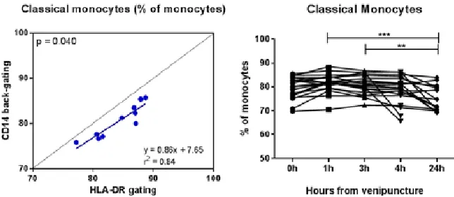
The stability of the blood sample as examined using 20 different patient blood samples. For the analysis, the HLA-DR based gating method is used to specify the different monocyte subsets. A significant decrease of classical monocytes was found after 24 hours, compared to 1h and 3h, with a p-value of 0.0006 and 0.0021 respectively (Figure 1, right). Using the same results of the stability test, the coefficients of variation (CV) values were calculated for all monocyte groups at time-points 1h, 3h, 4h and 24h, related to the 0h sample. The medians of all samples at the different time-points are within the 20% range. However, a large variation is observed, especially within the intermediate monocyte population.
Figure 1. Left: Linear regression analysis of the HLA-DR based gating method and the CD14 back-gating method for the fraction of classical monocytes. Right: stability of the classical monocyte population at different time-points.
Analytic Variable: CD16 Immunolabeling
classical and non-classical monocytes show a good identity between the two clones, whereas the intermediate monocyte fraction is slightly higher when using the B73.1 clone against CD16.
Analytic Variable: Reproducibility
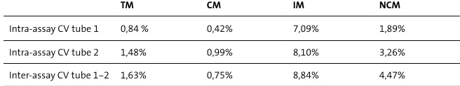
To determine the intra-assay variation of two measurements performed of the same flowcytometric tube, the CV values were calculated. All intra-assay CVs are beneath the margin of 20% with respect to the mean of both measurements (Table 1). The CVs belonging to the intermediate monocyte population show higher values compared to the other groups, but still below the 20% cut-off value. To determine the inter-assay variation, CV values were calculated of the measurements of two independently prepared flowcytometric tubes. The CVs for all monocyte groups are beneath the margin of 20% and the inter-assay CV of the intermediate monocyte population is again higher compared to other monocyte groups. Next to the CVs, the intra –and inter-assay correlation coefficients (ICC) were determined for all groups of monocytes. All ICC values are above 0.8 and therefore all denote excellent agreement across the repeated measures.
Table 1. Intra-assay CVs (%) of tube 1 and 2 and the inter-assay CV between tube 1 and 2 for the total monocyte (TM) population, classical monocytes (CM), intermediate monocytes (IM), and non-classical monocytes (NCM).
TM CM IM NCM
Intra-assay CV tube 1 0,84 % 0,42% 7,09% 1,89%
Intra-assay CV tube 2 1,48% 0,99% 8,10% 3,26%
Inter-assay CV tube 1-2 1,63% 0,75% 8,84% 4,47%
Discussion
Post-analytic Variable: Gating Strategy
In previous studies of our group (7) a back-gating method was used. A drawback of this method is the unwanted admixture of lymphocytes and neutrophils, which also express CD16. In order to identify the monocyte population and subsets more accurately, three different gating strategies were tested in this study. When comparing the CD14 back-gating method with the HLA-DR based back-gating method, the total monocyte population shows a decrease in the HLA-DR based gating method. The expected explanation for this is that HLA-DR negative cells, such as lymphocytes and neutrophils are excluded. The classical monocytes show a significant increase using the HLA-DR based gating method. A reason for this could be that not all classical monocytes are selected using the CD14 back-gating method. When creating the back-gate around the monocyte population, there may be some difficulties in distinguishing monocytes at the top of the monocyte population from granulocytes at the bottom of the granulocyte cloud.
The comparison between the CD14 back-gating method with the CD24 CD56 based gating method also shows a decrease of the classical monocyte population in the CD14 back-gate method. The fraction of intermediate monocytes is increased in the CD24 CD56 based gating method, which may be explained by the fact that this method assigns to many intermediate monocytes. The same increase in intermediate monocytes is observed in the CD24 CD56 gating method compared to the HLA-DR gating, which makes it more likely that this gating method may be less accurate in the determination of intermediate monocytes. Also in comparing these two gating methods, a decrease in total monocytes is observed when using the HLA-DR gating method, which may refer to the better purification of the monocyte population.
Pre-analytic Variable: Stability of the test
intermediate monocytes. This can be explained by the fact that it is a small cell population within the total monocyte population, and next to this, there is hardly consensus on how to gate and define this subpopulation (12).
Analytic Variable: CD16 Immunolabeling
The comparison of the B73.1 clone with the 3G8 clone used for CD16 labeling does not show prominent differences according to all monocyte groups. Although not significant, a somewhat higher percentage of total monocytes was observed when using the 3G8 clone, which may be the result of the higher staining level of the 3G8 clone. When staining the cell population with the 3G8 clone, granulocytes appeared to be more CD16 positive when compared to the B73.1 clone. This finding is already confirmed by Heimbeck et al., who described the strong neutrophil staining capacity of the 3G8 clone (13). With the appliance of the HLA-DR based gating method, this extremely CD16 positive granulocytes are much easier to identify in a HLA-DR/CD16 dot plot in comparison with the same dot plot with the B73.1 clone. Therefore, immunolabeling with the 3G8 antibody clone against CD16 results in a better distinction between granulocytes and monocytes when using the HLA-DR gating method. Some difficulties may arise in the determination of intermediate monocytes when using the 3G8 clone. Where granulocytes become more CD16 positive, non-classical and intermediate monocytes become less CD16 positive, which results in a down-shift the characteristic monocyte arc. This makes it harder to discriminate between the individual monocyte subsets, with especially the border between the classical and intermediate monocytes hard to establish.
Analytic Variable: Reproducibility
The CV values of the reproducibility experiment, as well as the CV values of the stability experiment demonstrate that the relative size of the intermediate monocytes shows a larger dispersion than the total monocyte population and the other subsets. This larger spread is probably due to the small population size of the intermediate monocytes, as described before. Overall, the CV values of the intra-assay as well as the inter-assay variability show good results with all CV values lower <10%. The ICC values of both variability tests are all above 0.8 and therefore denote excellent agreement.
previous results concerning the size of the different populations. Another limitation is the small size of the experimental groups used in this study. The use of more participants for each experiment will probably result in more significant and reliable finding.
From this study, it can be concluded that sample preparation and acquisition has to take place within 4 hours after venipuncture in order to make reliable determinations of the monocyte subsets. Next to this, the 3G8 clone is preferred over the B73.1 clone for CD16-epitope labeling and the HLA-DR based gating strategy is assumed to be the most accurate to identify the monocyte subsets. A good reproducibility was found, with all CV values <10%, when making use of the parameters described above.
Role of the student
Imke Demers was an undergraduate student biomedical sciences, with a specialization in molecular life sciences, working under the supervision of M.P.G Leers when the research in this report was performed. The topic was proposed by the supervisor. The processing and analysis of the blood samples, as well as all data analysis and interpretation were done by the student. The writing of the report was also done by the student.
Referencens
1. Abeles RD, McPhail MJ, Sowter D, Antoniades CG, Vergis N, Vijay GK, et al. CD14, CD16 and HLA-DR reliably identifies human monocytes and their subsets in the context of pathologically reduced HLA-DR expression by CD14(hi) /CD16(neg) monocytes: Expansion of CD14(hi) /CD16(pos) and contraction of CD14(lo) /CD16(pos) monocytes in acute liver failure. Cytometry A. 2012;81(10):823-34.
2. Ghattas A, Griffiths HR, Devitt A, Lip GY, Shantsila E. Monocytes in coronary artery disease and atherosclerosis: where are we now? J Am Coll Cardiol. 2013;62(17):1541-51.
3. Passlick B, Flieger D, Ziegler-Heitbrock HW. Identification and characterization of a novel monocyte subpopulation in human peripheral blood. Blood. 1989;74(7):2527-34.
4. Park BS, Lee JO. Recognition of lipopolysaccharide pattern by TLR4 complexes. Exp Mol Med. 2013;45:e66. 5. Janeway C. Appendix II. CD antigens. Immunobiology. 2001;5 ed.
6. Idzkowska E, Eljaszewicz A, Miklasz P, Musial WJ, Tycinska AM, Moniuszko M. The role of different monocyte subsets in the pathogenesis of atherosclerosis and acute coronary syndromes. Scand J Immunol. 2015. 7. Leers MP, Keuren JF, Frissen ME, Huts M, Kragten JA, Jie KS. The pro- and anticoagulant role of blood-borne
phagocytes in patients with acute coronary syndrome. Thromb Haemost. 2013;110(1):101-9.
8. Hübl W, Ziegler-Heitbrock LHW, Bayer PM. A Simple Method for Measurement of CD14weakCD16-strongMonocytes in Peripheral Blood. Immunobiology. 2000;202(1):2-10.
10. Hristov M, Schmitz S, Schuhmann C, Leyendecker T, von Hundelshausen P, Krotz F, et al. An optimized flow cytometry protocol for analysis of angiogenic monocytes and endothelial progenitor cells in peripheral blood. Cytometry A. 2009;75(10):848-53.
11. Selimoglu-Buet D, Wagner-Ballon O, Saada V, Bardet V, Itzykson R, Bencheikh L, et al. Characteristic repartition of monocyte subsets as a diagnostic signature of chronic myelomonocytic leukemia. Blood. 2015;125(23):3618-26.
12. Zawada AM, Fell LH, Untersteller K, Seiler S, Rogacev KS, Fliser D, et al. Comparison of two different strategies for human monocyte subsets gating within the large-scale prospective CARE FOR HOMe Study. Cytometry A. 2015.