O R I G I N A L R E S E A R C H
Open Access
Potential usefulness of
68
Ga-citrate PET/CT
in detecting infected lower limb prostheses
Jing-Ren Tseng
1,2, Yu-Han Chang
3, Lan-Yan Yang
4, Chen-Te Wu
5, Szu-Yuan Chen
3, Chih-Hsing Wan
6,
Ing-Tsung Hsiao
1,2*and Tzu-Chen Yen
1,2*Abstract
Background:Prosthetic joint infections may lead to failures of total joint arthroplasty. Radionuclide imaging can play a diagnostic role in identifying such infections, which require two-stage exchange arthroplasty (instead of simple revision surgery performed in non-infected cases). Although18F-FDG PET/CT has emerged as a novel diagnostic tool in this setting, the clinical usefulness of68Ga-citrate PET/CT has not been previously investigated. This single-center prospective study was designed to address this issue.
Methods:Between January 2016 and October 2017, we examined 34 patients with clinically proven or suspected prosthetic hip/knee joint infections scheduled to undergo surgery. All patients underwent68Ga-citrate PET/CT scans and sequential18F-FDG PET/CT imaging for comparative purposes. Intraoperative findings and the results of microbiological analyses of surgical specimens served as gold standard. The diagnostic results were examined according to (1) image interpretation based on radiotracer uptake patterns and (2) quantitative analysis using volumes of interest (VOIs) to calculate standard uptake values (SUVs) and metabolic volumes (MVs).
Results:A total of 26 (76%) patients were diagnosed as having infections. Based on radiotracer uptake pattern criteria, the sensitivity, specificity, and accuracy of68Ga-citrate PET/CT and18F-FDG PET/CT were 92%, 88%, and 91% and 100%, 38%, and 85%, respectively. MV was significantly higher in the infected group when68Ga-citrate PET/CT was used (422.45 vs. 303.65 cm3,p= 0.027), whereas no significant differences were observed on18F-FDG PET/CT. According to receiver operating characteristic (ROC) curve analysis, a cut-off value of 370.86 for MV resulted in a sensitivity of 61.5% and a specificity of 87.5% (area under curve: 0.75, 95% confidence interval: 0.57–0.88,p= 0.035). Conclusions:Subject to future confirmation, our data provide preliminary evidence that68Ga-citrate PET/CT may have a complimentary role to18F-FDG PET/CT in detecting prosthetic joint infections, being characterized by a higher specificity and the possibility to discriminate between an infectious condition and sterile inflammation.
Trial registration:This prospective study was registered at clinicaltrials.gov (registration number:NCT02855190).
Keywords:68Ga-citrate,18F-FDG, PET/CT, Prosthetic joint infections
Background
Total joint arthroplasty is one of most frequently per-formed and successful surgical procedures in orthope-dics [1]. However, between 0.4 and 4% of all joint replacement procedures ultimately develop infectious complications, with a 2–18% of cases showing aseptic loos-ening [2]. Numerous conventional nuclear medicine
techniques have been investigated as imaging tools to discriminate between an infectious condition and sterile in-flammation—including technetium-99m methylene dipho-sphonate (MDP) bone scintigraphy (BS), gallium (Ga)-67 citrate scans, and indium-111 or technetium-99m hexam-ethylpropylene amine oxime (HMPAO)-labeled white blood cell (WBC) scans [3]. However, each of these tech-niques has significant limitations, including (1) the lack of specificity typical of BS and Ga-67 citrate scans, (2) the suboptimal imaging characteristics of all conventional two-dimensional examinations, (3) the potentially hazard-ous preparation of radiopharmaceuticals required by WBC * Correspondence:[email protected];[email protected]
1Department of Nuclear Medicine and Center for Advanced Molecular Imaging and Translation, Chang Gung Memorial Hospital at Linkou, No. 5, Fu-Hsing ST., Kwei-Shan, Taoyuan, Taiwan
Full list of author information is available at the end of the article
scans, (4) the prolonged physical and biological half-life of indium-111- or Ga-67-labeled agents (resulting in a high absorbed radiation dose), and (5) the need of combining different traditional scanning techniques to obtain reliable diagnostic results. All of these caveats ultimately prevent their routine clinical use [4–7].
18
F-fluoro-2-deoxyglucose positron emission tomog-raphy/computed tomography (18F-FDG PET/CT) is in-creasingly emerging as a useful diagnostic tool for several infectious and inflammatory conditions— espe-cially in patients with chronic renal failure or in those who had undergone metal prosthesis implantation [8,
9]. Previous studies have shown that18F-FDG PET im-aging has a pooled sensitivity of 82.1–82.8% and a spe-cificity of 86.6–87.3% for the diagnosis of lower limb prosthetic joint infections [10, 11]. In this setting,
18
F-FDG PET imaging may also offer significant advan-tages over conventional nuclear medicine examinations (which not only are more complex and expensive but also potentially limited by safety issues) [12]. 68Gallium (68Ga) is a positron-emitting isotope that has been previ-ously used for PET imaging. Intriguingly, preliminary data on the potential usefulness of68Ga-citrate PET for identi-fying patients with bone infections have been promising [13]. In a head-to-head comparison,18F-FDG PET/CT has been superior to 68Ga-citrate PET/CT for diagnosing in-flammatory reactions elicited by metal debris [14]. How-ever, the question as to whether 68Ga-citrate PET/CT is either superior or may offer complimentary information to18F-FDG PET/CT in detecting lower limb prosthesis in-fections remains open. This prospective study was de-signed to address this research question.
Methods Ethics statement
This study had a prospective design. The protocol com-plied with the tenets of the Declaration of Helsinki, was approved by the Institutional Review Board of the Chang Gung Memorial Hospital (CGMH) at Linkou (approval number: 103-7266A), and was registered at
clinicaltrials.gov (NCT02855190). All patients gave their written informed consent. All data were securely protected (by delinking personal information from the main data sets), made available to investigators only, and analyzed anonymously.
68
Ga-citrate synthesis
68
Ga was obtained using a 68Ge/68Ga generator (ITG Isotope Technologies Garching GmbH, Garching, Germany) which was eluted with 2.5 ml of 0.05 mol/l hydrogen chloride (HCl) solution into a 20-ml sterile vial—which contained 2.5 ml of sterile sodium citrate (27 mg/ml) solution, and 5 ml of sterile injection water. The 68Ga sodium citrate solution was subsequently
transferred into the product vial through a sterile filter. The radiochemical purity of 68Ga-citrate was analyzed with an instant thin layer chromatography-silica-gel technique using methanol/acetic acid (9:1) as the mo-bile phase. The product pH was tested using indicator strips (pH range, 4.0–10.0), whereas the integrity of sterile filters was assessed with a bubble point test. The
68
Ga-citrate used in this study was characterized by (1) high radiochemical purity (≥97%), (2) a pH of 4.5–8.0, and (3) a strength≥11.1 MBq/ml.
Patient selection
Between January 2016 and October 2017, a total of 39 patients were deemed eligible for the study. In-clusion criteria were as follows: (1) clinically proven or suspected periprosthetic hip/knee joint infections and (2) scheduled surgery. Patients were excluded if they met one of the following criteria: (1) pregnancy or breastfeeding, (2) significantly abnormal labora-tory findings, and (3) critical illnesses or unstable vital signs that made the patient unsuitable for im-aging and surgical work-ups. Four patients were ex-cluded during the screening period, whereas one additional patient who completed the requested im-aging studies did not ultimately undergo surgery. After these exclusions, 34 patients were retained in the analysis. All of the study participants were followed up for at least 6 months.
PET/CT acquisition and processing
All of the patients underwent sequential 68Ga-citrate PET/CT and 18F-FDG PET/CT imaging performed within less than 1 week of each other. The order of the two scans was not predetermined. The injection dose was 111–185 MBq for 68Ga-citrate and 5.18 MBq per kilogram of body weight for 18F-FDG. Patients were required to fast for at least 4 h before injection of both radiotracers. Images were obtained 60 min after intravenous administration of each tracer via a Biograph mCT PET/CT system (Siemens Healthineers, Erlangen, Germany). Patients were scanned from the iliac crest to the toe. CT scanning parameters for 68Ga-citrate and 18F-FDG examina-tions were 120 kV; 0.5 s per rotation; collimation, 40 × 0.6; pitch, 1.5; quality reference mAs with CAR-EDose 4D, 100 mAs; and slice thickness, 5 mm. PET acquisition time per bed position was 3 min for
68
PET imaging interpretation by radiotracer uptake pattern All of the PET images were CT attenuation-corrected. The criterion for defining an examination as positive was the presence of an abnormal radiotracer uptake lo-cated at the bone-prosthesis interface and/or peripros-thetic soft tissue [10]. However, tracer uptake limited to soft tissues adjacent to the neck of the hip prosthesis was not considered sufficient to classify a scan as posi-tive [15, 16]. Two observers (J.R.T. and T.C.Y.)—who were blinded to clinical data—jointly interpreted all im-ages (including companion CT findings) and reached a diagnostic consensus through discussion. According to the definitions proposed by the Musculoskeletal Infec-tion Society [17], the final diagnosis was based on intra-operative findings and microbiological evaluations of surgical specimens.
PET imaging quantitative analysis
For each 18F-FDG and 68Ga-citrate image slide, a vol-ume of interest (VOI) was manually drawn by an expe-rienced nuclear medicine physician (J.R.T.) around the bone-prosthesis interface or periprosthetic soft tissue. To this aim, the PMOD 3.3 software package (PMOD Technologies Ltd., Zurich, Switzerland) was used. The standardized uptake value (SUV) was calculated as fol-lows: SUV = (tissue radioactivity [Bq/ml])/(injected radioactivity [Bq]/body weight [g]). Maximum SUV
(SUVmax), mean SUV (SUVmean), and metabolic volume
(MV) were subsequently calculated. The MV was defined as the number of voxel located inside the VOIs multiplied by voxel size. In this study, VOIs covered the entire bone-prosthesis interface as well as periprosthetic soft tissue (after the exclusion of vascular radioactivity). The isocontour was set at 20% of the maximum uptake observed within the corresponding focus.
Statistical analysis
Based on an expected prevalence of confirmed infection of 75%, we initially planned to enroll 40 patients (resulting in a total of 30 patients with confirmed infec-tion). However, recruitment was stopped at 39 cases be-cause of a higher than expected incidence of cases with confirmed infections. The general characteristics of the study participants were summarized using descriptive statistics. Continuous data were expressed as medians and ranges and compared with the Mann-Whitney U
test. Categorical variables were summarized as fre-quency counts and percentages. A 2 × 2 contingency table with four diagnostic outcomes (true positive, false positive, true negative, and false negative) was con-structed based on the final diagnostic results. Receiver operating characteristic (ROC) curve analysis was ap-plied to investigate the prediction accuracy. Optimal cut-off points that maximized prediction were
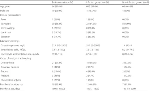
Table 1General characteristics of the study patients (n= 34)
Entire cohort (n= 34) Infected group (n= 26) Non-infected group (n= 8)
Age, years 64 (31–86) 68.5 (31–86) 66 (44–67)
Male sex 19 (55.9%) 15 (57.7%) 4 (50%)
Clinical presentations
Fever 1 (2.9%) 1 (3.8%) 0 (0%)
Joint pain 30 (88.2%) 22 (84.6%) 8 (100%)
Joint swelling 8 (23.5%) 8 (30.8%) 0 (0%)
Local heat 5 (14.7%) 5 (19.2%) 0 (0%)
Secretion 5 (14.7%) 5 (19.2%) 0 (0%)
Laboratory findings
C-reactive protein, mg/L 21.7 (0.2–250.9) 35.7 (2–250.9) 1.4 (0.2–3)
White blood cells, 103/μL 7.4 (1.8–18.0) 7.6 (1.8–18.0) 6.2 (4.4–9.1)
Erythrocyte sedimentation rate, mm/h 39 (2–116) 67 (2–116) 8 (18.5–36)
Cause of total joint arthroplasty
Osteoarthritis 21 (61.8%) 18 (69.2%) 3 (37.5%)
Avascular necrosis 3 (8.8%) 2 (7.7%) 1 (12.5%)
Trauma 6 (17.6%) 4 (15.4%) 2 (25%)
Fracture 3 (8.8%) 2 (7.7%) 1 (12.5%)
Rheumatoid arthritis 1 (2.9%) 1 (3.8%) 0 (0%)
Prosthesis location, hip 19 (55.9%) 12 (46.2%) 7 (87.5%)
Prosthesis age, days 180 (7–6000) 180 (7–1800) 135 (58–6000)
identified using the Youden’s index. All calculations were performed using the SPSS software (version 22.0; IBM, Armonk, NY, USA). A two-tailed p value < 0.05 was considered statistically significant.
Results
Table1 shows the baseline characteristics of the study participants. Nineteen (56%) patients were male, and
the median age was 64 years (range 31–86 years). Hypertension and diabetes were the most frequent comorbidities, accounting for 14 (41%) and 8 (23%) cases, respectively. Osteoarthritis was the most com-mon underlying condition that required total joint arthroplasty. A total of 19 (56%) patients received hip arthroplasty, whereas 8 (23%) cases underwent contralateral prosthesis replacement as well. Five
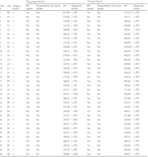
Table 2Diagnostic characteristics of68Ga-citrate PET/CT and18F-FDG PET/CT imaging in detecting infected lower limb prostheses
68Ga-citrate PET/CT 18F-FDG PET/CT
Case #
Site Surgical results
BPI uptake
Periprosthetic soft tissue uptake
MV Diagnostic results
BPI uptake
Periprosthetic soft tissue uptake
MV Diagnostic results
1 RH + Yes Yes 817.00 + (TP) Yes Yes 1212.13 + (TP)
2 LH + No Yes 576.90 + (TP) No Yes 781.12 + (TP)
3 RK + Yes No 310.80 + (TP) Yes No 560.66 + (TP)
4 LH − No No 147.18 −(TN) No Yes 469.88 + (FP)
5 LH − No No 366.66 −(TN) No No 402.01 −(TN)
6 LK + Yes No 584.26 + (TP) Yes Yes 955.45 + (TP)
7 RK + Yes Yes 473.44 + (TP) Yes Yes 666.63 + (TP)
8 LH + Yes Yes 377.26 + (TP) No Yes 626.08 + (TP)
9 RK + Yes Yes 595.68 + (TP) Yes Yes 1078.56 + (TP)
10 LK − No No 338.31 −(TN) Yes Yes 434.44 + (FP)
11 LH + Yes No 579.84 + (TP) Yes No 643.07 + (TP)
12 LH − No No 327.98 −(TN) No No 865.39 −(TN)
13 LH − Yes Yes 424.18 + (FP) Yes Yes 584.04 + (FP)
14 LH + Yes No 504.58 + (TP) Yes Yes 873.08 + (TP)
15 LK + Yes Yes 358.36 + (TP) Yes Yes 364.30 + (TP)
16 RH − No No 173.43 −(TN) Yes No 278.18 + (FP)
17 RH + Yes Yes 348.87 + (TP) Yes Yes 481.64 + (TP)
18 LK + Yes Yes 376.59 + (TP) Yes Yes 592.65 + (TP)
19 LH + Yes Yes 477.37 + (TP) Yes Yes 717.30 + (TP)
20 LH − No No 291.57 −(TN) No No 496.38 −(TN)
21 RH + Yes Yes 386.32 + (TP) Yes Yes 611.08 + (TP)
22 RK + Yes Yes 375.07 + (TP) Yes Yes 542.73 + (TP)
23 RK + Yes Yes 441.48 + (TP) Yes Yes 514.63 + (TP)
24 RH − No No 359.94 −(TN) Yes Yes 406.57 + (FP)
25 RH + Yes Yes 273.37 + (TP) Yes Yes 351.80 + (TP)
26 LH + No No 247.07 −(FN) No Yes 259.58 + (TP)
27 LK + Yes Yes 265.47 + (TP) Yes Yes 325.06 + (TP)
28 RH + Yes Yes 494.83 + (TP) Yes Yes 533.03 + (TP)
29 RK + Yes Yes 363.75 + (TP) Yes Yes 546.30 + (TP)
30 LK + Yes No 309.42 + (TP) Yes Yes 387.73 + (TP)
31 LK + Yes Yes 266.16 + (TP) Yes Yes 389.33 + (TP)
32 RK + Yes Yes 492.82 + (TP) Yes Yes 656.04 + (TP)
33 LK + Yes Yes 297.40 + (TP) Yes Yes 342.49 + (TP)
34 RK + No No 389.66 −(FN) Yes Yes 548.37 + (TP)
(15%) patients required a second debridement of the infected tissue during the follow-up period (median interval 60 days, range 20–120 days). The median interval between the last PET/CT scan and the collec-tion of surgical specimens was 9 days (range 1– 26 days). A total of 19 (57.6%) patients had a positive bacterial culture (Staphylococcus aureus, n= 6;
Staphylococcus epidermidis, n= 4; and Staphylococcus caprae, n= 2). Pseudomonas aeruginosa, Cellulosimi-crobium cellulans, Enterococcus faecalis, Streptococ-cus dysgalactiae, andStaphylococcus lugdunensiswere isolated only in one case each. Two patients were positive for two or more bacteria. Specifically, one was positive for both Staphylococcus aureus and
Staphylococcus lugdunensis, whereas the other was positive for four bacteria (Staphylococcus aureus, group B streptococcus, Enterococcus faecalis, and
Gemella morbillorum).
Table 2 depicts the diagnostic characteristics of
68
Ga-citrate PET/CT and 18F-FDG PET/CT scans, which are provided in Figs.1,2,3, and4. A total of 34 prostheses were examined in the study (19 hip pros-theses and 15 knee prospros-theses). Infections were diag-nosed in 26 (76%) patients. According to the radiotracer uptake pattern criteria, the number of true positive, true negative, false positive, false negative pa-tients was 24, 7, 1, 2 for68Ga-citrate PET/CT and 26, 3,
5, 0 for 18F-FDG PET/CT, respectively. There were five false positive cases on18F-FDG PET/CT, four of which were considered as true negative on 68Ga-citrate PET/ CT (cases # 4, 10, 16, and 24). The sensitivity, specifi-city, and accuracy of68Ga-citrate PET/CT and 18F-FDG PET/CT scans were 92%, 88%, and 91% and 100%, 38%, and 85%, respectively (Table3).
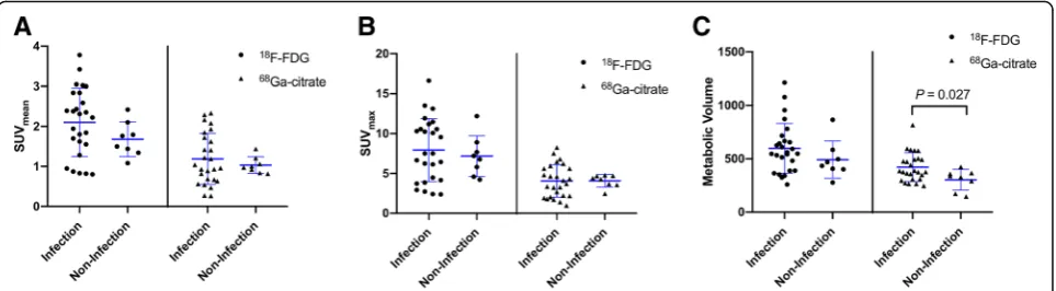
The median (range) SUVmean, SUVmax, and MV
values for the prostheses under examination were 0.99 (0.26–2.33), 4.20 (0.97–8.25), and 370.86 (147.18– 817.00) on68Ga-citrate PET/CT scans, respectively, and 1.89 (0.80–3.78), 7.36 (2.38–16.61), and 544.51 (259.58– 1212.13) on 18F-FDG PET/CT scans, respectively. The SUVmean, SUVmax, and MV values observed on 18F-FDG
PET/CT images were all significantly higher than those measured on 68Ga-citrate PET/CT scans (p< 0.001). In
68
Ga-citrate PET/CT scans, the MV of infected prostheses was significantly higher than that observed for non-infected prostheses (mean ± SD: 422.45 ± 133.87 vs. 303.65 ± 96.39, respectively,p= 0.027). However, all of the other measured values did not differ significantly between infected and non-infected prostheses on either 68 Ga-ci-trate PET/CT or18F-FDG PET/CT scans (Fig.5). Accord-ing to receiver operatAccord-ing characteristic (ROC) curve analysis, a cut-off value of 370.86 for MV resulted in a sensitivity of 61.5% and a specificity of 87.5% (area under curve 0.75, 95% confidence interval 0.57–0.88,p= 0.035).
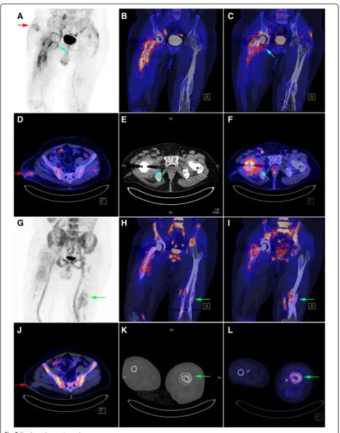
Fig. 1Patient (case # 16) with a history of right total hip arthroplasty performed 16 years before imaging. The results of18F-FDG PET/CT (upper
row) and68Ga-citrate PET/CT (lower row) are presented.a–dGreen arrows indicate an intense18F-FDG uptake surrounding the cup and the
proximal part of the hip prosthesis stem components. On CT images, osteolytic changes and the presence of residual cement in the right acetabulum were evident. Cup loosening with synovial hypertrophy and clear joint fluid were detected during surgery.18F-FDG PET/CT findings
were therefore classified as false positve.d–fFindings on68Ga-citrate PET/CT were true negative, without any obvious radiotracer accumulation at
Discussion
Data on the potential usefulness of 68Ga-citrate PET/ CT imaging in detecting infected lower limb prostheses remain limited. Although 18F-FDG PET has been shown to be highly sensitive in this clinical setting, re-sults on its specificity are less conclusive [18–20]. In the current prospective study, 68Ga-citrate PET/CT ap-peared superior to 18F-FDG PET/CT with respect to specificity (88% vs. 38%, respectively), being also suc-cessful in distinguishing between an infectious condi-tion and sterile inflammacondi-tion (Fig.1).
Non-specific 18F-FDG uptake can be persistently de-tected for several years following an arthroplasty [16]. In a previous experimental study, an increased uptake of both 18F-FDG and 68Ga was identified in infected bones; however, only 18F-FDG accumulation occurred in healing bones free from infections [21]. Owing to
different properties and uptake mechanisms for the two radiotracers, we hypothesized that 68Ga may be super-ior to 18F-FDG in distinguishing between infectious conditions and non-specific sterile inflammation.
Several mechanisms may potentially explain 68 Ga-cit-rate accumulation in infectious foci, including (1) higher Ga extravasation to the extracellular compartment as a result of an increased inflammation-related capillary per-meability, (2) binding of dissociated gallium ions to transferring followed by their subsequent sequestration in a highly stable state within an infectious site, (3) bind-ing to bacterial siderophores and activated lactoferrin in neutrophils, and (4) macrophage phagocytosis [22–24]. In contrast, 18F-FDG allows the identification of inflam-matory cells characterized by an increased glycolysis— ultimately serving as an “in vivo label” for activated in-flammatory cells at sites of infection/inflammation [25].
(See figure on previous page.)
Fig. 2Patient (case # 1) with a history of right hip prosthesis implantation performed 7 months before imaging. The results of18F-FDG PET/CT (upper two rows) and68Ga-citrate PET/CT (lower two rows) are presented.a–fThe red arrows clearly indicate18F-FDG uptake occurring at the sinus tract, which was absent on the corresponding68Ga-citrate PET/CT image. There was also evidence of swollen periarticular soft tissue (blue arrows) at the right ischiofemoral space on CT, which was accompanied by an increased18F-FDG uptake. A similar pattern of radiotracer uptake (b,h) was found to occur at the bone-prosthesis interface of the stem component, as well as in the adjacent soft tissue. The imaging results were considered positive, and the case was subsequently classified as true positive.g–lThe green arrows indicate an increased68Ga-citrate uptake occurring at the left femoral shaft. The CT scan revealed cortical hypertrophy caused by a prior traumatic fracture followed by bone reunion
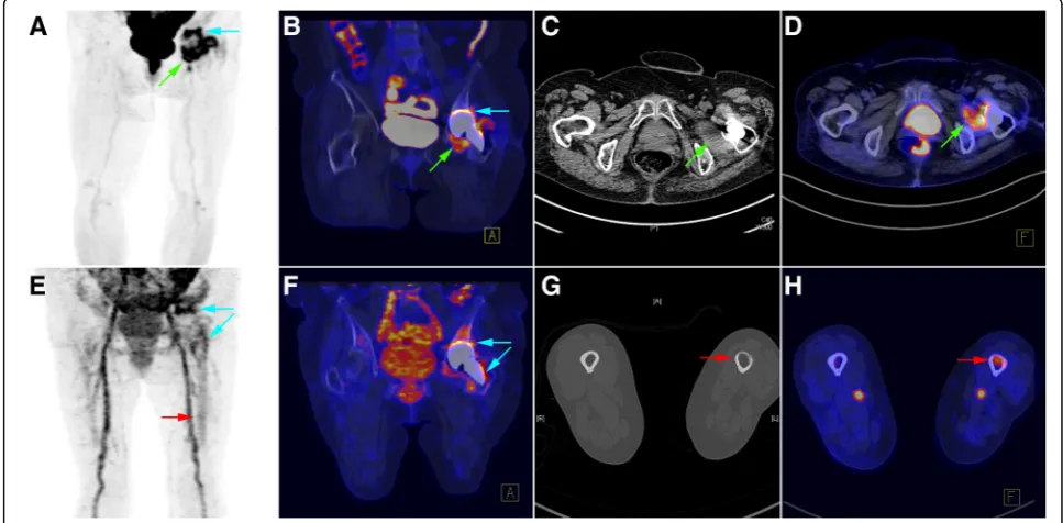
Fig. 3Patient (case # 14) with a history of hip prosthesis implantation performed 8 months before imaging. The results of18F-FDG PET/CT (upper
row) and68Ga-citrate PET/CT (lower row) are presented.a–dThe green arrows indicate an increased18F-FDG uptake occurring in necrotic tissue
located within a swollen left quadratus femoris muscle. There was also evidence of an increased18F-FDG uptake occurring at the bone-prosthesis
interface (cup component; blue arrow). Taken together, these findings were suggestive of an infectious process which was confirmed intraoperatively (presence of cloudy synovial fluid and necrotic tissue).e–hThe blue arrows indicate an increased68Ga-citrate uptake occurring at the bone-prosthesis
In a previous small cohort study that focused on
68
Ga-citrate and18F-FDG PET/CT in the detection of in-fectious foci [26], the use of18F-FDG resulted in a higher target-to-background ratio and higher signals at soft tissue infectious sites. Similar results were observed in the head-to-head comparisons performed in the current study. The SUVmax, SUVmean, and MV values were all
significantly higher for 18F-FDG PET/CT, which was also superior in diagnosing infections of the sinus tract or cold abscesses. In contrast, 68Ga-citrate PET/CT was more effective in identifying the bone remodeling
process related to fractures, bone unions, or osteolysis (Figs.2and3).
It is noteworthy that published studies have used dif-ferent criteria for positive imaging findings, as well as various reference standards [10]. In the current investi-gation, radiotracer uptake at the bone-prosthesis inter-face and/or periprosthetic soft tissue was the main criterion for positive results. We cannot exclude that this definition might have contributed to the high sen-sitivity and low specificity of 18F-FDG PET/CT ob-served in the current study. It should be also kept in mind that adverse reactions to metal debris can result in an increased 18F-FDG uptake that is indistinguish-able from that occurring at foci of infection [14]. In a previous study that used similar criteria for positive findings, the sensitivity and specificity for infected hip arthroplasties were 100% and 44.8%, respectively [15]. Although the interpretation criteria were identical, the specificity of68Ga-citrate PET/CT was markedly higher than that of 18F-FDG PET/CT (88% vs. 38%, respect-ively). Notably, 20 (58.8%) patients were identified based on the presence of 68Ga-citrate uptake in peri-prosthetic soft tissue. We therefore hypothesize that the high specificity of 68Ga-citrate PET/CT compared with 18F-FDG PET/CT is not directly linked to a soft tissue uptake underestimation (which should result in a lower likelihood of false positive results) but should ra-ther be ascribed to the intrinsic characteristics of
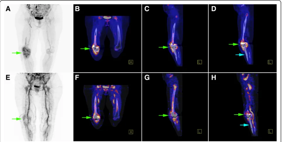
Fig. 4Patient (case # 3) with a history of knee prosthesis implantation performed 12 months before imaging. The results of18F-FDG PET/CT
(upper row) and68Ga-citrate PET/CT (lower row) are presented.a
–dThe green arrows indicate an increased18F-FDG uptake at both the bone
prosthesis interface and the knee synovium.e–hA similar pattern of68Ga-citrate uptake was evident. The imaging results were considered positive,
and the case was subsequently confirmed as true positive.d,hIn contrast to the femoral portion, there was no increased radiotracer uptake along the bone prosthesis interface of the elongated tibial stem component (indicating no loosening or infection of a specific prosthesis segment, blue arrows)
Table 3Diagnostic performances of68Ga-citrate PET/CT and 18
F-FDG PET/CT imaging in detecting infected prostheses according to the anatomical site
Sensitivity (%) Specificity (%) Accuracy (%)
Entire cohort (n= 34)
18F-FDG PET/CT 100 (26/26)a 38 (3/8)a 85
68Ga-citrate PET/CT 92 (24/26)a 88 (7/8)a 91
Hip prosthesis (n= 19)
18F-FDG PET/CT 100 (12/12)a 43 (3/7)a 79
68
Ga-citrate PET/CT 92 (11/12)a 86 (6/7)a 89
Knee prosthesis (n= 15) 18
F-FDG PET/CT 100 (14/14)a 0 (0/1)a 93 68
Ga-citrate PET/CT 93 (13/14)a 100 (1/1)a 93
a
68
Ga-citrate. Previous studies have shown that 18F-FDG PET may be less accurate in detecting knee prosthesis infections [10,27]. The question as to whether68 Ga-ci-trate PET/CT imaging can be sufficiently accurate even when the analysis is restricted to knee prostheses alone deserves further scrutiny.
Several authors have maintained that the key to PET/ CT diagnosis of prosthesis infections is mainly related to a specific uptake pattern (i.e., radiotracer localization in the bone-prosthesis interface) rather than to the in-tensity of uptake per se [11,15]. Our current data lend further support to this prevalent view. Accordingly, SUVmeanand SUVmax values did not differ significantly
between the infected and non-infected groups on both
18
F-FDG and 68Ga-citrate PET/CT imaging. However, we found significant differences in terms of MV on
68
Ga-citrate PET/CT imaging between infected and non-infected prostheses (422.45 vs. 303.65, respectively,
p= 0.027). The optimal cut-off value of 370.86 resulted in a sensitivity of 61.5% and specificity of 87.5%. Not-ably, only two non-infected cases were correctly identi-fiable beyond the MV range that overlapped with infected cases (Fig. 5). Further studies with larger sam-ple sizes are required to confirm the clinical usefulness of MV quantification on 68Ga-citrate PET/CT scans and to further investigate the optimal cut-off value.
Several limitations of our study merit comment. First, our results might not be generalizable owing to the lim-ited sample size and the single-center nature of the study. Second, we did not specifically assess the impact of attenuation-corrected, non-attenuation-corrected, or other different imaging reconstruction methods, which may pose some issues in standalone PET scanners. Third, this study was not designed to evaluate the dif-ferences between different interpretation criteria pro-posed in the literature. Finally, the specificity of
18
F-FDG PET/CT observed in our study was lower than that previously reported. Consequently, future studies should aim at validating the current data using
consistent interpretation criteria and combined PET/ CT scanners. Because diagnosis of prosthetic joint in-fections remains particularly challenging in the early postoperative period [20], we hypothesize that68 Ga-ci-trate PET/CT could serve as a complimentary diagnos-tic tool especially in patients with recently implanted prostheses.
Conclusions
Subject to future confirmation, our data provide pre-liminary evidence that68Ga-citrate PET/CT may have a complimentary role to 18F-FDG PET/CT in detecting prosthetic joint infections, being characterized by a higher specificity and the possibility to discriminate be-tween an infectious condition and sterile inflammation.
Acknowledgements
This study was financially supported by grants (CRRPG3E0031 and CRRPG3E0032) from the Chang Gung Memorial Hospital. The authors acknowledge the statistical assistance provided by the Clinical Trial Center, Chang Gung Memorial Hospital, Linkou, Taiwan (funded by the Ministry of Health and Welfare of Taiwan; grant MOHW107-TDU-B-212-123005).
Funding
This study was financially supported by grants (CRRPG3E0031 and CRRPG3E0032) from the Chang Gung Memorial Hospital (CGMH).
Availability of data and materials
This study was based on datasets included in the published article.
Authors’contributions
JRT, YHC, and TCY participated in the study design and drafted the manuscript; LYY was in charge of data analysis; CTW, SYC, and CHW were involved in data collection; ITH contributed to image analysis. All authors have read and approved the final version of the manuscript.
Ethics approval and consent to participate
This prospective study was performed in accordance with the 1964 Declaration of Helsinki and was approved by the Institutional Review Board of the Chang Gung Memorial Hospital (CGMH) at Linkou (approval number: 103-7266A). The study protocol was approved by the Institutional Review Board of the Chang Gung Memorial Hospital (CGMH) at Linkou.
Consent for publication
Written informed consent was obtained for all participants.
Fig. 5Scatter diagrams foraSUVmean,bSUVmax, andcmetabolic volume. Dots and triangles represent18F-FDG PET/CT and68Ga-citrate PET/CT
Competing interests
The authors declare that they have no competing interests.
Publisher’s Note
Springer Nature remains neutral with regard to jurisdictional claims in published maps and institutional affiliations.
Author details
1Department of Nuclear Medicine and Center for Advanced Molecular Imaging and Translation, Chang Gung Memorial Hospital at Linkou, No. 5, Fu-Hsing ST., Kwei-Shan, Taoyuan, Taiwan.2Department of Medical Imaging and Radiological Science and Healthy Aging Center, College of Medicine, Chang Gung University, Taoyuan, Taiwan.3Bone and Joint Research Center and Department of Orthopaedic Surgery, Chang Gung Memorial Hospital at Linkou, Taoyuan, Taiwan.4Biostatistics Unit, Clinical Trial Center, Chang Gung Memorial Hospital at Linkou, Taoyuan, Taiwan.5Department of Medical Imaging and Intervention, Chang Gung Memorial Hospital at Linkou, Taoyuan, Taiwan.6Department of Nuclear Medicine, Mackay Memorial Hospital, Taipei, Taiwan.
Received: 14 November 2018 Accepted: 12 December 2018
References
1. Ethgen O, Bruyere O, Richy F, et al. Health-related quality of life in total hip and total knee arthroplasty. A qualitative and systematic review of the literature. J Bone Joint Surg Am. 2004;86-A:963–74.
2. Phillips JE, Crane TP, Noy M, et al. The incidence of deep prosthetic infections in a specialist orthopaedic hospital: a 15-year prospective survey. J Bone Joint Surg Br. 2006;88:943–8.
3. Rennen HJ, Boerman OC, Oyen WJ, et al. Imaging infection/inflammation in the new millennium. Eur J Nucl Med. 2001;28:241–52.
4. King AD, Peters AM, Stuttle AW, et al. Imaging of bone infection with labelled white blood cells: role of contemporaneous bone marrow imaging. Eur J Nucl Med. 1990;17:148–51.
5. Kraemer WJ, Saplys R, Waddell JP, et al. Bone scan, gallium scan, and hip aspiration in the diagnosis of infected total hip arthroplasty. J Arthroplast. 1993;8:611–6.
6. Scher DM, Pak K, Lonner JH, et al. The predictive value of indium-111 leukocyte scans in the diagnosis of infected total hip, knee, or resection arthroplasties. J Arthroplast. 2000;15:295–300.
7. Larikka MJ, Ahonen AK, Junila JA, et al. Extended combined 99mTc-white blood cell and bone imaging improves the diagnostic accuracy in the detection of hip replacement infections. Eur J Nucl Med. 2001;28:288–93. 8. Haroon A, Zumla A, Bomanji J. Role of fluorine 18 fluorodeoxyglucose positron
emission tomography-computed tomography in focal and generalized infectious and inflammatory disorders. Clin Infect Dis. 2012;54:1333–41. 9. Tseng JR, Lin CW, Chen SH, et al. Clinical usefulness of (1)(8)F-FDG PET/CT
for the detection of infections of unknown origin in patients undergoing maintenance hemodialysis. J Nucl Med. 2015;56:681–7.
10. Kwee TC, Kwee RM, Alavi A. FDG-PET for diagnosing prosthetic joint infection: systematic review and metaanalysis. Eur J Nucl Med Mol Imaging. 2008;35:2122–32.
11. Zoccali C, Teori G, Salducca N. The role of FDG-PET in distinguishing between septic and aseptic loosening in hip prosthesis: a review of literature. Int Orthop. 2009;33:1–5.
12. Basu S, Kwee TC, Saboury B, et al. FDG PET for diagnosing infection in hip and knee prostheses: prospective study in 221 prostheses and subgroup comparison with combined (111)in-labeled leukocyte/(99m)Tc-sulfur colloid bone marrow imaging in 88 prostheses. Clin Nucl Med. 2014;39:609–15. 13. Nanni C, Errani C, Boriani L, et al. 68Ga-citrate PET/CT for evaluating patients
with infections of the bone: preliminary results. J Nucl Med. 2010;51:1932–6. 14. Aro E, Seppanen M, Makela KT, et al. PET/CT to detect adverse reactions to
metal debris in patients with metal-on-metal hip arthroplasty: an exploratory prospective study. Clin Physiol Funct Imaging. 2018;38:847–55. 15. Chacko TK, Zhuang H, Stevenson K, et al. The importance of the location of
fluorodeoxyglucose uptake in periprosthetic infection in painful hip prostheses. Nucl Med Commun. 2002;23:851–5.
16. Zhuang H, Chacko TK, Hickeson M, et al. Persistent non-specific FDG uptake on PET imaging following hip arthroplasty. Eur J Nucl Med Mol Imaging. 2002;29:1328–33.
17. Parvizi J, Zmistowski B, Berbari EF, et al. New definition for periprosthetic joint infection: from the workgroup of the musculoskeletal infection society. Clin Orthop Relat Res. 2011;469:2992–4.
18. Love C, Marwin SE, Tomas MB, et al. Diagnosing infection in the failed joint replacement: a comparison of coincidence detection 18F-FDG and 111In-labeled leukocyte/99mTc-sulfur colloid marrow imaging. J Nucl Med. 2004; 45:1864–71.
19. Love C, Marwin SE, Palestro CJ. Nuclear medicine and the infected joint replacement. Semin Nucl Med. 2009;39:66–78.
20. Palestro CJ, Love C. Role of nuclear medicine for diagnosing infection of recently implanted lower extremity arthroplasties. Semin Nucl Med. 2017;47:630–8. 21. Makinen TJ, Lankinen P, Poyhonen T, et al. Comparison of 18F-FDG and
68Ga PET imaging in the assessment of experimental osteomyelitis due to Staphylococcus aureus. Eur J Nucl Med Mol Imaging. 2005;32:1259–68. 22. Tsan MF. Mechanism of gallium-67 accumulation in inflammatory lesions. J
Nucl Med. 1985;26:88–92.
23. Roivainen A, Jalkanen S, Nanni C. Gallium-labelled peptides for imaging of inflammation. Eur J Nucl Med Mol Imaging. 2012;39(Suppl 1):S68–77. 24. Glaudemans AW, Prandini N, DIG M, et al. Hybrid imaging of
musculoskeletal infections. Q J Nucl Med Mol Imaging. 2018;62:3–13. 25. Jamar F, Buscombe J, Chiti A, et al. EANM/SNMMI guideline for 18F-FDG use
in inflammation and infection. J Nucl Med. 2013;54:647–58.
26. Salomaki SP, Kemppainen J, Hohenthal U, et al. Head-to-head comparison of (68)Ga-citrate and (18)F-FDG PET/CT for detection of infectious foci in patients with Staphylococcus aureus Bacteraemia. Contrast Media Mol Imaging. 2017;2017:3179607.