O R I G I N A L R E S E A R C H
Open Access
Improved quantitation and reproducibility
in multi-PET/CT lung studies by combining CT
information
Beverley F. Holman
1*, Vesna Cuplov
1, Lynn Millner
1, Raymond Endozo
1, Toby M. Maher
2,3,
Ashley M. Groves
1, Brian F. Hutton
1,4and Kris Thielemans
1*Correspondence: [email protected] 1Institute of Nuclear Medicine, University College London, UCLH (T-5), Euston Road, London, NW1 2BU UK
Full list of author information is available at the end of the article
Abstract
Background: Matched attenuation maps are vital for obtaining accurate and reproducible kinetic and static parameter estimates from PET data. With increased interest in PET/CT imaging of diffuse lung diseases for assessing disease progression and treatment effectiveness, understanding the extent of the effect of respiratory motion and establishing methods for correction are becoming more important. In a previous study, we have shown that using the wrong attenuation map leads to large errors due to density mismatches in the lung, especially in dynamic PET scans. Here, we extend this work to the case where the study is sub-divided into several scans, e.g. for patient comfort, each with its own CT (cine-CT and ‘snap shot’ CT). A method to combine multi-CT information into acombined-CThas then been developed, which averages the CT information from each study section to produce composite CT images with the lung density more representative of that in the PET data. This combined-CT was applied to nine patients with idiopathic pulmonary fibrosis, imaged with dynamic18F-FDG PET/CT to determine the improvement in the precision of the parameter estimates.
Results: Using XCAT simulations, errors in the influx rate constant were found to be as high as 60% in multi-PET/CT studies. Analysis of patient data identified displacements between study sections in the time activity curves, which led to an average standard error in the estimates of the influx rate constant of 53% with conventional methods. This reduced to within 5% after use of combined-CTs for attenuation correction of the study sections.
Conclusions: Use of combined-CTs to reconstruct the sections of a multi-PET/CT study, as opposed to using the individually acquired CTs at each study stage, produces more precise parameter estimates and may improve discrimination between diseased and normal lung.
Keywords: Lung, Density, PET/CT, Respiration, Attenuation correction, Quantitation, Dynamic PET
Background
PET/CT is becoming more popular for use in diffuse lung diseases such as chronic obstructive pulmonary disease (COPD), interstitial lung disease (ILD), infection and inflammation [1]. These studies commonly address disease progression and treatment effectiveness and make use of quantitative dynamic as well as static imaging [2–6].
Quantitative PET images can only be obtained with accurate attenuation correction (AC) maps. These AC maps are commonly derived from short CT acquisitions (i.e. a standard helical CT for AC), which are appropriate in regions where internal anatomical motion is unlikely [7,8]. However, in regions such as the thorax, these CT techniques only produce a ‘snap shot’ of the respiratory cycle and therefore determining an AC map matched to the PET data is difficult [9].
Respiratory AC map mismatches are due to two main contributions; motion, which leads to anatomical location mismatch, and density variations due to lung fractional air volume changes over the course of the breathing cycle [10], which could lead to attenu-ation coefficient mismatch. Mismatches due to motion have been widely explored in the literature [11,12] and although density variations due to the respiratory phase at the time of acquisition have long been acknowledged by the CT community [13,14], the effect on PET quantitation in the lung has only very recently been acknowledged [15].
In a previous publication [15], the effect of density variations between PET and CT on PET quantitation was explored. In this paper, we expand this work to multi-PET/CT (MPC) protocols. We define MPC protocols as those in which a dynamic PET/CT study is broken into two or more sections, allowing the patients to have short breaks for their comfort so as to improve patient compliance. Each section consists of a CT and a PET acquisition. The aim of the individual CTs is to allow for attenuation correction of the cor-responding PET data, while enabling accurate CT-based registration between the study sections. However, the CTs at the start of each study section may capture different phases of the breathing cycle, leading to variations in the degree of mismatch and resulting PET quantitation at each section. In turn, this will lead to errors in the extracted kinetic parameters.
This section-specific mismatch is likely to be most problematic if ‘snap-shot’ CTs are used. If an average of the cine-CTs can be used for attenuation correction [16] for all study sections, it is expected that these will better match the PET acquisitions, but differences can still occur due to changing breathing patterns. As a result, variation in quantification bias across the study sections is not necessarily rectified by using the higher dose cine-CT acquisitions.
In this paper, the errors in estimating kinetic parameters from ungated dynamic non time-of-flight (non-TOF) PET data associated with MPC protocols using ‘snap shot’ CTs are assessed using XCAT phantom simulations. A method is then suggested to reduce the errors in MPC studies (based on either ‘snap shot’ or averaged cine-CT) by combining all the available CT information. This combined-CT method is then tested on non-TOF18 F-FDG data from patients who have undergone MPC studies with a mixture of cine-CT and ‘snap shot’ CTs, to determine the improvement in the PET quantitation.
Methods
XCAT simulations
Initially, the possible errors in PET quantification as a result of using MPC protocols were investigated. A dynamic study with known kinetics was simulated using the XCAT phan-tom [17]. The time frames of this dynamic study were broken into three sections to match the MPC imaging protocol timings shown in Table1which describes the patient protocol used to acquire dynamic18F-FDG PET/CT of the thorax (see the “Patient acquisitions”
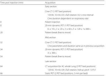
Table 1Patient acquisition protocol
Time post injection (min) Acquisition Early section
Cine-CT (1 PET bed position)
120 kV, 10 mA, 0.5 s full rotation, 0.2 s cine interval Cine duration dependent on respiratory rate
0 Patient injection
0 20-min dynamic PET (1 PET bed position) 6×10 s, 3×20 s, 3×60 s, 5×120 s, 1×300 s
20 Patient break (free to move)
Mid section
32 Cine-CT (1 PET bed position)
Cine parameters and duration same as in previous acquisition 34 20-min dynamic PET (1 PET bed position)
4×300 s
54 Patient break (free to move)
Late section
64 CT acquisition for AC whole Lung (2 PET bed positions) 120 kV, 10 mA, 0.8 s full rotation, helical, pitch 1.375:1 66 Static PET (2 PET bed positions, 3 min per bed)
Each section was then reconstructed with AC maps corresponding to different stages of the respiratory cycle as outlined below.
An ungated PET XCAT simulation, which contains AC map mismatches due to both motion and density (MD), was created as previously described [15] and outlined in brief here. First, tissue time activity curves (TTACs) and associated kinetic parameters (true parameters) for every organ in the thorax were determined from a patient dataset. These were used to create an organ-based activity distribution corresponding to each time frame in the dynamic sequence. For each time frame, XCAT phantoms of the thorax were cre-ated for 11 short time periods evenly spaced over a single breathing cycle (equivalent to respiratory phase gated periods), where the breathing motion was defined by the XCAT default diaphragm and anterior-posterior motion curves. An average of these 11 gates was used to form a motion averaged PET image. AC maps for the 11 gates were also sim-ulated varying the density in the lung regions from end expiration (μ = 0.0231 cm−1) to end inspiration (μ = 0.0194 cm−1) (representing a healthy patient). From these gates, an averaged PET-matched AC map was created by averaging all 11 gates (AVE). In addition, mismatched ‘snap shot’ AC maps from the extreme gates representing end inspi-ration (INS) and end expiinspi-ration (EXP) as well as a mid-breathing cycle (MID) gates were obtained. PET sinograms were created by using the analytic simulation capabilities of the ‘Software for Tomographic Image Reconstruction’ (STIR) [18] package, based on a model of the GE Discovery VCT scanner. PET volumes for each dynamic frame were forward projected taking into account single scatter and attenuation using the matched AC maps. These PET data were then reconstructed in STIR using OSEM (7 subsets, 40 iterations) with both the correct and mismatched AC maps for attenuation and scatter correction.
from each of the PET datasets. Following the protocol outlined in Table1, the derived lung TTACs were separated into early, mid and late study sections, each with three sets of frames derived from the different AC maps. All possible combinations were selected to create 27 full TTACs. An image-derived input function (IDIF) was determined from the left ventricle TAC by fitting a previously defined model [19]. An irreversible two-tissue compartment model [20] was then fit to each of the TTACs, and the influx rate constant
Ki2was determined (where the notation 2 is used to represent the influx rate constant
determined from the two-tissue compartment model) [21]. Patlak-Rutland analysis was also performed on the TTACs [22,23] to determineKiP(where thePrepresents the influx
rate constant from Patlak-Rutland analysis). For each TTAC, the percentage difference between the true and measuredKiwas determined.
Patient acquisitions
Nine patients, 5 males and 4 females aged 71±6 years, all with diagnosed idiopathic pul-monary fibrosis (IPF), underwent18F-FDG dynamic PET/CT with the protocol outlined in Table1using a GE Discovery VCT PET/CT scanner. All patients were imaged supine immediately post-injection using 209±23 MBq18F-FDG. The study was split into three stages (early, mid and late) to relieve patient discomfort during the long acquisition. The duration of the cine-CTs acquired for early and mid-cycle stages was set to the duration of the patient’s complete breathing cycle plus one extra second. A cine-CT obtains multi-ple CT acquisitions over time for each patient slice building up a 4D CT dataset [24]. The final stage of the study was a normal clinical static PET with a shallow breathing ‘snap shot’ CT study.
The patient data used in this study were the baseline scans (i.e. prior to patient dosing) obtained from a dose escalation study (NCT01725139) of omnipalisib (GSK2126458) in patients with IPF that included FDG-PET as one of the pharmacodynamics endpoints. Institutional Review Board permission, the UK Medicines and Healthcare Products Reg-ulatory Agency (MHRA) approval and informed patient consent were obtained for the study.
Patient data: multi-PET/CT method and analysis
In an initial study, the data from the nine imaged IPF patients (the “Patient acquisitions” section) were reconstructed without consideration of the effects of density variations in order to be consistent with the current methods used in the clinic. Each stage of the PET study was individually reconstructed using OSEM (7 subsets, 40 iterations, 6-mm FWHM Gaussian filter) with the corresponding CT for AC (either static CT or averaged cine-CT). GE proprietary software for off-line data processing of PET/CT data was used. The averaged cine-CT from the early and mid study sections were registered using non-rigid registration into the space of the late study (target image). Registration was achieved using the NiftyReg software [25,26], and the deformation fields from these registrations were used to register all the PET frames. Note that the non-rigid registration using the Niftyreg software only accounts for the repositioning of the patient between study sections; how-ever, density changes are not corrected. Therefore, the deformed images will match in terms of anatomical location, but the original lung density will be unchanged.
ROI was chosen to be smaller than the diameter of the ascending aorta in order to min-imise partial volume effects [28]. For each patient, the TAC for a selected aorta region was fit using a previously defined model [29] to create the IDIF.
The density in two regions of the lung on each of the registered CTs and two lung TTACs were determined, one from the high-density (HD) region of the IPF lung repre-senting fibrosis and one from the low-density (LD) ‘normal appearing’ region. To achieve this, HD and LD masks (HDMask and LDMask) were created by initially segmenting the whole lung using a thresholding technique where the lung is considered any voxel with
−900<HU<−150 within the body boundary on the target CT. For the LDMask, the whole lung mask was eroded by three voxels (12 mm) to avoid edge effects. The ‘normal appearing’ tissue was assumed to occupy any voxel within this mask with HU<−582. For the HDMask, the non-eroded whole mask is used, as the disease is most commonly seen in the lung periphery, and regions with HU> −530 are considered fibrosis. The HDMasks were then manually edited (where necessary) to remove any areas in the ROI not representative of fibrosis.
Lung TTACs were determined by application of the HD and LD masks to the registered PET data. A two-tissue irreversible compartment model (Eq.1) [20] was then fit to these lung TTACs using a weighted least squares method (Eq.2).
CR(t) = VBCP(t)+K1e−(k2+k3)t⊗CP(t)+
K1k3
k2+k3
1−e−(k2+k3)t⊗C
P(t) (1)
f(p) =
m
j=1
wj(ψj(p)−yj)2 (2)
where CR and CP are the concentrations of the radiotracer in the region of interest
and the plasma respectively;K1,k2andk3are the rate constants of the model;VBis the
fractional blood volume;⊗is the convolution operator;jindexes the time frames in the measured data;ψjis the model for each data point (computed as the numerical integral
of Eq.1over time framej),pare the parametersp=(VB,K1,k2,k3);yjare the measured
data;f(p) is the objective function for parametersp;mis the number of time frames; and
wjis a weighting factor equivalent to the inverse of the variance of the voxel values in the
ROI making up the data point.
The parametersKi2andKiP[21] along with their standard errors were determined and
compared. The standard error was calculated using the method outlined by Delforge et al. 1990 [30]. Briefly, the Hessian matrix (M) off(p)is estimated using the Jacobian (J), the matrix of first order derivatives of the modelψjwith respect to parametersp. Let W be a
diagonal matrix with thejth diagonal element equal to the weightswj, the approximation
of the Hessian can then be calculated:
M=JWJ (3)
withJthe transpose ofJ. The parameter covariance matrix can then be determined:
COV= f(p)
m−rM
−1 (4)
whereris the number of parameters in the kinetic model. The estimated standard error,
sk, on each parameterpkis then:
sk =
This standard error is an estimate of the parameter error assuming that the model used is correct and that the only cause for errors is noise.
Patient data: combined-CT method and analysis
The basic idea of the combined-CT method is to construct an AC map for each study section using all the available CT information. However, the process needs to take the changes in patient position into account.
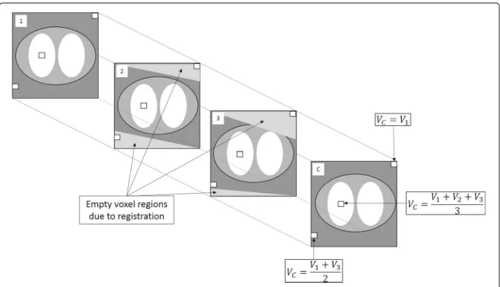
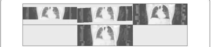
In a first step, the CTs from the different sections were registered to each other using the method previously described ( the “Patient data: multi-PET/CT method and analysis” section). After registration, not all areas in the CT images will overlap due to variable positioning at the start of each study section (see Fig. 5) and because the late section contains a greater number of bed positions. Therefore, to create the combined-CT for each study section, the method shown schematically in Fig.1 was used. This involved using a voxel-wise average across all voxel regions common to the CT for that section (the target CT) and the two registered CTs. Where the voxel was only present in two of the CT images, due to the registration, the average of the two voxel values was used to fill the appropriate voxel in the combined-CT image. Any remaining empty voxels were filled with the CT values from the target (unregistered) CT. This resulted in a new CT image where the entire field of view (FOV) was filled with a combination of the information from all the CT images.
The above process was repeated for each section. This enabled creation of a combined-CT in the space of each individual study section, avoiding possible issues that arise in registration due to mismatched field of view. The result is three combined-CT images, one in the space of each study section.
The raw PET data were then re-reconstructed and processed with the combined-CT images. TTACs were obtained using the HD and LDMasks defined in the
“Patient data: multi-PET/CT method and analysis” section. The TTACs from the combined-CT and original-CT processing were visually compared, andKi2andKiP
esti-mates and their associated standard errors were compared (the “Patient data: multi-PET/CT method and analysis” section). The density in each of the HD and LDMasks were also determined from each of the final combined-CTs after registration.
Results
XCAT simulations
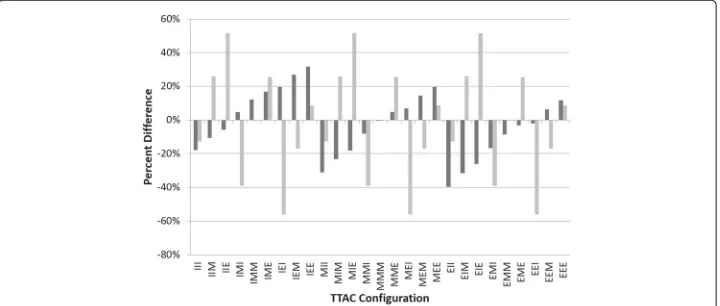
Figure2 displays the percentage differences between the true and the measured Ki for
each of the different configurations of the TTACs. The error trend was found to be dif-ferent for the compartment model and Patlak-Rutland methods of deriving the influx rate constant. As Patlak-Rutland only uses the final two study sections, the values ofKiPare
not affected by the initial scan section, provided the IDIF is minimally affected by the early variations in density. The maximum errors inKiPandKi2were found to be 55.9 and
39.7% respectively.
Patient analysis: multi-PET/CT
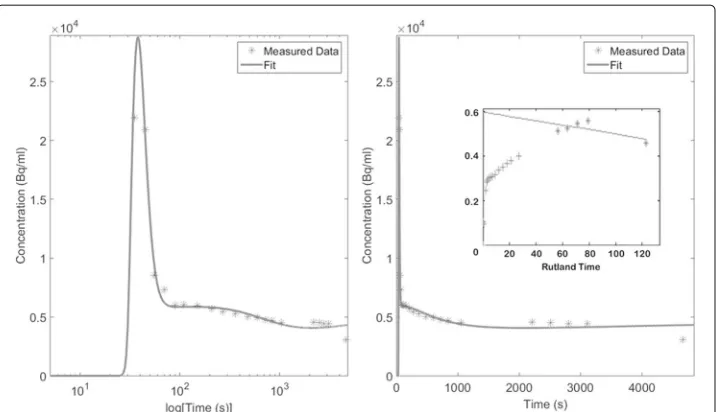
An example worst case (patient 5) FDG TTAC and the associated two-tissue compart-ment model and Patlak-Rutland fits are shown in Fig.3. Discontinuity between the study sections is observed, which leads to a poor fit of the compartment model. The effect of these discontinuities also leads to incorrect analysis when applying Patlak-Rutland analy-sis. In this case, this resulted in a negative slope, which is inconsistent with the fact that FDG is considered an irreversible tracer over the time course of the study.
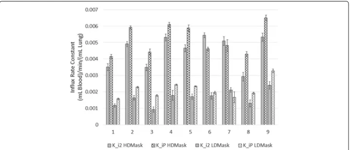
Figure4displays the measured influx rate constants from high- and low-density TTACs analysed with the two-tissue compartment model (Ki2) and with Patlak-Rutland analysis
(KiP) along with the associated standard errors. The percent error ((standard error/Ki)×
100%) forKi2andKiPwas averaged for all patients and regions and found to be 44±25%
and 13±17% respectively.
Fig. 2Variation in Kidue to respiration. The percentage difference between the true and measured Ki2
Fig. 3Example patient TTAC and fits with conventional analysis measured patient TTAC (stars) and the associated compartment model and Patlak-Rutland (inset to column 2) fits (filled line). Both columns show the same data. However, the left column has thex-axis in log(time) to allow better visualisation of the peak, while in the second column, thex-axis is linear in time
Theoretically, the two methods of calculating Ki (compartment model and
Patlak-Rutland analysis) should render the same results. However, the maximum percentage difference between the estimatedKiparameters was found to be 250%. The average and
standard deviation of the differences between the parameter estimates from the two anal-ysis methods for the derived TTACs in the high and low density regions are 51±102% and 43±54% respectively.
Patient analysis: combined-CT
Figure5 displays the three registered original CT acquisitions for the example patient shown in Fig.3 along with the combined late CT. On investigation of the CTs in this example patient, the lung density in the late acquisition was found to be up to 35% lower
Fig. 4Regional variation in the Kimeasurements with conventional analysis comparison of the fibrotic and normal appearing tissue influx rate constants using compartmental modelling (Ki2) and Patlak-Rutland
Fig. 5Example patient original and combined-CT images. The registered original CT images from the early (top left), mid (top middle) and late (top right) study sections and the resulting combined-CT for the late section (bottom middle) for an example patient. Grey scale−1350<HU <150
than that of the cine-CT acquisitions. Visual comparison of diaphragm position confirms that the CT was acquired at deep inspiration in the late stage of this study.
The density difference in the HD and LD lung between both the early and mid averaged cine-CTs and the late static CT are shown in Table2. The maximum difference in lung densities between the early and mid averaged cine-CTs were found to be 10.0 and 12.5% respectively for the HD and LD regions. In contrast, the maximum difference in lung density between the early and mid combined-CT images were found be 1.4 and 1.2% in the HD and LD regions respectively.
After reconstruction with the combined-CTs, all discontinuities in the PET-derived TTACs were reduced. Figure6displays the TTAC of the same example patient as shown in Fig.3 with associated compartment model and Patlak-Rutland analysis fits. Analysis led to more consistent results when the combined-CT was used for reconstruction.
Figure7 displays the measuredKi2 andKiP from the high- and low-density TTACs
along with the associated standard errors after the data have been reconstructed with the combined-CT for comparison with the original method shown in Fig.4.
Table3displays the comparison between the estimates ofKi2andKiP. There is much
better agreement using the combined-CT; a factor 2.5 and 1.7 for the high- and low-density regions respectively.
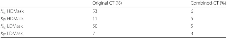
Table4displays the change in the standard errors for the original and new methods of reconstructing the PET data. For bothKi2andKiPthe data errors reduce to within 6%.
Discussion
Quantitative imaging in the thorax is known to be complex due to respiratory effects. While motion correction has been extensively reviewed in the literature, little attention has been given to density changes. In previous work [15], we have shown, for the lung, that failure to account for CT density changes within the lung can lead to significant errors in
Table 2Average density variation in the HD and LD regions for all patients in the early and mid cine-CT studies with respect to the late ‘snap shot’ CT study (((CTx/CTL)−1)×100%) whereCTxis
eitherCTEorCTM)
Original CT Combined-CT
Early cine-CT HDMask −2.96±9.09% −1.58±4.96%
Early cine-CT LDMask 10.99±16.66% −1.68±4.84%
Mid cine-CT HDMask 2.80±9.15% 1.06±0.70%
Fig. 6Example patient TTAC and fits with using combined-CT reconstructions for analysis. An FDG example TTAC with associated compartment model and Patlak-Rutland (inset) fits. This is the same FDG patient as shown in Fig.3for comparison. Both columns show the same data using different time axes as before (Fig.3)
estimated kinetic parameters, and in this paper, we have extended this work to investigate the effect of density variation in multi-PET/CT studies.
In simulations with ‘snap shot’ CTs, the largest errors in the parameter estimates are seen when the AC maps for each study section are acquired at different phases of the breathing cycle (Fig.2). Accounting for CT density changes to ensure parameter stability is therefore essential for MPC studies including those that involve monitoring of disease progression over time.
With simulations appropriate for non-TOF18F-FDG studies, it has been shown that MPC protocols can lead to errors inKi of up to 57% with ‘snap shot’ CTs. The
Patlak-Rutland influx rate constant is determined from the tail of the TTACs only and is largely unaffected by early study section errors. This was reflected in the fact that the Patlak-Rutland derived parameters were more precise and stable than the compartment model
Fig. 7Regional variation in the Kimeasurements using combined-CT reconstructions for analysis. Comparison of the fibrotic and normal appearing tissue influx rate constants determined after the PET data were reconstructed with the combined-CT, using compartmental modelling (Ki2) and Patlak-Rutland analysis
Table 3Average±standard deviation of the percentage differences between the compartment model and Patlak-Rutland analysis derivedKiestimates using the reconstructions with the actual
acquired CTs (original CT) and using the reconstructions with the combined-CTs
Original CT Combined-CT
HDMask 51±102% 20±10%
LDMask 43±54% 26±11%
estimates. This was also found to be the case in the patient data (Table4) where the standard errors were found to be 5 times greater for Ki2 thanKiP before use of the
combined-CT for AC.
The18F-FDG protocol outlined in the “Patient acquisitions” section was split into three sections to encourage patient compliance. The derived TTACs from these data were found to have discontinuities between the study sections. The evidence suggests that this is associated with a variation in density across the CT acquisitions used for the AC at each stage. This variation in density could be due to timing of the short ‘snap-shot’ CT scans or, for averaged cine-CTs, possible variation in the breathing pattern between acquisitions.
For MPC studies, we propose that an approach for improving the match between the PET and CT in terms of density is to use an average of all the CT information to pro-duce a combined-CT image (Fig. 5) for each study section. Results from applying the combined-CTs to each study section of the MPC protocols rectified TTAC discontinuity and improved parameter estimation (Fig.6).
It can be observed from Figs.4and7that use of the combined-CTs produces absolute
Kivalues that are different to those obtained using the multiple CTs. This is due to the
derived TTACs being very different and therefore producing very different parameter estimates. This is especially true as the model used to fit the TTAC from the original method could be considered to be wrong in this case.
Using the combined-CT approach, the average ratio of rate constants increased to 2.8 compared to 1.7 when using the original approach. This suggests that the proposed anal-ysis may result in better discrimination of diseased and normal lung tissue, which would be important in evaluation of treatment.
Combining the CTs results in all the CTs used for AC having almost the same density in the regions where all the CTs overlap. However, it is noted that due to the larger FOV in the late section, there will be regions of lung that will not benefit from the combined-CT but nor can these regions be used for dynamic analysis.
Creating combined-CT images from ‘snap shot’ CT acquisitions has the advantage that a lower radiation dose will be given to the patient than use of cine-CTs. Good PET
Table 4The average parameter estimate errors as a fraction of the average parameter estimate for the FDG patients using the original acquired CTs and the combined-CTs for AC in the PET reconstruction at each study stage
Original CT (%) Combined-CT (%)
Ki2HDMask 53 6
KiPHDMask 11 5
Ki2LDMask 50 5
matching, however, is dependent on those ‘snap shot’ CTs being acquired at different stages of the breathing cycle, which cannot be guaranteed, unless instructed breathing is part of the clinical protocol, but patient compliance can be quite difficult to achieve, even with training [12]. Averaged cine-CTs, as available in the early and mid stages of this study, have the advantage that they more closely match the longer acquisition PET and would therefore be recommended as they represent entire respiratory cycles. However, in this work, the averaged cine-CTs from the early and mid study sections were found to still have variable densities in the lung. Moreover, the higher dose may mean that this method is not suitable for routine use.
The results in this paper show that the proposed approach of combining CTs is rec-ommended for MPC studies involving ‘snap shot’ CTs, cine-CTs or a mixture of the two. However, it is noted that due to the irregularity of breathing, the combined-CTs may not be perfectly matched to the PET. The result is that the possibility of remaining mismatch artefacts and parameter bias still exists.
As an alternative to using the combined-CT method, our simulations indicate that training the patient such that a MID-CT can be acquired would work; however, as previously discussed, this may be difficult to achieve.
This paper has discussed mismatch between ungated PET and CT only. It should also be possible to gate the PET study and only reconstruct the gate that best matches the acquired CT. However, reconstructing gated dynamic PET data is challenging due to noise, especially in the early study frames. In addition, this approach would need to ensure that CTs in each section are acquired in the same respiratory state. Another alternative method is to use a joint registration/reconstruction technique that accounts for motion and misaligned attenuation, such as that outlined by Bousse et al. 2016 [31]. However, this technique would need adaptation to include corrections for the density changes for both the CT and gated PET data [32], but shows strong promise for the future of accurate quantitative pulmonary PET/CT imaging.
Conclusions
Changes in respiratory induced lung density between PET and CT acquisitions contribute to the errors in lung tissue PET quantitation, especially in multi-PET/CT protocols, resulting in discontinuities between study sections in the time activity curves. When reconstructing the PET data for each study section, use of a combined-CTs for AC can improve the precision of the estimated parameters and may discriminate better between diseased and normal lung.
Acknowledgements
The authors would like to thank General Electric Healthcare for the use of their software and Robert Shortman for his assistance with this study.
Funding
We acknowledge funding support from GlaxoSmithKline (studentship STU100028576 and study funding for NCT01725139) and the Engineering and Physical Sciences Research Council (EP/K0052781/1). Also, this project is supported by researchers at the National Institute for Health Research, University College London Hospitals Biomedical Research Centre.
Authors’ contributions
Ethics approval and consent to participate
All procedures performed in studies involving human participants were in accordance with ICH Good Clinical Practice (GCP), the ethical standards of the institutional and/or national research committee and with the 1964 Helsinki declaration and its later amendments or comparable ethical standards.
Informed consent was obtained from all individual participants included in the study.
Consent for publication
Informed consent was obtained from all individual participants included in the study.
Competing interests
The authors declare that they have no competing interests.
Publisher’s Note
Springer Nature remains neutral with regard to jurisdictional claims in published maps and institutional affiliations.
Author details
1Institute of Nuclear Medicine, University College London, UCLH (T-5), Euston Road, London, NW1 2BU UK.2National
Institute for Health Research Respiratory Biomedical Research Unit, Royal Brompton Hospital, Sydney St, London, SW3 6NP UK.3Fibrosis Research Group, Inflammation, Repair and Development Section, NHLI, Sir Alexander Flemming
Building, Imperial College London, London, SW7 2AZ UK.4Centre for Medical Radiation Physics, University of
Wollongong, Wollongong, Australia.
Received: 14 September 2017 Accepted: 9 April 2018
References
1. Scherer PM, Chen DL. Imaging pulmonary fibrosis. J Nucl Med. 2016;57(11):1764–70.
2. Groves AM, Win T, Screaton NJ, Berovic M, Endozo R, Booth H, Kayani I, Menezes LJ, Dickson JC, Ell PJ. Idiopathic pulmonary fibrosis and diffuse parenchymal lung disease: implications from initial experience with 18F-FDG PET/CT. J Nucl Med. 2009;50(4):538.
3. Win T, Screaton NJ, Porter J, Endozo R, Wild D, Kayani I, Dickson J, Shortman RI, Reubi JC, Ell PJ, Groves AM. Novel positron emission tomography/computed tomography of diffuse parenchymal lung disease combining a labeled somatostatin receptor analogue and 2-deoxy-2[18F]fluoro-D-glucose. Mol Imaging. 2012;11(2):91. 4. Schroeder T, Vidal Melo MF, Musch G, Harris RS, Venegas JG, Winkler T. Image-derived input function for
assessment of 18F-FDG uptake by the inflamed lung. J Nucl Med. 2007;48(11):1889–96.
5. Jones HA, Cadwallader KA, White JF, Uddin M, Peters AM, Chilvers ER. Dissociation between respiratory burst activity and deoxyglucose uptake in human neutrophil granulocytes: implications for interpretation of (18)F-FDG PET images. J Nucl Med. 2002;43(5):652–7.
6. Chen DL, Mintun MA, Schuster DP. Comparison of methods to quantitate 18F-FDG uptake with PET during experimental acute lung injury. J Nucl Med. 2004;45(9):1583–90.
7. Beyer T, Antoch G, Blodgett T, Freudenberg LF, Akhurst T, Mueller S. Dual-modality PET/CT imaging: the effect of respiratory motion on combined image quality in clinical oncology. Eur J Nucl Med Mol Imaging. 2003;30(4):588–96. 8. Osman MM, Cohade C, Nakamoto Y, Wahl RL. Respiratory motion artifacts on PET emission images obtained using
CT attenuation correction on PET-CT. Eur J Nucl Med Mol Imaging. 2003;30(4):603–6.
9. Pepin A, Daouk J, Bailly P, Hapdey S, Meyer ME. Management of respiratory motion in PET/computed tomography: the state of the art. Nucl Med Commun. 2014;35(2):113–22.
10. Verschakelen JA, Van fraeyenhoven L, Laureys G, Demedts M, Baert AL. Differences in CT density between dependent and nondependent portions of the lung: influence of lung volume. AJR Am J Roentgenol. 1993;161(4):713–7.
11. Nehmeh SA, Erdi YE. Respiratory motion in positron emission tomography/computed tomography: a review. Semin Nucl Med. 2008;38(3):167–76.
12. Sun T, Mok GS. Techniques for respiration-induced artifacts reductions in thoracic PET/CT. Quant Imaging Med Surg. 2012;2(1):46–52.
13. Shaker SB, Dirksen A, Laursen LC, Skovgaard LT, Holstein-Rathlou NH. Volume adjustment of lung density by computed tomography scans in patients with emphysema. Acta Radiol. 2004;45(4):417–23.
14. Gorbunova V, Sporring J, Lo P, Loeve M, Tiddens HA, Nielsen M, Dirksen A, de Bruijne M. Mass preserving image registration for lung CT. Med Image Anal. 2012;16(4):786–95.
15. Holman BF, Cuplov V, Hutton BF, Groves AM, Thielemans K. The effect of respiratory induced density variations on non-TOF PET quantitation in the lung. Phys Med Biol. 2016;61(8):3148–63.
16. Pan T, Mawlawi O, Nehmeh SA, Erdi YE, Luo D, Liu HH, Castillo R, Mohan R, Liao Z, Macapinlac HA. Attenuation correction of PET images with respiration-averaged CT images in PET/CT. J Nucl Med. 2005;46(9):1481–7. 17. Segars WP, Sturgeon G, Mendonca S, Grimes J, Tsui BM. 4D XCAT phantom for multimodality imaging research.
Med Phys. 2010;37(9):4902–15.
18. Thielemans K, Tsoumpas C, Mustafovic S, Beisel T, Aguiar P, Dikaios N, Jacobson MW. STIR: software for tomographic image reconstruction release 2. Phys Med Biol. 2012;57(4):867–3.
19. Feng D, Wang X, Yan H. A computer simulation study on the input function sampling schedules in tracer kinetic modeling with positron emission tomography (PET). Comput Methods Programs Biomed. 1994;45(3):175–86. 20. Sokoloff L, Reivich M, Kennedy C, Des Rosiers MH, Patlak CS, Pettigrew KD, Sakurada O, Shinohara M. The
21. Gunn RN, Gunn SR, Cunningham VJ. Positron emission tomography compartmental models. J Cereb Blood Flow Metab. 2001;21(6):635–52.
22. Patlak CS, Blasberg RG, Fenstermacher JD. Graphical evaluation of blood-to-brain transfer constants from multiple-time uptake data. J Cereb Blood Flow Metab. 1983;3(1):1–7.
23. Rutland MD. A single injection technique for subtraction of blood background in 131I-hippuran renograms. Br J Radiol. 1979;52(614):134–7.
24. Alessio AM, Kohlmyer S, Branch K, Chen G, Caldwell J, Kinahan P. Cine CT for attenuation correction in cardiac PET/CT. J Nucl Med. 2007;48(5):794–801.
25. Ourselin S, Roche A, Subsol G, Pennec X, Ayache N. Reconstructing a 3D structure from serial histological sections. Image Vis Comput. 2001;19(1–2):25–31.
26. Modat M, Ridgway GR, Taylor ZA, Lehmann M, Barnes J, Hawkes DJ, Fox NC, Ourselin S. Fast free-form deformation using graphics processing units. Comput Methods Prog Biomed. 2010;98(3):278–84.
27. de Geus-Oei LF, Visser EP, Krabbe PF, van Hoorn BA, Koenders EB, Willemsen AT, Pruim J, Corstens FH, Oyen WJ. Comparison of image-derived and arterial input functions for estimating the rate of glucose metabolism in therapy-monitoring 18F-FDG PET studies. J Nucl Med. 2006;47(6):945–9.
28. van der Weerdt AP, Klein LJ, Boellaard R, Visser CA, Visser FC, Lammertsma AA. Image-derived input functions for determination of MRGlu in cardiac (18)F-FDG PET scans. J Nucl Med. 2001;42(11):1622–9.
29. Feng D, Huang SC, Wang X. Models for computer simulation studies of input functions for tracer kinetic modeling with positron emission tomography. Int J Biomed Comput. 1993;32(2):95–110.
30. Delforge J, Syrota A, Mazoyer BM. Identifiability analysis and parameter identification of an in vivo ligand-receptor model from PET data. IEEE Trans Biomed Eng. 1990;37(7):653–61.
31. Bousse A, Bertolli O, Atkinson D, Arridge S, Ourselin S, Hutton B, Thielemans K. Maximum-likelihood joint image reconstruction/motion estimation in attenuation-corrected respiratory gated PET/CT using a single attenuation map. IEEE Trans Med Imaging. 2016;35(1):217–8.