P R O C E E D I N G S
Open Access
The role of SH3BP2 in the pathophysiology of
cherubism
Ernst J Reichenberger
1*, Michael A Levine
2, Bjorn R Olsen
3, Maria E Papadaki
4, Steven A Lietman
5From
International Meeting on Fibrous Dysplasia/McCune-Albright Syndrome and Cherubism: Best Clinical
Practice and Future Research
Bethesda, MD, USA. 3-5 October 2010
Abstract
Cherubism is a rare bone dysplasia that is characterized by symmetrical bone resorption limited to the jaws. Bone lesions are filled with soft fibrous giant cell-rich tissue that can expand and cause severe facial deformity. The disorder typically begins in children at ages of 2-5 years and the bone resorption and facial swelling continues until puberty; in most cases the lesions regress spontaneously thereafter. Most patients with cherubism have germline mutations in the gene encoding SH3BP2, an adapter protein involved in adaptive and innate immune response signaling. A mouse model carrying a Pro416Arg mutation in SH3BP2 develops osteopenia and expansile lytic lesions in bone and some soft tissue organs. In this review we discuss the genetics of cherubism, the biological functions of SH3BP2 and the analysis of the mouse model. The data suggest that the underlying cause for cherubism is a systemic autoinflammatory response to physiologic challenges despite the localized appearance of bone resorption and fibrous expansion to the jaws in humans.
Introduction
“Bone dystrophies paint queer and irregular pictures throughout the skeleton and have been reported in most bones”W.A. Jones begins his 1950 review, where he pro-posed the name“cherubism” for the multilocular cystic disease of the jaws that he had first described 17 years ear-lier [1,2]. In 2011 we still lack good explanations for the bilateral expression of cherubism [MIM 602104] lesions. Other areas of investigation are the limitation of the aggressive bone resorption and expansion of fibrous tissues in the maxilla and mandible as well as the age-dependent onset in children at age 2-5 years, and in most cases the spontaneous regression of the fibrous growths after puberty [3]. Cherubism typically begins with a swel-ling of submandibular lymph nodes. The phenotype comes to the attention of health care providers, often den-tists, at its early stages when excessive bone resorption in the jaws causes characteristic symmetrical cystic lesions
that can be detected by routine panoramic radiographs. The“cherubic”swelling of cheeks occurs when the fibrous tissue filling the cysts expands and deforms the cortical shell.
Clinical management of cherubism has progressed sig-nificantly but therapeutic approaches to inhibit or delay the progression of cherubic lesions are not available. The gaps in our understanding of the natural history of cher-ubism, and the molecular mechanism that initiates and maintains bone resorption as well as the replacement of bone with tumor-like fibrous tissue are now being addressed by several research groups. In this review we will assess the many functions of the cherubism gene
SH3BP2[MIM 118400] in immune cells and osteoclasts and discuss how animal models and in vitro studies can help to understand the human disease.
SH3BP2: genetic aspects
Cherubism is classically transmitted as an autosomal dominant trait, but there are indications that a recessive form may also exist. Based on a thorough statistical ana-lysis of 21 previously published families by Anderson and McClendon, 100% penetrance in males and reduced
* Correspondence: [email protected]
1University of Connecticut Health Center, Department of Reconstructive
Sciences, Center for Regenerative Medicine and Skeletal Development, Farmington, CT, USA
Full list of author information is available at the end of the article
penetrance (70 - 50%) in females has been reported [4]. However, the authors concede in this retrospective study that only 50% of the adult female family members which were considered unaffected underwent radiographic examination. The apparently reduced female penetrance may also be due to examination of some children before they developed clinical signs of cherubism. Unfortu-nately, this paper has been cited many times since then without acknowledging these caveats. In the experience of our group, we cannot confirm incomplete penetrance but we have seen variable expressivity within families. It should be noted that older patients with a mild form of cherubism may have bone lesions that have been remo-deled with normal mandibular bone and therefore signs of cherubism may no longer be detected by radiographs [5]. Based on published case reports of cherubism as well as patients referred to our clinics and research environ-ment there appears to be no obvious difference in the prevalence of the disorder among different racial or eth-nic groups. Adequate epidemiologic data for cherubism do not exist.
Approximately 50% of cases seen in our laboratory at UCHC are sporadic and represent de novo mutations. The genetic interval for the autosomal dominant form of cherubism was first identified in 1999 by linkage and haplotype analysis to be on chromosome 4p16.3 [6,7]. The 1.5 Mb cherubism locus is contained within the locus for Wolf-Hirschhorn disease [8].
Wolf-Hirschhorn syndrome is caused by heterozygous chromosomal deletions that cause craniofacial malforma-tions, intellectual disability, muscle hypotonia and heart defects [9]. This chromosomal region is also commonly deleted in bladder cancer [10]. Since a cherubism-like phenotype is not part of the Wolf-Hirschhorn syndrome, Tiziani at al. concluded that a cherubism mutation must be a gain-of-function mutation [6]. In 2001 Ueki at al. identified heterozygous mutations for cherubism in 12 families in the gene for the signaling adapterSH3-domain binding protein 2(SH3BP2) [11].
SH3BP2 was initially identified as a c-Abl binding protein in mice and humans [10,12]. TheSH3BP2gene product is expressed in most cell types. It acts as an adap-ter protein to control intracellular signaling by inadap-teracting and forming complexes with binding proteins [13] and with scaffolding proteins [14,15]. The 561 amino acid (aa) protein (559 aa in mouse) is highly conserved in mammals with 87% amino acid sequence homology between human and mouse [10] and 84% homology on the nucleotide level. The 48kb SH3BP2 gene contains 13 exons that code for a 62 kDa protein with 561 amino acids (Figure 1). As is the case with most adapter proteins, SH3BP2 has a modular domain structure and consists of an N-terminal pleckstrin homology (PH) domain, a proline-rich (PR)
domain and a C-terminal Src-homology 2 domain (SH2). SH3BP2 is thought to bind to cell membrane lipids via its PH domain and to interact with the SH3 domains of bind-ing partners via SH3 bindbind-ing motives in the proline-rich domain. The SH2 domain can interact with a number of binding partners carrying a Tyr-Glu-Asn (YEN) binding motif (reviewed in [13]).
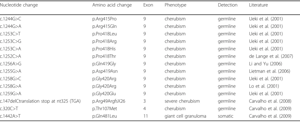
The mutations identified by Ueki et al. were located in exon 9, within a 6 amino acid interval (RSPPDG) in the proline-rich domain proximal to the SH2 domain of SH3BP2 (Figure 1; Table 1) [11]. All mutations were transitions or transversions of single nucleotides that led to the substitution of amino acids Arg415, Pro418 or Gly420. These mutations account for 100% of the muta-tions detected in the laboratory at UCHC. Additional sin-gle nucleotide substitutions were found in Gly420, Pro418 and Asp419 (Table 1; see also http://fmf.igh.cnrs. fr/ISSAID/infevers/) [16-19]. Carvalho et al. described unusual mutations in the pleckstrin homology domain in two Brazilian cherubism patients. A point mutation in exon 4 resulted in a Thr107Met substitution that was detected in blood (germline) and in tumor tissue [20]. In the tumor tissue of another patient the same group found a variant of what appears to be a deletion of nucleotide 147 (c.147delC) which led to a frame shift over 26 aa and a premature stop codon at position 325 (p.Arg49ArgfsX26) [21]. This patient suffered from a severe case of cherubism and is to our knowledge the only patient who had a fatal form of cherubism [22]. The mutation found in this patient could conceivably have led to a severe and rapidly progressing form of cherubism if the partial gene product (the N-terminal 48 amino acids) is translated. A truncated protein may have a dominant negative effect on disease mechanisms or exacerbate the disease progression by activating expression of certain (yet unknown) proteins. It is unlikely that the mutant protein is not expressed because hemizygosity, as in Wolf-Hirschhorn syndrome, is not expected to cause any cherubism-like phenotype. For all other patients with commonly detected cherubism mutations in SH3BP2 seen in our clinics or in the research laboratory we were unable to establish any genotype–phenotype correlation.
NS/MGCLS have been found in the SHP2-coding gene
PTPN11and inSOS1[24,27-31]. Both gene products act in the RAS-mitogen-activated protein kinase signaling pathway and it is therefore conceivable that SH3BP2 may also play a role in this pathway. It may be worth-while to test whether those patients who were diagnosed with cherubism and were negative for a mutation in
SH3BP2have mutations in other genes within the RAS-MAPK axis. Interestingly, bilateral mandibular cherub-ism-like lesions and giant cell lesions in the mandible and in long bones have been described in neurofibroma-tosis patients [32,33], and are associated with mutations in the neurofibromin gene,NF1. NF1 is known as a reg-ulator of the RAS pathway and mutations in NF1are
associated with neurofibromatosis and Noonan syn-drome [34,35].
To date there is only one report of a somatic mutation of SH3BP2 in a central giant cell lesion (CGCL) [20]. The described mutation is not identical with canonical cherubism mutations in exon 9 but is a point mutation in exon 11 leading to a Glutamine 481 to Leucine exchange in the SH2 domain of SH3BP2.
Alternative splicing variants ofSH3BP2have been iden-tified experimentally and by computational delineations. However, it is not known whether any of these variants are biologically relevant [10,36] (see also http://genecards. org). Regulation of SH3BP2 transcription is largely unknown but recently evidence emerged thatSH3BP2 Figure 1Gene map and protein structure of human SH3BP2 indicating mutations in the canonical cherubism mutation interval (amino acids 415-420) and mutations reported in the pleckstrin homology (PH) domain. The mutation in the SH2 domain has been found in tumor tissue of a patient with giant cell tumor. (Modified after Ueki et al., 2001)
Table 1 Mutations in SH3BP2
Nucleotide change Amino acid change Exon Phenotype Detection Literature
c.1244G>C p.Arg415Pro 9 cherubism germline Ueki et al. (2001)
c.1244G>A p.Arg415Gln 9 cherubism germline Ueki et al. (2001)
c.1253C>T p.Pro418Leu 9 cherubism germline Ueki et al. (2001)
c.1253C>G p.Pro418Arg 9 cherubism germline Ueki et al. (2001)
c.1253C>A p.Pro418His 9 cherubism germline Ueki et al. (2001)
c.1252C>A p.Pro418Thr 9 cherubism germline de Lange et al. (2007)
c.1256A>G p.Gln419Gly 9 cherubism germline Li and Yu (2006)
c.1255G>A p.Asp419Asn 9 cherubism germline Lietman et al. (2006)
c.1258G>C p.Gly420Arg 9 cherubism germline Ueki et al. (2001)
c.1258G>A p.Gly420Arg 9 cherubism germline Lo et al. (2001)
c.1259G>A p.Gly420Glu 9 cherubism germline Ueki et al. (2001)
c.147delCtranslation stop at nt325 (TGA) p.Arg49ArgfsX26 3 severe cherubism germline Carvalho et al. (2008)
c.320C>T p.Thr107Met 4 cherubism germline Carvalho et al. (2009)
expression is differentially regulated by hypoxic conditions in tumor cells [37]. More is known about the role its gene product plays during immune response.
SH3BP2 function in immune cells
Before its identification as the principal disease-causing gene for cherubism, SH3BP2 had been of interest to immunologists because of its multiple roles in hemato-poietic and immune cells. Therefore a number of aliases (SH3-domain binding protein 2; SH3BP2; 3BP2; CRBM; CRPM; RES4-23; FLJ42079; FLJ54978) and various pro-tein names (SH3BP2; Abl-SH3 binding propro-tein 2; TNFAIP3 interacting protein 2) can be found in the literature.
Early investigations examined the function of SH3BP2 in hematopoietic cells and found that SH3BP2 induced B cell receptor activation, NK cell mediated cytotoxicity and basophilic cell degranulation [38-43]. The modular struc-ture of SH3BP2 suggests that it may function as an adap-tor protein [11,39,40,44] particularly as it lacks known catalytic activity. In various studies, investigators have examined the proteins that interact with SH3BP2 to derive clues about its function(s). A direct interaction between SH3BP2 and Syk was identified in a yeast 2-hybrid screen of a T lymphocyte library for Syk kinase-interacting pro-teins, and the role of SH3BP2 in modulating Syk activity has been examined in lymphocytes and Jurkat TAg cells [44]. In lymphocytes, SH3BP2 binds to 14-3-3, Vav1 and 2 and PLCg1 [40,44]. In addition, an SH3BP2 mutant incapable of binding to 14-3-3 showed increased NFAT (nuclear factor of activated T cells) activation, indicating that the interaction of 14-3-3 with SH3BP2 can block its function [40]. Vav proteins are guanine nucleotide exchange factors that activate the small GTPases Ras and Rac1, which in turn activate AP-1 and NFAT, respectively [39,40,45,46]. Vav1 and Vav2 functionally cooperate with SH3BP2 in Jurkat TAg cells [39] and Vav3 is known to regulate osteoclast function [45,47].
Cbl and the Cbl interacting protein CIN85 have also been identified as proteins which directly or indirectly bind to SH3BP2 [15,44]. Cbl expression is enriched in the podosome belt in osteoclasts at sites of cell attachment and as a result c-Cbl-/-osteoclasts have impaired motility [48]. CIN85 overexpression decreases intracellular calcium signaling and decreases PLCg1 and 2 phosphorylation [49]. SH3BP2 can be modified by tyrosine and serine phos-phorylation and therefore alter its activity and binding properties. SH3BP2 phosphorylation of Tyr183is required for interaction with Vav1 and phosphorylation of Tyr446 of SH3BP2 is required for SH3BP2 interaction with the SH2 domain of Lck [39,46]. Phosphorylation of Ser225 and Ser277 are required for 14-3-3 binding, and a SH3BP2 protein lacking these serines was shown to have increased activity in Jurkat TAg cells [40]. In T cells,
SH3BP2 is phosphorylated on tyrosine448in response to T cell receptor stimulation and this phosphorylation is required for T cell signaling as indicated by NFAT acti-viation [50]. Further, phosphorylation of SHP1 phospha-tase causes recruitment and dephosphorylation of SH3BP2 and termination of T cell signaling [50]. SH3BP2 phosphorylation is also induced by CD244 liga-tion and tyrosine337phosphorylation of CD244 regulates its interaction with SH3BP2 in NK cells [51]. Mutant SH3BP2 alters the phosphorylation of other proteins. For example, replacement of amino acids Tyr183and Tyr446 or Arg486, which are phosphorylation sites, with other amino acids reduces the ability of SH3BP2 to respond to signals that activate NFAT. Moreover, heterozygous and homozygous Sh3bp2 knockin cells that contain the P416R mutation found in cherubism patients show increased phosphorylation of ERK1/2 and Syk (at Tyr346) after stimulation with M-CSF and RANKL [52].
In summary, SH3BP2 can be differentially phosphory-lated depending on the functions it fulfills in the various immune cell types thus attracting specific protein binding partners and regulating downstream signaling pathways. In osteoclasts, another cell type of hematopoietic origin, SH3BP2 is a major regulator of bone resorption. Muta-tions in SH3BP2 result in osteoclasts that lead to increased bone resorption in jaws of cherubism patients, whereas in a mouse model bone resorption is more general [11,52].
SH3BP2 in osteoclasts
The limited distribution of bone lesions in patients with cherubism is unexpected as the disorder is associated with the heterozygous germline mutations in SH3BP2, which is widely expressed throughout the osteoimmune system. The precise function of the six-amino acid region where most of the known mutations occur remains unclear, but recent work suggests that the cherubism missense mutations lead to a gain-of-function rather than a loss of activity [16,52,53]. Mutations in cherubism that result in a gain-of-function for SH3BP2 is consistent with prior observations that deletions of 4p16.3 in patients with Wolf-Hirschhorn syndrome, which result in loss of one copy ofSH3BP2, do not cause a bone resorp-tive phenotype [54-56].
mature osteoclasts requires RANKL, indicating that this cytokine, in addition to colony-stimulating factor 1 (CSF-1)/macrophage colony-stimulating factor (M-CSF), is a cri-tical differentiation factor that specifies the osteoclast maturation program, and hence induction of bone resorp-tion. Although RANKL (in conjunction with M-CSF) has been recognized as one of the key osteoclastogenic signals expressed by osteoblasts and stromal cells, the down-stream signaling pathways activated by this cytokine have not been fully characterized.
RANKL induces osteoclast formation via transcription and activation of NFATc1, the master“switch”for osteo-clastogenesis [57-59]. NFATc1 is activated by calcineurin, a calcium-calmodulin dependent phosphatase, via depho-sphorylation, which facilitates translocation of NFATc1 into the nucleus [57-62]. In addition to NFATc1 there are other NFAT isoforms, termed NFATc2, NFATc3, and NFATc4, but these proteins are not expressed at signifi-cant levels in pre-osteoclast cells [59].
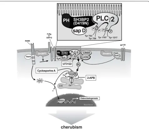
RANKL can induce intracellular calcium oscillations to activate calcineurin in bone marrow macrophages (BMMs, BMM cells) [57] and the mouse osteoclast precursor cell line RAW 264.7 [61]. However, it is increasingly clear that other signaling pathways can also increase concentrations of cytosolic Ca2+, and can also activate calcineurin and NFATc1. For example, membrane proteins with immu-noreceptor tyrosine-based activating motifs (ITAMs), such as FcRg1 and DAP12 interact with their own ligands as well as activated RANK to increase cytosolic Ca2+ [57,63-65]. Mechanistically, activation of these immunore-ceptors in concert with RANK signaling leads to phos-phorylation of the ITAM domains, which in turn recruit Syk to the membrane with subsequent activation of PLCg. Activation of PLCgleads to the generation of IP3, which releases Ca2+from the endoplasmic reticulum and thereby stimulates calcineurin-dependent dephosphorylation of NFATc1 and consequently translocation of NFATc1 into the cell nucleus [63,65].
Overexpression of wild-type and mutant SH3BP2 in B and T cells leads to transactivation of a luciferase reporter gene that is under the control of the NFAT binding sequence from the interleukin 2 (IL-2) gene promoter [16,39,40,44]. Moreover, overexpression of a constitutively active form of NFATc1 in the RAW 264.7 osteoclast pre-cursor cell line is sufficient to induce osteoclast differentia-tion [11,57,59,63]. Based on these observadifferentia-tions Lietman and coworkers examined whether wild-type SH3BP2 increased NFAT translocation, and activation and TRAP activation in RAW 264.7 cells and whether SH3BP2 mutants found in cherubism patients further increased NFAT and TRAP activation to induce the osteoclastic bone lesions of cherubism [53,66]. Indeed, wild-type SH3BP2 increased NFAT and TRAP activation in RAW 264.7 cells [66]. This effect was dependent upon sRANKL,
which induced expression of endogenous NFATc1 and was inhibited by 2-APB, U73122, and cyclosporine A, which act upstream of NFATc1 activation [57] (Figure 2). SH3BP2 specifically stimulated translocation of NFATc1 into the nucleus [66]. Moreover, isoforms of SH3BP2 car-rying cherubism mutations further increased NFAT and TRAP activation and therefore these mutant forms may be a sufficient stimulus to induce the osteoclastic bone lesions of cherubism in a manner consistent with a gain-of-function mutation. At low concentrations, mutant SH3BP2 led to higher increases of NFATc1 than wild-type SH3BP2 until NFAT activity reached a plateau, which sug-gests that mutant SH3BP2 is more efficient in inducing osteoclastogenesis [67].
Because nuclear translocation of NFAT requires depho-sphorylation by calcineurin, one may hypothesize that SH3PB2, which lacks catalytic activity, requires intermedi-aries to stimulate calcineurin activity. One such candidate is the SH3BP2 binding partner PLCg. PLCg1 is phosphory-lated by sRANKL [15,39,66,68]. PLCg, as well as other forms of PLC, cleave the membrane phospholipid phos-phatidyl inositol-4,5-biphosphate (PIP2) into the second messenger molecules inositol-1,4,5-triphosphate (IP3) and diacylglycerol (DAG) [69]. IP3 directly increases intracellu-lar calcium levels by inducing the release of endoplasmic reticulum calcium stores, which leads to activation of cal-cineurin. There are two forms of PLCg (1 and 2) [68,70-72]. While PLCg1 is widely distributed, expression of PLCg2 is primarily limited to cells of hematopoietic lineage [70]. Both PLCgisoforms require phosphorylation on specific tyrosine residues for their catalytic activity [71]. Targeted deletion ofPlcg2but notPlcg1in mice results in anin vivoosteopetrotic phenotype [68], suggesting that PLCg2 is the critical isoform for sRANKL-induced osteoclastogenesis. PLCg2 has four tyrosine phosphoryla-tion sites (Tyr753, Tyr759, Tyr1197, Tyr1217) [73-75]. In separate experiments the mutation of all four of these tyrosines had a dramatic effect on PLCg2 activation as measured by intracellular calcium mobilization in B cells [73]. Forced expression of wild-type and mutant SH3BP2 in RAW 264.7 cells led to an increase in the relative amount of both phospho-PLCg1 and phospho-PLCg2, with no alteration in the total amount of either protein, and mutant SH3BP2 was more active than the wild-type [57,63,76]. Overexpression of SH3BP2 also augmented sRANKL-dependent phosphorylation of SYK, but there were no differences between wild-type and mutant SH3BP2 proteins in SYK phosphorylation. However in the SH3BP2 knockin mouse there were increases in SYK phosphorylation relative to wild-type mice [52]. Similarly, both wild-type and mutant SH3BP2 produced compar-able increases in sRANKL-induced activation of VAV3 in
these interacting proteins is enhanced by SH3BP2, but under the conditions that were used to replicate cherub-ism i.e. low dose transfections [66], mutant SH3BP2 pro-teins have a specific activating effect that appears to be limited to PLCg1 and PLCg2. The increase of PLCg2 phosphorylation (and by inference activation) by the mutant forms of SH3BP2 compared to the wild-type is consistent with the recent finding that PLCg2 activation can be dependent on Tec nonreceptor kinases rather than Syk [77]. Thus the effect of mutant SH3BP2 on increased osteoclastogenesis could be downstream of Syk activation (since Syk stimulation is not further increased but PLCgis in this in vitro model) [66]. No SH3BP2 mutant was consistently more active than the others in terms of phosphorylation of PLCg2, and stimulation of NFAT and TRAP or TRAP staining of multinucleated
cells [66] (Figure 2). Based on these findings we think that SH3BP2 functions in the cytoplasm most directly by increasing phosphorylation of PLCg2 at critical tyrosine residues. The mechanism for the PLCg2 activation and the NFATc1 activation by SH3BP2 remains unknown.
Our knowledge of SH3BP2 in the various cell types that contribute to the cherubism phenotype is still only frag-mentary. Whilein vitrostudies offer valuable insights into the regulation, modification and molecular interaction of a protein, animal models are needed to investigate disease mechanisms, which in turn can be tested by in vitro
experiments.
Animal models
Ueki et al., created a mouse model for cherubism by using homologous recombination to introduce a
proline-to-arginine substitution in SH3BP2 codon 416 that corresponds to Pro418 in humans [52]. Knockin mice were bred into a C57Bl6/J background to avoid variability due to strain differences. Heterozygous mice looked and behaved like wild type mice on gross exami-nation. Although heterozygous mice developed osteope-nia of all bones, they did not show cherubic lesions or detectable swellings of lymph nodes as the homozygous mice did. Homozygous mice were smaller at birth and failed to thrive [52,78]. They were smaller, weighed less than wild-type littermates and had an average life span of 6 months. In contrast to heterozygous littermates they developed cystic lesions with fibrous inflammatory infiltrates in the skeleton as well as in organs such as lung and liver [52].
Cherubism occurs as an autosomal dominant (AD) trait in humans whereas mice express cherubic lesions only as homozygotes. Severe phenotypes in mouse mod-els for autosomal dominant human disorders are fre-quently found only in homozygote mice [79-82]. This apparent contradiction may be due to species-specific phenotypic thresholds, genetic redundancy and lifespan.
The bone-loss phenotype in homozygous mice was manifested by significant reduction of bone volume in calvaria, jaws and long bones. Exogenous bone resorption (pitting) was especially pronounced in jaw bones and at the distal end of femurs. Excessive bone resorption at the metaphyses of long bones affected cortical as well as tra-becular bone and already became apparent at young age. Static histomorphometry of long bones indicated that the number of osteoblasts in homozygous mice tripled and the number of osteoclasts doubled, which suggests a pos-sible increase in osteoblast and osteoclast activities. In vitro studies showed that mutant osteoclasts not only respond to much lower levels of the inductive cytokines RANKL and MCSF, but respond to the signals with highly increased osteoclast numbers, increased number of nuclei per osteoclast and subsequently with greater bone resorption [52]. The increased bone resorption is attributed to increased osteoclastogenesis and resorptive activity of osteoclasts and not to increased numbers of osteoclast progenitors. Osteoclast progenitor numbers are not changed between wild-type, heterozygous and homozygous mutant mice [78].
Heterozygous and homozygous mice lack sufficient numbers of mature osteoblasts [83]. The authors investi-gated the ratio of mature osteoblasts to immature osteo-blastsin vivoin crosses ofSh3bp2KI/KI
mice with mice expressing GFP driven by a 3.6 kb promoter of collagen I (indicator of immature osteoblasts; pOBCol3.6GFPtpz) to crosses with a marker for mature osteoblasts (pOB-Col2.3GFPemd) [84]. They found a 3-fold increase in osteoblast perimeter to bone perimeter due to overex-pression of immature osteoblasts and that the mature
form of osteoblasts (2.3GFP positive) is actually almost 20% lower than in wild-type mice. Similar results were seenin vitroin calvarial osteoblast cell culture experi-ments. As a result of insufficient osteoblast differentia-tion, mutant osteoblasts lay down undermineralized bone matrix in the mouse model [52,83]. Gene expression pro-filing in mutant mice showed some important differences in mutant osteoblasts, one of which was the reduced expression of osteoprotegerin, the soluble RANKL decoy receptor. The difference in the RANKL/OPG ratio may be the reason for increased osteoclastogenesis in wild-type and in knock-in osteoclasts when co-cultured with knock-in osteoblasts [83]. The studies by both groups showed thatSh3bp2has different functions in osteoblasts and osteoclasts. To test the relevance of thein vivoand
in vitroosteoblast studies that have been performed in the mouse model it would be interesting to study osteo-clasts and osteoblasts isolated from cherubism patients.
Infiltrative lesions in bone and soft-tissue organs were rich in spindle-shaped fibroblastoid cells, macrophages and TRAP-positive multinucleated osteoclast-like cells [52] and closely resembled human cherubism lesions. Because macrophages are known to produce the pro-inflammatory cytokine tumor necrosis factor-alpha (TNF-a), the authors measured TNF-alevels in serum and in isolated peritoneal macrophage populations and discov-ered highly increased TNF-alevels in homozygous mice while levels in heterozygous mice and wild-type mice were not measurable. In macrophage cultures, however, the het-erozygous macrophages began to secrete similarly high TNF-alevels within 2 days of culture. While studying downstream effects of increased TNF-alevels, the authors found that mutant macrophages expressed higher levels of the intracellular signaling components ERK, p38, and IқBa and showed increased phosphorylation of SYK, which is a regulator of osteoclastogenesis. Additional experiments conducted in differentiating osteoclasts showed similar results and suggested that the Sh3bp2
mutation indeed elicits a gain-of-function effect.
To study the influence of possible immune reactions on the development of inflammatory lesions, Sh3bp2KI/KI
mice were crossed with RAG1-deficient mice, which lack B- and T cells. Mice homozygous for both mutations had the same bone phenotype and inflammatory infiltrates in bones and soft-tissue organs, which suggested that immunoregulation by B- and T-cells is not involved in the cherubism phenotype. WhenSh3bp2KI/KImice were crossed with mice lacking the cytokine M-CSF (op/op) the authors could show that bone loss and tissue infil-trates were virtually non-existent but TNF-aexpression was still high. This strongly suggested that macrophage differentiation in this mouse model must be regulated by an M-CSF-independent pathway. When Sh3bp2KI/KI
infiltrative lesions disappeared and the bone phenotype was partially rescued, although bone marrow stromal cells from double mutants still responded with increased osteoclastogenesis to M-CSF and RANKL stimulation. The double mutant Sh3bp2KI/KI / TNF-a-/- mice resembled heterozygoteSh3bp2KI/+mice and had a nor-mal life span.
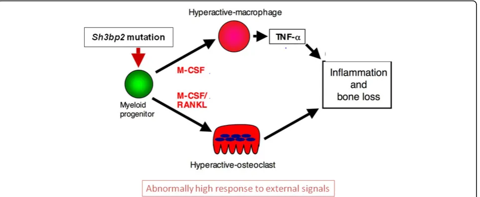
These results point to the existence of at least 2 mechanisms that are involved in the phenotype of the
Sh3bp2KI/KImouse. The authors hypothesize that the effect of the mutation elicits macrophage hyper-reactivity through ERK signaling via a positive autocrine feedback loop, which leads to the increased TNF-a production and inflammatory reactions (Figure 3). The other effect is the generation of hyper-reactive osteoclasts via a Syk-related pathway that leads to increased bone resorption. While TNF-amay have a direct effect on osteoblast dif-ferentiation in vivo, there is also a cell-autonomous effect on osteoblast precursors that can be seen when mutant osteoblasts are cultured in the absence of TNF-a -producing cells [83].
As already discussed in the previous section, NFATc1 is a downstream target of RANKL signaling and a master regulator of osteoclastogenesis. The role of NFATc1 in the cherubism phenotype has been examined by crossing
Sh3bp2KI/KImice withNfatc1conditional knockout mice [85]. Cre-mediated deletion ofNfatc1withMx1-Crein all myeloid cells of 10-day-old mice resulted in an osteo-petrotic phenotype due to lack of osteoclastogenesis. However, the skeletalSh3bp2KI/KIphenotype in double mutant mice was fully rescued in the absence of NFATc1 and the mice actually displayed an osteopetrosis-like phe-notype. The authors showed that NFATc1 is a target of
SH3bp2. NFATc1 is upregulated in RANKL/M-CSF-sti-mulated osteoclast precursors by mutant SH3BP2, which led to the formation of excessive numbers of osteoclasts. In the absence of NFATc1 there was noin vitro osteo-clast formation. However, the Sh3bp2KI/KI /Nfatc1
-/-double mutants still developed inflammatory infiltrates in lungs, livers and other soft-tissue organs as TNF-alevels were still high in those mice.
These experiments confirmed that theSh3bp2KI/KI phe-notype is caused by at least two mechanisms. Mutant SH3BP2 stimulates excessive osteoclastogenesis by increasing NFATc1 expression, which leads to increased bone resorption. Since TNF-alevels are still high in dou-ble mutants but osteoclastogenesis is disrupted, one can conclude that any effect of TNF-aon bone resorption in the cherubism model must go through NFATc1 while signs of inflammatory reactions without osteoclast invol-vement are independent of NFATc1. TNF-ais regulated by SH3BP2 through a mechanism not involving NFATc1 but possibly other NFAT family members [86].
Aliprantis and coworkers also showed that NFATc1 has an inhibitory function on the expression of osteopro-tegerin in stimulated bone marrow osteoclast precursor cells. It is still to be determined whether the reduced level of OPG in osteoblasts ofSh3bp2KI/KImice [83] also depends on NFATc1.
Mice in whichSh3bp2was ablated showed deficiencies mainly in the adaptive immune system.Sh3bp2is required for functional B-cell receptor (BCR) signaling while it is not needed for T-cell receptor (TCR) signaling [38]. The delayed B-cell response may be explained in part by reduced proliferation and increased apoptosis induced by B-cell receptor signaling [87]. Investigating skeletal
responses toSh3bp2ablation may further illuminate the functions ofSh3bp2although results have not yet been made public.
While initial investigations of the cherubism mouse model focused on the skeletal phenotype and abnormal osteoclast and osteoclast differentiation, it became soon apparent that the phenotype in theSh3bp2KI/KImice is at least in part based on abnormal immune response. Then, Ueki and coworkers showed that the generalized chronic inflammation in theSh3bp2KI/KI mouse is elicited by TNF-aand is independent of B- or T-cell involvement. The disease phenotype can be transferred by myeloid cells (monocytes, macrophages) and it can therefore be argued that the disease phenotype is mediated by abnor-mal innate immune response and should be included in the list of autoinflammatory diseases with known genetic origin [88].
Cherubism as an inflammatory disorder
Autoinflammatory disorders are defined by multisystem inflammation without the production of high-titer auto-antibodies or identifiable pathogens [89-91]. Cherubism fulfills these criteria in the mouse model where infiltrat-ing inflammatory lesions are found in many organs and in human patients where bone lesions are limited to the jaws but swelling of lymph nodes is found during or prior to cherubic episodes. Because the process is (at least in the mouse) driven by high levels of TNF-a it could be argued that cherubism is as much a systemic disorder of myeloid cells as it is a matrix disorder [92]. Pro-TNF-ais a plasma membrane protein and the solu-ble form of TNF-ais released by matrix metalloprotei-nases. The various responses to membrane-associated and soluble TNF-aare elicited upon binding of TNF-a to its transmembrane receptors TNFR1 and TNFR2 and the subsequent activation of distinct signaling pathways [93].
TNF-ais also a key player in the host defense to bacter-ial, viral and parasitic infections [93] where it mediates the normal response to the infective agent. However, excessive TNF-aexpression or a temporally or spatially inappropri-ate expression can have damaging effects to the organism, which results in osteopenia and infiltrative inflammatory lesions in theSh3bp2KI/KImouse.
It has long been hypothesized that the limitation of bone-resorptive lesions to the jaws in human cherubism patients is connected to rapid bone remodeling during the development and eruption of the secondary dentition in children [2,11]. The bone remodeling needed in the process of tooth eruption elicits the expression and recruitment of a host of cytokines. It could be those cyto-kines and the hypersensitivity of myeloid cells that trigger a self-sustaining loop of TNF-aexpression that leads to osteoclastogenesis, soft fibrous tissue proliferation and
swollen lymph nodes. In an ongoing study, Ueki and co-workers offer a new hypothesis for the restriction of cher-ubism lesions to the jaws. They suspect that the trigger for cherubism in patients that are heterozygous for a
Sh3bp2mutation could be a hyper-reactive host response to oral pathogens or physical damage that occurs on a regular basis in the oral cavity [94].
Lipopolysaccharide (LPS) produced by Gram-negative commensal bacteria is known to induce osteoclastogen-esis, TNF-aexpression and bone loss [95]. It is conceiva-ble that cherubism patients are predisposed to osteolytic reactions in the jaws once a certain threshold for indu-cing agents (from intense bone remodeling in addition to commensal bacterial load) has been reached. LPS can enhance osteoclastogenesis in RANKL -induced osteo-clast precursors [96]. LPS can also inhibit osteoblast dif-ferentiation [97,98] through the Toll-like receptor expressed on osteoblasts and its interaction with myeloid differentiation factor 88 (MyD88) [99]. The myeloid dif-ferentiation marker MyD88 is an adaptor protein that mediates host response to damage- and pathogen-asso-ciated molecular events. MyD88 is known to act down-stream of Toll-like receptors and the interleukin-1 receptor by interacting with their intracellular Toll/IL-1 receptor homology domains [100]. Current literature suggests that the role of MyD88 in LPS-stimulated clastogenesis is mainly via RANKL stimulation in osteo-blasts and by supporting the survival of differentiated osteoclasts [101].
Ueki and coworkers are now investigating why crosses of Sh3bp2KI/KIand MyD88 deficient mice show less inflammatory infiltrates in bone and other organs and significant improvement of facial swellings and bone resorption [94]. While the importance of LPS or other bacterial products in this partial “rescue” is not yet known, it is obvious that MyD88 plays a major role in the cherubism phenotype of the mouse model and MyD88-independent pathways are likely to contribute as well. Future research will show whether this TLR/IF-1 pathway is needed only for the early stage of cherubism to generate sufficient pro-inflammatory signals and whether some auto-stimulatory loop takes over or whether it is required to maintain the phenotype. What-ever the outcome of this exciting work in progress may be, it is likely to lead to new targets for treatment or pre-vention of cherubism.
of NFATc1 and are likely to be the major drivers for con-tinued bone resorption. There is no current evidence that suggests that immune response in cherubism patients is abnormal. However, cherubic bone resorption is preceded or accompanied by submandibular lymph node swelling, which has not yet been thoroughly investigated. Further immunologic research is needed to study the initiation of bone resorption in the mouse model and how the extra-skeletal inflammatory infiltrations develop. The ultimate goal is to test those findings in cherubism patients and to identify ways to treat or better still, to prevent the disease.
Abbreviations
kDa: kiloDalton; aa: amino acid; SH3BP2: src homology 3 binding protein 2; PH: pleckstrin homology domain; PR: proline-rich domain; SH2: Src-homology 2 domain; Tyr: Tyrosine; Glu: Glutamic Acid; Asn: Asparagine; NS/ MGCLS: Noonan syndrome/multiple giant-cell lesion syndrome; PTPN11: gene encoding the protein tyrosine phosphatase (PTP) Shp2; SOS1: gene encoding the son of sevenless homolog 1 protein; CGCL: central giant cell lesion; NFAT: nuclear factor of activated T cells; PLCγ: phospholipase Cγ; TRAP: tartrate resistant acid phosphatase; sRANKL: soluble receptor activator of NFκB ligand; OPG: osteoprotegerin; TNF-α: tumor necrosis factor-alpha; ERK: extracellular-signal-regulated kinases; SFK: src family kinase; GFP: green fluorescent protein; Jurkat T Ag: Jurkat T Antigen; NFAT-luc: NFAT luciferase; WT: wild-type; OMIM: online mendelian inheritance in man; M-CSF: macrophage-colony stimulating factor; PKC: protein kinase C; TNFR: tumor necrosis factor receptor; BMM: bone marrow macrophages; ITAM: immunoreceptor tyrosine-based activating motifs; MYD88: myeloid differentiation primary response gene (88).
Acknowledgments
This article was developed as part of the Proceedings of the International Meeting on Fibrous Dysplasia/McCune-Albright Syndrome and Cherubism that took place at the National Institutes of Health, Bethesda, MD, October 3-5, 2010. The meeting was supported by funding from the National Institute of Dental and Craniofacial Research and Office of Rare Diseases, NIH, and the Fibrous Dysplasia Foundation. The publication of this manuscript was supported by the Fibrous Dysplasia Foundation and an unrestricted grant from Zimmer, Inc.
This article has been published as part ofOrphanet Journal of Rare Diseases Volume 7 Supplement 1, 2012: International Meeting on Fibrous Dysplasia/ McCune-Albright Syndrome and Cherubism. The full contents of the supplement are available online at http://www.ojrd.com/supplements/7/S1. Publication of the proceedings was funded by the Fibrous Dysplasia Foundation and an unrestricted grant from Zimmer.
Author details
1University of Connecticut Health Center, Department of Reconstructive
Sciences, Center for Regenerative Medicine and Skeletal Development, Farmington, CT, USA.2Division of Endocrinology and Diabetes, The Children’s Hospital of Philadelphia and Department of Pediatrics, University of Pennsylvania School of Medicine, Philadelphia, PA, USA.3Department of Developmental Biology, Harvard School of Dental Medicine, Boston, MA, USA.4Department of Oral and Maxillofacial Surgery, Massachusetts General Hospital, Harvard School of Dental Medicine, Boston, MA, USA.5The Departments of Orthopaedic Surgery and Biomedical Engineering, Cleveland Clinic Lerner Research Institute, Cleveland, OH, USA.
Authors’contributions
EJR and SL have drafted the manuscript. All authors were involved in the critical review of the manuscript. All authors read and approved the final manuscript.
Competing interests
The authors declare that they have no competing interests.
Published: 24 May 2012
References
1. Jones WA:Familial multilocular cystic disease of the jaws.American Journal of Cancer1933,17:946-950.
2. Jones WA, Gerrie J, Pritchard J:Cherubism–familial fibrous dysplasia of the jaws.J Bone Joint Surg Br1950,32-B:334-347.
3. Papadaki ME, Lietman SA, Levine MA, Olsen BR, Kaban LB, Reichenberger EJ: Cherubism: Best Clinical Practice Orphanet.Orphanet Journal of Rare Diseases2012,7(Suppl 1):S6.
4. Anderson DE, McClendon JL:Cherubism - hereditary fibrous dysplasia of the jaws. I. Genetic considerations.Oral Surgery Oral Medicine Oral Pathology1962,15:5-16.
5. Von Wowern N:Cherubism: a 36-year long-term follow-up of 2 generations in different families and review of the literature.Oral Surg Oral Med Oral Pathol Oral Radiol Endod2000,90:765-772.
6. Tiziani V, Reichenberger E, Buzzo CL, Niazi S, Fukai N, Stiller M, Peters H, Salzano FM, Raposo do Amaral CM, Olsen BR:The gene for cherubism maps to chromosome 4p16.Am J Hum Genet1999,65:158-166. 7. Mangion J, Rahman N, Edkins S, Barfoot R, Nguyen T, Sigurdsson A,
Townend JV, Fitzpatrick DR, Flanagan AM, Stratton MR:The gene for cherubism maps to chromosome 4p16.3.American Journal of Human Genetics1999,65:151-157.
8. Hadano S, Ishida Y, Ikeda JE:The primary structure and genomic organization of five novel transcripts located close to the Huntington’s disease gene on human chromosome 4p16.3.DNA Res1998,5:177-186. 9. Zollino M, DS C, Zampino G, Mastroiacovo P, Wright TJ, Sorge G,
Selicorni A, Tenconi R, Zappala A, Battaglia A, Di Rocco M, Palka G, Pallotta R, Altherr MR, Neri G:Genotype-phenotype correlations and clinical diagnostic criteria in wolf-hirschhorn syndrome.Am J Med Genet 2000,94:254-261.
10. Bell SM, Shaw M, Jou YS, Myers RM, Knowles MA:Identification and characterization of the human homologue of SH3BP2, an SH3 binding domain protein within a common region of deletion at 4p16.3 involved in bladder cancer.Genomics1997,44:163-170.
11. Ueki Y, Tiziani V, Santanna C,et al:Mutations in the gene encoding c-Abl-binding protein SH3BP2 cause cherubism.Nat Genet2001,28:125-126. 12. Ren R, Mayer BJ, Cicchetti P, Baltimore D:Identification of a ten-amino
acid proline-rich SH3 binding site.Science1993,259:1157-1161. 13. Deckert M, Rottapel R:The adapter 3BP2: how it plugs into leukocyte
signaling.Adv Exp Med Biol2006,584:107-114.
14. Le Bras S, Foucault I, Foussat A, Brignone C, Acuto O, Deckert M: Recruitment of the actin-binding protein HIP-55 to the immunological synapse regulates T cell receptor signaling and endocytosis.J Biol Chem 2004,279:15550-15560.
15. Le Bras S, Moon C, Foucault I, Breittmayer JP, Deckert M:Abl-SH3 binding protein 2, 3BP2, interacts with CIN85 and HIP-55.FEBS Lett2007,581:967-974. 16. Lietman SA, Kalinchinko N, Deng X, Kohanski R, Levine MA:Identification
of a novel mutation of SH3BP2 in cherubism and demonstration that SH3BP2 mutations lead to increased NFAT activation.Hum Mutat2006, 27:717-718.
17. de Lange J, van Maarle MC, van den Akker HP, Redeker EJ:A new mutation in the SH3BP2 gene showing reduced penetrance in a family affected with cherubism.Oral Surg Oral Med Oral Pathol Oral Radiol Endod 2007,103:378-381.
18. Lo B, Faiyaz-Ul-Haque M, Kennedy S, Aviv R, Tsui LC, Teebi AS:Novel mutation in the gene encoding c-Abl-binding protein SH3BP2 causes cherubism.Am J Med Genet A2003,121A:37-40.
19. Li CY, Yu SF:A novel mutation in the SH3BP2 gene causes cherubism: case report.BMC Med Genet2006,7:84.
20. Carvalho VM, Perdigao PF, Amaral FR, de Souza PE, De Marco L, Gomez RS: Novel mutations in the SH3BP2 gene associated with sporadic central giant cell lesions and cherubism.Oral Dis2009,15:106-110.
21. Carvalho VM, Perdigao PF, Pimenta FJ, de Souza PE, Gomez RS, De Marco L: A novel mutation of the SH3BP2 gene in an aggressive case of cherubism.Oral Oncol2008,44:153-155.
22. Silva EC, de Souza PE, Barreto DC, Dias RP, Gomez RS:An extreme case of cherubism.Br J Oral Maxillofac Surg2002,40:45-48.
23. Cohen MM Jr., Gorlin RJ:Noonan-like/multiple giant cell lesion syndrome. Am J Med Genet1991,40:159-166.
25. Tartaglia M, Zampino G, Gelb BD:Noonan syndrome: clinical aspects and molecular pathogenesis.Mol Syndromol2010,1:2-26.
26. Bufalino A, Carrera M, Carlos R, Coletta RD:Giant cell lesions in noonan syndrome: case report and review of the literature.Head Neck Pathol 2010,4:174-177.
27. Jafarov T, Ferimazova N, Reichenberger E:Noonan-like syndrome mutations in PTPN11 in patients diagnosed with cherubism.Clin Genet 2005,68:190-191.
28. Lee JS, Tartaglia M, Gelb BD, Fridrich K, Sachs S, Stratakis CA, Muenke M, Robey PG, Collins MT, Slavotinek A:Phenotypic and genotypic characterisation of Noonan-like/multiple giant cell lesion syndrome.J Med Genet2005,42:e11.
29. Sarkozy A, Obregon MG, Conti E, Esposito G, Mingarelli R, Pizzuti A, Dallapiccola B:A novel PTPN11 gene mutation bridges Noonan syndrome, multiple lentigines/LEOPARD syndrome and Noonan-like/ multiple giant cell lesion syndrome.Eur J Hum Genet2004,12:1069-1072. 30. Beneteau C, Cave H, Moncla A, Dorison N, Munnich A, Verloes A, Leheup B:
SOS1 and PTPN11 mutations in five cases of Noonan syndrome with multiple giant cell lesions.Eur J Hum Genet2009,17:1216-1221. 31. Hanna N, Parfait B, Talaat IM, Vidaud M, Elsedfy HH:SOS1: a new player in
the Noonan-like/multiple giant cell lesion syndrome.Clin Genet2009, 75:568-571.
32. van Capelle CI, Hogeman PH, van der Sijs-Bos CJ, Heggelman BG, Idowu B, Slootweg PJ, Wittkampf AR, Flanagan AM:Neurofibromatosis presenting with a cherubism phenotype.Eur J Pediatr2007,166:905-909.
33. Ruggieri M, Pavone V, Polizzi A, Albanese S, Magro G, Merino M, Duray PH: Unusual form of recurrent giant cell granuloma of the mandible and lower extremities in a patient with neurofibromatosis type 1.Oral Surg Oral Med Oral Pathol Oral Radiol Endod1999,87:67-72.
34. De Luca A, Bottillo I, Sarkozy A,et al:NF1 gene mutations represent the major molecular event underlying neurofibromatosis-Noonan syndrome. Am J Hum Genet2005,77:1092-1101.
35. Bertola DR, Pereira AC, Passetti F, de Oliveira PS, Messiaen L, Gelb BD, Kim CA, Krieger JE:Neurofibromatosis-Noonan syndrome: molecular evidence of the concurrence of both disorders in a patient.Am J Med Genet A2005,136:242-245.
36. Stamm S, Riethoven JJ, Le Texier V, Gopalakrishnan C, Kumanduri V, Tang Y, Barbosa-Morais NL, Thanaraj TA:ASD: a bioinformatics resource on alternative splicing.Nucleic Acids Res2006,34:D46-55.
37. Proulx-Bonneau S, Guezguez A, Annabi B:A concerted HIF-1α/MT1-MMP signalling axis regulates the expression of the 3BP2 adaptor protein in hypoxic mesenchymal stromal cells.PLoS One2011,6:e21511. 38. de la Fuente MA, Kumar L, Lu B, Geha RS:3BP2 deficiency impairs the
response of B cells, but not T cells, to antigen receptor ligation.Mol Cell Biol2006,26:5214-5225.
39. Foucault I, Le Bras S, Charvet C, Moon C, Altman A, Deckert M:The adaptor protein 3BP2 associates with VAV guanine nucleotide exchange factors to regulate NFAT activation by the B-cell antigen receptor.Blood2005, 105:1106-1113.
40. Foucault I, Liu YC, Bernard A, Deckert M:The chaperone protein 14-3-3 interacts with 3BP2/SH3BP2 and regulates its adapter function.J Biol Chem2003,278:7146-7153.
41. Jevremovic D, Billadeau DD, Schoon RA, Dick CJ, Leibson PJ:Regulation of NK cell-mediated cytotoxicity by the adaptor protein 3BP2.J Immunol 2001,166:7219-7228.
42. Maeno K, Sada K, Kyo S, Miah SM, Kawauchi-Kamata K, Qu X, Shi Y, Yamamura H:Adaptor protein 3BP2 is a potential ligand of Src homology 2 and 3 domains of Lyn protein-tyrosine kinase.J Biol Chem 2003,278:24912-24920.
43. Sada K, Miah SM, Maeno K, Kyo S, Qu X, Yamamura H:Regulation of FcepsilonRI-mediated degranulation by an adaptor protein 3BP2 in rat basophilic leukemia RBL-2H3 cells.Blood2002,100:2138-2144. 44. Deckert M, Tartare-Deckert S, Hernandez J, Rottapel R, Altman A:Adaptor
function for the Syk kinases-interacting protein 3BP2 in IL-2 gene activation.Immunity1998,9:595-605.
45. Faccio R, Teitelbaum SL, Fujikawa K, Chappel J, Zallone A, Tybulewicz VL, Ross FP, Swat W:Vav3 regulates osteoclast function and bone mass.Nat Med2005,11:284-290.
46. Qu X, Kawauchi-Kamata K, Miah SM, Hatani T, Yamamura H, Sada K: Tyrosine phosphorylation of adaptor protein 3BP2 induces T cell
receptor-mediated activation of transcription factor.Biochemistry2005, 44:3891-3898.
47. Zou W, Teitelbaum SL:Integrins, growth factors, and the osteoclast cytoskeleton.Ann N Y Acad Sci2010,1192:27-31.
48. Chiusaroli R, Sanjay A, Henriksen K, Engsig MT, Horne WC, Gu H, Baron R: Deletion of the gene encoding c-Cbl alters the ability of osteoclasts to migrate, delaying resorption and ossification of cartilage during the development of long bones.Dev Biol2003,261:537-547.
49. Peruzzi G, Molfetta R, Gasparrini F, Vian L, Morrone S, Piccoli M, Frati L, Santoni A, Paolini R:The adaptor molecule CIN85 regulates Syk tyrosine kinase level by activating the ubiquitin-proteasome degradation pathway.J Immunol2007,179:2089-2096.
50. Yu Z, Maoui M, Zhao ZJ, Li Y, Shen SH:SHP-1 dephosphorylates 3BP2 and potentially downregulates 3BP2-mediated T cell antigen receptor signaling.FEBS J2006,273:2195-2205.
51. Saborit-Villarroya I, Del Valle JM, Romero X, Esplugues E, Lauzurica P, Engel P, Martin M:The adaptor protein 3BP2 binds human CD244 and links this receptor to Vav signaling, ERK activation, and NK cell killing.J Immunol2005,175:4226-4235.
52. Ueki Y, Lin CY, Senoo M,et al:Increased Myeloid Cell Responses to M-CSF and RANKL Cause Bone Loss and Inflammation in SH3BP2“Cherubism” Mice.Cell2007,128:71-83.
53. GuezGuez A, Prod’homme V, Mouska X, Baudot A, Blin-Wakkach C, Rottapel R, Deckert M:3BP2 Adapter protein is required for receptor activator of NFkappaB ligand (RANKL)-induced osteoclast differentiation of RAW264.7 cells.J Biol Chem2010,285:20952-20963.
54. Bergemann AD, Cole F, Hirschhorn K:The etiology of Wolf-Hirschhorn syndrome.Trends Genet2005,21:188-195.
55. Battaglia A, Carey JC:Health supervision and anticipatory guidance of individuals with Wolf-Hirschhorn syndrome.Am J Med Genet1999, 89:111-115.
56. Battaglia A, Carey JC, Cederholm P, Viskochil DH, Brothman AR, Galasso C: Natural history of Wolf-Hirschhorn syndrome: experience with 15 cases. Pediatrics1999,103:830-836.
57. Takayanagi H, Kim S, Koga T,et al:Induction and activation of the transcription factor NFATc1 (NFAT2) integrate RANKL signaling in terminal differentiation of osteoclasts.Dev Cell2002,3:889-901. 58. Ishida N, Hayashi K, Hoshijima M, Ogawa T, Koga S, Miyatake Y,
Kumegawa M, Kimura T, Takeya T:Large scale gene expression analysis of osteoclastogenesis in vitro and elucidation of NFAT2 as a key regulator. J Biol Chem2002,277:41147-41156.
59. Hirotani H, Tuohy NA, Woo JT, Stern PH, Clipstone NA:The calcineurin/ nuclear factor of activated T cells signaling pathway regulates osteoclastogenesis in RAW264.7 cells.J Biol Chem2004,279:13984-13992. 60. Bellows CG, Ishida H, Aubin JE, Heersche JN:Parathyroid hormone
reversibly suppresses the differentiation of osteoprogenitor cells into functional osteoblasts.Endocrinology1990,127:3111-3116.
61. Komarova SV, Pereverzev A, Shum JW, Sims SM, Dixon SJ:Convergent signaling by acidosis and receptor activator of NF-kappaB ligand (RANKL) on the calcium/calcineurin/NFAT pathway in osteoclasts.Proc Natl Acad Sci U S A2005,102:2643-2648.
62. Uhlen P, Burch PM, Zito CI, Estrada M, Ehrlich BE, Bennett AM: Gain-of-function/Noonan syndrome SHP-2/Ptpn11 mutants enhance calcium oscillations and impair NFAT signaling.Proc Natl Acad Sci U S A2006, 103:2160-2165.
63. Takayanagi H:Mechanistic insight into osteoclast differentiation in osteoimmunology.J Mol Med2005,83:170-179.
64. Billadeau DD, Upshaw JL, Schoon RA, Dick CJ, Leibson PJ:NKG2D-DAP10 triggers human NK cell-mediated killing via a Syk-independent regulatory pathway.Nat Immunol2003,4:557-564.
65. Koga T, Inui M, Inoue K,et al:Costimulatory signals mediated by the ITAM motif cooperate with RANKL for bone homeostasis.Nature2004, 428:758-763.
66. Lietman SA, Yin L, Levine MA:SH3BP2 is an activator of NFAT activity and osteoclastogenesis.Biochem Biophys Res Commun2008,371:644-648. 67. Lietman SA, Yin L, Levine MA:SH3BP2 mutations potentiate
osteoclastogenesis via PLCgamma.J Orthop Res2010,28:1425-1430. 68. Mao D, Epple H, Uthgenannt B, Novack DV, Faccio R:PLCgamma2
69. Patterson RL, van Rossum DB, Ford DL, Hurt KJ, Bae SS, Suh PG, Kurosaki T, Snyder SH, Gill DL:Phospholipase C-gamma is required for agonist-induced Ca2+ entry.Cell2002,111:529-541.
70. Wilde JI, Watson SP:Regulation of phospholipase C gamma isoforms in haematopoietic cells: why one, not the other?Cell Signal2001, 13:691-701.
71. Rebecchi MJ, Pentyala SN:Structure, function, and control of phosphoinositide-specific phospholipase C.Physiol Rev2000, 80:1291-1335.
72. Katan M:Families of phosphoinositide-specific phospholipase C: structure and function.Biochim Biophys Acta1998,1436:5-17. 73. Watanabe D, Hashimoto S, Ishiai M, Matsushita M, Baba Y, Kishimoto T,
Kurosaki T, Tsukada S:Four tyrosine residues in phospholipase C-gamma 2, identified as Btk-dependent phosphorylation sites, are required for B cell antigen receptor-coupled calcium signaling.J Biol Chem2001, 276:38595-38601.
74. Humphries LA, Dangelmaier C, Sommer K, Kipp K, Kato RM, Griffith N, Bakman I, Turk CW, Daniel JL, Rawlings DJ:Tec kinases mediate sustained calcium influx via site-specific tyrosine phosphorylation of the phospholipase Cgamma Src homology 2-Src homology 3 linker.J Biol Chem2004,279:37651-37661.
75. Kim YJ, Sekiya F, Poulin B, Bae YS, Rhee SG:Mechanism of B-cell receptor-induced phosphorylation and activation of phospholipase C-gamma2. Mol Cell Biol2004,24:9986-9999.
76. Hur EM, Park YS, Lee BD, Jang IH, Kim HS, Kim TD, Suh PG, Ryu SH, Kim KT: Sensitization of epidermal growth factor-induced signaling by bradykinin is mediated by c-Src. Implications for a role of lipid microdomains.J Biol Chem2004,279:5852-5860.
77. Jongstra-Bilen J, Puig Cano A, Hasija M, Xiao H, Smith CI, Cybulsky MI:Dual functions of Bruton’s tyrosine kinase and Tec kinase during Fcgamma receptor-induced signaling and phagocytosis.J Immunol2008, 181:288-298.
78. Mukherjee PM, Wang CJ, Chen IP, Jafarov T, Olsen BR, Ueki Y,
Reichenberger EJ:Cherubism gene Sh3bp2 is important for optimal bone formation, osteoblast differentiation, and function.Am J Orthod Dentofacial Orthop2010,138:140, e141-140 e111; discussion 140-141. 79. Chipman SD, Sweet HO, McBride DJ Jr., Davisson MT, Marks SC Jr.,
Shuldiner AR, Wenstrup RJ, Rowe DW, Shapiro JR:Defective pro alpha 2(I) collagen synthesis in a recessive mutation in mice: a model of human osteogenesis imperfecta.Proc Natl Acad Sci U S A1993,90:1701-1705. 80. Chen IP, Wang CJ, Strecker S, Koczon-Jaremko B, Boskey A,
Reichenberger EJ:Introduction of a Phe377del mutation in ANK creates a mouse model for craniometaphyseal dysplasia.J Bone Miner Res2009, 24:1206-1215.
81. Adler CP, Harle F:23. Zur Differentialdiagnose osteo-fibroser
Kieferekrankungen.Verhandlungen der Deutschen Gesellschaft fur Pathologie 1974,58:308-314.
82. Liao BY, Zhang J:Null mutations in human and mouse orthologs frequently result in different phenotypes.Proc Natl Acad Sci U S A2008, 105:6987-6992.
83. Wang CJ, Chen IP, Koczon-Jaremko B, Boskey AL, Ueki Y, Kuhn L, Reichenberger EJ:Pro416Arg cherubism mutation in Sh3bp2 knock-in mice affects osteoblasts and alters bone mineral and matrix properties. Bone2010,46:1306-1315.
84. Kalajzic I, Kalajzic Z, Kaliterna M, Gronowicz G, Clark SH, Lichtler AC, Rowe D: Use of type I collagen green fluorescent protein transgenes to identify subpopulations of cells at different stages of the osteoblast lineage.J Bone Miner Res2002,17:15-25.
85. Aliprantis AO, Ueki Y, Sulyanto R, Park A, Sigrist KS, Sharma SM, Ostrowski MC, Olsen BR, Glimcher LH:NFATc1 in mice represses osteoprotegerin during osteoclastogenesis and dissociates systemic osteopenia from inflammation in cherubism.J Clin Invest2008, 118:3775-3789.
86. Kaminuma O, Kitamura F, Kitamura N, Hiroi T, Miyoshi H, Miyawaki A, Miyatake S:Differential contribution of NFATc2 and NFATc1 to TNF-alpha gene expression in T cells.J Immunol2008,180:319-326.
87. Chen G, Dimitriou ID, La Rose J, Ilangumaran S, Yeh WC, Doody G, Turner M, Gommerman J, Rottapel R:The 3BP2 adapter protein is required for optimal B-cell activation and thymus-independent type 2 humoral response.Mol Cell Biol2007,27:3109-3122.
88. Ferguson PJ, El-Shanti HI:Autoinflammatory bone disorders.Curr Opin Rheumatol2007,19:492-498.
89. Chitkara P, Stojanov S, Kastner DL:The hereditary autoinflammatory syndromes.Pediatr Infect Dis J2007,26:353-354.
90. McGonagle D, Aziz A, Dickie LJ, McDermott MF:An integrated
classification of pediatric inflammatory diseases, based on the concepts of autoinflammation and the immunological disease continuum.Pediatr Res2009,65:38R-45R.
91. McGonagle D, McDermott MF:A proposed classification of the immunological diseases.PLoS Med2006,3:e297.
92. Nicolae C, Olsen BR:Unexpected matrix diseases and novel therapeutic strategies.Cell Tissue Res2010,339:155-165.
93. Bradley JR:TNF-mediated inflammatory disease.J Pathol2008, 214:149-160.
94. Ueki Y, Mukai T, Yoshitaka T:Mechanism of inflammation in cherubism.In J Bone Miner Res2010,25(Suppl 1), Available at [http://www.asbmr.org/ Meetings/AnnualMeeting/AbstractDetail.aspx?aid=9553097c-08c8-4d7e-897a-6ed4927d0475] Accessed [12-24-2010].
95. Abu-Amer Y, Ross FP, Edwards J, Teitelbaum SL: Lipopolysaccharide-stimulated osteoclastogenesis is mediated by tumor necrosis factor via its P55 receptor.J Clin Invest1997,100:1557-1565.
96. Liu J, Wang S, Zhang P, Said-Al-Naief N, Michalek SM, Feng X:Molecular mechanism of the bifunctional role of lipopolysaccharide in osteoclastogenesis.J Biol Chem2009,284:12512-12523.
97. Tomomatsu N, Aoki K, Alles N, Soysa NS, Hussain A, Nakachi H, Kita S, Shimokawa H, Ohya K, Amagasa T:LPS-induced inhibition of osteogenesis is TNF-alpha dependent in a murine tooth extraction model.J Bone Miner Res2009,24:1770-1781.
98. Kadono H, Kido J, Kataoka M, Yamauchi N, Nagata T:Inhibition of osteoblastic cell differentiation by lipopolysaccharide extract from Porphyromonas gingivalis.Infect Immun1999,67:2841-2846. 99. Bandow K, Maeda A, Kakimoto K, Kusuyama J, Shamoto M, Ohnishi T,
Matsuguchi T:Molecular mechanisms of the inhibitory effect of lipopolysaccharide (LPS) on osteoblast differentiation.Biochem Biophys Res Commun2010,402:755-761.
100. Akira S:Toll-like receptor signaling.J Biol Chem2003,278:38105-38108. 101. Sato N, Takahashi N, Suda K,et al:MyD88 but not TRIF is essential for
osteoclastogenesis induced by lipopolysaccharide, diacyl lipopeptide, and IL-1alpha.J Exp Med2004,200:601-611.
doi:10.1186/1750-1172-7-S1-S5
Cite this article as:Reichenbergeret al.:The role of SH3BP2 in the
pathophysiology of cherubism.Orphanet Journal of Rare Diseases20127
(Suppl 1):S5.
Submit your next manuscript to BioMed Central and take full advantage of:
• Convenient online submission
• Thorough peer review
• No space constraints or color figure charges
• Immediate publication on acceptance
• Inclusion in PubMed, CAS, Scopus and Google Scholar
• Research which is freely available for redistribution