Abstract— The present work proposes a methodology for detecting plant diseases early and accurately, using diverse image processing techniques. Farmers experience great difficulties in changing from one disease control policy to another. Relying on pure naked- eye observation to detect and classify diseases can be expensive various plant diseases pose a great threat to the agricultural sector by reducing the life of the plants. The aim is to develop a simple disease detection system for plant diseases. The work begins with capturing the images. Filtered and segmented using median filtering method. Then, color and texture features are extracted from the result of segmentation. Classification is done to detect the type of disease the leaf has been affected. Index Terms— Red Green Blue, Gray Level Co-occurrence Matrix, Alternaria disease, Bacterial Blight
1) INTRODUCTION
Agriculture is the mother of all cultures. It has played a key role in the development of human civilization.Agricultural practices such as irrigation, crop rotation, fertilizers and pesticides were developed long ago, but have made great strides in the past century. By the early 19th century, agricultural techniques had so improved that yield per land unit was many times that seen in the middle ages. Therefore, judicious management of all the inputs is essential for the sustainability of a complex system. The focus on enhancing the productivity, without considering the ecological impacts has resulted into environmental degradation.Plant disease is one of the crucial causes that reduces quantity and degrades quality of the agricultural products. Diseases and insect pests are the major problems that threaten pomegranate cultivation. These require careful diagnosis and timely handling to protect the crops from heavy loses. In pomegranate plant diseases can be found in various parts such as fruit, stem and leaves. Major diseases that affect pomegranate fruit are Bacterial Blight, Alternaria and Phomopsis.The disease symptoms can be initially found on stem part which gradually pervades to leaves and then to fruits. On leaves, the disease starts with small, irregular, water soaked spots that are 2 to 5 mm in size with necrotic centre of pin head size. Spots are translucent against light. Later, these spots turn light to dark brown and are surrounded by prominent water soaked
margins. Numerous spots may coalesce to form bigger patches. Severely infected leaves may drop off. High temperature and high relative humidity favors the disease. The disease spreads to healthy plants through wind splashed rains and in new area through infected cuttings.
Leaf area plays an important role in plant growth analysis and photosynthesis. Leaf area is measured using different destructive and non-destructive methods. In destructive methods first the leaf is removed from plant and then measured. In non-destructive methods dimensions of leaf are measured without removing the leaf.The technology leverage farmers can take up to asses the leaf, look at the possibility of diseases at early stages take decision on possible treatment and the like. A methodology is developed to determine the type of disease the leaf is affected.
2) PROBLEMSTATEMENT
When the pomegranate had been infected or attacked by some disease, the other areas had been exposed to be infected. Thus, it will decrease pomegranate yield and it also reduces farmer’s income. Currently, the pomegranate farmer determines the type of disease manually. The errors might occur in order to determine the type of diseases. Pomegranate farmers also have to spend a lot of time to detect the type of disease. It also takes a time as the pomegranate farmers manually check the disease since the pomegranate field is in wide area.
3) OBJECTIVES
Creation of our own leaf image data set.
Exploring different segmentation methods to achieve best classification of accuracy.
To determine the type of disease the leaf has been affected.
4) EXISTINGSYSTEM
The emergence and spreading of diseases have become more common because of climate and environmental factors.The naked eye observation is the main approach adopted so far.Due to factors like different working conditions, personal judgment and level of fatigue, grading results often turn out to be inconsistent among individual inspectors.It is clear that some approaches are found to be inaccurate, primarily because the input image contains noise.To achieve this
Disease Detection in Pomegranate Leaf
Using Image Processing Technique
Sowmya GM1, Chandan V2, Sampath Kini3
1PG Student,Dept Of CSE,NMAM Institute Of Technology,Nitte,India
2Assistant Professor, Dept Of ISE, Bapuji Institute Of Engineering Technology,Davanagere,India
objective, the current visual inspection methods need to be automated.
5) PROPOSEDSYSTEM
Proposed system starts with the acquisition of image sample. The image sample is pre-processed to remove noise and enhance it.It is then converted into binary image and its texture features are extracted. Later classification is done based on which we are going to detect the type of disease the leaf has been affected.
6) LITERATURESURVEY
H. Al-Hiary, S. Bani-Ahmad, M. Reyalat, M. Braik
and A. Rahamneh: We are proposed Fast and
Accurate Detection and Classification of Plant diseases; we propose and experimentally evaluate a software solution for automatic detection and classification of plant leaf diseases. The proposed solution is an improvement to the solution proposed as it provides faster and more accurate solution. The developed processing scheme consists of four main phases. The following two steps are added successively after the segmentation phase. In the first step we identify the mostly-green colored pixels. Next, these pixels are masked based on specific threshold values that are computed using Otsu's method, then those mostly green pixels are masked. The other additional step is that the pixels with zeros red, green and blue values and the pixels on the boundaries of the infected cluster (object) were completely removed. The experimental results demonstrate that the proposed technique is a robust technique for the detection of plant leaves diseases.
B. S. Anami and D. G. Savakar: we are proposed the Improved Method for Identification and Classification of Foreign bodies, mixed food grains, automated food processing and evaluation is considered a significant research area in computer vision. The development of automated cooking and food serving by robots is envisaged as part of automated food processing and temperature plays a major role in cooking Indian foods. The delicious Indian foods are generally boiled or fried with other ingredients. The boiled grains like Bengal Gram, Black Gram, Green Gram, Red Gram and Toor Dal are part of typical Indian foods and taste differently, when boiled or cooked at different temperatures and periods of time. Therefore, identifying the effect of boiling and automatic recognition of images of boiled food grains is presented in this paper. The boiling temperatures chosen are 400 C, 500 C, 600 C, 800 C and 1000 C. A color feature centered knowledge based classifier is proposed. The classification accuracy observed is high at lower and higher temperatures and low at medium temperatures. The work finds applications in automatic inspection of food preparations in food industries, drug preparation in pharmaceutical industries, automatic serving, cooking and monitoring of foods in restaurants and motels.
C. C. Tuker and K. Chakraborty: we are proposed Quantitative Assessment of Lesion Characteristics and Disease Severity Using Digital Image Processing, This work describes dedicated software which detects and characterizes disease lesions on leaves to provide data on the number and type of lesions and the percentage of leaf area diseased (severity). The software, written in C2+, can be used with a standard computer in combination with a colour CCD camera and a frame grabber for image acquisition. The usefulness and adaptability of the software was evaluated using two foliar diseases, Alternaria blight of sunflower and oat leaf rust, which differ in symptoms. Using image segmentation and classification techniques, the software discriminated disease symptoms from the healthy leaf area. The number and size of lesions and severity, obtained using the image processing software, were compared with those calculated using a software planimeter or visual assessment. Significant linear relationships between planimeter and the imaging software were obtained for lesion number and severity in oat leaf rust and for severity in sunflower blight. Artifacts, mistakenly classified as blight lesions by the imaging software resulted in an over-estimation of the number of lesions. Future research is aimed at improving accuracy through better illumination during image capture. A dedicated, compact and portable hardware is currently being developed for field use as a self-contained device for disease assessment.
Z. May and M. H. Amaran: we are proposed Automated Oil Palm Fruit Grading System using Artificial Intelligence, This project deals with the ripeness of oil palm fruit. The current procedure in the palm oil mills is grading the oil palm fruit manually using human graders. This method is subjective and inconsistence because each graders has its own techniques and may vary from each other's. Hence, it affects the quality and quantity of the oil that can be extracted. In this project, a new model of automated grading system for oil palm fruit is developed using the RGB color model and artificial fuzzy logic.
7) METHODOLOGY
7.1 Image Acquisition:
Image Acquisition is the process of collecting the image samples. Images are taken from the digital camera and samples are stored in dataset.
function main()
[file,path]=uigetfile('*.*','Select input image'); I=imread(strcat(path,file)); Io=I; % if size(I,3)==3 % I=rgb2gray(I); % end I=I(:,:,1); I=double(I); subplot(2,2,1),imshow(I,[]); title('ORIGINAL IMAGE');
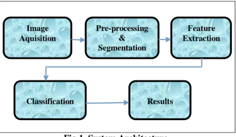
Fig 1. System Architecture 7.2 Pre-processing and Segmentation:
Segmentation refers to the process of clustering the pixels with certain properties into salient regions and these regions correspond to individual surfaces, objects or natural parts of the objects. More precisely, image segmentation is the process of assigning a label to every pixel in an image so that pixels with the same label share certain visual characteristics. The ultimate goal of segmentation is to simplify and/or change the representation of an image into a form more meaningful and easier to analyze. We propose the following techniques to segment the affected area.
%% HE=im2bw(uint8(I),0.2); I=(~HE.*I); [J,ass2]=kmeans(I,2); %% KM(:,:,1)=J; KM(:,:,2)=ass2; l1=length(find(J)); l2=length(find(ass2)); cv=[l1 l2]; cv=cv(find(cv>0)); [a,b]=min(cv); J=bwareaopen(KM(:,:,b),2); subplot(2,2,3),imshow(J,[]); title('SEGMENTED IMAGE'); 7.3 Feature Extraction:
In this step we extract the desired features from the sample image for the analysis of pathogenic affected region of the fruit. Color feature extraction & Texture Feature Extraction is employed.
7.3.1 Color Feature Extraction:
There are four color models namely RGB, HIS, CMY, YIQ. We have used RGB color model for analysis. Red, Green and Blue component of the image sample is extracted. We also extract the total count of red, green and blue pixels in the pomegranate leaf. We also extract the total count of pixels occupied by entire pomegranate, which gives the total area of pomegranate.
7.3.2 Texture Features Extraction:
Important texture features like correlation, homogeneity, entropy and contrast are extracted along with color features. We use Gray Level Co- occurrence Matrix for this purpose. Gray Level Co-occurrence Matrix: This method was proposed by Haralick. GLCM is a texture description
method, which is based on the repeated occurrence of some gray-level configuration in the texture. Haralick features calculation is done in two phases:
i) Calculation of the Co-occurrence Matrices,
ii) Calculation of the features based on the Co-occurrence Matrix.
7.5 Classification:Classification is done based on the Euclidian distance and different types of disease is detected like Alternaria, Bacterial Blight and Phomopsis.
handles.output = hObject; jk=handles.jk; if jk==1 load out.mat load group.mat fe1=out; gr=group; else fe1=handles.out; gr=handles.group; end fe=handles.feature; J=handles.J; for i=1:29 s(i)=sum((fe1(i,:)-fe').^2); end [a,b]=min(s); dc=gr(b); if dc==1
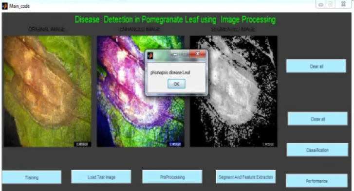
msgbox('Alternaria disease Leaf');
end if dc==2
msgbox('bacterial blight disease Leaf');
end if dc==3
msgbox('Normal Leaf');
end if dc==4
msgbox('phomopsis disease Leaf');
end GLCM2 = co_occurent_matrix(J,'Offset',[2 0;0 2]); out = GLCM_Features(GLCM2,0); feature(1,:)=out.maxpr; feature(2,:)=out.energ; feature(3,:)=out.entro; feature(4,:)=out.contr; feature(5,:)=out.dissi; feature(6,:)=out.homom; feature(7,:)=out.idmnc; [r,c]=find(J); Aarea=length(r); Tarea=size(I,1)*size(I,2); Image Aquisition Pre-processing & Segmentation Feature Extraction Classification Results
8) RESULTS AND SCREENSHOTS
The below figure 2 illustrates the interface design of image processing technique. When we open the MAT LAB software for the code execution and run the code the output screen will be displayed as in the figure 2. The front end used in our project is Graphical User Interface(GUI).
Here training refers to the initial stage,where the image samples are collected from the dataset. We are considering four group labels where each group label contains image samples
which are affected by different diseases. Group 1 contains image samples affected by Alternaria disease, Group 2 contains samples affected by Bacterial Blight disease,where Group 3 contains normal leaf images and Group 4 contains image samples affected by Phomopsis.
.
Figure 2. Image Acquisition of Altenaria disease In case, if we consider any query image sample, during classification when the query image sample matches to the features of any one of the groups,then the corresponding disease is displayed, if not the image is considered as healthy leaf sample.
Figure 3: Enhanced Image of Alternaria disease Figure 3 shows the enhanced and intensified image where the RGB image is converted to gray scale image. The main objective of this process is to obtain the binary image. This process includes filtering, intensifying the image and Binary form. We apply median filtering method, which is a non-linear method of removing noise from the image & preserving the edges. The image sample is now intensified and converted to binary image.
Figure 4: Segmented Image of Alternaria disease In the above Figure 4 the preprocessed image is segmented using K-means clustering algorithm. Segmentation refers to the process of clustering the pixels with certain properties into salient regions and these regions correspond to individual surfaces, objects or natural parts of the objects. More precisely, image segmentation is the process of assigning a label to every pixel in an image so that pixels with the same label share certain visual characteristics.
Figure 5: Classified Image of alternaria disease The K-means clustering technique is a technique which aims to partition n observations into k clusters in which each observation belongs to the cluster with the nearest mean. In this method, k is the number of clusters in the segmented image.
The below Figure 5 and 6 are the classified images of the bacterial blight and phomosis identified disease of pomegranate leaves respectively, where it undergoes preprocessing and segmentation techniques then classified for the affected disease.
Figure 7: Classified image of Phomopsis leaf
9) CONCLUSION
A system for diagnosis the leaf disease has been developed using the Matlab application. The image processing techniques is applied to improve and enhance the image to a better quality. This methodology involves image acquisition, pre-processing and segmentation, analysis and classification of the pomegranate leaf. All the leaf image samples will be passing through the segmentation techniques like thresholding, region growing, K-means clustering are used to separate affected lesion area from normal area and then feature extraction is done through GLCM technique. Later it undergoes classification to identify various types of diseases. ADVANTAGES
Shows preliminary and final results instantly.
User-friendly, accurate, fast, efficient and effective solution to the problem.
Provides an instant analysis of the quality of product by finding different diseases in pomegranate leaf. DISADVANTAGES
This requires continuous monitoring of experts which might be prohibitively expensive in large farms.
It is very costly depending on the system used, the number of detectors purchased.
This makes consulting experts too expensive and time consuming and more over farmers are unaware of non-native diseases
FUTURE ENHANCEMENT
This prototype has a very great potential to be further improved in the future. The work carried out has relevance to real world classification of fruits disease using different classifier to get better accuracy rate and it involves both image processing and pattern recognition techniques.
.
REFERENCES
1) D. Al Bheesh, M. Braik and S. Bani-Ahmad, “Detection and Classification of leaf diseases using K-means-based segmentation and Neural-networks based Classification”, Information Technology Journal, (2011), pp. 265-267.
2) J. K. Patil and R. Kumar, “Advances in image processing in image processing for detection of plant diseases”, Journal of Advanced Bioinformatics Applications and Research, vol. 2, Issue 2, (2011), pp. 135-141.
3) Z. May and M. H. Amaran, “Automated Oil Palm Fruit Grading System using Artificial Intelligence”,
International Journal of Video & Image Processing and Network Security, vol. 11, no. 03, (2011). 4) N. A. Lili, F. Khalid and N. M. Borhan,
“Classification of Herbs Plant Diseases via Hierarchical Dynamic Artificial Neural Network after Image Removal using Kernel Regression Framework”, International Journal on Computer Science and Engineering, vol. 3, no. 1, (2011). 5) D. S. Guru, P. B. Mallikarjuna and S. Manjunath,
“Segmentation and Classification of Tobacco Seedling Diseases”, COMPUTE '11 Proceedings of the Fourth Annual ACM Bangalore Conference, (2011).