Disease Detection of Cotton crop using Image
Processing Technique: A Survey
Naik Durgesh Manikrao1, Dr. Prof. A.J.Vyavahare2
1
P. G. Student at Department of E&TC, 2Associate professor at Department of E&TC Savitribai Phule University Pune, India
Abstract— Farming is important sector in India for human being, as near about 55-60% people are depends directly and indirectly on it. Among all crops, Cotton is main cash crop in India gives more income to the farmer. Due to diseases on cotton there may be chances of decrease in production and drastic change is occurred on crop. The fungal diseases like Verticilium Wilt, Bacterial blight, Red spot, Alternaria, Downy Mildew are responsible for production loss. So, this paper presents various types of diseases and control on it using image processing technique. The comparative study of artificial neural network, Support vector machine is discussed
Keywords— Image Processing, Agriculture, leaf diseases, Artificial Neural network, Support vector machine.
I. INTRODUCTION
No one knows exactly how old cotton is Scientists searching caves in china found bits of cotton bolls and pieces of cotton cloth that proved that it may be about 7,000 years old. They also get that the cotton itself was much like that grown in India today; In the Indus River in Pakistan, cotton was being made, produced and woven into cloth 3,000 years BC. At about the same duration, natives of Egypt’s Nile valley were making and wearing cotton clothing. Diseases on the cotton are the main problem that decreases the productivity of the cotton. The main source for the disease is the leaves of the cotton plant. About 80 to 90 % of disease on the cotton plant is on its leaf. So f our study of interest is the leaf of the cotton tree rather than whole cotton plant the cotton leaves is mainly suffered from diseases like insecticide(tudtude,
mawa) fungus, Foliar leaf on leaf of cotton, Alternaria leaf spot of cotton. The machine vision system now a day is normally consists of computer, digital camera and application software. Various types of algorithms are integrated in the application. Image processing is one important method that helps segment image into objects and background image. One of the key steps in image analysis is feature detection. Study of diseases on the cotton leaf can robustly studied by the image processing toolbox and also the diagnosis by using MATLAB helps us to suggest necessary remedy for that disease arises on the leaf of cotton plant. Image recognition has attracted many researchers in the area of pattern recognition, similar flow of concept are applied to the field of pattern recognition of plant leaf, that is used in diagnosing the cotton leaves diseases. There are numerous methods have been proposed in the last two decades which are not fully solved. However this is challenging problems. The critical issue is how to extract the discriminative and stable feature for classification. It is found that linear subspace analysis has been extensively studied and becomes a popular feature extraction method.
II. ORIGIN OF RESEARCH PROBLEM
A. Diseases on Cotton
The diseases on the cotton leaves are classified as
1) Bacterial disease: e.g. Bacterial Blight, Crown Gall,
2) Fungal diseases: e.g. Anthracnose, Leaf Spot., Alternaria
3) Viral disease: e.g. Leaf Curl, Leaf Crumple, Leaf Roll.
4) Diseases: Due To insects: e.g. White flies Leaf insects.
Above types of disease these are dramatically affect the leaf of cotton plant and its Leaves. We go through the selective type of diseases on the cotton leaves. And further we discuss the ANN image segmentation method to detect the diseases on cotton plant by scanning of cotton leaves through our portable dedicated scanner.
So many diseases are found on the cotton plant out of this we discuss the disease some of the major diseases which are often found on the leaves of cotton, those are viz.
As shown in Fig 1, small, brown, round or irregular spots measuring 0.5 - 3 mm in diameter and broken centers appears on the affected leaves on the plant. Affected leaf become dry and fall off [3], [4],. The disease may cause cracks on the stem. The infection spreads to the fruit finally falls off.
Figure No.1: Alternaria Leaf Spot
C. Curl Gemini virus
Cotton leaf curl Gemini virus (CLCuV) causes a major disease of cotton in Asia and Africa [2],[3],[4]. Leaves of infected cotton curl upward Fig 3. and bear leaf-like enations on the underside along with vein thicken Fig 3. Plants infected earlier in the season are stunted and yield is reduced drastically. Severe epidemic of CLCuV have occurred in Pakistan in the past few years, with yield losses as high as 100% in fields where infection occurred early in the growing season. Another cotton Gemini virus, cotton leaf crumple virus (CLCrV),
[image:3.612.185.432.311.379.2]Occurs in Arizona, California, and Mexico. CLCrV symptoms are distinguishable from CLCuV symptoms in that infected leaves curl downward accompanied
Figure No. 2: Curl Gemini virus
D. Bacterial Blight
Xanthomonas campestis pv. Malvacearum Bacterial blight starts out as angular leaf spot with a red to brown border [2],[3],[4]. The angular appearance is due to restriction of the lesion by fine veins of the cotton leaves. Spots on infected leafs may spread along the major leaf veins as disease progresses, leaf petioles as shown in Fig 3. The angular leaf spot, results in premature defoliation and stems may become infected resulting in premature defoliation
Figure No. 3: Bacterial Blight
E. Cerco Spora-leaf Spot Cerco Spora
The disease affects older leaves of mature plants The spots are round/ irregular in shape yellowish brown, with purple, dark brown or blackish and white centres affected leaves become pale in colour and finally fall off [2],[3],[4] as shown in Fig 4.
[image:3.612.235.380.456.553.2] [image:3.612.238.374.602.697.2]F. Grey Mildew on Cotton
Plasmopara viticola is the pathogen responsible for Downey mildew disease. It creates the white spots on the back side of leaves. As seen in first stage white spots are present on backside of leaf. Exactly on the opposite side (upper side) of white spot
[image:4.612.167.445.122.240.2](downside).yellow spot comes. Primary stage of yellow spot development is shown in figure
Figure No. 5: Grey Mildew on Cotton
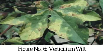
G. Verticilium Wilt
[image:4.612.218.395.327.414.2]Verticillium wilt of cotton is caused by Verticillium dahlia, a soil borne fungus that enters the roots and grows into the vascular system of the plant. Vascular discoloration or browning extending throughout the stem and into the petioles. Plants rarely wilt but may defoliate immaturely at the end of the season. Leaves develop a characteristic yellow mottle, at the edges and between the veins. Lower leaves are usually affected first. Dead tissue develops at the leaf edges and may replace the mottled areas. The mottle can be diffuse or angular. Environmental factors Favored by cool seasons.
Figure No. 6: Verticilium Wilt
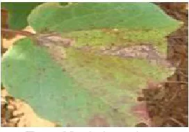
H. Leaf Red spot (lalya) on cotton
As shown in above figures the, the disease is known as Leaf Red spot(lalya) arises out as angular leaf spot with red to brown border [2],[3],[4]. The early stage of this disease is as shown in fig, now if the more spots of this disease results into the final stage of this plant where the plant leaf is get fall so it is called as Foliar disease of the cotton plant as shown in fig. The leaf is having multiple no of spots which clearly denotes more potassium deficiency in the plant. Due to this disease, it will damage plant 80-90% of total plant of production.
Figure No. 7: Leaf Red spot (lalya) on cottonBottom
III. REVIEWOFRESEARCHANDDEVELOPMENT
The proposed work described the cotton leaf spot diseases using various techniques suggesting the various development ways as illustrated in this part.
Gulhane V.A et al., (2011) This proposed work described in the features that could be extracted using the Self organizing feature map with a back propagation neural network which is used to recognize the color of the images [2].
[image:4.612.168.448.499.599.2]linear contrast stretch, intensity adjustment and filtering [7]. Hui Li et al., (2011) This proposed work based on the Web-Based Intelligent Diagnosis System for Cotton Diseases Control system used the proposed method in a BP neural network as a decision making system.
Syed A. Health et al., The proposed work discussed the automated system that can identify the pest or disease affecting a cotton leaf, boll or flower by using image analysis. In this proposed method CMYK based image cleaning technique is used to remove shadows, hands and other impurities from images.
Bernardes A. A. et al., (2011). This method proposed for automatic classification of cotton diseases through feature extraction of leaf symptoms from digital images. Wavelet transform energy has been used for feature extraction while SVM has been used for classification. Yan Cheng Zhang et al., (2007) The proposed paper discussed the fuzzy feature selection approach -fuzzy curves and Fuzzy surfaces are to select features of cotton leaf disease.
Ajay. A, et al., (2012) this work addresses that disease analysis is possible for the cotton leaf disease. The analysis of the various diseases present on the cotton leaves can be effectively detected in the early stage before it will injure the whole crop. Initially we are able to detect three types of diseases of the cotton leaves by the methodology of Eigen feature regularization and extraction technique [4]
Yan-cheng zang Han-Ping Mao, Bo Hu, Ming-xi Li in paper titled Feature Selection of Cotton Disease Leaves Image Based on Fuzzy Feature Selection Techniques[1] proposed the fuzzy feature selection approach -fuzzy curves (FC) and surfaces (FS) – for cotton leaves disease image feature selection. This research is done in two steps .Firstly to automatically and quickly isolate a small set of significant features from a set of original features according to their significance and to eliminate spurious features they make use of FC. Secondly to isolate the features dependent on the significant features, utilize FS. This approach is useful for practical classification applications which reduce the dimensionality of the feature space.
Ajay A. Gurjar, Viraj A. Gulhane describes Eigen feature regularization and extraction technique by this detection of three diseases can be done. This system is having more accuracy, than that of the other feature detection techniques. With this method about 90% of detection of Red spot i.e. fungal disease is detected.
P. Revathi M. Hemalatha detected Cotton leaf spot diseases in [7] by using Homogenous Segmentation based Edge Detection Techniques. This system is analyzed with eight types of cotton leaf diseases they are Fusarium wilt, Verticillium wilt,Root rot,Boll rot,Grey mildew ,Leaf blight ,Bacterial blight,Leaf curl.In these work symptoms of cotton leaf spot images are captured by mobile and classification is done by using neural network.
In [4], diagnosis system for grape leaf diseases is proposed. The proposed system is composed of three main parts: Firstly grape leaf color extraction from complex background, secondly grape leaf disease color extraction and finally grape leaf disease classification. In this analysis back-propagation neural network with a self-organizing feature map together is utilize to recognize colors of grape leaf. Further MSOFM and GA deployed for grape leaf disease segmentation and SVM for classification. Finally filtration of resulting segmented image is done by Gabor Wavelet and then SVM is again applied to classify the types of grape leaf diseases. This system can classify the grape leaf diseases into three classes: Scab disease, rust disease and no disease. Even though there are some limitations of extracting ambiguous color pixels from the background of the image. The system demonstrates very promising performance for any agricultural product analysis.
In the next step, the segmentation is done by using K-means clustering technique. After that the mostly green pixels are masked. Further the pixels with zero green, red and blue values and the pixels on the boundaries of the infected object were completely removed. Then the infected cluster was converted into HIS format from RGB format. In the next step, for each pixel map of the image for only HIS images the SGDM matrices were generated. Finally the extracted feature was recognized through a pre-trained neural network. The results show that the proposed system can successfully detect and classify the diseases with a precision between 83% and 94%.
IV. INTERDISCIPLINARYRELEVANCE
The machine vision system now a day is normally consists of computer, digital camera and application software. Various kinds of algorithms are integrated in the application software. Image analysis is one important method that helps segment image into objects and background. One of the key steps in image analysis is feature detection. Study of diseases on the cotton leaf can robustly studied by the image processing toolbox and also the diagnosis by using MATLAB helps us to suggest necessary remedy for that disease arises on the leaf of cotton plant.
A. INTERNATIONAL STATUS
Breeding and Application of Zhongzhi Cotton Cultivars against Verticillium Wilt.
B. NATIONAL STATUS
In agricultural laboratory of India, In the research of identifying and diagnosing cotton disease using computer vision intellectively in the agriculture, feature selection is a key question in pattern recognition and affects the design and performance of the classifier.
V. SIGNIFICANCEOFTHESTUDY
This project will help us to
A. Generating free agriculture atmosphere to sustain the farmers to simply identify the diseases and get control of it.
B. Attempting to automatic disease recognition process using Advance colour image processing.
C. Easy way to take decision support system to farmers.
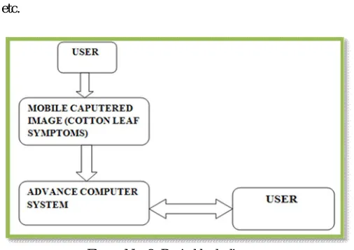
VI. METHODOLOGY The methodologies/techniques are going to implement.
Image RGB feature pixel counting technique.[1] Advanced computing technique
PCA [4]
[image:6.612.184.437.305.484.2]HPCCDD proposed Algorithm, etc. ANN/SVM as a classifier.
Figure No. 8: Basic block diagram
A. IMAGE ACQUISITION
The color images of cotton leaves are captured using a digital camera or mobile phones camera. Images are stored in BMP format.
B. IMAGE PRE-PROCESSING
In the next step, Input image is pre-processed to improve the data that suppress undesired distortions, enhances some image features important for further processing and analysis task. It including color space conversion, image enhancement for contrast improvement, and image segmentation. The color images of leaf are transforming into color space representation. The purpose of the color space is to indicate the specification of colors in some standard accepted way. RGB images transformed into Hue Saturation Value (HSV) color space representation. Because RGB is for color generation and his for color descriptor. HSV model is an important tool for color perception. Hue is a color that describes pure color as perceived by an observer. Saturation termed in to relative purity or the amount of white light added to hue and value means amplitude of light. After the color transformation process, hue component used for further analysis. Saturation and value are dropped since it does not give extra information.
C. IMAGE SEGMENTATION:
approaches of digital image processing. There are various techniques for image segmentation discuss below.
D. FEATURE EXTRACTION
After segmentation the area of interest i.e. diseased part extracted. In the next step, significant features are extracted and those features can be used to determine the meaning of a given sample. Actually, image features usually includes color, shape and texture features. Currently most of the researchers targeting plant leaf texture as the most important feature in classifying plants. With the help of texture features, plant diseases are classified into different types. There are various methods for feature extraction as discussed below.
E. CLASSIFICATION
The Concept of SVM (Support Vector Machine) was introduced by Vapnik and co-workers. It gains popularity because it offers the attractive features and powerful machinery to tackle the problem of classification i.e., we need to know which belongs to which group and promising empirical performance. The SVM is based on statistical learning theory. SVM’s better generalization performance is based on the principle of Structural Risk Minimization (SRM) .The concept of SRM is to maximize the margin of class separation. The SVM was defined for two-class problem and it looked for optimal hyper-plane, which maximized the distance, the margin, between the nearest examples of both classes, named SVM. At present SVM is popular classification tool used for pattern recognition and other classification purposes. Support vector machines (SVM) are a group of supervised learning methods that can be applied to classification or regression. The standard SVM classifier takes the set of input data and predicts to classify them in one of the only two distinct classes. SVM classifier is trained by a given set of training data and a model is prepared to classify test data based upon this model. For multiclass classification problem, we decompose multiclass problem into multiple binary class problems, and we design suitable combined multiple binary SVM classifiers
Most traditional classification models are based on the empirical risk minimization principle. SVM implements the structural risk minimization principle which seeks to minimize the training error and a confidence interval term. A number of applications showed that SVM hold the better classification ability in dealing with small sample, nonlinearity and high dimensionality pattern recognition. Support Vector Machines are based on the concept of decision planes that define decision
F. Artificial Neural Network (ANN)
An Artificial Neuron is basically an engineering approach of biological neuron.ANN consists of a number of nodes, called neurons. Neural networks are typically organized in layers. In neural network each neuron in hidden layer receives signals from all the neurons in the input layer. The strength of each signal and the biases are represented by weights and constants, which are calculated through the training phase. After the inputs are weighted and added, the result is then transformed by a transfer function into the output. The transfer functions used are Sigmoid, hyperbolic tangent functions or a step. Back propagation is a neural network learning algorithm (Rumelhart and McClelland, 1986) is used in layered feed-forward Artificial Neural Networks. Back propagation is a form of supervised training [21][17]. Originally, ANNs started in the form of a single neuron, proposed in the McCulloch and Pitts model in the 1940s (McCulloch & Pitts, 1943). In 1958, frank Rosenblatt proposed Perceptron, is the simplest single layer networks whose weights and biases could be trained to produce a correct target vector when presented with the corresponding input vector. This network made up of only input neurons and output neurons. It can solve only linear problems. Multi Layer perceptron (MLP) is one of the feed forward neural networks with one or more layers between input and output layer. Feedforward means that data flows in one direction from input to output layer (forward). Multiple layers of neurons with nonlinear transfer functions allow the network to learn nonlinear and linear relationships between input and output vectors. Multilayer perceptrons (MLPs), which can be trained using a back propagation algorithm (Rumelhart and McClelland, 1986), is a very popular choice for many researchers.
VI.CONCLUSION
REFERENCES
[1] P. Revathi & M. Hemalatha,” Advance Computing Enrichment Evaluation of Cotton Leaf Spot Disease Detection U sing Image Edge detection”, IEEE-20180 (ICCCNT), Coimbatore, India, 2012.
[2] Mr. V. A. Gulhane & Dr. A. A. Gurjar," Detection of Diseases on Cotton Leaves and its Possible Diagnosis", (IJIP), Volume (5), Issue (5), 2011, Pg: No: 591-598.
[3] P. Revathi & M. Hemalatha,” Classification of Cotton Leaf Spot Diseases Using Image Processing Edge Detection Techniques”, IEEE, International conference on emerging Trends in science, engineering & technology, 2012, pg: No: 169-173.
[4] Ajay A. Gurjar and Viraj A. Gulhane,“ Disease Detection On Cotton Leaves by Eigen feature Regularization and Extraction Technique”, (IJECSCSE 1) 2012, pp 1-4.
[5] Yan Cheng Zhang, Han Ping Mao, Bo Hu, Ming Xili “features selection of Cotton disease leaves image based on fuzzy feature selection techniques” IEEE Proceedings of the 2007 International Conference on Wavelet Analysis and Pattern Recognition, Beijing, China, 2-4 Nov. 2007.
[6] Fu, K.S., Mui, J.K., 1981.” A survey on image segmentation. Pattern Recognition” 13(1).
[7] Rothe.P.R and R.V. Kshirsagar,”A Study on the Method of Image Preprocessing for Recognition of Crop Diseases,” ICBEST and IJCA, 2012,pp:8-10. [8] PradnyaRavindra Narvekar1, Mahesh Manik Kumbhar2, S. N. Patil3,” Grape Leaf Diseases Detection & Analysis
using SGDM Matrix Method”, International Journal of Innovative Research in Computer and Communication Engineering [9] http://cotton.ces.ncsu.edu/
[10] http://www.cicr.org.in/
[11] http://aiccip.cicr.org.in/Nanded.html