IDENTIFICATION AND VALIDATION OF GERMLINE GENETIC VARIANTS THAT ASSOCIATE WITH SORAFENIB CLINICAL OUTCOMES AND CYTOTOXICITY
Daniel James Crona
A dissertation submitted to the faculty of the University of North Carolina at Chapel Hill in partial fulfillment of the requirements for the degree of Doctor of Philosophy in the
Eshelman School of Pharmacy (Pharmacotherapy and Experimental Therapeutics).
Chapel Hill 2015
ABSTRACT
Daniel James Crona: Identification and validation of germline variations that associate with overall survival in metastatic renal cell carcinoma patients treated with sorafenib
(Under the direction of Federico Innocenti)
Sorafenib is a potent inhibitor of multiple oncogenic, stromal and angiogenic receptor tyrosine kinases. Germline variants in VEGF-pathway genes and in sorafenib pharmacology genes might associate with prognosis and/or sorafenib efficacy in metastatic renal cell carcinoma (mRCC patients). A total of 295 mRCC patients from the phase III TARGET trial were genotyped using candidate germline variants from 56 candidate genes implicated in angiogenesis, sorafenib pharmacology and/or RCC prognosis/pathogenesis. Seven variants that significantly associated with overall survival (OS) in mRCC patients treated with sorafenib, and an additional two variants associated with OS in a combined analysis of both treatment arms.
Statistical associations between genetic variants and outcomes in cancer studies should be supported with molecular mechanistic evidence of variant function to aid in
biomarker validation. Variants identified in Aim 1 that significantly associated with OS were analyzed using in silico bioinformatic tools to prioritize in vitro validation assays. Cell viability assays validated one non-synonymous variant in FLT-4, and dual reporter gene luciferase assays validated two intronic VEGFA variants in three different cell lines.
cytotoxicity utilizing high content imaging and simultaneous evaluation of cell health parameters (cell viability, membrane permeability, mitochondrial membrane potential, and cytochrome C release). One quantitative locus (QTL) on chromosome 9, which reached genome-wide significance and significantly associated with cytochrome C release, was identified. A total of nine genes, expressed in MEF cells at mRNA level, were present in this QTL. A second QTL associated with cell viability was also identified. A total of 13 candidate genes, expressed in MEF cells at mRNA level, were present in this QTL. In the future,
I would like to dedicate this work to my one and only true love: Dr. Lana Crona. Thank you for sticking by me through all of this, and for being my rock. I love you, my dear Nannybelle.
ACKNOWLEDGEMENTS
Federico Innocenti, you were precisely what I needed in a mentor. You taught me self-reliance, critical thinking skills, and self-confidence. You helped me see the big picture of why I am here and why I want to continue as a clinical pharmacologist and
pharmacogeneticist in oncology: to make impactful changes and advancements that better the lives of our patients. I truly value your hands-off mentoring approach and that you did not micro-manage my every move. I appreciate that, from the beginning, you treated me like you would a post-doctoral researcher and gave me the latitude to pursue new ideas and test new hypotheses. As a result, you engendered within me a sense of independence. I hope I met your high expectations for a first graduate student. Amy Etheridge, you have certainly been a great teacher and mentor to me as well. Thank you for helping me design and develop my assays, for making sure supplies were always available, for babysitting my cells, for
providing me with sage career advice, for talking about biking and running, and for listening to all of my thoughts and concerns on so many different subjects. I literally could not have done this without you. My friend and colleague from “down under,” Dylan Glubb, I only have one thing to say to you: shot bro. I could not have asked for a better colleague, and role model when you moved here from Chicago. I appreciate all of your advice on
one of our collaborators from University of Chicago, Andrew Skol. You are an amazing statistician, and were integral to the analysis of the data and my overall learning in the early stages of my dissertation. I cannot thank you enough. I also would like to thank Carol Peña from Bayer for providing me access to the TARGET patient samples, as well as to thank Habibul Ahsan at the University of Chicago and Jason Luo from UNC for their assistance with the genotyping portion of my research.
Tim Wiltshire, I sincerely could not have asked for a better Chair of my Dissertation Committee. You are so kind, intelligent, easy to talk with, and truly personify the words “mentor” and “teacher.” I would not be at UNC if not for you because, as you know, you literally convinced me to attend UNC while I was waiting in the lift line while snowboarding in Colorado. You were my first lab PI here, and I am so thankful that I will soon get the opportunity to be your collaborator and colleague. A million times thank you. I cannot say enough good things about two more members of the Wiltshire lab: Oscar Suzuki and Amber Frick. Oscar, you are simply amazing. I wish I knew half as much about genetics,
bioinformatics, and molecular biology (you are also one heck of a swimmer). And Amber, you are no less amazing. You are so smart, so kind, are an incredible teacher, and wonderful at explaining difficult concepts (and you are also on heck of a baker). I am also very grateful to Rusty Thomas, Bethany Parks, Joe Trask, and all of the members of the Hamner Institute for collaborating with me on the MEF study. And, thank you very much to Tammy Havener for helping guide me on cell viability assays, and molecular biology. I learned so much from each and every one of you. Thank you for being integral to my learning.
oncology topics, your advice on my different HOPS presentations, you advice on my dissertation, your explanations regarding supportive care topics, as well as how you’ve advised me extensively on career and on life. You have truly made this training experience unique and special for me. You are an oncology pharmacist of the highest quality, an
insightful pharmacogenetics researcher, and have incredible perspective. You are everything I want to be as a clinician, as a clinical researcher and as a person. Thank you so very much for taking a chance on me.
Billy Kim, I wanted to thank you for being such a great part of my Dissertation Committee and for being a mentor to me in clinic. I will always remember the first time you introduced me to one of your patients as “Dr. Crona.” That was such a validating moment for me. Thank you for challenging me to think deeply in my Dissertation Committee meetings, and for all of our discussions about science while in clinic. Thank you so much for your generosity. Giving me the Caki-1 cells REALLY helped me, and all of your lab members were so kind and giving of their time. You are clearly one of the most intelligent people I have ever met, and I really look forward to working together in the future, both in clinic and hopefully through lab collaborations.
hope that you will continue to mentor me in the future. I value you and your expertise so very much.
As part of my education, I have been lucky enough to engage in a longitudinal clinic experience. I have essentially served as the clinical pharmacist specialist in the outpatient GU oncology clinic of over four years. During that time, I have gotten to meet some really
extraordinary individuals who have truly shaped me as a clinician and researcher. Matt Milowsky, I cannot adequately express just how much I gain from you as a mentor. You are a brilliant clinician, and an even better clinical researcher. Thank you for providing me with opportunities to collaborate with researchers at Memorial Sloan Kettering, and thank you for all you do for me (which I am sure is more than I know). Mary Dunn, what would I ever do without you? Thank you so very much for teaching me on all topics GU, thank you for making me laugh daily, for all the trips to Starbucks, and for being my friend. Young Whang, I have learned perhaps most from you. You are such an excellent teacher, and I have gained immensely from every single one of our conversations on medicine, medications, and science. Thank you for collaborating with me on the PRES-enzalutamide case report. Paul Godley and Ethan Basch, thank you both so much for supporting me, and for allowing me to be so integrated in the care of your patients. I know both of you have supported me
During my clinical experience, I was also fortunate to have my own “committee” of pharmacists that served as clinical advisors. I cannot say thank you enough times to Aimee Faso, Benyam Muluneh, and Meredith Keisler for being excellent role models of exemplary outpatient oncology pharmacists and CPPs. I want to be you guys when I a finally grow up and get the job of my dreams. I would also like to thank Benyam and Maurice Alexander for being my HOPS mentors. And, finally I would like to thank Lindsey Amerine, the members of the COG and all of the members of the CHIP pharmacy for helping navigate my clinical experience. It has been such a meaningful and positive adventure!
I am also extremely thankful for being able to go through this process in such a wonderful training environment at the UNC Eshelman School of Pharmacy. I would like to thank all of my mentors in DPET, most notably Herb Patterson, Heyward Hull, Craig Lee and Dhiren Thakker. At one point or another during my training, you all have provided me with excellent mentoring on myriad topics ranging from science to professional development. I would also like to thank our exemplary administrative staff, including: Arlo Brown, Kathy Maboll, Jessie Bishop, Aaron Todd, Anna Crollman, and members of the ITSOP staff. I would like to thank Kelly Scolaro for providing me with the opportunities to work with SHAC and SHAC Outreach. These have allowed me to hone mentoring skills and grow as a person. Thank you so much. Finally, I would like to convey my deepest appreciation to Brian Rybarczyk. You are so committed to professional development of graduate students, and really helped me to become a better person (not to mention a viable candidate for faculty positions).
Brouwer as an important DPET mentor to me, but also as the person who brought me into the T32 training program. I would also like to thank Daniel Benjamin, Paul Watkins, Angela Kashuba, and Robert Noveck for this great opportunity to work with some great trainees in such a neat multi-disciplinary training environment. Thank you, as well, to the T32
administrators: Lisa Phillippe and Kirsten Leysieffer. It has been so wonderful, and I cannot thank all of you enough. I would also like to thank all of the organizations that helped fund my research and/or my stipend: the American Foundation for Pharmaceutical Education, GlaxoSmithKline, Khalid Ishaq, and the National Institutes of Health/National Institute for General Medicine Sciences.
I would like to thank all of the collaborators I was lucky enough to work with on the irinotecan pharmacogenetics replication project. Thank you so much to Jackie Ramirez and Mark Ratain from University of Chicago, to Gary Rosner from Johns Hopkins, to Wei Qiao from MD Anderson, and to Ron van Schaik, Ron Mathijssen, and Anne-Joy de Graan from Erasmus Medical Center in the Netherlands.
I would like to sincerely thank all of the mentors in my life outside of UNC that have helped shape and guide me through the years, and have all helped me complete my
dissertation. James West, what can I say about you that gives you the proper credit for setting me on this path? Whatever words I use will fall desperately short. You are, without question, the most intelligent human being I have ever met. But it’s not just that. You believed in me from day 1 and have continued to be one of my most ardent supporters and trusted mentors. You have impacted my life so positively in so many ways that I fear I can really never repay all of your support and kindness. I loved being your “mouse wrangler” back at CU, I
now I am pleased to be able to call you one of my colleagues. You are truly one of the
hoopiest froods I know. Jules Harral and Michelle Carr, you both helped provide me with the basics of molecular biology research, and helped ignite in me a passion for the work we do in the lab. Even though we don’t still work together, I think you are two of the finest scientists I have ever met, and I am so privileged to call you my friends. If my experiences with you both at the Center for Genetic Lung Disease hadn’t been so positive, I am not sure I would even be a scientist today. And someday, I hope we can get the lab back together…..I just need to figure out how to bridge oncology with PAH.
Michelle Rudek, and my UNC Alumni mentor, Nader Moniri, for all of the hours they spent on the phone with me advising me on all facets of my science, professional development, and on planning for next steps in my career.
I could not have made it through this process without the love, kindness and support of the many friends and colleagues I have made since moving to North Carolina: Bob Schuck and Brittney Wright, thank you for being here in North Carolina and being our friends. We miss you every day. Colin Sheffield, thank you so much for being my friend and going to all the concerts with me. As you know, music fuels my soul. It has been so wonderful getting to know you, and I am so thankful you and Amanda Corbett have become such great friends. Kevin Watt, thank you for being the other “old man” in the DPET PhD program, and thanks for becoming one of my best friends here. I want to sincerely thank all of my other friends I’ve met while in North Carolina that I have not already mentioned, including: Jessica (Adams) and Ian Murphy, James Huckle and Ericka Mallow, Dylan Glatt (despite the fact that he went to Dakota Ridge), Paul Kim, Keith Carr, Dan and Taryn Golin (despite the fact she went to Chatfield), Jenna (Siskey) and VJ Nigro, Jai Patel, Bob Wittorf, Andy Madden, and Allison Schorzman. And a special shout-out goes to the best next-door neighbors and friends I could ask for: thank you so much Mike and Keiko Bury. An extra special shout-out goes out to my Russian friend, Olga Galeyeva. For just being you. You are very good. Stay weird.
being there for me. Jill (Kennedy) Klem, you are one of the absolute funniest people I know and constantly make me laugh. My life is better with you in it. A special thank you goes out to my wonderful cousins Kelly Dimond and Erin Marvin (both of whom are more than just cousins, and have become two of my best friends). I love you both immensely and really appreciate all of your support over the years. And to any other friends I am forgetting, THANK YOU TOO!
Above all, I would like to thank my family for your unconditional love and
unwavering support of me through all of the good times and bad. Julie and Jim Crona (aka Mom and Dad), the two of you set excellent examples for me as people that value education, and instilled in me a tenacious work ethic. It doesn’t hurt that I also inherited some pretty great genes from you both! Meghan, Katie and Kevin (aka my siblings), I would like to thank you for being loving supporters of me throughout this process. Meghan, you provide me with an example of a person who is adventurous, and takes the risks I cannot. I am so jealous of your life in Ireland. Kevin, I am so proud of you and am quite honored that you chose pharmacy as a career (in part) because of me. That is quite a compliment. Katie, I want to give you an extra shout-out for being my running partner. I don’t think I would have made it through this PhD without running becoming my stress reliever. I love that we have run marathons together! I love all three of you very, very much. A very special thank you also goes out to my godparents, Duane and Susie Bollig. I love you both very much. I would also like to thank my four-legged, furry family members for their unconditional love and support: Duke, Sweet Dee, Cooper and Danger- I love all of you and thank you so much.
TABLE OF CONTENTS
LIST OF TABLES ...xx
LIST OF FIGURES ...xxi
LIST OF ABBREVIATIONS ... xxiv
CHAPTER 1: RENAL CELL CARCINOMA: ANGIOGENESIS, VEGF-PATHWAY INHIBITORS AND BIOMARKERS ...1
1.1. Overview ...1
1.2. Renal Cell Carcinoma ...3
1.3. Renal Cell Carcinoma and Angiogenesis ...4
1.4. The Treatment of Advanced or Metastatic Renal Cell Carcinoma ...6
1.5. Sorafenib ...7
1.6. Renal Cell Carcinoma Biomarkers ... 10
1.7. Purpose of the Research ... 12
1.8. Specific Aims ... 13
TABLES ... 17
FIGURES ... 18
REFERENCES ... 22
CHAPTER 2: TARGET TRIAL PATIENTS: IDENTIFICATION OF PREDICTIVE AND PROGNOSTIC GERMLINE VARIANTS ASSOCIATED WITH OVERALL SURVIVAL ... 28
2.1. Overview ... 28
2.3. Patients, Material and Methods... 33
2.4. Results ... 40
2.5. Discussion ... 47
TABLES ... 57
FIGURES ... 61
REFERENCES ... 79
CHAPTER 3: VALIDATION OF GERMLINE VARIANTS THAT ASSOCIATE WITH OVERALL SURVIVAL IN TARGET TRIAL PATIENTS... 86
3.1. Overview ... 86
3.2. Introduction ... 88
3.3. Materials and Methods ... 89
3.4. Results ... 95
3.5. Discussion ...100
TABLES ...107
FIGURES ...114
REFERENCES ...119
CHAPTER 4: IDENTIFYING GENETIC MARKERS FOR CYTOTOXIC RESPONSE TO SORAFENIB IN MOUSE EMBRYONIC FIBROBLAST CELLS ...122
4.1. Overview ...122
4.2. Introduction ...124
4.3. Materials and Methods ...127
4.4. Results ...134
4.5. Discussion ...138
FIGURES ...150
REFERENCES ...167
CHAPTER 5: DISCUSSION, PERSPECTIVE AND FUTURE DIRECTIONS ...172
5.1. Summary and Scope ...172
5.2. Key Findings ...174
5.3. Future Directions ...178
5.4. Conclusions ...182
REFERENCES ...184
APPENDIX 1: CAN KNOWLEDGE OF GERMLINE MARKERS OF TOXICITY OPTIMIZE DOSING AND EFFICACY OF CANCER THERAPY? ...188
A1.1. Overview ...188
A1.2. Introduction ...188
A1.3. Establishing the Relationship Between Germline Variants and Toxicity ...193
A1.4. The Effect of the Germline Variant-Toxicity Relationship on Chemotherapy Dosing ...197
A1.5. The Downstream Effect of Germline Variants of Toxicity on Dosing and Efficacy ...200
A1.6. Conclusions ...205
A1.7. Future Perspectives ...206
A1.8. Executive Summary ...209
REFERENCES ...213
APPENDIX 2: CLINICAL VALIDITY OF NEW GENETIC BIOMARKERS OF IRINOTECAN NEUTROPENIA: AN INDEPENDENT REPLICATION STUDY ...222
A2.3. Materials Methods...224
A2.4. Results ...229
A2.5. Discussion ...231
TABLES ...235
FIGURES ...239
LIST OF TABLES
Table 1.1. Sorafenib inhibits angiogenic kinases and RAF/MEK/ERK
pathway oncogenic kinases. ...17
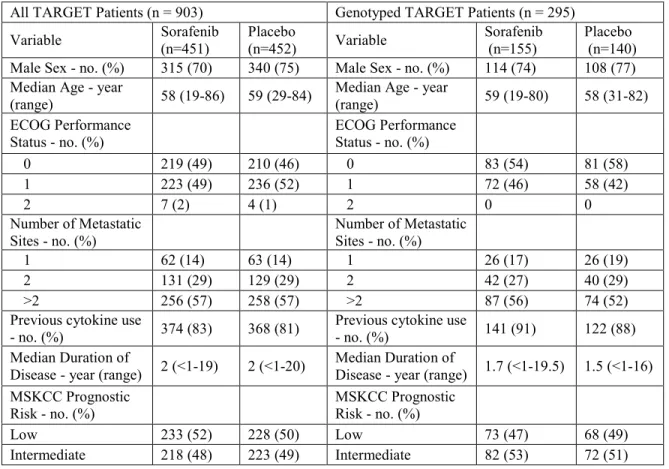
Table 2.1. Patient characteristics for the entire TARGET population versus genotyped ... 57
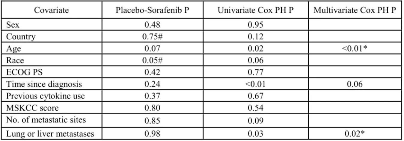
Table 2.2. Covariate selection for the OS multivariate model. ... 58
Table 2.3. Covariate selection for the PFS multivariate model ... 59
Table 2.4. Significant Variants associated with OS in TARGET patients. ... 60
Table 3.1. HaploReg output for rs1885657 and rs3024987 ...107
Table 3.2. Regulome DB scoring system ...108
Table 3.3. HaploReg output for rs3816375...109
Table 3.4. HaploReg output for rs8047917...110
Table 3.5. HaploReg output for rs307826...111
Table 3.6. Summary of key results ...112
Table 4.1. Description of the high content imaging output data features ...143
Table 4.2. MEF cell expression of candidate genes within the QTL associated with Cytochrome C release ...144
Table 4.3. MEF cell expression of candidate genes within the QTL associated with Valid Object Count ...145
Table 4.4. Non-synonymous coding SNPs and deleterious protein effects for genes associated with Cytochrome C release. ...146
Table 4.5. Non-synonymous coding SNPs and deleterious protein effects for genes associated with Valid Object Count ...148
Table A2.1. Baseline patient characteristics and pharmacokinetic data from the discovery and the replication cohorts ...235
LIST OF FIGURES
Figure 1.1. Overview of the RCC pathway with VEGF and therapeutic
targets. ... 18
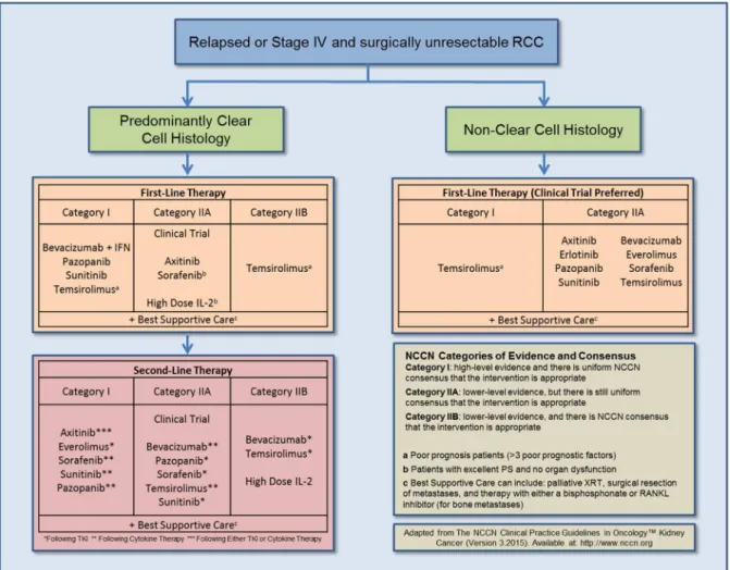
Figure 1.2. First and second line therapy recommendations for relapsed or Stage IV and surgically unresectable RCC... 20
Figure 1.3. Chemical structure of sorafenib ... 21
Figure 2.1. TARGET trial design ... 61
Figure 2.2. First and second line therapy recommendations for relapsed or Stage IV and surgically unresectable RCC... 62
Figure 2.3. Illumina GenomeStudio output from GoldenGate assay ... 63
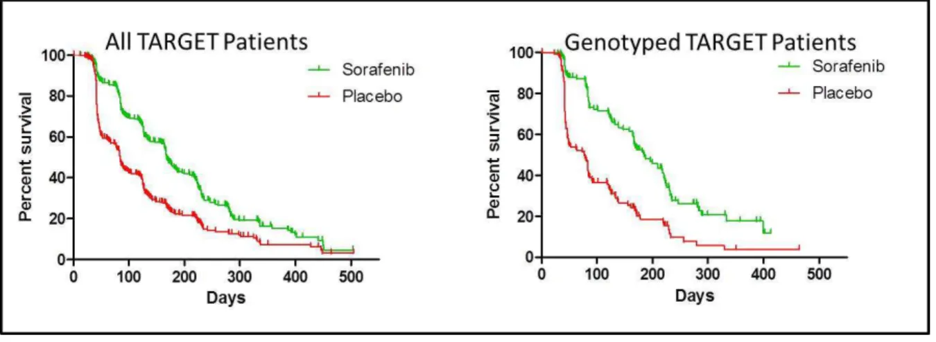
Figure 2.4. Kaplan-Meier analysis of OS in the entire TARGET population versus the genotyped TARGET patients ... 64
Figure 2.5. Kaplan-Meier analysis of PFS in the entire TARGET population versus the genotyped TARGET patients ... 65
Figure 2.6. TARGET genotyping study schematics... 66
Figure 2.7. Results from confirmatory PCR assays ... 67
Figure 2.8. Kaplan-Meier analysis of OS for rs1885657 (VEGFA). ... 68
Figure 2.9. Kaplan-Meier analysis of OS for rs3816375 (ITGAV). ... 69
Figure 2.10. Kaplan-Meier analysis of OS for rs8047917 (WWOX)... 70
Figure 2.11. Kaplan-Meier analysis of OS for rs6719561 (3’ of UGT1A9) ... 71
Figure 2.12. Kaplan-Meier analysis of OS for rs200809375 (3’ of NRP-1) ... 72
Figure 2.13. Kaplan-Meier analysis of OS for rs307826 (FLT-4) ... 73
Figure 2.14. Kaplan-Meier analysis of OS for rs3024987 (VEGFA) ... 74
Figure 2.15. Correlation analyses between OS and PFS ... 75
Figure 2.16. Kaplan-Meier analysis of PFS for rs1885657 (VEGFA). ... 76
Figure 2.17. Kaplan-Meier analysis of PFS for three variants previously with OS ... 77
Figure 3.1. UCSC Browser’s ENCODE output for VEGFA variants ...114 Figure 3.2. Histone marks in HUVEC cells for VEGFA variants ...115 Figure 3.3. Effects of rs307826 on cell viability in WT-transfected,
and mutant-transfected HEK-293 cells ...116 Figure 3.4. Dual reporter gene assay results for luciferase activity
of SNPs in VEGFA...117 Figure 3.5. Dual reporter gene assay results for luciferase activity
of rs3024987 ...118 Figure 4.1. A high-throughput cellular genetics approach to identify
QTLs and candidate genes that associate with sorafenib
response...150 Figure 4.2. Experimental workflow for high-throughput screening
of MEF lines in a concentration-response format after
administration of sorafenib ...151 Figure 4.3. Dose response curves and IC50 values generated using
a Brain-Cousens model ...153 Figure 4.4. Example of QTL interval selection ...154 Figure 4.5. Example of candidate gene selection from GWAS ...155 Figure 4.6. A multi-step and multi-faceted schematic for candidate
gene selection ...156 Figure 4.7. Distribution of IC50 values associated with Cytochrome C
across 32 MEF strains ...157 Figure 4.8. Distribution of IC50 values associated with Valid Object
Count across 32 MEF strains ...158 Figure 4.9. Manhattan plots for Cytochrome C release ...159 Figure 4.10. Genes under the QTL associated with Cytochrome C release ...160 Figure 4.11. Manhattan plots for Valid Object Count ...161 Figure 4.12. Genes under the QTL associated with Valid Object Count ...162 Figure 4.13. Haplotype structure for genes associated with the
Figure 4.14. Ingenuity pathway analysis for genes with the QTL associated
with Cytochrome C release ...164 Figure 4.15. Haplotype structure for genes associated with the Valid Object
Count QTL ...165 Figure 4.16. Ingenuity pathway analysis for genes with the QTL associated
with Valid Object Count ...166 Figure A2.1. Associations between SLCO1B1*1b and ANC nadir (A),
UGT1A1*93 and ANC nadir (B), and ABCC2 -24C>T
LIST OF ABBREVIATIONS
%CV Percent coefficient of variation (interpatient variability)
3C Chromosome conformation capture
ABCB1 ATP-binding cassette, sub-family B (MDR/TAP), member 1
ABCC1 ATP-binding cassette, sub-family C (MRP), member 1
ABCC2 ATP-binding cassette, sub-family C (MRP), member 2
ADRs Adverse drug reactions
AKT Protein kinase B
ALL Acute lymphoblastic leukemia
ALT Alanine transaminase
ANC Absolute neutrophil count ANOVA Analysis of variance AST Aspartate aminotransferase
AUC Area under the curve
AXIS Axitinib Versus Sorafenib in Advanced Renal Cell Carcinoma
BID Twice per day
BRAF v-Raf murine sarcoma viral oncogene homolog B
BSA Body surface area
Cas-9 CRISPR-associated nuclease protein 9 CDK-1 Cyclin dependent kinase 1
cDNA Complementary deoxyribonucleic acid ChIP-Seq Chromatin immunoprecipitation sequencing
CI Confidence interval
c-MET MET proto-oncogene, receptor tyrosine kinase
CNV Copy number variation
CPIC The Clinical Pharmacogenetics Implementation Consortium CRISPR Clustered regularly interspaced short palindromic repeats
CT Computed tomography
CTCAE Common Terminology Criteria for Adverse Events
CYP Cytochrome P450
dbSNP SNP Database
DLT Dose limiting toxicity
DMEM Dulbecco’s modified Eagle's medium
DMSO Dimethyl sulfoxide
DNA Deoxyribonucleic acid
DPD/DPYD Dihydropyrimidine dehydrogenase EBM-2 Endothelial growth basal medium
EBP50 Ezrin-radixin-moesin-binding phosphoprotein-50 EC50 Half maximal effective concentration
EGFR Epidermal growth factor receptor
ECM Extra cellular matrix
ERM Ezrin-radixin-moesin
FBS Fetal bovine serum
FDA Food and Drug Administration
FDR False discovery rate
FGFR-1 Fibroblast growth factor receptor-1
FLT-3 FMS-like tyrosine kinase-3
FLT-4 FMS-like tyrosine kinase-4
FOLFIRI 5-fluorouracil, leucovorin and irinotecan chemotherapy regimen
GWA Genome wide association
GWAS Genome wide association study/studies
HapMap Haplotype Map
HCC Hepatocellular carcinoma
HER-2 Human epidermal growth factor receptor 2 HIF1α Hypoxia Inducible Factor-1α
HCS High-content cell-based screening
HEATR7B1 Maestro heat-like repeat family member 2A (MROH2A)
HEK Human embryonic kidney
HR Hazard ratio
HUVEC Human umbilical vein endothelial cells
HWE Hardy–Weinberg equilibrium
IC50 Half maximal inhibitory concentration
IFN Interferon
IGFR Insulin growth factor receptor
IL Interleukin
ITGAV Integrin alpha-V
KIT v-kit Hardy-Zuckerman 4 feline sarcoma viral oncogene homolog
LD Linkage disequilibrium
LDH Lactate dehydrogenase
LPEC Liver parenchyma endothelial cells
LRT Likelihood ratio test
MAF Minor allele frequency
MAPK Mitogen-activated protein kinases
MCL-1 Myeloid leukemia-1
MDP Mouse Diversity Panel
MDR1 Multidrug resistance protein 1 MEFs Mouse embryonic fibroblasts MEK Mitogen-activated protein kinase
MGI The Mouse Genome Informatics Database
MRI Magnetic resonance imaging
mRNA Messenger ribonucleic acid
MSKCC Memorial Sloan Kettering Cancer Center mRCC Metastatic renal cell carcinoma
MTD Maximum tolerated dose
MTHFR Methylene tetrahydrofolate reductase
mTOR Mammalian target of rapamycin
MVD Microvascular density
NCCN National Comprehensive Cancer Network NCI National Cancer Institute
NIH National Institutes of Health
NRP-1 Neuropilin-1
NSCLC Non-small cell lung cancer
OBD Optimal biologic dose
OCT Organic cation transporter
OATP Organic anion-transporting polypeptide
OR Odds ratio
OS Overall survival
pAKT Phosphorylated protein kinase B
PANTHER Protein Analysis Through Evolutionary Relationships PBS Phosphate buffered saline
PCR Polymerase chain reaction PDGF Platelet derived growth factor
PDGFR Platelet derived growth factor receptor PFS Progression free survival
Pgp P-glycoprotein
PI3K Phosphatidylinositol-4,5-bisphosphate 3-kinase PGENI Pharmacogenomics for Every Nation Initiative
PH Proportional hazards
PharmGKB Pharmacogenomics Knowledge Base
PK Pharmacokinetics
PIM-1 Proviral integration site 1
PKA Protein kinase A
PKB Protein kinase B (also known as AKT)
PROVEAN Protein Variation Effect Analyzer
PS Performance status
QC Quality control
QTL Quantitative trait locus/loci RAF Rapidly accelerated fibrosarcoma
RANKL Ligand of receptor activator of nuclear factor-κB
RCC Renal cell carcinoma
RECIST Response Evaluation Criteria in Solid Tumors RFU Relative fluorescent units
RNA Ribonucleic acid
RR Relative risk
SCAN SNP and CNV Annotation
SE Standard error
SEM Standard error of the mean SIFT Sorting Intolerant From Tolerant siRNA Small interfering RNA
SLC22A1 Solute carrier family 22 (organic cation transporter), member 1
SLCO1B1 Solute carrier organic anion transporter family, member 1B1
SLCO1B3 Solute carrier organic anion transporter family, member 1B3
SN-38G SN-38 glucuronide
SNP Single nucleotide polymorphism
TALEN Transcription activator-like effector nuclease
TARGET Treatment Approaches in Renal Cancer Global Evaluation Trial
TF Transcription factor
TGN Thioguanine nucleotides
TIME Telomerase immortalized microvascular endothelial
TPMT Thiopurine-S-methyltransferase
TTP Time to progression
TYMS Thymidylate synthetase
UCSC University of California Santa Cruz
UGT Uridine-5’-diphospho-glucuronosyltransferase
UGT1A9 Uridine 5'-diphospho-glucuronosyltransferase 1 family,
polypeptide A9
ULN Upper limit of normal
UTR Untranslated region
VEGF Vascular endothelial growth factor
VEGFA Vascular endothelial growth factor A
VEGFC Vascular endothelial growth factor C VEGFR Vascular endothelial growth factor receptor VHL/VHL von Hippel-Lindau
VOC Valid object count
WT Wild type
CHAPTER 1: RENAL CELL CARCINOMA:
ANGIOGENESIS, VEGF-PATHWAY INHIBITORS AND BIOMARKERS
1.1 Overview
Cancers of the kidney and renal pelvis account for approximately 2-3% of all adult malignancies, and have increased in overall incidence over the past few decades. The most common subtype of kidney cancer arises from the renal parenchyma in the proximal tubules of the kidney and is classified as renal cell carcinoma (RCC). Approximately 30% of RCC patients will present initially with metastatic disease, and another 30% will relapse after surgical resection of their primary tumor. RCC responds poorly to standard cytotoxic chemotherapy and, prior to advent of targeted multikinase inhibitor therapies, interleukin-2 (IL-2) and interferon-α (INF-α) were the only systemic therapies commonly used for the treatment of advanced or metastatic RCC (mRCC).
However, over the past decade the treatment landscape for mRCC has changed dramatically due to the U.S. Food and Drug Administration (FDA) approval of multiple agents that target tumorigenic and angiogenic pathways. The approval of seven agents, which target angiogenic and/or oncogenic signaling pathways, has helped increase median survival time amongst mRCC patients. Nevertheless, despite these major advancements, most patients experience disease progression while on treatment and mRCC is eventually their cause of death.
leads to inhibition of tumor proliferation and angiogenesis. Data from the pivotal phase III randomized, placebo-controlled, multicenter Treatment Approaches in Renal Cancer Global Evaluation Trial (TARGET) confirmed a significant overall survival (OS) and progression-free survival (PFS) benefit. These data led to its U.S. FDA approval in December 2005 for the treatment of patients with advanced or metastatic RCC.
Despite the recent U.S. FDA approval of several additional multikinase inhibitors for the treatment of mRCC, there is a clear unmet need to identify and validate prognostic and predictive biomarkers that associate with improved survival. Because anti-angiogenic multikinase inhibitors target, in addition to the tumor itself, non-malignant endothelial cells and tumor microenvironment, germline DNA variations likely affect the treatment efficacy and/or toxicity profiles of these drugs. In addition, because RCC is a highly vascularized tumor type and considerable interindividual variability in response to sorafenib is observed clinically, identification and validation of germline genetic variants that associate with sorafenib response may help determine which patients should be treated with sorafenib. In a crowded landscape of targeted agents for the treatment of mRCC, identification of predictive pharmacogenetic variants could certainly impact clinician treatment decisions and improve patient outcomes.
Additionally, the identification of novel prognostic markers may provide insight into RCC pathogenesis/prognosis, or identify patients who would benefit from more intensive therapies and/or monitoring. Germline single-nucleotide polymorphisms (SNPs) in
angiogenesis pathway genes have associated with patient outcome in numerous tumor types, but the results are often inconsistent across studies and rarely validated. Furthermore,
only interrogated a small number of candidate genes or SNPs. Moreover, for the variants identified in these studies, little information regarding their effects on angiogenesis, at the molecular and cellular level, is available.
This research stems from the hypothesis that germline genetic variants in mRCC patients will help explain the interindividual differences in sorafenib response and patient survival. This hypothesis will be addressed through three aims, described in detail below. The overall goal of this dissertation research is to identify and validate predictive germline genetic markers of sorafenib efficacy, and prognostic germline genetic markers that associate with RCC pathogenesis and/or prognosis.
1.2 Renal Cell Carcinoma
Cancers of the kidney and renal pelvis account for approximately 2-3% of all adult tumors, and the overall incidence has increased over the past few decades.1,2 Renal cell
carcinomas (RCC) arise from the epithelia that lines the renal tubules, and at least 85-90% of all malignancies arising in the kidney and renal pelvis can be classified as RCC.3 The most
common histological subtype of RCC is clear-cell RCC (70-80% of all cases of RCC).4-6 Clear-cell RCC often presents as a single solid tumor located at the periphery of the renal parenchyma, and is defined by its optically clear cytoplasm, with nested clusters of cells surrounded by a dense endothelial network.7,8
approximately 63,920 new RCC cases were diagnosed, and 13,860 deaths (8,900 men and 4,960 women) occurred due to RCC in the U.S. in 2014.1
Median overall survival rates for RCC have improved over the past two decades, which could be attributed to improved screening and early detection of smaller tumors, the use of cytoreductive nephrectomy prior to the use of systemic therapy in advanced disease, and/or the U.S. FDA approval of multiple agents that target angiogenic and oncogenic signaling pathways. The 5-year and 10-year relative survival rates for kidney cancer are 72% and 62%, respectively. A majority of RCC cases are diagnosed at an early stage when disease is localized (64%), and the 5-year relative survival rate for these patients is 92%. Overall, the 5-year survival for all patients with RCC is 74%, and as high as 96% when patients present with stage I disease.11,12 However, approximately 30% of RCC patients will present initially
with metastatic disease, and an additional 30-50% of RCC patients, initially thought to be curable through nephrectomy, will relapse.13-16 The median survival time for patients with
metastatic disease is 10-12 months,17 the 5-year survival rate for these patients is
approximately 23%, and the 10-year survival is only 12.3%.2,10,12
1.3 Renal Cell Carcinoma and Angiogenesis
RCC arises from a series of mutations and selection events in cells of the proximal tubules of the nephron. These events ultimately result in the formation of cells that possess characteristics that are consistent with the hallmarks of cancer: unregulated cellular
A seminal event in the pathogenesis of clear-cell RCC is loss of function of the von Hippel-Lindau (VHL) tumor suppressor gene. VHL was identified in 1993,19 contains three exons, and is located on the short arm of chromosome 3 (3p25). Germline inheritance of mutated or deleted VHL alleles is the primary etiology for inherited clear-cell RCC. In addition, at least 75% of sporadic clear-cell RCC cases also occur as a consequence of
aberrant VHL function.18-21 Indeed, biallelic gene inactivation of VHL is a hallmark event that
promotes clear-cell RCC tumor development, and it classically conforms to the Knudson 2-hit carcinogenesis model in cases of sporadic clear-cell RCC.22 A deletion of one VHL allele
results in a loss of heterozygosity in more than 90% of cases of sporadic clear-cell RCC.23
Subsequently, the second allele can be inactivated through additional gene mutations,24 or through gene silencing secondary to hypermethylation.25,26 In contrast to inherited clear-cell
RCC, both the first and second “hits” occur as a result of somatic mutations, rather than germline mutations.20
VHL is an upstream mediator of a family of transcription factors, known as hypoxia inducible factors. Under normoxic conditions, VHL marks hypoxia inducible factor-1α (HIF-1α) for ubiquitination and proteasomal degradation.27 Ultimately, dysfunctional VHL protein
inhibitors that target distal HIF-1α effectors, primarily pro-angiogenic effectors in the VEGF-pathway, to patients with mRCC.
1.4 The Treatment of Advanced or Metastatic Renal Cell Carcinoma
RCC responds poorly to traditional cytotoxic chemotherapy. While multiple agents (e.g. gemcitabine, vinblastine, and 5-fluorouracil) have been tested in patients with mRCC, response rates are extremely poor (4% to 6%).3,30 One main mechanism of resistance to traditional chemotherapy could be related to the expression of MDR1, which encodes for the P-glycoprotein (Pgp) drug efflux transporter, in the proximal tubules of the kidney.3 Prior to
2005, pharmacotherapeutic options for mRCC patients were limited to immunotherapies (IL-2 and INF-α). However, IL-(IL-2 and/or INF-α are highly toxic to patients, only a subset of RCC patients adequately respond to these therapies,31 and prognosis for patients with mRCC receiving immunotherapy was poor with less than 10% achieving durable and complete remissions.5
Over the past decade the treatment landscape for mRCC has changed dramatically due to the U.S. FDA approval of multiple agents that target tumorigenic and angiogenic pathways. The approval of seven agents (Figure 1.2), which target angiogenic and/or
oncogenic signaling pathways (notably, inhibitors of the VEGF-pathway and the mammalian target of rapamycin [mTOR] pathway) based on the pathophysiology of the disease (Figure 1.1), has helped increase median survival time amongst mRCC patients.2,32-38
a positive impact on patient overall survival. Nevertheless, despite these major
advancements, most patients experience disease progression while on treatment and mRCC is eventually their cause of death.7 In 2014, it was estimated that over 13,000 patients in the
U.S. died from RCC,2 and these statistics highlight the need for mRCC treatment
optimization.
1.5 Sorafenib
Sorafenib tosylate (Nexavar; Bayer HealthCare Pharmaceuticals Corporation, Wayne, NJ; Onyx Pharmaceuticals, South San Francisco, CA) is an orally administered biaryl urea agent that is also a potent multikinase inhibitor. The chemical name of sorafenib is 4-[4-[[4-
chloro-3-(trifluoromethyl)phenyl]carbamoylamino]phenoxy]-N-methyl-pyridine-2-carboxamide (Figure 1.3). It has a broad spectrum of activity in angiogenic, oncogenic, and stromal kinases, as well as the RAF/MEK/ERK signaling pathway. Sorafenib was originally developed as a RAF kinase inhibitor, but was subsequently shown to effectively inhibit VEGFR-1, -2 and -3, PDGFR-β, FMS-like tyrosine kinase-3 (FLT-3), fibroblast growth factor receptor-1 (FGFR-1), RAF-1, BRAF (wild-type and mutant BRAFV600E), and c-KIT (cellular homolog of the feline sarcoma viral oncogene v-kit) receptor tyrosine kinases in multiple tumor cell lines (Table 1.1).39-42 Sorafenib also exhibited broad-spectrum,
dose-dependent inhibitory activity in multiple mouse xenograft models, including: breast, colon, lung, thyroid, and kidney tumors, as well as melanoma.39,41 The anti-proliferative effects of
sorafenib are largely dependent on the inhibition of oncogenic signaling pathways that regulate tumor proliferation.41 Sorafenib has also been shown to induce apoptosis in
numerous cell lines.41 While the mechanisms underlying its pro-apoptotic effects are not well
phosphorylation of initiation factor eIF4E combined with the loss of the anti-apoptotic myeloid leukemia-1 (MCL-1) protein.43 Dynamic contrast-enhanced magnetic resonance imaging (MRI) in RCC patients revealed that sorafenib also significantly altered vascular permeability and tumor perfusion.44
Data from four dose-escalation phase I trials revealed that sorafenib was relatively safe at its maximum tolerated dose (MTD) of 400 mg twice daily (BID).45-48 However, there
was also a high degree of interpatient variability in the sorafenib pharmacokinetic profile for patients enrolled on these trials. The mean elimination half-life of sorafenib is approximately 25–48 hours. Multiple dosing at the MTD for seven days resulted in sorafenib accumulation levels 2.5- to 7-fold higher than when a single dose was administered.40,47 Steady-state concentrations of sorafenib were reached after seven days of dosing, and no additional accumulation observed after steady-state was reached.48 In the non-continuous trials, the mean peak plasma concentration (Cmax) and area under the concentration–time curve (AUC)
values were substantially greater on the last day than they were after sorafenib administration on the first day.49 And, at 200 mg BID and at the MTD of 400 mg BID, the interpatient variability (%CV) in sorafenib exposure (measured by its AUC) ranged from 5 to 83%, and from 33 to 88% for sorafenib Cmax.49,50 Fortunately, even with this wide interpatient
variability in sorafenib pharmacokinetics, there was not an observed association between increased sorafenib exposure and increased toxicities (notably: fatigue, diarrhea and dermatologic toxicities).49
prevalent circulating analyte detected in the plasma is parent sorafenib (70-85%); however, the main pyridine N-oxide is still detected at high levels, and has been shown to be as potent as the parent drug.40,51 In patients with mild or moderate hepatic dysfunction (Child Pugh A
and Child Pugh B) who received sorafenib twice daily at the MTD, AUC values for the N-oxide metabolite were 23-65% lower than for patients without hepatic impairment.52
As a target of glucuronide conjugation, sorafenib is believed to undergo extensive enterohepatic recirculation, as evidenced by occurrence of observable double peaks in the concentration–time profiles among patients treated with sorafenib. This is supported by population pharmacokinetic modeling, which adequately described sorafenib disposition when accounting for enterohepatic recirculation in the model.50 Sorafenib is highly bound to plasma proteins (99.5%), and because of its lipophilic characteristics, it is widely distributed to tissues. However, recent studies have also shown that sorafenib also undergoes OCT1, OATP1B1, and OATP1B3-mediated active transport.53,54
Data from a phase II randomized discontinuation trial showed that sorafenib
significantly improved PFS in mRCC patients.55 Data from the pivotal phase III randomized, placebo-controlled, multicenter TARGET confirmed a significant PFS benefit, and showed a trend towards an OS benefit in patients with mRCC treated with sorafenib. Median PFS was significantly improved for patients treated with sorafenib,33 and final survival analyses
revealed improved OS for patients treated with sorafenib.56 Based on these clinical trial data,
1.6 Renal Cell Carcinoma Biomarkers
The development and approval of oral multikinase inhibitors, such as sorafenib, that target the angiogenesis and the VEGF-pathway have improved overall survival for many patients with mRCC. However, there is a significant interindividual variability when it comes to the benefit of these medications. And, the overall response rate, defined generally as the proportion of patients with reduction in tumor burden of a predefined amount, only ranged from 10-44% in patients that received front-line VEGF-pathway inhibitor therapy.33,36,37,57 The identification of prognostic and predictive biomarkers is an important next step in the evolution of mRCC treatment, and will help clinicians prioritize the use and sequence of the seven targeted agents approved over the past decade.
Prognostic biomarkers are used to evaluate phenotypes, which correlate with survival outcomes, independent of treatment.12,58 Clinical prognostic biomarkers have been used extensively to estimate RCC prognosis. The Memorial Sloan Kettering Cancer Center (MSKCC) risk criteria score for estimating survival has been incorporated into routine clinical practice, and categorizes mRCC patients into low, intermediate and high risk categories. The MSKCC risk score examined five prognostic factors (serum hemoglobin levels, corrected serum calcium levels, serum lactate dehydrogenase (LDH) levels, interval between diagnosis and the start of treatment, and Karnofsky performance status) in mRCC patients.59,60 More recently, newer prognostic models have been developed subsequent to the
U.S. FDA approval of VEGF-targeting agents.61,62 And, additional histological, molecular (e.g. circulating tumor cells, serum amyloid A protein, C-reactive protein, HIF-1α,
Predictive biomarkers are used to predict the clinical benefit and/or response to medications, and can be followed throughout the course of treatment.12,58 No clinically validated biomarkers for RCC are utilized. However, types of predictive molecular biomarkers have been investigated, including: circulating biomarkers (e.g. VEGFA, sVEGFR2 and sVEGFR3), cytokine angiogenic factors (e.g. baseline IL-6 and elevated LDH), tissue-based biomarkers (e.g. VHL mutations), and factors in the mTOR pathway (e.g. elevated phosphor-S6 expression, and elevated phosphorylated protein kinase B [pAKT] expression).58 In addition, single nucleotide polymorphisms (SNPs) that associate with
differences in pharmacokinetics/pharmacodynamics, and that associate with differences in survival have been postulated to be predictive biomarkers of mRCC treatments.63-68
To date, mRCC remains incurable, despite the approval of several targeted therapies, and there is a clear unmet need to identify and validate markers that associate with improved survival. And, despite the U.S. FDA approval of multiple VEGF pathway inhibitors that have become the mainstay for pharmacotherapeutic treatment of mRCC, many unanswered
questions remain regarding the choice of drug for an individual patient, the timing of and dose at treatment initiation, and the optimal sequencing of these agents for an individual patient. But, there are currently no validated molecular/genetic prognostic or predictive biomarkers that have been incorporated into routine clinical practice to help answer these questions and help clinician decision making. The identification, validation and clinical implementation of novel prognostic and predictive biomarkers are important towards
1.7 Purpose of the Research
Despite the recent U.S. FDA approval of several multikinase inhibitors for the treatment of mRCC, there is a clear unmet need to identify and validate prognostic and predictive markers that associate with improved survival. Because anti-angiogenic
multikinase inhibitors target, in addition to the tumor itself, non-malignant endothelial cells and tumor microenvironment, germline variations likely affect the treatment efficacy and/or toxicity profiles of these drugs. In addition, because RCC is a highly vascularized tumor type and considerable interindividual variability in response to sorafenib is observed clinically, identification and validation of germline genetic variants that associate with sorafenib response may help determine which patients should be treated with sorafenib. In a crowded landscape of targeted agents for the treatment of mRCC, identification of predictive
pharmacogenetic variants will certainly impact clinician treatment decisions.
Additionally, the identification of novel prognostic markers may provide insight into RCC pathogenesis/prognosis, or identify patients who would benefit from more intensive therapies and/or monitoring. Germline single-nucleotide polymorphisms (SNPs) in
angiogenesis pathway genes have associated with patient outcomes in numerous tumor types, but the results are often inconsistent across studies and results are rarely validated.
1.8 Specific Aims
The central hypothesis of this research is that identification and validation of germline genetic variants in mRCC patients will help explain the interindividual differences in
sorafenib response and OS. This hypothesis will be addressed through three aims, described in detail below. The overall goal of these studies is to identify and validate predictive germline genetic markers of sorafenib efficacy and/or pharmacology, and prognostic germline genetic markers that associate with RCC pathogenesis and/or prognosis.
Aim 1. To genotype candidate SNPs from 56 candidate genes, using available genomic DNA from TARGET patients, and test associations with OS.
Hypothesis: Germline variants in genes related to RCC prognosis/pathogenesis, the
angiogenesis pathway and/or sorafenib pharmacology will associate with OS in patients with mRCC enrolled on the phase III TARGET trial.
Significance: The oral multikinase inhibitor sorafenib helped revolutionize the treatment of mRCC, but mRCC remains incurable, even for patients with stage IV disease who have been treated with sorafenib.69 In addition, wide interindividual variation in response to sorafenib
Rationale: Anti-angiogenic multikinase inhibitors target tumor cells, host endothelial cells, pericytes, and even the tumor microenvironment rather than simply targeting the tumor cell alone. Therefore, germline variation is likely an important determinant of drug response in multikinase inhibitors.70Identification and validation of germline genetic variants that
significantly associate with survival will help identify patients who are optimal candidates for sorafenib therapy.
Aim 2. To validate functionality of germline variants (identified in Aim 1) that associate with OS in TARGET patients.
Hypothesis: Functional validation of germline variants that significantly associate with OS in TARGET patients can help elucidate the molecular effects of these variants on RCC pathogenesis/prognosis, angiogenesis and/or sorafenib pharmacology.
Significance: Findings from pharmacogenetic and pharmacogenomic studies (both candidate gene and genome wide association studies [GWAS]) continue to provide a plethora of
information about genetic variation that underlies both disease pathology and responses to pharmacotherapy. However, a clear understanding of the molecular effects of candidate variants (selected from significant associations between genotype and clinical phenotypes) is often absent. Since a tagging SNP approach was employed to select SNPs for genotyping TARGET patient DNA, it is imperative that the causal variant(s) is identified. Therefore, it is important that a series of validation assays characterize the effect(s) of the variant on
provide a basic explanation of the mechanistic processes that underlie the genotype-phenotype associations. Validation of these germline variants (in the absence of, or in conjunction with replicative genotyping in an independent, external cohort) is essential to their translation into potentially useful biomarkers that will inform treatment decisions concerning sorafenib therapy for mRCC patients.
Rationale: Many significant genotype-phenotype associations, derived from multikinase inhibitor pharmacogenetic studies, lack validation.63-68 Information regarding the molecular effects, which underlie disease pathogenesis and/or response to therapy, is direly needed. Therefore, a sequential in silico in vitro approach to validate germline variants of interest (garnered from Aim 1) will be employed. Functional validation of genetic variants that reveal significant associations with clinical phenotypes and drug response can increase the validity of the observed associations.71,72 Elucidation of the molecular effects of variants will help translate the genotype-phenotype associations derived from pharmacogenetic studies into clinically useful biomarkers.
Aim 3. To use in vitro cell models to discover novel candidate genes and signaling
pathways related to sorafenib cytotoxicity
Hypothesis: Differential cell health and response data (e.g. EC50 or IC50 values) from 32
MEF cells lines treated with sorafenib, can be used in GWAS to identify candidate genes associated with sorafenib response, which will ultimately lead to the discovery of novel genes for future pharmacogenetic testing in patients treated with sorafenib.
about mRCC pathogenesis/prognosis, angiogenesis and/or sorafenib pharmacology, there is little chance that novel and previously unidentified signaling pathways or candidate genes will be discovered. This aim will use a cellular genetics approach, using high-content cellular imaging and genetic mapping, and will help discover novel genes and pathways involved with sorafenib cytotoxicity and provide a better understanding of the variability observed with this phenotype.
Rationale: Previous studies have shown that GWAS mapping can be successfully performed in a panel of diverse inbred strains of mice to identify genetic loci that contain candidate genes that modulate both single gene and polygenic traits.73-76 But to date, there have been
few examples of animal GWAS pharmacogenetics, and even those that have been published have not analyzed the contribution of genetics to multikinase inhibitor (e.g. sorafenib) cytotoxicity.77,78 The use of genetically well-characterized inbred mouse strains provides a viable model system to analyze the genetic basis for cytotoxicity variability. Mouse
TABLES
Table 1.1. Sorafenib inhibits angiogenic kinases and RAF/MEK/ERK pathway oncogenic kinases. Table adapted from Wilhelm, et al. Cancer Res. 2004;64:7099-7109. Abbreviations: BRAF, v-Raf murine sarcoma viral oncogene homolog B; c-KIT, v-Kit Hardy-Zuckerman 4 feline sarcoma viral oncogene homolog; c-MET, MET proto-oncogene, receptor tyrosine kinase; CDK1, cyclin dependent kinase 1; EGFR, epidermal growth factor receptor, ERK, extracellular-signal-regulated kinase; FGFR-1, fibroblast growth factor receptor-1; FLT-3, fms-related tyrosine kinase 3; HER-2, human epidermal growth factor receptor 2; IC50, half maximal inhibitory concentration; IGFR, insulin growth factor receptor
; MEK, ; mitogen-activated protein kinase kinase; PDGFR- β, platelet derived growth factor receptor β; PIM-1, proviral integration site 1; PKA, protein kinase A; PKB, protein kinase B; PKC, protein kinase C; RAF, rapidly accelerated fibrosarcoma; SD, standard deviation; VEGFR, vascular endothelial growth factor receptor.
Molecular Target Biochemical Activity Sorafenib IC50 (mmol/L) ± SD
VEGFR-1 NA
VEGFR-2 90 ± 15
mVEGFR-2 15 ± 6
mVEGFR-3 20 ± 6
RAF-1 6 ± 3
BRAF WT 22 ± 6
BRAFV600E 38 ± 9
FGFR-1 580 ± 100
mPDGFR-β 57 ± 20
c-KIT 68 ± 21
FLT-3 58 ± 20
FIGURES
Figure 1.1. Overview of the RCC pathway with VEGF and therapeutic targets. Under normoxic conditions and with normal VHL function, the VHL protein is an integral part of the E3 ubiquitin ligase complex that marks HIF-1α for proteasomal degradation. Under hypoxic conditions and/or mutated VHL, HIF-1α is allowed to accumulate, which leads to the accumulation of HIF-1α transcription factors. HIF-1α can also accumulate secondary to activation of mTOR by PI3K/AKT signaling. Activated HIF-1α translocate to the nucleus and promotes transcription of pro-angiogenic genes, such as VEGF and PDGF.
Transcriptional activation of these genes subsequently leads to the production of
REFERENCES
1. Siegel R, Ma J, Zou Z, Jemal A. Cancer statistics, 2014. CA Cancer J Clin. 2014;64:9-29.
2. Hackzell A, Uramoto H, Izumi H, Kohno K, Funa K. p73 independent of c-Myc represses transcription of platelet-derived growth factor beta-receptor through interaction with NF-Y. J Biol Chem. 2002;277:39769-39776.
3. Cohen HT, McGovern FJ. Renal-cell carcinoma. N Engl J Med. 2005;353:2477-2490. 4. Moch H, Gasser T, Amin MB, Torhorst J, Sauter G, Mihatsch MJ. Prognostic utility of the recently recommended histologic classification and revised TNM staging system of renal cell carcinoma: a Swiss experience with 588 tumors. Cancer. 2000;89:604-614. 5. Rini BI, Campbell SC, Escudier B. Renal cell carcinoma. Lancet. 2009;373:1119-1132.
6. Leibovich BC, Lohse CM, Crispen PL, et al. Histological subtype is an independent predictor of outcome for patients with renal cell carcinoma. J Urol. 2010;183:1309-1315. 7. Jonasch E, Gao J, Rathmell WK. Renal cell carcinoma. BMJ. 2014;349:g4797. 8. Sircar KaT, P. Pathologic Considerations. In: Lara Jr. PaJE, ed. Kidney Cancer: Principles and Practice. Heidelberg: Springer; 2012:17-28.
9. International Agency for Research on Cancer. GLOBOCAN. Kidney- estimated incidence, all ages: both sexes. (Accessed March 24, 2015. Available at:
http://globocan.iarc.fr/ Pages/fact_sheets_population.aspx.)
10. Motzer RJ, Jonasch E, Agarwal N, et al. Kidney cancer, version 3.2015. J Natl Compr Canc Netw. 2015;13:151-159.
11. American Cancer Society. Cancer facts & figures. 2015. (Accessed March 20, 2015. Available at:
http://www.cancer.org/acs/groups/content/@editorial/documents/document/acspc-044552.pdf.)
12. Li MaRW. Biomarkers for Renal Cell Carcinoma. In: Lara Jr. PaJE, ed. Kidney Cancer: Principles and Practice. Heidelberg: Springer; 2012:47-65.
13. Kane RC, Farrell AT, Saber H, et al. Sorafenib for the treatment of advanced renal cell carcinoma. Clin Cancer Res. 2006;12:7271-7278.
15. Leibovich BC, Blute ML, Cheville JC, et al. Prediction of progression after radical nephrectomy for patients with clear cell renal cell carcinoma: a stratification tool for prospective clinical trials. Cancer. 2003;97:1663-1671.
16. Motzer RJ, Bander NH, Nanus DM. Renal-cell carcinoma. N Engl J Med. 1996;335:865-875.
17. Motzer RJ, Bacik J, Mazumdar M. Prognostic factors for survival of patients with stage IV renal cell carcinoma: memorial sloan-kettering cancer center experience. Clin Cancer Res. 2004;10:6302S-6303S.
18. Patel PH, Chadalavada RS, Chaganti RS, Motzer RJ. Targeting von Hippel-Lindau pathway in renal cell carcinoma. Clin Cancer Res. 2006;12:7215-7220.
19. Latif F, Tory K, Gnarra J, et al. Identification of the von Hippel-Lindau disease tumor suppressor gene. Science. 1993;260:1317-1320.
20. Kim WY, Kaelin WG. Role of VHL gene mutation in human cancer. J Clin Oncol. 2004;22:4991-5004.
21. Albiges L, Salem M, Rini B, Escudier B. Vascular endothelial growth factor-targeted therapies in advanced renal cell carcinoma. Hematol Oncol Clin North Am. 2011;25:813-33. 22. Knudson AG, Jr., Strong LC. Mutation and cancer: neuroblastoma and
pheochromocytoma. Am J Hum Genet. 1972;24:514-532.
23. Gnarra JR, Tory K, Weng Y, et al. Mutations of the VHL tumour suppressor gene in renal carcinoma. Nat Genet. 1994;7:85-90.
24. Gallou C, Joly D, Mejean A, et al. Mutations of the VHL gene in sporadic renal cell carcinoma: definition of a risk factor for VHL patients to develop an RCC. Hum Mutat. 1999;13:464-475.
25. Schraml P, Struckmann K, Hatz F, et al. VHL mutations and their correlation with tumour cell proliferation, microvessel density, and patient prognosis in clear cell renal cell carcinoma. J Pathol. 2002;196:186-193.
26. Herman JG, Latif F, Weng Y, et al. Silencing of the VHL tumor-suppressor gene by DNA methylation in renal carcinoma. Proc Natl Acad Sci U S A. 1994;91:9700-9704. 27. Kamura T, Sato S, Iwai K, Czyzyk-Krzeska M, Conaway RC, Conaway JW.
Activation of HIF1alpha ubiquitination by a reconstituted von Hippel-Lindau (VHL) tumor suppressor complex. Proc Natl Acad Sci U S A. 2000;97:10430-10435.
29. Baldewijns MM, van Vlodrop IJ, Vermeulen PB, Soetekouw PM, van Engeland M, de Bruine AP. VHL and HIF signalling in renal cell carcinogenesis. J Pathol. 2010;221:125-138.
30. Yagoda A, Abi-Rached B, Petrylak D. Chemotherapy for advanced renal-cell carcinoma: 1983-1993. Semin Oncol. 1995;22:42-60.
31. Coppin C, Porzsolt F, Awa A, Kumpf J, Coldman A, Wilt T. Immunotherapy for advanced renal cell cancer. Cochrane Database Syst Rev. 2005:CD001425.
32. Hudes G, Carducci M, Tomczak P, et al. Temsirolimus, interferon alfa, or both for advanced renal-cell carcinoma. N Engl J Med. 2007;356:2271-2281.
33. Escudier B, Eisen T, Stadler WM, et al. Sorafenib in advanced clear-cell renal-cell carcinoma. N Engl J Med. 2007;356:125-134.
34. Escudier B, Pluzanska A, Koralewski P, et al. Bevacizumab plus interferon alfa-2a for treatment of metastatic renal cell carcinoma: a randomised, double-blind phase III trial.
Lancet. 2007;370:2103-2111.
35. Motzer RJ, Escudier B, Oudard S, et al. Efficacy of everolimus in advanced renal cell carcinoma: a double-blind, randomised, placebo-controlled phase III trial. Lancet.
2008;372:449-456.
36. Motzer RJ, Hutson TE, Tomczak P, et al. Sunitinib versus interferon alfa in metastatic renal-cell carcinoma. N Engl J Med. 2007;356:115-124.
37. Sternberg CN, Davis ID, Mardiak J, et al. Pazopanib in locally advanced or metastatic renal cell carcinoma: results of a randomized phase III trial. J Clin Oncol. 2010;28:1061-1068.
38. Rini BI, Escudier B, Tomczak P, et al. Comparative effectiveness of axitinib versus sorafenib in advanced renal cell carcinoma (AXIS): a randomised phase 3 trial. Lancet. 2011;378:1931-1939.
39. Wilhelm SM, Carter C, Tang L, et al. BAY 43-9006 exhibits broad spectrum oral antitumor activity and targets the RAF/MEK/ERK pathway and receptor tyrosine kinases involved in tumor progression and angiogenesis. Cancer Res. 2004;64:7099-7109.
40. Product information. Nexavar (sorafenib). Wayne NBHP, Inc. June 2013. (Accessed January 29, 2015. Available at: http://berlex.bayerhealthcare.com/html/products/pi/
Nexavar_PI.pdf.)
42. Wilhelm S, Chien DS. BAY 43-9006: preclinical data. Curr Pharm Des. 2002;8:2255-2257.
43. Yu C, Bruzek LM, Meng XW, et al. The role of Mcl-1 downregulation in the proapoptotic activity of the multikinase inhibitor BAY 43-9006. Oncogene. 2005;24:6861-6969.
44. Strumberg D. Sorafenib for the treatment of renal cancer. Expert Opin Pharmacother. 2012;13:407-419.
45. Clark JW, Eder JP, Ryan D, Lathia C, Lenz HJ. Safety and pharmacokinetics of the dual action Raf kinase and vascular endothelial growth factor receptor inhibitor, BAY 43-9006, in patients with advanced, refractory solid tumors. Clin Cancer Res. 2005;11:5472-5480.
46. Moore M, Hirte HW, Siu L, et al. Phase I study to determine the safety and
pharmacokinetics of the novel Raf kinase and VEGFR inhibitor BAY 43-9006, administered for 28 days on/7 days off in patients with advanced, refractory solid tumors. Ann Oncol. 2005;16:1688-1694.
47. Strumberg D, Richly H, Hilger RA, et al. Phase I clinical and pharmacokinetic study of the Novel Raf kinase and vascular endothelial growth factor receptor inhibitor BAY 43-9006 in patients with advanced refractory solid tumors. J Clin Oncol. 2005;23:965-972. 48. Awada A, Hendlisz A, Gil T, et al. Phase I safety and pharmacokinetics of BAY 43-9006 administered for 21 days on/7 days off in patients with advanced, refractory solid tumours. Br J Cancer. 2005;92:1855-1861.
49. Strumberg D, Clark JW, Awada A, et al. Safety, pharmacokinetics, and preliminary antitumor activity of sorafenib: a review of four phase I trials in patients with advanced refractory solid tumors. Oncologist. 2007;12:426-437.
50. Jain L, Woo S, Gardner ER, et al. Population pharmacokinetic analysis of sorafenib in patients with solid tumours. Br J Clin Pharmacol. 2011;72:294-305.
51. Iyer R, Fetterly G, Lugade A, Thanavala Y. Sorafenib: a clinical and pharmacologic review. Expert Opin Pharmacother. 2010;11:1943-1955.
52. Miller AA, Murry DJ, Owzar K, et al. Phase I and pharmacokinetic study of sorafenib in patients with hepatic or renal dysfunction: CALGB 60301. J Clin Oncol. 2009;27:1800-1805.
53. Swift B, Nebot N, Lee JK, et al. Sorafenib hepatobiliary disposition: mechanisms of hepatic uptake and disposition of generated metabolites. Drug Metab Dispos. 2013;41:1179-1186.
55. Ratain MJ, Eisen T, Stadler WM, et al. Phase II placebo-controlled randomized discontinuation trial of sorafenib in patients with metastatic renal cell carcinoma. J Clin Oncol. 2006;24:2505-2512.
56. Escudier B, Eisen T, Stadler WM, et al. Sorafenib for treatment of renal cell carcinoma: Final efficacy and safety results of the phase III treatment approaches in renal cancer global evaluation trial. J Clin Oncol. 2009;27:3312-3318.
57. Rixe O, Bukowski RM, Michaelson MD, et al. Axitinib treatment in patients with cytokine-refractory metastatic renal-cell cancer: a phase II study. Lancet Oncol. 2007;8:975-984.
58. Maroto P, Rini B. Molecular biomarkers in advanced renal cell carcinoma. Clin Cancer Res. 2014;20:2060-2071.
59. Motzer RJ, Bukowski RM, Figlin RA, et al. Prognostic nomogram for sunitinib in patients with metastatic renal cell carcinoma. Cancer. 2008;113:1552-1558.
60. Motzer RJ, Bacik J, Murphy BA, Russo P, Mazumdar M. Interferon-alfa as a comparative treatment for clinical trials of new therapies against advanced renal cell carcinoma. J Clin Oncol. 2002;20:289-296.
61. Heng DY, Xie W, Regan MM, et al. Prognostic factors for overall survival in patients with metastatic renal cell carcinoma treated with vascular endothelial growth factor-targeted agents: results from a large, multicenter study. J Clin Oncol. 2009;27:5794-5799.
62. Choueiri TK, Garcia JA, Elson P, et al. Clinical factors associated with outcome in patients with metastatic clear-cell renal cell carcinoma treated with vascular endothelial growth factor-targeted therapy. Cancer. 2007;110:543-550.
63. Xu CF, Bing NX, Ball HA, et al. Pazopanib efficacy in renal cell carcinoma: evidence for predictive genetic markers in angiogenesis-related and exposure-related genes. J Clin Oncol. 2011;29:2557-2564.
64. van Erp NP, Eechoute K, van der Veldt AA, et al. Pharmacogenetic pathway analysis for determination of sunitinib-induced toxicity. J Clin Oncol. 2009;27:4406-4412.
65. Peer CJ, Sissung TM, Kim A, et al. Sorafenib is an inhibitor of UGT1A1 but is metabolized by UGT1A9: implications of genetic variants on pharmacokinetics and hyperbilirubinemia. Clin Cancer Res. 2012;18:2099-2107.
66. Beuselinck B, Karadimou A, Lambrechts D, et al. Single-nucleotide polymorphisms associated with outcome in metastatic renal cell carcinoma treated with sunitinib. Br J Cancer. 2013;108:887-900.
67. Garcia-Donas J, Esteban E, Leandro-Garcia LJ, et al. Single nucleotide