Pupillary Response as a Physiological Indication of Neural Resource Utilization in Those With and Without a Concussion History
By Kou Yang
A thesis submitted to the faculty of the University of North Carolina at Chapel Hill in partial fulfillment of the requirements for graduation with honors in the Department of Exercise and Sport Science
Chapel Hill 2019
Approved by:
________________________ Johna K. Register Mihalik ________________________ Jason P. Mihalik
©2019 Kou Yang
ABSTRACT
KOU YANG: Pupillary Response as a Physiological Indication of Neural Resource Utilization in Those With and Without a Concussion History
(Under the direction of Christina B. Vander Vegt and Johna K. Register-Mihalik)
TABLE OF CONTENTS
CHAPTER I ... 1
Research Questions ... 3
Research Hypothesis ... 4
CHAPTER II ... 8
LITERATURE REVIEW ... 8
Concussion Prevalence ... 8
Pathophysiology ... 8
Concussion Assessment and Recovery ... 9
Pupillary Response to Cognitive Demand ... 11
Rationale ... 15
CHAPTER III ... 16
METHODS ... 16
Design and Participants ... 16
Laboratory Procedure ... 16
Overload Digit-span Task ... 17
Task Presentation ... 18
Statistical Analysis ... 20
Power Analysis ... 20
CHAPTER IV ... 22
Manuscript ... 22
Tables and Figures ... 35
Table 2 – Demographics from the Study Sample ... 36
Table 3 – Statistical Analysis Overview ... 37
Table 4 – Statistical results for concussion history group, sequence length, digit-span accuracy, and the Interaction effect of concussion history group*digit-span sequence length ... 38
Figure 1 – Task Design ... 39
Figure 2 – Stimulus Presentation For the Digit-Span Task ... 40
Appendix A ... 41
References ... 43
CHAPTER V ... 48
LIST OF TABLES AND FIGURES
CHAPTER 1: BACKGROUND
Introduction
Interest in proper concussion assessment and management has risen significantly over the past 10 years with the rise in concern regarding collision sports and head injuries. The 5th international conference on concussions in Berlin, defines sport-related concussions as functional brain injuries induced by
biomechanical forces typically resulting in short-term neurological impairment, and not always involving a loss of consciousness.1 The pathophysiological changes—often referred to as the neurometabolic cascade— following concussive injury involve an initial release of excitatory neurotransmitters and ionic imbalance, leading to increased metabolic demand during a time of decreased cerebral blood flow
ultimately causing an energy crisis and secondary injury in the brain.2 The result of these imbalances lead to various clinical neurological, motor, cognitive, and behavioral impairments that typically follow a gradual decrease in severity.3,1,4 The majority of patients demonstrate concussion symptom resolution within a 5 to 7 day period following injury.5
Currently, a diagnostic gold standard for concussion diagnosis does not exist , therefore clinicians rely on a comprehensive clinical assessment battery that enlists several tools for proper assessment and management.1 The current recommended battery includes a symptom inventory, balance and
A variety of neuroimaging techniques have been used to investigate the physiological response and recovery following concussion (e.g., functional magnetic resonance imaging (fMRI) and
electroencephalography (EEG), cerebral blood flow (CBF)) though there remains a dearth of evidence to fully characterize this response given inhibiting factor related to cost and applicability. A recent
systematic review suggests that persistent physiological deficits extend beyond normalization of clinical assessments and represents an over-excitatory period during which the brain remains physiologically compromised and at an increased risk for re-injury.9,10 However, application and generalizability of these findings are limited due to extensive training, high cost, and time intensive factors. Recent studies using eye tracking technology—specifically pupillary responses to cognitive demands as an indicator of neural resource utilization— may provide a clinically feasible, valid, and sensitive mechanism by which researchers and clinicians can understand physiological aspects of concussive injury. Prior studies have revealed a relation between brain activity and pupil diameter with one study correlating pupil diameter to blood-oxygen-level-dependent fMRI activity in the locus coeruleus of the human brain (a nucleus in the dorsal pons that secretes neuromodulator noradrenaline).11 A recent review have also suggest a strong link in pupil dilation and positive going even-related potentials seen in EEG studies, another marker of LC-NE activity.12
Pupillary responses to cognitive demands or mental activity is modulated by the noradrenergic locus coeruleus (LC) where increasing cognitive demands yield greater changes in pupil dilation until individuals reach their neural resource capacity for a specific demand.12 The LC is a small nucleus in the brainstem, that propagates various brain functions through the synthesis and secretion of the
neuromodulator noradrenaline and is important in various cognitive demands.11 Many studies consistently indicate that as cognitive demands increase in difficulty—or present a greater cognitive demand— greater pupil dilation responses are exhibited.9,12,13 Kahneman and Beatty were the first to describe this
utilized in concussion assessments as a form of working memory5 but no previous study has investigated digit-span task presentation and pupillary response in a concussion population.
Changes in pupillary responsivity have been validated as an index of neural resource utilization— via fMRI and EEG—and often examined in cognitive testing paradigms of attention, processing speed and working memory.14 Common neurocognitive testing paradigms used to examine these cognitive constructs include the stroop task and the digit-span task where during incongruent trials of the stroop and presentation of digit-spans of greater lengths are directly related to increases in pupil diameter in healthy individuals.15 The pupillary response to cognitive demands has also shown discriminatory ability beyond task performance or accuracy outcomes in those with and without neurocognitive impairment (e.g., Alzheimer’s16 and Parkinsons17). Specifically, these studies have demonstrated that those with neurological impairment exhibit greater neural resource utilization in the form of greater pupillary dilation response, in order to produce the same level of task performance for a given task. Therefore, the true value of pupil responsivity as a physiological index of neural resource utilization during a cognitive task may be a cost effective, noninvasive, assessment of physiological response and recovery following concussion. Therefore, the purpose of this study was to examine pupillary responsivity to various levels of sequence lengths during a digit-span task, in those with a concussion history versus healthy
recreationally active individuals with no prior concussion diagnoses. We hypothesiz ed that those with a concussion history would exhibit greater average change in pupil diameter during the digit-span task compared to healthy participants, while controlling for behavioral response accuracy, indicating greater neural resource utilization to produce similar task performance.
Research Question
Research Hypothesis
We hypothesize that those with a concussion history would exhibit greater average change in pupil diameter throughout the digit-span task compared to healthy participants, while considering behavioral response accuracy, indicating greater neural resource utilization to produce similar task performance throughout the task.
Definition of Terms
1. Behavioral Response - Accuracy: single trial accuracy required the participant to correctly report all digits in exact reverse order. Accuracy for each sequence length (4-14) required at least 3/6 (50%) accurate trials.
2. Sequence length: the numbers of digits in a digit-span.
3. Neural Resource Utilization: brain activity used to accomplish a cognitive task.
4. Baseline Period: two seconds prior to each digit-span being presented to allow for pupils to rest and stabilize.
5. Loading Phase: the portion of the digit-span task in which participants are presented with a sequence of digits at the rate of 1 per second and asked to remember the number sequence. 6. Retention Period: three second period after each digit-span is presented when participants
process/encode the information and prepare to recall.
7. Unloading Phase: the portion of the digit-span task during which participants recite the previous sequence of digits in the reverse order in which they were presented.
Operational Definitions
1. Healthy: individuals physically active at least 30 minutes a day, 3 times a week of moderate to vigorous activity between the ages of 18 and 30, with no previous concussion history.
2. History of Concussion: physically active individuals between the ages of 18 and 30, with at least one previous concussion diagnosed by a physician.
3. Behavioral response—Accuracy: response accuracy will be defined by trial and across digit-span lengths (loads).
a. Task performance—Accuracy within trial: requires 100% accurate digit-span recall (in reverse order as presented) in both content and position.
b. Task performance—Accuracy across sequence lengths: requires trial accuracy on 4 of the 6 trials for each sequence length.
Delimitations
1. Individuals with concussions within the past 6 months, or not diagnosed by a physician were excluded.
2. Individuals who were not physically activate at least 30 minutes of moderate to vigorous physical activity 3 times a week.
3. Individuals with permanent vision loss, strabismus, amblyopia, or eye surgery in the last 6 months were not included in this study.
4. Individuals who participate in visual or vestibular therapy were excluded to prevent confounding variables.
Limitations
2. Participants were responsible for reporting their own activity level and thus some participants may have been included into the study that did not meet the physical activity criteria.
Assumptions
1. Participants accurately reported past medical history.
2. Participants accurately reported typical physical activity levels.
3. Participants remembered and accurately reported all concussion injuries with recurrent symptoms.
4. Participants remained engaged and gave full effort during the task.
Variables
1. Dependent
a. Average change in pupil diameter between the retention and baseline periods. Calculated by taking the difference between average pupil size during the first 120 samples of the retention period and the last 15 samples of the baseline period for each trial, then averaging across sequence lengths.
2. Independent a. Group
i. Concussion history ii. No prior concussions b. Behavioral response
i. Accurate ii. Inaccurate c. Sequence Lengths
CHAPTER II: LITERATURE REVIEW Concussion Prevalence
Sport related concussion are functional brain injuries that are typically induced by biomechanical forces (generally a direct or indirect blow to the head, neck, or body) resulting in an impulsive force to the head which results in a cascade of short-lived neurological and neuropathological changes.18 According to the CDC, sport related traumatic brain injury (TBI) that resulted in reported loss of consciousness (LOC) accounted for approximately 300,000 incidents annually. As sport related TBI/sport related concussions do not necessarily have to result in LOC (as LOC only accounts for 8-19.2% of TBI), a better estimate of sport related TBI/concussions is between 1.6 million to 3.8 million per year.19 This estimated number, however, is believed to be lower than the actual prevalence as many injuries go unrecognized due to concussion assessments being dependent on athlete reporting.19 The difficulty in this particular injury is the variability in symptomology resulting from concussion pathophysiology.
Concussion Pathophysiology
Concussion is a complex pathophysiological condition that results in various functional
neurological impairments. Biomechanically induced head trauma via linear and rotational accelerations creates neuronal shear stress and strain that triggers what has been described by Giza et al. as the neurometabolic cascade. During the acute phase of concussion injury and start of the neurometabolic cascade, there is a release of excitatory neurotransmitters in the brain, resulting in neuronal
mitochondrial dysfunction with decreased glucose utilization and metabolism. Increased glucose demand with decreased in oxidation leads to an energy crisis.2,20 Reduced cerebral blood flow also contributes to secondary injury. These physiological deficits are reflected in athlete/patient presentation though have been shown to persist beyond clinical recovery and timeframe of the current neurometabolic cascade. These findings, outline by neuroimaging studies suggest what has been termed as a ‘window of neural vulnerability’ during which time the brain remains physiologically-compromised and at risk for repeated trauma which may be detrimental long term.20
Concussion Assessment and Recovery Clinical Recovery
The pathophysiological response to injury results in various signs and symptoms and therefore requires clinicians to use a comprehensive assessment battery that includes a symptom inventory, and balance and neurocognitive assessment.1 A symptom check list is used to account for the variety of common symptoms following concussions including headache, balance deficits, dizziness, and fatigue.6 As balance and/or postural control is also commonly effected, clinicians commonly use the Balance Error Scoring System (BESS).7,21,22 Computerized neurocognitive tests assess common neurocognitive
Persistent physiological deficits
Physiological response and recovery from concussion remains poorly characterized24 though, advanced neuroimaging technology has been used to expand our understanding. A recent systematic review by Kamins et al. suggests that the many physiological changes makes the development of a single ‘physiological time window for concussion recovery’ impossible. Rather, more research should use imaging techniques to link symptoms with physiological changes, and potential risk for subsequent injury.24 Examples of these advanced imaging include: Diffusion tensor imaging (DTI), magnetic resonance spectroscopy (MRS), cerebral blood flow (CBF), electrophysiology (EEG), fluid biomarkers, and transcranial magnetic stimulation (TMS).24 A recent systematic review on neuroimaging and physiological response to concussion investigated some of the most popular neuroimaging techniques24 and revealed prolonged physiological deficits beyond clinical recovery time. The review of 18 fMRI studies (imaging technology used to measure brain activation at various regions of the brain), reported changes in cerebral activation from 3 to 23 months after return to play, and suggest that this may be due to ‘network modifications’ that are separate from the acute injury.24 Neurocognitive testing paradigms that assess attention, processing speed and working memory are used during these neuroimaging studies to characterize regional brain activity and neural resource utilization as an indication of physiological response and recovery following concussion.
Neurocognitive Testing Paradigms
Digit-span tasks are some of the most common assessments of working memory and are
commonly examined in concussion patients requiring individuals to remember, process, and synthesize a collection of pieces of information (e.g., words, digits, letters) for recall.30 Our working memory has a limit, known as working memory capacity (WMC) which is thought to hold around between 5 and 9 pieces of information at a time.31 A subset of digit-span tasks used to assess cognitive demand and neuro-resource utilization is the n-back test. Rondell et al. used this paradigm in relation to pupillary response to assess for resource utilization (the portion of the brain that is called upon during cognitive processing).14 The amount of resources that a person allocates in order to complete a complex task is suggested to be indicative of ability cognitive control.14,32
Concussion Assessment Battery Limitations
A large limitation to the current concussion battery is the lack of clinically useful physiological evaluations/ biomarkers despite knowing a prolonged physiological time of recovery. These physiological alteration persist beyond typical return to play and research is limited on how these alterations could potentially correlate with injury vulnerability.24 There is not enough evidence to suggest a change in the current return to play progression, but future studies should investigate the physiological biomarkers with longitudinal analysis (especially to account for multiple blows).24 However, this suggestion is difficult due to expensive imaging equipment, extensive training necessary to run tools, and lack of portability of equipment. These factors can be challenging in conducting studies at secondary schools where there is large youth concussions occur and where it may be needed the most. One possible solution to this predicament is the utility of eye tracking techniques, specifically changes in pupil diameter, to obtain physiological changes in brain function.
Pupillary Response to Cognitive Demand Anatomy and Physiology of Pupillary Response
Two muscles, the dilator pupillae and the sphincter pupillae control changes in pupil size, via dilation and constriction respectively. They are both directly controlled by sympathetic and
noradrenergic locus coeruleus (LC). The LC is a small nucleus in the brain stem that plays a role in physiological arousal, cognitive function, and is central for release of norepinephrine (NE also called noradrenaline) throughout the brain. NE is important for central and peripheral nervous system activity and for proper brain development. This LC-NE activity directly communicates with the parasympathetic Edinger-Westphal nucleus that controls the constricting fibers. Inhibition signals from LC activity will directly inhibit the Edinger-Westphal nucleus and induce the constricting fibers to inhibit, causing pupil dilation.12 Various studies have confirmed the relationship linking the pupillary constriction/dilation system and LC-NE activity. 12
Pupillary Response to Cognitive Demand
Pupillary response has been studied under various testing paradigms inducing cognitive demand. In Beatty and Kahneman’s research, they examined the relationship between a number recall task and pupil dilation and found dilation was related to string length and level of difficulty. In their experiment, participants recalled phone numbers that they were familiar with compared with novel phone numbers that they provided. Their findings concluded that participants had greater dilation when they were required to remember a new phone number when compared to recalling a familiar phone number. This experiment was later expanded upon in Klinger’s et al. research in which participant pupil responses were obtained while given either a visual or an aural presentation of a digit recall (digit-span) task. They concluded that visual presentation of the digit recall task had higher accuracy and auditory presentation led to larger pupillary dilations. 9 In Piquado’s research, younger and older populations recalled various digit-span lengths and also found that pupil sizes were “sensitive” to task difficulty (i.e., digit length) with older populations showing a larger effect. This indicated a greater effort for older populations during similar task difficulty levels.33
Locus Coeruleus Norepinephrine Activity
The relationship between LC activity and pupil changes have well been research. In a study by Murphy et al., pupil diameter was shown to be correlated with activity in the locus
completing two class oddball paradigms and while at rest. Brain activity was recorded using pupillometry and blood-oxygen-level-dependent (BOLD) fMRI to compare changes in pupil diameter with LC activity. In summary, their results concluded that changes in pupil diameter was a traceable and non-invasive tool in determine LC-NA activity of the brain.11 The utility in pupil diameter changes could potentially be used to evaluate brain activity in various concussion settings without expensive imaging techniques and provide consistent data points for longitudinal testing.
Joshi et al. research further expands and solidifies the pupillary response as a reliable tool for assessing cognitive demand. In the study, adult rhesus monkeys performed various task for varied amount of time while being probed with an electrical microstimulant in the LC and other brain regions connecting to it. During the first portion of the test, passive fixation, they found fluctuations in pupil dilations which they account to pupil noise, spontaneous pupil oscillations, and pupillary unrest.34 Subjects were
presented with a brief, loud, startling tone at varying times while observing their pupillary response and found that the sound caused dilation in all of the monkeys (showing responses to the tone) within the LC and other brain regions connecting to it. They concluded that changes in pupil diameter can reflect neural activity of the locus coeruleus.34 To further solidify the connection between the LC activation and pupil response, Joshi et al. also used electrical moicrostimulation on the LC and found that the sites reliably evoked increases in pupil diameter within 1,000ms of stimulation.34
participants, non-amnestic single-domain mild-cognitive impaired, and cognitively normal participants, amnestic participants had greater compensatory effort to maintain consistent performance.16
Instrumentation and Measurement
Prior to the link between pupil response and LC-NE activity, pupil responses were used in measuring cognitive ability via various paradigms and tools. Cognitive ability has been assed using various paradigms and tools. Past research have specifically targeted skills like engagement,35 inhibititon,36 task switching ability,36 cognitive effort,16,33 cognitive demand,9,17,37 and resource utilization.32 Testing paradigms are cognitive exertional test like a short-term memory task33, reaction time task32, digit-span task9,16, or an arithmetic problem9,37 which are completed under a pupil measuring device like eye trackers9,17,33,35–37 and pupillometers.16,32 Notability, the most popular cognitive task used with pupillometry is the digit-span task.9,13
Beatty and Kahneman was the first to report the use of this paradigm with pupil recording and established the typical pupil response pattern to the task. In their study, participants were given unfamiliar telephones numbers (one digit at a time) to remember and asked to recall the information after a brief pause. 5 phases were described in the experiment, an early control phase (5 seconds where a pupil baseline diameter was taken), a listen phase (3 seconds where telephone numbers were presented), a pause phase (2 seconds of silence), a reporting phase (participants recited telephone numbers), followed by another control phase (an extended period where pupils were recorded). Their results first
demonstrated pupil dilation/construction pattern throughout the paradigm with pupil stabilization during the control phase, dilation during the listening phase, maximum dilation during the pause, and
Rationale for Current Study
CHAPTER III: METHODS Design and Participants
This quasi-experimental, cross-sectional study recruited physically active, college aged individuals with and without a concussion history. All participants were recruited from a single university, were between 18 and 30 years of age, and had normal or corrected-to-normal vision. Additionally, concussion history group participants were included if their most recent physician
diagnosed concussion occurred more than 6 months prior to their participation date. Physically active was operationally defined as participating in at least 30 minutes of moderate to vigorous physical activity, 3 times per week. Concussion was defined using the Consensus Statement on Concussion in Sport from the 5th International Conference on Concussion in Sport as a functional brain injuries that are typically induced by biomechanical a cascade of short-lived neurological and neuropathological changes.18
All participants reported for one testing session and provided written informed consent prior to any study participation. The study was approved by the institution’s Office of Human Research Ethics board. Upon study completion, participants were compensated $10 for their time.
Laboratory Procedures
Participants completed a demographic and medical history questionnaire that included specific questions regarding their concussion injury history. Participants were then seated in a chair and fitted with the FOVE Virtual Reality (VR) head-mounted display (FOVE, Inc. Silicon Valley, San Mateo, CA) while wearing a disposable sanitary face cover mask. The positioning camera was vertically aligned parallel to top of the headset to ensure participants were vertically aligned and facing forward in the VR
task by completing practice trials (all a 4-digit sequence) until study staff had determined that the participant demonstrated adequate understanding and compliance with task requirements. Participants were instructed to keep their head still with their eyes focused on the testing wall in front of them, refrain from closing their eyes and/or looking around the room while trying to remember the digits.
Overload Digit-span Task Task Design
The backward digit-span task designed to overload working memory used in the present study was adapted from Johnson et al. 13 to accommodate a university sample. Our adaptation of the task utilized a randomized blocked design containing 6 consecutive testing blocks of 11 random
digit-sequence lengths between 4 and 14 digits long—each including the numbers 1 through 9—as depicted in Figure 1. These digit sequence lengths were chosen based on previous concussion literature, in order to
overload individuals within this age range beyond they’re working memory capacity sequences would need to be beyond 12 digits long.5 A Latin Square was used to complete task presentation randomization with respect to sequence presentation order and individual digit presentation within each sequence—this approach ensured appropriate counterbalancing and exclusion of duplicate and consecutive integers.38 A custom random number generator determined initial sequences for each Latin Square model. All
participants completed the same task as designed, which took approximately 40 minutes to complete, including task familiarization.
Task Performance
Task Presentation FOVE VR Headset
The digit-span task and virtual reality (VR) environment were custom developed using Unity® software and presented using the FOVE VR head-mounted display (FOVE, Inc. Silicon Valley, San Mateo, CA) with imbedded infrared eye tracking. The head-mounted display simultaneously recorded pupillary responses in both eyes at 60Hz and a Logitech keyboard was used by participants to record when they were finished reporting all digits they remember from each trial and to initiate each trial. Two infrared illumination eye tracking sensors within the VR headset measured participant pupils by fitting an ellipse to the infrared pupil image—producing pupil radius measurements in millimeters. The headset display consisted of wide quad high definition organic light-emitting diode (WQHD OLED) 2560 x 1440, displaying up to 70 frames per second and having up to 100 degrees of viewing angle. To control for shifts in gaze, participants were asked to look straight ahead throughout testing. The task was displayed on a fixed wall within the VR environment to eliminate peripheral and distant distractions, reducing potential influence of the accommodation reflex.
Unity and Task Environment
The task presentation environment was a square shaped room (10x10x10 meters, with grey walls (R: 169, G: 169, B: 169, A: 255), built using Unity –a cross platform game development engine. To account for major confounding variables, with the largest being the pupillary light reflex, the scene utilized the “Default-Skybox” light, with an intensity multiplier of 1 and remained consistent to
Stimulus Presentation
Participants initiated each trial by pressing the “right direction arrow.” Each digit-span trial began with a baseline/accommodation period that lasted 3 seconds, indicated by an “X”. Each digit was then displayed at a rate of 1 digit per second. Final digit presentation for each trial, was followed by a 3 second retention period before participants were prompted to verbally recall the numbers in the reverse order in which they were presented for that trial. Stimulus presentation is shown in sequence in Figure 1. When the participant finished their response, they pressed the “space bar” to notify that they finished their response and then they pressed the “right directional arrow” to proceed to the next trail. Participants continued through all 66 trials until reaching the ‘end of trial’ slide, without reinforcement from research assistants.
Data Reduction and Processing
6) segment averages were used to determine pupil size change for each trial (i.e., average pupil size during retention – average baseline pupil size).39
Processed data (i.e., change in pupil diameter for each trial) were exported from Matlab and reduced using SAS 9.4 (Cary, NC). Trails were binned by sequence length (4-14) and averaged within each sequence length (6 trials per sequence length) to obtain 11 average change in pupil diameter per sequence length. Ensembled average changes in pupil diameter were calculated across all trials and served as our dependent variable (primary outcome) compared between healthy and participants with a concussion history summarized in Table 1.
Statistical Analysis
Continuous data were summarized using means and standard deviations and frequencies and associated percentages summarized our categorical variables (Table 2). A generalized linear mixed model examined average pupil response changes across digit-sequence lengths, while considering summarized accuracy. The model compared effects across testing groups (healthy to concussion history and accurate to inaccurate) and random intercepts, representing individual average pupillary response change for each digit-sequence length.
Power analysis
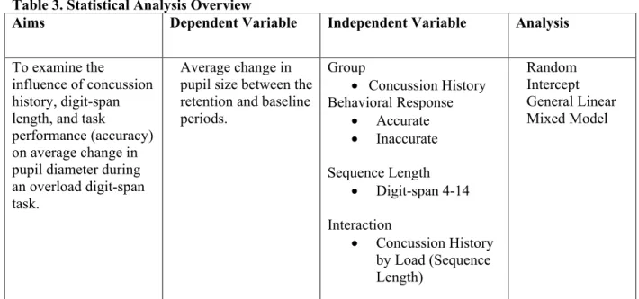
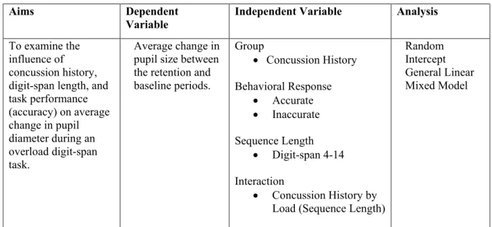
Table 3. Statistical Analysis Overview
Aims Dependent Variable Independent Variable Analysis
To examine the
influence of concussion history, digit-span length, and task
performance (accuracy) on average change in pupil diameter during an overload digit-span task.
Average change in pupil size between the retention and baseline periods.
Group
• Concussion History Behavioral Response
• Accurate • Inaccurate Sequence Length
• Digit-span 4-14 Interaction
• Concussion History by Load (Sequence Length)
CHAPTER IV: MANUSCRIPT Introduction
Concussion is a complex injury induced by biomechanical forces and characterized by a cascade of relatively short-lived neurological and neuropathological changes.18 These changes result in clinical presentation of a variety of symptoms and deficits that most individuals report recovery within 10 to 14 days.18 Previous research reported that mental status, balance, and symptoms typically return to baseline within 2 days,40 3-5 days,21,41 and 7 days,21 respectively using validated clinical test. However,
neuroimaging studies show that physiological markers of concussion persist beyond clinical
normalization18,24 and suggest that the current battery of assessments lack a clinically useful physiological component. A clinically useful physiological assessment would improve our understanding of physiologic function across the recovery continuum and help clinicians with more objective return to play decisions.
Pupillary dilation response to a cognitive demand is a relatively low resource physiologic measure that has not currently been examined post-concussion. The pupillary dilation response is modulated by the noradrenergic Locus Coeruleus neuromodulatory system (LC-NE). Measuring pupil dilation in response to a cognitive demand is a validated indirect physiological measure of neural resource utilization.11,34 Pupil dilation may provide a feasible and cost-effective physiological alternative to more expensive non-invasive counterparts like electroencephalography (EEG) and functional magnetic resonance imaging (fMRI). EEG is a tool that functionally measures electrical brain cortical
this relationship between a number recall task/digit-span task and pupil dilation and concluded that dilation was related to string length.13 A more recent study in TBI by Hershaw comparing percent change in pupil diameter in a cued-attention task found that those with a TBI history had a higher cognitive load compared to controlled counterparts.44 Despite the research support for the use of the pupillary response as a physiological indicator, it is limited clinically, as pupils are sensitive to the light reflex and testing must be conducted in a light controlled environment. However, recent advancements in eye tracking technology have introduced Virtual Reality (VR) headsets with pupil tracking capabilities that could overcome this limitation.
METHODS
Design and Participants
This quasi-experimental, cross-sectional study recruited physically active, college aged individuals with and without a concussion history. All participants were recruited from a single university, were between 18 and 30 years of age, and had normal or corrected-to-normal vision. Additionally, concussion history group participants were included if their most recent physician
diagnosed concussion occurred more than 6 months prior to their participation date. Physically active was operationally defined as participating in at least 30 minutes of moderate to vigorous physical activity, 3 times per week. Concussion was defined using the Consensus Statement on Concussion in Sport from the 5th International Conference on Concussion in Sport as a functional brain injuries that are typically induced by biomechanical a cascade of short-lived neurological and neuropathological changes.18 All participants reported for one testing session and provided written informed consent prior to any study participation. The study was approved by the institution’s Office of Human Research Ethics board. Upon study completion, participants were compensated $10 for their time.
Laboratory Procedures
Participants completed a demographic and medical history questionnaire that included specific questions regarding their concussion injury history. Participants were then seated in a chair and fitted with the FOVE Virtual Reality (VR) head-mounted display (FOVE, Inc. Silicon Valley, San Mateo, CA) while wearing a disposable sanitary face cover mask. The positioning camera was vertically aligned parallel to top of the headset to ensure participants were vertically aligned and facing forward in the VR
environment. Adjustments were made so 6 corneal glints were made visible participant pupils. Research assistants then started a 5-point calibration in which participants followed a highlighted point using only their eyes to accurately measure pupil size and gaze direction. Participants were then familiarized with the task by completing practice trials (all a 4-digit sequence) until study staff had determined that the
were instructed to keep their head still with their eyes focused on the testing wall in front of them, refrain from closing their eyes and/or looking around the room while trying to remember the digits.
Overload Digit-span Task Task Design
The backward digit-span task designed to overload working memory used in the present study was adapted from Johnson et al. 13 to accommodate a university sample. Our adaptation of the task utilized a randomized blocked design containing 6 consecutive testing blocks of 11 random
digit-sequence lengths between 4 and 14 digits long—each including the numbers 1 through 9—as depicted in Figure 1. These digit sequence lengths were chosen based on previous concussion literature, in order to overload individuals within this age range beyond they’re working memory capacity sequences would need to be beyond 12 digits long.5 A Latin Square was used to complete task presentation randomization with respect to sequence presentation order and individual digit presentation within each sequence—this approach ensured appropriate counterbalancing and exclusion of duplicate and consecutive integers.38 A custom random number generator determined initial sequences for each Latin Square model. All
participants completed the same task as designed, which took approximately 40 minutes to complete, including task familiarization.
Task Performance
Participants responded verbally and study personnel entered participants’ responses into a digital survey—back-up audio recordings were also collected. Task performance was determined for each trial and summarized across digit-sequence lengths; specifically, trial accuracy required participants to correctly report all digits in exact reverse order and summarized accuracy required at least 50% (3 of 6 trials) accuracy.
Task Presentation FOVE VR Headset
Mateo, CA) with imbedded infrared eye tracking. The head-mounted display simultaneously recorded pupillary responses in both eyes at 60Hz and a Logitech keyboard was used by participants to record when they were finished reporting all digits they remember from each trial and to initiate each trial. Two infrared illumination eye tracking sensors within the VR headset measured participant pupils by fitting an ellipse to the infrared pupil image—producing pupil radius measurements in millimeters. The headset display consisted of wide quad high definition organic light-emitting diode (WQHD OLED) 2560 x 1440, displaying up to 70 frames per second and having up to 100 degrees of viewing angle. To control for shifts in gaze, participants were asked to look straight ahead throughout testing. The task was displayed on a fixed wall within the VR environment to eliminate peripheral and distant distractions, reducing potential influence of the accommodation reflex.
Unity and Task Environment
The task presentation environment was a square shaped room (10x10x10 meters, with grey walls (R: 169, G: 169, B: 169, A: 255), built using Unity –a cross platform game development engine. To account for major confounding variables, with the largest being the pupillary light reflex, the scene utilized the “Default-Skybox” light, with an intensity multiplier of 1 and remained consistent to
standardize luminance between participants. Participants’ vision within the VR environment was directed toward the forward-facing wall (3 meters away), on a blank canvas measuring 0.6m X 0.34m, at a scale of 0.083m. Stimulus cue text on the canvas was standardized across all aspects of the task using Arial text (size= 40, color= R:46 G:46 B:46 A: 255) and scaled in the x-axis by 0.0015, in the y-axis by 0.0015, and in the z-axis by 0.005 to ensure the best clarity. Data recording was initiated by study personnel prior to any stimulus presentation.
Stimulus Presentation
which they were presented for that trial. Stimulus presentation is shown in sequence in Figure 1. When the participant finished their response, they pressed the “space bar” to notify that they finished their response and then they pressed the “right directional arrow” to proceed to the next trail. Participants continued through all 66 trials until reaching the ‘end of trial’ slide, without reinforcement from research assistants.
Data Reduction and Processing
The variables obtained from the raw data included left and right pupil radius (mm) and testing time (s) each sampled at 60Hz. Timestamp were recorded for: 1) trial number (1-66), 2) span length (4-14), 3) digit presentation (s), 4) retention X display, 5) response box display, and 6) participant finished response. Participant data files were directly exported into a spreadsheet and imported into Matlab (MATLAB and Statistic Toolbox Release 2017b, The MathWorks, Inc., Natick, MA, USA). The study team used a custom Matlab program adapted from the suggested procedures for processing pupil size data by Kret and colleagues in 201839 to complete all pupil size data processing and reduction. The adapted processing program employed 6 procedural levels as follows: 1) raw pupil radius measures for each eye were converted into diameter and averaged across both eyes; 2) raw data were prepared for filtering by removing identified blinks (recorded as .000mm or .0001mm); 3) additional artifacts were identified and removed (e.g., signal noise, longer eye closures, etc.) to identify valid samples (i.e., pupil measurements not interrupted by blinks or noise); 4) the remaining valid samples were then up-sampled and smoothed using linear interpolation; 5) pupil response regions of interest were segmented (i.e., the last 15 samples of each baseline period and the first 120 samples of each retention period) and individually averaged; and 6) segment averages were used to determine pupil size change for each trial (i.e., average pupil size during retention – average baseline pupil size).39
served as our dependent variable (primary outcome) compared between healthy and participants with a concussion history summarized in Table 3.
Statistical Analysis
SAS 9.4 (Cary, NC) was used to analyze processed data. Change in pupillary diameter was quantified using the subtraction method whereby the averaged pupil size in the first 120 samples of the retention period was subtracted from the averaged 15 last samples from the baseline period to obtain our average change in pupil diameter per trial. Trails were binned by sequence length (4-14) and averaged within each sequence length (6 trials per sequence length) to obtain 11 average change in pupil diameter per sequence length. Ensembled average changes in pupil diameter were calculated across all trials and served as our dependent variable (primary outcome) compared between healthy and participants with a concussion history summarized in Table 3.
Continuous data were summarized using means and standard deviations for all as described in Table 1. Generalized linear mixed model examined average pupil response changes during the retention period compared to baseline, across digit-spans, considering for accuracy. The model compared effects across testing groups (healthy to concussion history and accurate to inaccurate) by sequence length with random intercepts representing individual average pupillary response change during the retention period for each digit-span
RESULTS
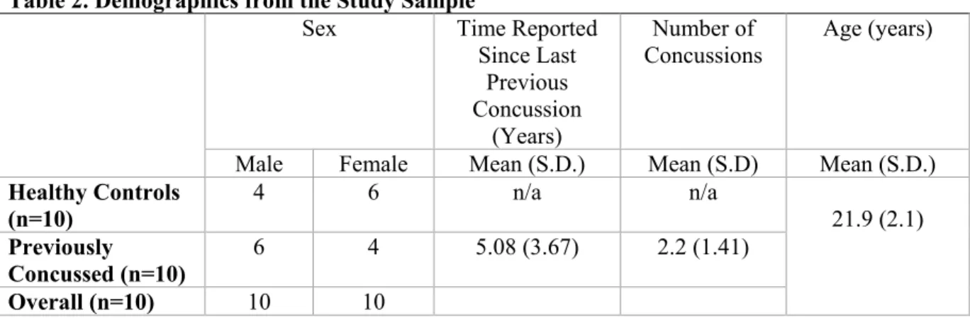
for both concussion history and accuracy groups across digit-span sequence length are summarized in Table 2. The average numbers of years since the last concussion incident for concussion history
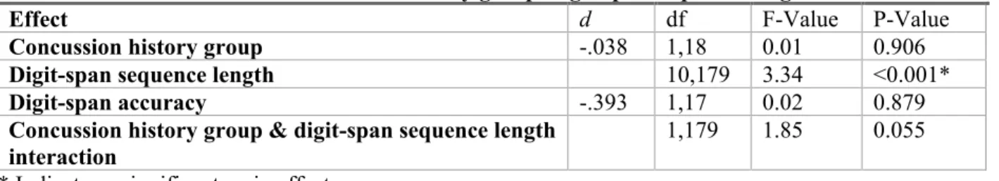
participants was 5.08 ± 3.67 years, summarized in Table 2. Test statistics for the generalized linear mixed model are summarized in Table 4. These data suggest no main effects for concussion history (F1,18=0.01, P=0.906) or accuracy (F1,17=0.02, p=0.879). There was a main effect of sequence length (F10,179=3.34, p<0.001). This main effect was driven by significant differences between spans 5 and 13, 5 and 14, 7 and 14, as well as 10 and 14; whereby, spans at the higher load had smaller average change in pupil diameter. We did not observe a significant interaction effect between concussion history and digit-span load (length); however, this omnibus finding was trending towards clinical significance (F10,179=1.85, p=0.0.056).
DISCUSSION
Concussion history had no significant effect on average change in pupil diameter, which suggests proper brain resource utilization recovery and efficiency within our concussion history participants. Physiologically, Kamins et al. summarized that physiological changes after a concussion have been reported in EEG studies up to 4 years post-concussion, up to 9 months post-concussion in transcranial magnetic stimulation, and up to 23 months post-concussion in fMRI.24 As many of our concussion history participants reported their most recent concussion being 5.08 ± 3.67 years ago, which exceed previously proposed time frames of reported changes and may account for the lack of significant effect. Additionally, our study was limited in the number of recruited participants and a larger sample would lead to a more concrete conclusion.
Our current findings contradict Hershaw’s conclusions in which he investigated pupillary response of 76 participants to a cognitive demand (cued attention paradigm) in individuals with a concussion history (median years since previous concussion was 6.91 years) and concluded that those with a prior history of a traumatic brain injury had a greater cognitive demand during a controlled attention task.44 Although our results differ in findings, Hershaw’s study investigated a different dependent variable (baseline pupil diameter and variability) and a different cognitive demand (cued attention task). These differences in task design, outcome measure, and time since previous concussion could account for discrepancies between our results. Future studies should investigate outcomes reported in Hershaw’s study.
lengths of 5 to 13, 5 to 14, 7 to 14, and from 10 to 14. Similar findings were reported by Johnson et al. who found that children tasked with a forward digit-span test from the Wecshler Intelligence Scale for Children had constricting pupil sizes after the 6 sequences. He suggested that the children had reached their cognitive limits and began to disengage with the task.45 Likewise, the drop in average change in pupil diameter reported could be due to participants reaching their cognitive limits and disengaging with the task at higher sequence lengths. Future studies could encourage more task engagement by decreasing overall task difficulty.
Accuracy was found to have no significant effect with average change in pupil diameter, further suggesting no effect on neural resource utilization. This implied that our participants, regardless of whether they were able to meet our accuracy standards at a specific span length, utilized the same amount of resources to accomplish the task. Additionally, the healthy group outperformed the concussion history group in relation to task accuracy; however, these results were not significantly different and both groups decreased in accuracy as task difficulty increased. Further examinations including larger sample sizes may provide additional information with respect to the influence of task performance on pupil response changes. Additionally, researchers should consider accounting for accuracy by trial to further describe this relationship.
Clinical Significance: Why does this matter?
Ultimately, pupillary response to a digit-span inside a VR environment may add viable
knowledge to the current literature on the physiological considerations post-concussion injury. McCrory and colleagues list the current measures of physiological change after concussions into 9 different categories: 1) functional MRI (fMRI), 2) diffusion tensor imaging (DTI), 3) magnetic resonance spectroscopy (MRS), 4) cerebral blood flow (CBF), 5) electrophysiology, 6) heart rate, 7) exercise performance, 8) fluid biomarkers, and 9) transcranial magnetic stimulation.18 These physiological assessment tools commonly used in concussion literature are essential tools in exploring concussion recovery. However, many are limited in the availability and cost. The pupillary dilation response as an indirect measure of neural resource utilization has been reliably used in psychophysiology research and this study serves as one of the first studies to control for the pupillary light reflex. Because the
constricting sphincter muscles of the pupil are influenced from light stimulus, cognitive, and autonomic activity, controlling for the light reflex is crucial in controlling environmental confounds. This would make the application more versatile for clinicians who work in various sport settings. The current study support the use of pupil response as an indirect measure of brain resource utilization and help in understanding physiological recovery in concussion populations.
Limitations
The study was limited in the number of participants. The average time since the last previous concussion was 5.08 ± 3.67 years which would increase the potential recovery time and decrease potential differences between groups. Future studies should examine the effects of multiple concussions and control for recovery time. The protocol was also number oriented, which may have given an advantage to students with an arithmetic background. Testing length may have led to a decrease in effort across the protocol due to disengagement; however, we aimed to control for this by allowing participant initiated adequate breaks in between trials. Although some studies that have found significant differences between healthy and concussion groups using pupillary response, these studies also looked a drastically different cognitive populations, like Alzheimer’s 16 that had significant known differences in cognitive function. 16 Conclusions
Pupillary response to a cognitive is a relatively new tool in the concussion literature. Our findings conclude that concussion history has no significant effect on the average change in pupil diameter during an overload digit-span task. However, digit-span length, at specific loads, had significant effects on average change in pupil diameter during an overload digit-span task, consistent with findings from other sources. These findings contradict one recent article by Hershaw44, but are consistent with some other previous literature on cognitively impaired states and future studies could potentially utilize this tool in better understanding the physiological recovery post concussions.
should also investigate changes in utilization between age groups (high school to college), and changes from baseline throughout concussion recovery.
Acknowledgements
This study was funded by the Sarah Steel Danhoff Undergraduate Research Award at the
University of North Carolina at Chapel Hill. We would like to acknowledge and thank the scientific team at the Matthew Gfeller Sport-Related Traumatic Brain Injury Research Center for their contribution and unyielding support. Additionally, we would like to thank Emily Barron and Joe Benson for their
MANUSCRIPT TABLES AND FIGURES
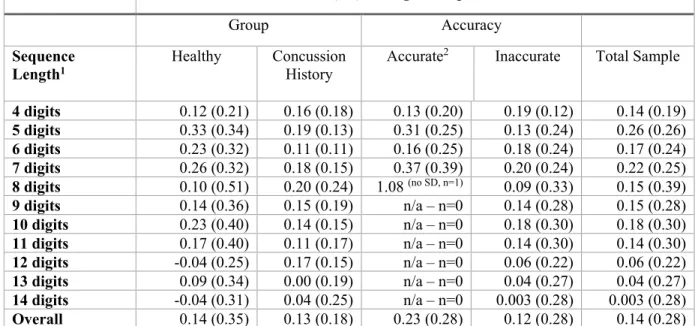
Table 1. Average change in pupil diameter (mm) between healthy and previously concussed participants and across accurate and inaccurate responses at each sequence lengths
Mean (SD) Change in Pupil Diameter
Group Accuracy
Sequence
Length1 Healthy Concussion History Accurate
2 Inaccurate Total Sample
4 digits 0.12 (0.21) 0.16 (0.18) 0.13 (0.20) 0.19 (0.12) 0.14 (0.19) 5 digits 0.33 (0.34) 0.19 (0.13) 0.31 (0.25) 0.13 (0.24) 0.26 (0.26) 6 digits 0.23 (0.32) 0.11 (0.11) 0.16 (0.25) 0.18 (0.24) 0.17 (0.24) 7 digits 0.26 (0.32) 0.18 (0.15) 0.37 (0.39) 0.20 (0.24) 0.22 (0.25) 8 digits 0.10 (0.51) 0.20 (0.24) 1.08 (no SD, n=1) 0.09 (0.33) 0.15 (0.39) 9 digits 0.14 (0.36) 0.15 (0.19) n/a – n=0 0.14 (0.28) 0.15 (0.28) 10 digits 0.23 (0.40) 0.14 (0.15) n/a – n=0 0.18 (0.30) 0.18 (0.30) 11 digits 0.17 (0.40) 0.11 (0.17) n/a – n=0 0.14 (0.30) 0.14 (0.30) 12 digits -0.04 (0.25) 0.17 (0.15) n/a – n=0 0.06 (0.22) 0.06 (0.22) 13 digits 0.09 (0.34) 0.00 (0.19) n/a – n=0 0.04 (0.27) 0.04 (0.27) 14 digits -0.04 (0.31) 0.04 (0.25) n/a – n=0 0.003 (0.28) 0.003 (0.28) Overall 0.14 (0.35) 0.13 (0.18) 0.23 (0.28) 0.12 (0.28) 0.14 (0.28) 1Sequence length refers to the number of digits at each provided at each level.
Table 2. Demographics from the Study Sample
Sex Time Reported
Since Last Previous Concussion
(Years)
Number of
Concussions Age (years)
Male Female Mean (S.D.) Mean (S.D) Mean (S.D.) Healthy Controls
(n=10) 4 6 n/a n/a 21.9 (2.1)
Previously
Concussed (n=10) 6 4 5.08 (3.67) 2.2 (1.41)
Table 3. Statistical Analysis Overview
Aims Dependent
Variable Independent Variable Analysis To examine the
influence of concussion history, digit-span length, and task performance (accuracy) on average change in pupil diameter during an overload digit-span task.
Average change in pupil size between the retention and baseline periods.
Group
• Concussion History Behavioral Response
• Accurate • Inaccurate Sequence Length
• Digit-span 4-14 Interaction
• Concussion History by Load (Sequence Length)
Table 4. Statistical results for concussion history group, sequence length, digit-span accuracy, and the Interaction effect of concussion history group*digit-span sequence length
Effect d df F-Value P-Value
Concussion history group -.038 1,18 0.01 0.906
Digit-span sequence length 10,179 3.34 <0.001*
Digit-span accuracy -.393 1,17 0.02 0.879
Concussion history group & digit-span sequence length
interaction 1,179 1.85 0.055
Figure 2. Stimulus Presentation For the Digit-Span Task
APPENDIX A Questionnaire Subject ID: ____________________
Date: ________________________
Start Time: ____________________AM/PM End Time: _____________________AM/PM
Medical History
Have you been diagnosed with a concussion? Yes No If yes, how many?
Have you been diagnosed with ADHD? Yes No
If yes, list medications you are taking: Have you been diagnosed with any other
learning disabilities? Yes No
If Yes, please elaborate:
Do you have a history of consistent migraines? Yes No
How many hours of sleep do you typically get? ________ hrs. Fri-Sat? _______ hrs. Sun- Thurs?
Have you been diagnosed with Anxiety? Yes No
If yes, list medications you are taking:
Have you been diagnosed with Depression? Yes No If yes, list medications you are taking:
Sport History
From birth indicated, have you participated in any of the following organized sports? (Check all that apply)
Sport Circle all that apply Level of sport How many years did you play? Football High School Club College
Hockey High School Club College Demographics Age Sex Demographics Height Weight Concussion History Date of prior
concussion
(mm/yyyy) Time to recovery
Loss of
consciousness? Mechanism of injury
1
Within 2 weeks Within a month > One month > One year
Yes No Sports? ________________ Other? ________________ 2
Within 2 weeks Within a month > One month > One year
References
1. McCrory P, Meeuwisse W, Dvořák J, et al. Consensus statement on concussion in sport—the
5thinternational conference on concussion in sport held in Berlin, October 2016. Br J Sports Med. 2017;51(11):838-847. doi:10.1136/bjsports-2017-097699.
2. Giza CC, Hovda DA. The new neurometabloic cascade of concussion. Neurosurgery. 2015;75(0 4):S24-S33. doi:10.1227/NEU.0000000000000505.The.
3. Mccrory P, Meeuwisse WH, Aubry M, et al. Consensus statement on concussion in sport: the 4th International Conference on Concussion in Sport held in Zurich, November 2012. Br J Sport Med. 2013;47:250-258. http://dx.doi. Accessed December 2, 2017.
4. Choe MC, Babikian T, Difiori J, Hovda DA, Giza CC. A pediatric perspective on concussion pathophysiology. Curr Opin Pediatr. 2012;24(6):689-695. doi:10.1097/MOP.0b013e32835a1a44. 5. Guskiewicz KM, Ross SE, Marshall SW, KM G, SE R, SW M. Postural stability and
neuropsychological deficits after concussion in collegiate athletes. J Athl Train (National Athl Trainers’ Assoc. 2001;36(3):263-273.
http://myaccess.library.utoronto.ca/login?url=http://search.ebscohost.com/login.aspx?direct=true&
db=rzh&AN=106900629&site=ehost-live%0Ahttp://ovidsp.ovid.com/ovidweb.cgi?T=JS&PAGE=reference&D=medp&NEWS=N&AN =12937495.
6. Jeter CB, Hergenroeder GW, Hylin MJ, Redell JB, Moore AN, Dash PK. Biomarkers for the Diagnosis and Prognosis of Mild Traumatic Brain Injury/Concussion. J Neurotrauma. 2013;30(8):657-670. doi:10.1089/neu.2012.2439.
7. Resch JE, McCrea MA, Cullum CM. Computerized neurocognitive testing in the management of sport-related concussion: An update. Neuropsychol Rev. 2013;23(4):335-349. doi:10.1007/s11065-013-9242-5.
http://bjsm.bmj.com/content/38/3/369.short.
9. Klingner J, Tversky B, Hanrahan PAT. Effects of visual and verbal presentation on cognitive load in vigilance , memory , and arithmetic tasks. 2010:1-10. doi:10.1111/j.1469-8986.2010.01069.x. 10. Mccrea M, Meier T, Huber D, et al. Role of advanced neuroimaging , fluid biomarkers and genetic
testing in the assessment of sport-related concussion : a systematic review. 2017:919-929. doi:10.1136/bjsports-2016-097447.
11. Murphy PR, O’Connell RG, O’Sullivan M, Robertson IH, Balsters JH. Pupil diameter covaries with BOLD activity in human locus coeruleus. Hum Brain Mapp. 2014;35(8):4140-4154. doi:10.1002/hbm.22466.
12. Eckstein MK, Guerra-carrillo B, Miller AT, Bunge SA. Developmental Cognitive Neuroscience
Beyond eye gaze : What else can eyetracking reveal about cognition and cognitive development ?
Accid Anal Prev. 2017;25:69-91. doi:10.1016/j.dcn.2016.11.001.
13. Beatty J, Kahneman D. Pupillary changes In Two Memory Tasks. 1966;5(10):371-372. 14. Just MA, Carpenter PA, Miyake A. Neuroindices of cognitive workload: Neuroimaging,
pupillometric and event-related potential studies of brain work. Theor Issues Ergon Sci. 2003;4(1-2):56-88. doi:10.1080/14639220210159735.
15. Laeng B, Ørbo M, Holmlund T, Miozzo M. Pupillary stroop effects. Cogn Process. 2011;12(1):13-21. doi:10.1007/s10339-010-0370-z.
16. Granholm EL, Panizzon MS, Elman JA, et al. Pupillary Responses as a Biomarker of Early Risk for Alzheimer’s Disease. J Alzheimers Dis. 2017;56(4):1419-1428. doi:10.3233/JAD-161078. 17. Kahya M, Moon S, Lyons KE, Pahwa R, Akinwuntan AE, Devos H. Pupillary response to
cognitive demand in Parkinson’s disease: A pilot study. Front Aging Neurosci. 2018;10(APR):1-8. doi:10.3389/fnagi.2018.00090.
19. Langlois JA, Rutland-Brown W, Wald MM. The Epidemiology and Impact of Traumatic Brain Injury A Brief Overview. J Head Trauma Rehabil. 2006;21(5):375-378.
doi:00001199-200609000-00001 [pii].
20. Ellis MJ, Leddy JJ, Willer B. Physiological , vestibulo-ocular and cervicogenic post-concussion
disorders : An evidence-based classification system with directions for treatment.
2015;9052(2):238-248. doi:10.3109/02699052.2014.965207.
21. McCrea M, Guskiewicz KM, Marshall SW, et al. Acute Effects and Recovery Time Following. J Am Med Assoc. 2003;290(19):2556-2563. doi:10.1001/jama.290.19.2556.
22. Guskiewicz KM, Perrin DH, Gansneder BM. Effect of mild head injury on postural stability in athletes. J Athl Train. 1996;31(4):300-306. http://www.ncbi.nlm.nih.gov/pubmed/16558414. Accessed October 31, 2018.
23. Cole WR, Arrieux JP, Schwab K, Ivins BJ, Qashu FM, Lewis SC. Test-retest reliability of four computerized neurocognitive assessment tools in an active duty military population. Arch Clin Neuropsychol. 2013;28(7):732-742. doi:10.1093/arclin/act040.
24. Kamins J, Bigler E, Covassin T, et al. What is the physiological time to recovery after
concussion ? A systematic review. 2017:935-940. doi:10.1136/bjsports-2016-097464.
25. CHANDLER P, SWELLER J. THE SPLIT-ATTENTION EFFECT AS A FACTOR IN THE DESIGN OF INSTRUCTION. Br J Educ Psychol. 1992;62(2):233-246. doi:10.1111/j.2044-8279.1992.tb01017.x.
26. Chandler P, Sweller J. Cognitive Load Theory and the Format of Instruction Linked references are
available on JSTOR for this article : Cognitive Load Theory and the Format of Instruction. Cogn
Instr. 1991;8(4):293-332.
27. Ayres P, Sweller J. Locus Of Dififculty in Multistage Mathematics Problems. Am J Psychol. 1990;103(2):167-193.
doi:10.1016/S0022-5371(82)90709-5.
29. Kerr B. Processing Demands During Mental Operations. Mem Cognit. 1973;1(4):401-412. doi:10.3758/BF03208899.
30. Sweller J, Ayres P, Kalyuga S. Cognitive Load Theory.; 2011. doi:10.1007/978-1-4419-8126-4. 31. Miller GA. The Magical Number Seven, Plus or Minus One: Some Limits on our Capacity for
Processing Musical Information. Psyghological Revie. 1994;101(2):343-352. doi:10.1177/102986490200600205.
32. Van Der Meer E, Beyer R, Horn J, et al. Resource allocation and fluid intelligence: Insights from pupillometry. Psychophysiology. 2010;47(1):158-169. doi:10.1111/j.1469-8986.2009.00884.x. 33. Piquado T, Isaacowitz D, Wingfield A. Pupillometry as a measure of cognitive effort in younger
and older adults. Psychophysiology. 2010;47(3):560-569. doi:10.1111/j.1469-8986.2009.00947.x. 34. Joshi S, Li Y, Kalwani RM, Gold JI. Relationships between Pupil Diameter and Neuronal Activity
in the Locus Coeruleus , Colliculi , and Cingulate Cortex Article Relationships between Pupil Diameter and Neuronal Activity in the Locus Coeruleus , Colliculi , and Cingulate Cortex.
Neuron. 2016;89(1):221-234. doi:10.1016/j.neuron.2015.11.028.
35. Gilzenrat MS, Cohen JD. Pupil diameter tracks changes in control state predicted by the adaptive gain theory of locus coeruleus function. Cogn Affect Behav Neurosci. 2010;10(2):252-269. doi:10.3758/CABN.10.2.252.
36. Rondeel EWM, Steenbergen H Van, Holland RW. A closer look at cognitive control : differences
in resource allocation during updating , inhibition and switching as revealed by pupillometry. 2015;9(September):1-13. doi:10.3389/fnhum.2015.00494.
37. Id KK, Duchowski AT, Niedzielska A, Biele C. Eye tracking cognitive load using pupil diameter and microsaccades with fixed gaze. 2018:1-24. doi:10.1371/journal.pone.0203629.
38. Richardson JTE. The use of Latin-square designs in educational and psychological research. Educ Res Rev. 2018;24(March):84-97. doi:10.1016/j.edurev.2018.03.003.
2018:1-7. doi:10.3758/s13428-018-1075-y.
40. McCrea M. Standardized Mental Status Testing on the Sideline after Sport-Related Concussion. J Athl Train. 2001;36(3):274-279. doi:10.1016/S1388-2457(02)00239-0.
41. Guskiewicz KM. Postural Stability Assessment Following Concussion: One Piece of the Puzzle.
Clin J Sport Med. 2001;11(3):182-189. doi:10.1097/00042752-200107000-00009. 42. André-Obadia N, Sauleau P, Cheliout-Heraut F, et al. Recommandations françaises sur
l’électroencéphalogramme. Neurophysiol Clin. 2014;44(6):515-612. doi:10.1016/j.neucli.2014.10.001.
43. Lazar N. The Big Picture: Functional Magnetic Resonance Imaging—Introduction to a Neuroimaging Modality. Chance. 2015;25(4):42-45. doi:10.1080/09332480.2012.752288. 44. Hershaw JN. Cognitive Load in Mild traumatic Brain Injury: A Pupillometric Assessment of
Multiple Attentional Processes. 2016.
45. Johnson EL, Miller Singley AT, Peckham AD, Johnson SL, Bunge SA. Task-evoked pupillometry provides a window into the development of short-term memory capacity. Front Psychol.
CHAPTER V: SUMMARY & REFLECTION Purpose
The purpose of this study was to examine the effect of concussion history, digit sequence length and task performance pupillary response magnitudes during a backward digit-span task. A total of 20 participants were included in the study, 10 healthy and 10 with a concussion history, and completed a digit-span task in a virtual reality environment. We will continue with this line of work until we meet 50 participants (25 in each concussion history group), beyond this Honor’s Thesis project.
Summary Aim
Aim: To examine the influence of concussion history, digit-span length, and task performance (accuracy) on average change in pupil diameter during an overload digit-span task.
Hypothesis: We anticipate that those with a concussion history would exhibit greater average change in pupil diameter during the digit-span task compared to healthy participants, while considering for behavioral response accuracy, indicating greater neural resource utilization to produce similar task performance.
Results: Our results concluded that concussion history had no effect on average change in pupil diameter, but that digit-span load did have a significant effect and there was a trending interaction between
concussion history and load. As many of our concussion history participants exceed previously proposed time frames of reported changes, this may account for the lack of significant effect. Additionally, our study was limited in the number of recruited participants and future work will continue to recruit up to 50 participants to have a solid conclusion and to fully determine what interaction effects may exists.
Lessons Learned
Reflecting on this past year, my honor’s thesis has been a long and enduring process that
looks like. I was challenged and learned so many things about myself personally, in my struggle to manage so many responsibilities this entire year. I had learn to humble myself and accept the fact that letting go was sometimes better than holding onto my worries, and in those times of difficulty, I learned how to take a breath and push forward the best I could. This was a hard lesson to learn, as there were so many times where the project was not progressing and it was frustrating to handle every setback that occurred. Every obstacle tested my patience and I often contemplated on giving up on the project. However, it was because of the hardships and struggles that we encountered with this project that made it worthwhile and satisfying. In fact, it’s quite remarkable how in less than a year, an unknown headset with little instructions, would become a full functioning assessment, and I’m excited to continue the project beyond the end of my thesis. Ultimately, this experience not only pushed my time management abilities, but honed my mentality and resilience to the difficulties faced before me.
Teamwork was also one of the greatest virtues that made this project successful. None of this would be possible without the hard work and effort from everyone who helped in troubleshooting our development problems, our recruiting and task management, statistical analysis, and especially in encouraging us to keep moving forward despite the setbacks. I learned that working cohesively to a common goal was difficult to manage but essential as research is a team effort that cannot be accomplished alone.
During the calibration process, we had to constantly troubleshoot how to get good readings as we struggled to get the system to work sometimes. Finally, I learned that it is important to review my own work time and time again. When we were running the first pilot test for the system, I had to build the code to run the system, and I forgot to double check my work and make sure that it would run. Unfortunately, the pilot testing went on without me and crashed, leaving us delayed in the development process. Had I double checked my work, I would have not wasted time with our pilots.
Future Research
Future studies should continue the use of this tool or similar in concussion protocols and