The Podospora
rmp1
Gene Implicated in Nucleus-Mitochondria
Cross-Talk Encodes an Essential Protein Whose Subcellular
Location Is Developmentally Regulated
Ve´ronique Contamine, Denise Zickler and Marguerite Picard
1Institut de Ge´ne´tique et Microbiologie, Universite´ Paris-Sud, UMR 8621, Orsay, France
Manuscript received June 26, 2003 Accepted for publication October 3, 2003
ABSTRACT
It has been previously reported that, at the time of death, thePodospora anserina AS1-4mutant strains accumulate specific deleted forms of the mitochondrial genome and that their life spans depend on two natural alleles (variants) of thermp1gene:AS1-4 rmp1-2strains exhibit life spans strikingly longer than those ofAS1-4 rmp1-1. Here, we show thatrmp1is an essential gene.In silicoanalyses of eightrmp1natural alleles present in Podospora isolates and of the putative homologs of this orphan gene in other filamentous fungi suggest thatrmp1evolves rapidly. The RMP1 protein is localized in the mitochondrial and/or the cytosolic compartment, depending on cell type and developmental stage. Strains producing RMP1 without its mitochondrial targeting peptide are viable but exhibit vegetative and sexual defects.
T
HE number of mitochondrial proteins has been esti- in the nuclearAS1gene, which encodes a cytosolic ribo-somal protein. Consequently, the effect of these muta-mated at 700–1000 (Schatz 1995; Wallace 1999;Kumar et al. 2002). The vast majority are encoded by tions (e.g., AS1-4) is indirect (Dequard-Chablat and Sellem 1994). This process exhibits similarities with nuclear genes and synthesized in the cytosol. Studies
have focused on these “nuclear-mitochondrial genes” human diseases characterized by the accumulation of mtDNA deletions. While some of these diseases are spo-(Chinnery2003) not only because of their
fundamen-tal interest in understanding the biogenesis and func- radic, others are inherited in a Mendelian fashion, thus implicating mutations in nuclear genes (reviewed in tion of the organelle, but also in applied research to
address the molecular bases of human diseases due to LarssonandClayton1995). Four genes for these dis-orders have been characterized, and all are nuclear-mitochondrial dysfunctions and mutations in nuclear
genes. The yeastSaccharomyces cerevisiaehas been a pow- mitochondrial genes. They encode a thymidine phos-phorylase (Nishinoet al.1999), an adenine nucleotide erful model for these studies (e.g., reviewed in Foury
andKucej2001;Chinnery2003). However,S. cerevisiae translocator (Kaukonenet al.2000), a putative helicase (Spelbrink et al. 2001), and the mtDNA polymerase exhibits intrinsic weaknesses in this difficult path: it is
a facultative aerobe and is unicellular. Thus, it is not gamma (Goethemet al.2001). InP. anserina, our aim has been to seek genes whose mutations or gene-dosage surprising that some human nuclear-mitochondrial
genes have no yeast homologs (reviewed inFouryand modifications can delay (or even abolish) the accumula-tion of the specific deleted mtDNA molecules and con-Kucej2001;Chinnery2003). Filamentous fungi, which
are both strict aerobes and multicellular organisms, ap- sequently increase the life span of the relevant AS1-4 strains (Contamine and Picard 1998; Dequard-pear to be good complementary models. The genome
sequences of several of these fungi are now available ChablatandAlland2002). To date, four genes have been characterized, all of which are nuclear-mitochon-and it has been stressed that the number of human
genes with homologs in the entire fungal kingdom is drial genes. It is noteworthy that one, pol G, encodes the mtDNA polymerase gamma (M.Dequard-Chablat, double that found withS. cerevisiae alone (Zenget al.
2001). personal communication). Another ismthmg1(
Dequard-ChablatandAlland2002), encoding a HMG-like pro-In the filamentous ascomycetePodospora anserina, we
tein, which is probably the homolog of the human tran-have been interested in a degenerative process linked
scription factor mtTFA/mtTF1 (Parisi and Clayton to the accumulation of mitochondrial genomes carrying
1991). Interestingly, this factor is necessary for mtDNA specific deletions (mtDNA deletions;Belcouret al.1991;
maintenance in mice (Larsson et al. 1998). The two reviewed inBelcouret al.1999). These deleted mtDNA
final genes, identified by our screening procedures in molecules accumulate only in strains bearing mutations
P. anserina, encode outer mitochondrial membrane pro-teins (Jamet-Viernyet al.1997). These include TOM70, a well-conserved component of the receptor for protein
1Corresponding author:IGM, Baˆt. 400, Universite´ Paris-Sud, 91405
Orsay Cedex, France. E-mail: [email protected] import into mitochondria (Pfanneret al.1996), as well
Bacterial strains, cosmids, plasmids, and plasmid
construc-as MDM10, a protein previously identified inS. cerevisiae
tions: Cosmid and plasmid preparations were performed in and involved in mitochondrial morphology and
distri-eitherEscherichia coliDH5␣(Hanahan1983) or CM5␣(Camonis
bution (SogoandYaffe1994). et al.1990).
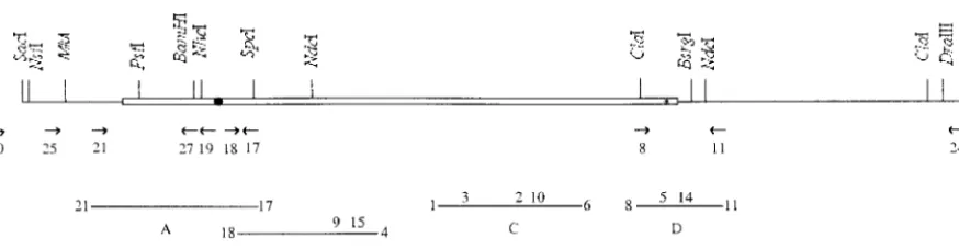
Another gene,rmp1, was initially identified inP. anse- The genomic library used in this study was constructed from args12 rmp1-1strain (Dequard-ChablatandAlland2002) rina due to the presence of two natural alleles in our
in the integrative cosmid vector pMOcosX, with the bacterial reference strains:rmp1-1(formerlyrmp⫺) was found in
hygromycin resistance gene under the control of the cpc1 the strain bearing the mating-type mat⫺ information,
promoter ofNeurospora crassaas selectable marker (Orbach while rmp1-2 (formerly rmp⫹) was found in themat⫹ 1994). The integrative plasmids used for constructions are strain. BothAS1-4 rmp1-1andAS1-4 rmp1-2strains accu- derived from pCB1004 (Carrollet al.1994) or pPaBle ( Cop-pin andDebuchy2000), which carry hygromycin or phleo-mulate the specific deleted mtDNA molecules at the
mycin resistance genes, respectively (Table 1). The pHSS and time of death, but are strikingly different in their life
pPMB plasmids (Table 1) contain genomic fragments of 5.2 spans, which are ⵑ2 and 80 cm, respectively (
Conta-and 3.4 kb, respectively, both of which encompass thermp1-1
mine et al. 1996). Here, we describe the cloning and allele (Figure 1).
analysis ofrmp1by a multidisciplinary approach: search- To obtain a frameshift mutation inrmp1-1, the pHSS plasmid was digested byNheI (Figure 1), successively followed by Klenow ing for homologs in databases and construction of new
and ligation treatments. We thus obtained a ⫹1 frameshift alleles and expression studies throughout the life cycle.
mutation and a stop codon between the two ATGs of the open The results presented below demonstrate thatrmp1is an
reading frame (ORF) in position 138 (Figure 2), which yielded essential nuclear-mitochondrial gene, which probably pHfs (Table 1). The modification in pHfs was confirmed by evolves rapidly and exhibits a complex expression pat- sequencing with primer 17 (Figure 1).
The pLSS plasmid (Table 1), used to obtain a null allele tern strictly regulated during development.
ofrmp1, was constructed from pHSS, whose hygromycin resis-tance gene was deleted. Thermp1-1coding sequence was ex-cised between theNheI site and the 3⬘terminalNdeI site (Fig-MATERIALS AND METHODS
ure 1) and replaced by aXbaI-HindIII fragment carrying the
leu1gene prepared from the pUL plasmid (Debuchy et al.
P. anserinastrains, growth conditions, and transformation:
1993). A purifiedNsiI-DraIII fragment (Figure 1) from pLSS
P. anserina is a heterothallic filamentous ascomycete whose
was used for transformation-mediated gene replacement. life cycle and general methods for genetic analysis have been
Constructions of the pHG, pHN, pHNG plasmids were described (Rizet and Engelmann1949). The asci contain
achieved as follows. Two fragments were obtained by amplifi-four binucleate spores, each formed around two nonsister
cation from a bacterial artificial chromosome (BAC) con-nuclei after a postmeiotic mitosis. A few asci contain five
taining thermp1-2allele (provided by A. Billault, R. Debuchy, spores, two of which are smaller and uninucleate. They give
and P. Silar). The first was amplified with primers 21 and 17, rise to homocaryotic mycelia, used mainly for genetic analyses.
followed by digestion with BamHI andSpeI (Figure 1) and These strains develop both female organs (ascogonia) and
cloning into pHSS* (Table 1) digested by the same enzymes. male gametes (microconidia), but are unable to self-fertilize.
This procedure gave rise to pHG. The second fragment was The binucleate ascospores are used as tools, especially when
amplified with primers 8 and 11, followed by enzymatic diges-heterocaryotic strains are required for complementation
anal-tion withClaI andBsrgI (Figure 1). The resultant product was yses. Crosses were performed by spermatization: a suspension
cloned into pHSS* digested byBsrgI and partially digested by of microconidia obtained from one strain (the male culture)
ClaI (which has only one site inrmp1-1), giving rise to pHN. was poured onto a homocaryotic mycelium (the female
cul-pHNG was constructed from pHN digested by BamH1 and ture) of opposite mating type. Self-fertilization of
heterocary-SpeI, followed by ligation with the initial PCR fragment di-oticmat⫹/mat⫺strains was also examined. Cultures were
usu-gested by the same enzymes. The constructions were con-ally performed at 27⬚. All media,i.e., corn-meal extract (MR),
firmed by sequencing, using primers 17 and 18 for pHG and minimal synthetic (M2), and germination (G) media were
pHNG and primer 11 for pHN (Figure 1). as described (Esser 1974). When required, G medium was
The rmp1-1::GFP gene fusion was obtained as follows. A supplemented with 1% yeast extract. M1 is the protoplast
fragment of pHSS* (Table 1) was amplified with primers 8 regeneration medium. When necessary, hygromycin (Roche
(Figure 1) and Rgfp (5⬘-GAGGGTACCTCGCGATTACCGCG Diagnostics), phleomycin (Sigma, St. Louis), or leucine were
GAAGTTTTTCCCCGGCCCCCACCCAGT-3⬘). In this PCR
added to M1 at a concentration of 100, 5, and 100 g/ml,
product, the TAA stop codon ofrmp1-1is replaced by CTT respectively. Life spans were measured on M2 according to
(leu), which is immediately followed by three restriction sites Contamineet al.(1996). Protoplast preparation and
transfor-(SacII,NruI, andAcc65I). An in-frame TAA is present between mation experiments were performed as described previously
SacII andNruI. In an initial step, the PCR fragment was di-(Berteaux-Lecellieret al.1998).
gested byClaI andAcc65I and cloned into pHSS* (Table 1), Thermp1gene is tightly linked to thematlocus. Thermp1-1
andrmp1-2(formerlyrmp⫺andrmp⫹, respectively) are linked digested by BsrgI and partially digested by ClaI (Figure 1). This construction was sequenced using primer 11 (Figure 1). tomat⫺andmat⫹, respectively. Previous recombination data
demonstrated that the genetic distance betweenmatandrmp1 We also verified that this modified form ofrmp1-1retained the ability to complement the absence of aerial hyphae at 37⬚ wasⵑ0.25 cM (Contamineet al.1996). Thus,matcan be used
as a reliable marker ofrmp1. All mutant strains are derived of a rmp1-2 strain. Second, the pEGFP-1 plasmid (CLON-TECH, Palo Alto, CA) was digested by ClaI and SacII. The from the S strain (Rizet1952). Theleu1-1mutant is
auxotro-phic for leucine. The AS1-4 mutation was identified as an restriction fragment containing EGFP was ligated to the previ-ous construction digested by SacII and NruI, giving rise to antisuppressor mutation (Picard-Bennoun1976). The origin
and main features of theP. anserinawild-type isolates s and pHRGFP (Table 1).
pHRGFP was used to construct pHNRGFP, which contains A and of theP. comataspecies were previously described (
of the ORF (⌬3-23). A pHSS* fragment was amplified (Table1) oplan photomicroscope. Fluorescence images were captured by a CDD Princeton camera system. Mitochondria were stained using primers 25 (Figure 1) and Nrmp (5⬘-TGCACTGCAGT
GAGCATTTGATTTGGTGCTTTCCT-3⬘). The PCR product with the vital mitochodrion-specific dye 2-(4-dimethylamino-styryl)-1-methylpyridinium iodide (DASPMI; Sigma), ac-was digested by MluI and PstI (Figure 1) and cloned in
pHRGFP digested by the same enzymes. This construction, cording to the procedure described previously (Jamet-Vierny et al.1997), after growth of the relevant strains at 27⬚and 37⬚. bearing thermp1-1⌬(3-23)::GFP allele, was sequenced using
primers 19 and 25 (Figure 1). In addition to the deletion⌬(3- Database search analyses:In addition to the general data-bases, we used specific databases for N. crassa (http:// 23), this form ofrmp1-1also replaces the ala 24 codon with
a threonine codon. pedant.gsf.de/),Aspergillus fumigatus(http://www.sanger.ac.uk/
Projects/A_fumigatus/), Magnaporthe grisea (http://www. Isolation of strains bearing the ⌬rmp1 allele: The
gene-replacement experiment yielding a genome bearing the⌬rmp1 genome.wi.mit.edu/annotation/fungi/magnaporthe/), His-toplasma capsulatum (http://genome.wustl.edu/projects/ allele was performed by a strategy that permitted us to obtain
the relevant transformants without phenotypic screening, even hcapsulatum/),Schizosaccharomyces pombe(http://www.sanger. ac.uk/Projects/S_pombe/), andS. cerevisiae (http://genome-if the inactivation ofrmp1were lethal. A transgenicrmp1-1strain
(SS2, Table 2), carrying an ectopic functional copy ofrmp1-1 www.stanford.edu/Saccharomyces/). (carried by pHSS) was crossed with aleu1-1strain and aleu1-1
rmp1-1(rmp1-1) (transgenes in parentheses) strain was recovered and used as recipient in transformation experiments. As described
RESULTS above, theNsiI-DraIII transforming fragment contains theleu1⫹
gene, which replaces rmp1-1. Thus, the transformants were The rmp1 gene was cloned by complementation of screened for leucine prototrophy. They were then tested by PCR
thermp1-2defects:Two possible cloning strategies were analysis for the integration of the relevant fragment at thermp1
available forrmp1.The first was based on the dominance locus. For this purpose, we used primer 30, localized upstream
ofrmp1-1 overrmp1-2 (Contamine et al. 1996): AS1-4 of theSacI restriction site in the genomic sequence (Figure 1),
and a primer localized in the expected neighboring 3⬘region of strains, heterocaryotic for the two rmp1 alleles, show theleu1gene. This test was performed on pools containing mycelia a very short life span, characteristic of rmp1-1. Thus, from 10 transformants, followed by a sib-selection procedure ap- transformation of aAS1-4 rmp1-2strain with a cosmidic plied on positive pools, using a method developed by E.Coppin
library issued from a rmp1-1 strain should lead to the (personal communication). Three transformants among the 193
recovery of thermp1-1 sequence in transformants dis-tested gave an amplification of a fragment with the expected size.
A second PCR test was then performed using primer 24 (Figure playing a short life span. Unfortunately, AS1-4 rmp1-2 1) and a primer localized in the expected neighboring 5⬘region strains sporadically exhibit short life spans (Contamine of theleu1gene. For two of these transformants, it was established et al. 1996). Consequently, this transformation proce-that thermp1-1sequence was not replaced by the deletedrmp1
dure implies long and tedious analyses to eliminate false-copy. The unique candidate,⌬rmp1 leu1-1(rmp1-1) (leu1⫹), was
positive transformants, especially because we use the crossed with aleu1-1 rmp1-2strain, and 12 asci were analyzed to
control the segregation of the leu⫹phenotype. As expected, due sib-selection method (AkinsandLambowiz1985). The to the tight genetic link betweenrmp1and thematlocus, the leu⫹ second strategy was less time consuming and based on ascospores were allmat⫺, but only those carrying the ectopic copy the following observation. AS1⫹ strains bearing the ofrmp1-1were able to germinate. The inability of⌬rmp1ascospores
rmp1-1andrmp1-2alleles, respectively, exhibit a mycelial to germinate was deduced from ascal analyses with respect to
difference, which cannot be seen in a AS1-4 context: segregation of thematlocus (linked tormp1) and of the selective
after 2 days of growth at 37⬚, AS1⫹ rmp1-1 thalli show marker associated with the rmp1-1 transgenic copy. A ⌬rmp1
(rmp1-1) strain devoid of theleu1-1mutation was obtained by aerial hyphae whileAS1⫹rmp1-2thalli do not. Since the
crosses. rmp1-1 haplotype is again dominant over the rmp1-2
To isolate a balanced heterocaryon bearing⌬rmp1in one haplotype, the gene responsible for aerial hyphae for-of its two nuclei, armp1-2 mat⫹leu1-1strain was crossed with
mation at 37⬚could be cloned by complementation of a⌬rmp1 mat⫺leu1⫹(rmp1-1) strain (MB15, Table 2), in which
armp1-2strain. Although we knew that this gene could thermp1-1ectopic copy was carried by pPMB (Table 1). Marker
segregation was controlled in asci issued from this cross. Candi- be eitherrmp1or another tightly linked gene, we chose date heterocaryotic strains of the genotype being sought (is- the second strategy. A genomic library, constructed sued from binucleate ascospores) were submitted to genetic from armp1-1 strain (see materials and methods), analysis to confirm their genotype, i.e., ⌬rmp1 mat⫺ leu1⫹/
was used to transformAS1⫹rmp1-2with pools containing rmp1-2 mat⫹leu1-1.
96 cosmids. Restoration of aerial hyphae at 37⬚was
ob-Sequencing:Genomic DNA was prepared according to
Lec-ellierand Silar (1994). The localization of primers used served in 2 transformants (among 250) in the eighth for PCR performed on genomic DNAs is shown in Figure 1. cosmid pool tested. Crosses between these 2 trans-In contrast to the other alleles,rmp1-1was sequenced from formants and a AS1-4 strain suggested that the inte-subclones of the 5.2-kbSacI fragment, initially by the universal
grated cosmid carried thermp1-1allele because all the and reverse primers, followed by oligonucleotides deduced
AS1-4 rmp1-2ascospores carrying the cosmidic marker from the sequence. Cloned fragments (rmp1-1) or PCR
prod-ucts (otherrmp1alleles) were sequenced using the ABI PRISM gave rise to thalli exhibiting a short life span. The myce-ready reaction dye deoxy terminator cycle sequencing kit (Ap- lial phenotype was used to identify the relevant cosmid plied Biosystems, Foster City, CA), with automatic sequencing through successive rounds of sib selection. In the final machines (373 A or 310 DNA sequencer; Applied Biosystems).
step (in the single cosmid transformation), we obtained
Cytological analyses:Processing of cells for meiocyte
stain-10 transformants (among 20) exhibiting the expected ing was as described previously (Berteaux-Lecellieret al.
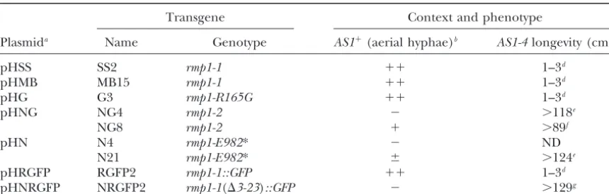
TABLE 1
Plasmids used in this study
Selectable
Plasmids Comments phenotype Source
pCB1004 Used to create pHSS HygroR Carrollet al.(1994)
pPaBle Used to create pPMB PhleoR CoppinandDebuchy
(2000) pHSS From pCB1004; contains theSacI 5.2-kb fragment encompassingrmp1-1 HygroR This study
pPMB From pPaBle; contains theMluI-BsrgI 3.4-kb fragment encompassingrmp1-1 PhleoR This study
pHfs From pHSS withrmp1-1bearing a frameshift mutation HygroR This study
pLSS From pHSS deleted of thehphgene and in whichrmp1-1was replaced by Leu⫹ This study theleu1⫹gene
pHSS* pHSS deleted of its polylinker HygroR This study
pHG pHSS* in whichrmp1-1bears the R165G missense mutation HygroR This study
pHN pHSS* in whichrmp1-1bears the UAG stop codon at position 982 (E982*) HygroR This study
pHNG pHSS* carrying a reconstructedrmp1-2allele (rmp1-1bearing R165G and HygroR This study
E982*)
pHRGFP From pHSS* withrmp1-1::GFPgene fusion HygroR This study
pHNRGFP From pHRGFP withrmp1-1::GFPbearing a deletion of 21 codons (⌬3-23) HygroR This study
in the 5⬘part of the ORF
Seematerials and methodsfor further comments. *, indicates, according to the international nomenclature, that the codon for glutamic acid (E) in position 982 is replaced by a stop codon (see Figures 1–3).
for this phenotype was localized in the cosmid accord- mids (pHSS and pPMB) were crossed withAS1-4,AS1-4 rmp1-2 strains recovered in the progeny and bearing ing to the procedure developed byTurcqet al.(1990).
The ability to complement the mycelial phenotype of a the transgenic sequences exhibited the short life span characteristic of AS1-4 rmp1-1. Second, when a AS1-4 rmp1-2strain was found in a 5.2-kbSacI fragment, which
was cloned into pCB1004, giving rise to pHSS (Table rmp1-2 strain was directly transformed with pHSS, this short-life-span phenotype was observed in 20 (of 54) 1). A smaller fragment,MluI-BsrgI of 3.4 kb (Figure 1),
bearing the same complementing ability, was cloned transformants. Crosses of 3 transformants confirmed that the relevant phenotype was linked to the transgenic into pPaBle and named pPMB (Table 1). Two lines of
evidence demonstrated that the gene involved in the sequence. These data permitted us to use the mycelial phenotype as an easy marker of rmp1in some of the mycelial phenotype seen in the AS1⫹ context and the
gene involved in the longevity feature in theAS1-4back- further analyses.
The sequence of the complementing 5.2-kbSacI frag-ground are one and the same gene, namelyrmp1. First,
when transformants carrying either of the two plas- ment revealed an uninterrupted ORF encoding a
Figure 2.—Comparison of the predicted sequence of RMP1 (pa) with its puta-tive homologs in N. crassa
(nc) andA. fumigatus (af). Identical amino acids are in a black background; similar amino acids are in a shaded background. The positions of the introns in theN. crassa
sequence are indicated by a triangle. The position of the frameshift mutation created in pHfs (Table 1) is indicated by a cross. Positions of the mis-sense (R165G) and nonmis-sense (UAG) mutations in the
rmp1-2 allele are indicated by a circle and a star, respec-tively. The alignment was obtained by the PIMA 1.4 algorithm (http://search l a u n c h e r.b c m.t m c.e d u/ m u l t i-a l i g n/m u l t i-a l i g n. html). The DDBJ/EMBL/ GenBank accession number of the rmp1-1 sequence is AJ581793. TheN. crassaand
A. fumigatussequences can be found at the relevant web sites indicated inmaterials and methods.
tive protein of 1000 amino acids. To demonstrate that into aAS1⫹rmp1-2strain and the transformants checked for their mycelial phenotype after growth at 37⬚: 24 of this ORF corresponded to thermp1-1allele, a frameshift
mutation was introduced at codon position 138 (Figure 28 transformants exhibited the flat mycelium character-istic ofAS1⫹rmp1-2whereas 4 transformants exhibited 2), which simultaneously created a stop codon at the
reconstruction of armp1-1allele through recombination MR) at any temperature tested (18⬚, 27⬚, 37⬚). This
⌬rmp1defect was confirmed repeatedly in the numerous between the transgenic sequence and the endogenous
rmp1-2 allele. In the control experiment, performed crosses performed in this study (see below): among
⬎150 ⌬rmp1 ascospores tested, either AS1⫹ or AS1-4, with the unmodified form ofrmp1-1, complementation
of the recipient strain was observed in most trans- none was viable even on a germination medium en-riched with yeast extract. This lethality can be comple-formants (i.e., 28 of 33). Thus, the fact that a frameshift
mutation at the beginning of the ORF led to loss of its mented by an ectopic insertion, not only of pHSS but also of pPMB, which, respectively, carry the 5.2- and the complementing ability demonstrated that this ORF was
rmp1-1. 3.2-kb fragments encompassing thermp1-1allele (Tables
1 and 2). In contrast, the pHfs plasmid, which carries In silicoanalysis of the RMP1 protein and its putative
homologs:Database searches using the BLAST program rmp1-1 with a frameshift mutation (see Table 1 and above), was unable to complement the lethality of⌬rmp. revealed that the RMP1 protein had putative homologs
in four fungal species for which genomic sequences To test if⌬rmp1could be complemented by a functional rmp1allele present in another nucleus, a heterocaryotic were available:N. crassa,M. grisea,A. fumigatus, andH.
capsulatum. Identity percentages are 39, 32, 25, and 27% strain was constructed (seematerials and methods). In such a heterocaryotic⌬rmp1 mat⫺leu1⫹/rmp1-2 mat⫹ (BESTFIT program) between RMP1 and the four other
sequences, respectively. Furthermore, the parameter leu1-1 strain, the rmp1-2 nucleus should complement the lethality of the⌬rmp1nucleus, which, in turn, com-termed “quality of the alignment” was compared with
the average quality of 100 alignments of random permu- plements the auxotrophy due to the leu1-1 mutation present in thermp1-2nucleus. Indeed, ascospores bear-tations in each case. The reduced deviation (Z
parame-ter), calculated as (cognate quality⫺ average quality/ ing this genotype germinate normally and the issuing strains grow on minimal medium. This result indicates standard deviation of quality of random permutations),
was 205, 106, 21, and 22 for the four combinations, clearly that⌬rmp1internuclear complementation takes place.
respectively. All these values, including the lowest, are
highly significant (Slonimski and Brouillet 1993). To ensure that⌬rmp1lethality was not restricted to the ascospore germination stage, we used the hetero-Overall, these data reflect the phylogeny of those fungi.
All five are filamentous ascomycetes. P. anserina, N. caryotic strain to examine the regeneration capacity of
⌬rmp1 protoplasts. Three types of protoplasts are ex-crassa, andM. griseaare Pyrenomycetes whileA.
fumiga-tusandH. capsulatumare Plectomycetes. In addition,P. pected: those in which the heterocaryotic state is main-tained, homocaryotic protoplasts carrying theleu1-1 nu-anserinaandN. crassaare closer to one another than to
M. grisea. The PSORT program (Nakai2000) disclosed cleus, and, finally, homocaryotic protoplasts containing the ⌬rmp1 nucleus. As shown in Table 3, on leucine-nuclear localization signals (NLS) in three of the five
sequences. RMP1 contains three monopartite and one supplemented medium, 92% of the regenerating proto-plasts were homocaryotic leu1-1; this indicates a clear bipartite signal while N. crassa and A. fumigatus
se-quences contain one and two monopartite signals, re- loss of the heterocaryotic state. In contrast, on medium devoid of leucine, 89% of the regenerating protoplasts spectively. Further studies (see below) did not help us
to understand why RMP1 contains NLS. Interestingly, were heterocaryotic. The lack of homocaryotic⌬rmp1 protoplasts in this experiment, in which the number of the unique feature shared by the five sequences is a
mitochondrial targeting peptide (mTP) as predicted by ⌬rmp1 nuclei was high enough to be easily detected, shows clearly that they did not regenerate. Interestingly, the Target P program (Emanuelsson et al.2000). An
alignment of RMP1 and its putative homologs in N. a fewleu1-1protoplasts were recovered from the selec-tive medium. Their regeneration ability can be ex-crassaandA. fumigatusis shown in Figure 2.
rmp1is an essential gene:Amat⫺strain bearing the plained by sufficient amounts of the LEU1 protein pro-vided in the original heterocaryon. In contrast, none
⌬rmp1 allele was obtained as described in materials
and methods. The recipient strain, used for the trans- of the 98 protoplasts tested from both regenerating conditions displayed themat⫺ (⌬rmp1) genotype. It is formation-mediated gene replacement, carried an
ec-topic copy (SS2, Table 2) of thermp1-1allele carried by not excluded that the two degenerative thalli of un-known genotype recovered in this experiment con-pHSS (Table 1). As expected, the primary transformants
and the purified strains, recovered through crosses, tained a ⌬rmp1nucleus, but even if so, they were not able to regenerate beyond a small filament, like the which exhibited the ⌬rmp1 (rmp1-1) genotype
(trans-gene in parentheses), displayed a rmp1-1 phenotype. ⌬rmp1ascospores (see above). These observations pro-vide strong epro-vidence that both ⌬rmp1protoplasts and These crosses also gave rise to ascospores bearing the
⌬rmp1allele without the complementing ectopic rmp1-1 ⌬rmp1ascospores are unable to give rise to viable thalli and thus thatrmp1is an essential gene.
copy, but none was viable. Careful microscopic
examina-tion revealed that ⌬rmp1 ascospores did in fact form Wild-type rmp1 alleles are polymorphic: The rmp1 gene is tightly linked to thematlocus: the genetic dis-one or two small filaments, which did not continue
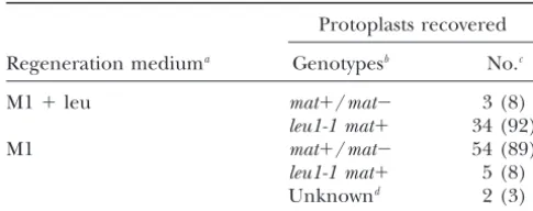
TABLE 2
Expression of different forms ofrmp1in the⌬rmp1background
Transgene Context and phenotype
Plasmida Name Genotype AS1⫹(aerial hyphae)b AS1-4longevity (cm)c
pHSS SS2 rmp1-1 ⫹⫹ 1–3d
pHMB MB15 rmp1-1 ⫹⫹ 1–3d
pHG G3 rmp1-R165G ⫹⫹ 1–3d
pHNG NG4 rmp1-2 ⫺ ⬎118e
NG8 rmp1-2 ⫹ ⬎89f
pHN N4 rmp1-E982* ⫺ ND
N21 rmp1-E982* ⫾ ⬎124e
pHRGFP RGFP2 rmp1-1::GFP ⫹⫹ 1–3d
pHNRGFP NRGFP2 rmp1-1(⌬3-23)::GFP ⫺ ⬎129g
ND, not determined.
aSee Table 1 for more details. Note thatrmp1-2was reconstructed fromrmp1-1.
bFormation of aerial hyphae is observed after growth at 37⬚. The three symbols⫹⫹,⫹, and⫾describe the thickness of aerial hyphae in comparison to theAS1⫹rmp1-1reference strain (⫹⫹);⫺indicates absence of aerial hyphae.
cSeematerials and methods.
dShort longevities were determined on 5 (SS2), 18 (MB15), 4(G3), and 7 (RGFP2) subcultures issued from
one ascospore of each genotype. These values do not differ from those obtained with the reference AS1-4 rmp1-1strain.
eLongevities of the NG4 and N21 strains were measured in the same experiment. In the first case, 10
subcultures, issued from three ascospores were used; only 1 subculture stopped growing (at 9 cm). In the second case, 18 subcultures, issued from four ascospores were used; only 1 subculture stopped growing (at 66 cm). In this experiment, theAS1-4 rmp1-2reference strain showed a mean life span of⬎124 cm (16 subcultures issued from four ascospores; 1 subculture stopped growing at 68 cm).
fMeasurements were made on 15 subcultures, issued from four ascospores. Only 1 subculture was still growing
(⬎171 cm) at the end of the study. In this experiment, theAS1-4 rmp1-2reference strain had a mean life span of⬎135 cm (6 subcultures, issued from two ascospores, with 2 subcultures still growing⬎171 cm).
gMeasurements were made on 11 subcultures issued from four ascospores. Six subcultures were still growing
when the experiment ended. In this series, the shortest longevity was observed at 88 cm. In this experiment, theAS1-4 rmp1-2reference strain displayed a mean life span of⬎150 cm (no dead subculture among 17 issued from five ascospores).
et al.1996). Thermp1-1andrmp1-2alleles are linked to drawn. First, a number of changes, scattered over the entire ORF, are found when theP. comata rmp1alleles mat⫺andmat⫹, respectively. Genetic experiments have
also previously shown that isolates ofP. anserinaandP. are compared to those of theP. anserinaisolates. Second, five sites appear polymorphic among the P. anserina comata(closely related toP. anserina) can be separated
into two classes. The first class (e.g., s) bears twormp1 isolates. Third, in all cases, roughly half of the substitu-tions are nonsynonymous. Finally and importantly, com-alleles similar tormp1-1andrmp1-2with respect to life
spans in aAS1-4context: thermp1-1-like andrmp1-2-like parison between S2 (i.e.,rmp1-2) and s2, on one hand, and all otherrmp1alleles on the other hand, discloses alleles linked tomat⫺andmat⫹, respectively (and thus
like our reference wild-type S strain). In the second the molecular differences responsible for the functional differences betweenrmp1-1andrmp1-2. S2 and s2 share class, the isolates (e.g., A and P. comata) carry a single
type ofrmp1allele, which exhibits the features character- a premature stop (UAG) codon at position 982, which yields a protein lacking its last 19 amino acids. The two istic ofrmp1-1: these alleles confer a short life span to
aAS1-4strain regardless of the associated mating type alleles also differ from S1 (i.e., rmp1-1) and s1 by a missense mutation at position 165. However, this R165G (Contamineet al.1996). We thus decided to sequence
thermp1gene frommat⫹andmat⫺strains of the s and is also found in A2 and in the two rmp1 alleles of P. comata, which display the functional status of rmp1-1. A isolates and ofP. comata(Pc), in addition to thermp1-2
allele of our S strain (seematerials and methodsand Therefore, the longevity differences observed between AS1-4 rmp1-1 and AS1-4 rmp1-2 can be due to either Figure 1).
The data are reported in Figure 3. Each rmp1allele the stop codon alone or its association with the R165G substitution.
was named according to both its origin (S, s, A, or
Pc) and its presence in themat⫺(number 1) ormat⫹ Dissection of the rmp1-2 allele: An ectopic copy of rmp1-1fully complementsrmp1-2and⌬rmp1. When the (number 2) haplotypes. Thermp1-1allele (S1) was used
TABLE 3 surprising phenotype was maintained through crosses. It was dominant overrmp1-2, as observed in the primary
Protoplasts recovered from the heterocaryotic strain
transformants, and recessive with respect tormp1-1. It
⌬rmp1 mat⫺leu1ⴙ/rmp1-2 mat⫹leu1-1
was also observed in a ⌬rmp1 context (Table 2). Al-though these data remain unexplained, the important Protoplasts recovered
point is that the AS1-4 strains bearing either one of
Regeneration mediuma Genotypesb No.c
the two constructs (pHNG or pHN) exhibited the high
M1⫹leu mat⫹/mat⫺ 3 (8) longevity characteristic ofrmp1-2(Table 2). On the basis
leu1-1 mat⫹ 34 (92) of this criterion, we conclude that theE982* mutation
M1 mat⫹/mat⫺ 54 (89) alone leads to the same phenotype as that of the original
leu1-1 mat⫹ 5 (8) rmp1-2allele.
Unknownd 2 (3)
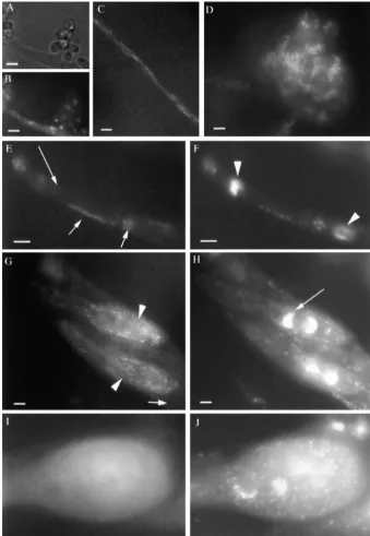
Cellular location of the RMP1-GFP protein shows that
Numbers in parentheses are percentages. thermp1gene is developmentally regulated:To localize
aM1 is the minimal medium used for protoplast
regenera-RMP1, the protein encoded byrmp1-1was tagged exactly tion. M1⫹leu is supplemented with leucine (100g/ml). at its carboxy terminus with green fluorescent protein
bOnly the relevant genetic markers are shown. Note that
(GFP; seematerials and methods) and the relevant
⌬rmp1is present in themat⫺nucleus.
construct was introduced into armp1-2recipient strain.
cThe number of protoplasts capable of regeneration is
much higher on M1⫹leu than on M1. Few were tested from Primary transformants were screened, initially for the M1⫹leu. Seematerials and methodsfor more details. presence of aerial hyphae at 37⬚, and then for GFP
dDegenerative thalli, which did not grow beyond the M1
expression. Aerial hyphae were observed in half of the plates.
transformants (8/18). However, their density varied among transformants, suggesting that complementa-tion of rmp1-2 was more or less efficient. The level of 37⬚ in the AS1⫹ context and a very short life span in
theAS1-4context,AS1⫹rmp1-2(rmp1-1) orAS1⫹⌬rmp1 complementation correlated with the intensity of GFP fluorescence. One transformant, which exhibited the (rmp1-1) do not differ from AS1⫹ rmp1-1, and AS1-4
rmp1-2(rmp1-1) orAS1-4⌬rmp1(rmp1-1) do not differ highest expression ofrmp1-1::GFP, was chosen for further analysis. As shown in Table 2, the transgene (RGFP2) fromAS1-4 rmp1-1(see above and Table 2). Sequence
comparisons ofrmp1-1andrmp1-2revealed two nonsyn- displays the features of abona fide rmp1-1allele: it fully complements⌬rmp1and confers a very short life span onymous substitutions leading to a missense (R165G)
and a nonsense (E982*) mutation inrmp1-2. To address toAS1-4strains.
GFP expression was examined during vegetative the question of the roles of these mutations in the
phe-notypic features due tormp1-2, three plasmids derived growth of the ⌬rmp1(rmp1-1::GFP) strain RGFP2 and over the sexual cycle in perithecia obtained by crossing from pHSS were constructed. They contain armp1allele,
which bears the missense mutation alone (pHG), the this strain (used as the female partner) with a rmp1-2 strain. During early mycelial growth (⬍3 days), GFP nonsense mutation alone (pHN), or both mutations
(pHNG; Table 1 andmaterials and methods). These labeling was not found along all filaments but only along the filaments giving rise to microconidia (conidio-plasmids were introduced singly into armp1-2recipient
strain. Mycelia of the primary transformants were care- phores) in those surrounding the female organs (asco-gonia) and in the ascogonia. In contrast, fluorescence fully examined for the presence or absence of aerial
hyphae after growth at 37⬚. One transformant represen- was detected in microconidia (Figure 4, A and B) and in the vegetative filaments (Figure 4C) only after 3 days tative of each phenotypic class was then crossed to
intro-duce the newrmp1alleles into the⌬rmp1context with of growth. The GFP signal was first seen in the apical cells and then extended to the entire thallus (data not or without theAS1-4mutation.
The data are reported in Table 2. The rmp1-R165G shown). The snake-like forms of the fluorescent bodies are mitochondria, as demonstrated by staining with the allele is not different fromrmp1-1. (i) Of the 10 primary
transformants recovered, 9 exhibited aerial hyphae after vital mitochondrion-specific dye DASPMI (compare Fig-ures 4C and 5A). Note that DASPMI is currently used growth at 37⬚. (ii) The⌬rmp1(rmp1-R165G) strains
ex-hibited the two characteristic features ofrmp1-1(Table to specifically label mitochondria since the pioneer work ofSogoandYaffe(1994) in yeast. We have pre-2). The results obtained with the reconstructedrmp1-2
allele (carried by pHNG) and rmp1-E982* (carried by viously shown that this dye works well in Podospora (Jamet-Viernyet al.1997). In contrast to the mycelium, pHN) are less clear-cut. With both plasmids, the primary
transformants showed two different phenotypes. About which exhibited a GFP staining restricted to mitochon-dria, the ascogonia showed both cytosolic and punctate half of the transformants exhibited no aerial hyphae
after growth at 37⬚, as observed for thermp1-2allele. The or reticular bright staining (see Figure 4D). Similarly, such a punctate pattern was also seen in all paraphysae second half showed aerial hyphae that were less dense
Figure3.—Polymorphism of different wild-typermp1alleles. Polymorphic codons are numbered vertically by position (from 5 to 991). The sequence of each codon is presented vertically, according to the rmp1-1sequence. The polymorphic base is indicated by a shaded background. The first line with boldface type gives the amino acids translated from thermp1-1sequence. The boldface characters of the second line correspond to the residues translated from the sequences that differ fromrmp1-1. Each strain from whichrmp1was sequenced is named according to its origin (S, s, and A are wild-type isolates ofP. anserina, with S being the reference strain used; Pc stands forP. comata) and its mating type (1,mat⫺; 2,mat⫹). For eachrmp1allele, nucleotide differences with respect tormp1-1are shown under the relevant codon positions. For instance, the CGA codon at position 160 inrmp1-1is changed to CGG in five rmp1alleles. This synonymous substitution maintains an arg residue at this position in the polypeptide. In contrast, the CGC codon at position 165 is changed into GGC in the same five alleles. This nonsynonymous substitution leads to a gly rather than an arg residue in the polypeptides (R165G). Codon GAG at position 982 is changed to a UAG (amber) codon in S2 and s2. Note that S1 and S2 give the sequence ofrmp1-1andrmp1-2, respectively, and that s2 is armp1-2-like allele with respect toAS1-4longevity. The two positions in which bothrmp1-2and s2 differ fromrmp1-1
are framed.
the punctate staining, these cells were stained with 4⬘,6- the rmp1-1::GFP construct do not show fluorescence. This is especially striking in the ascospores because the diamidino-2-phenylindole (DAPI) and DASPMI. The
fluorescent bodies observed in the cytoplasm are mito- crosses are heterozygous with respect to this construct, which therefore segregates in the ascospores. The rele-chondria, as seen by DAPI (compare Figure 4, E and
F) and by DASPMI (data not shown). In contrast, RMP1- vant cross was performed several times and we can esti-mate that at least 30 ascospores have been examined in GFP was never detected in the nuclei. During perithecial
development, RMP1-GFP appears only in heterocaryotic each case (early, more mature, and fully mature asco-spores).
basal cells, which contain numerous copies of the two
parental nuclei. In contrast, no signal was detected in These observations lead to three main conclusions. First, thermp1gene is developmentally regulated. Sec-the dicaryotic cells (croziers), which emerge from Sec-the
basal cells and contain one copy of each parental nu- ond, the RMP1-GFP protein is seen in both cytosol and mitochondria. Third, depending on cell type and devel-cleus. Similarly, no fluorescence was seen in asci
under-going caryogamy, meiosis, and postmeiotic mitoses opmental stage, RMP1 either can be seen in both com-partments or is restricted to one or the other compart-(data not shown). However, the signal reappeared in the
ascospores. In early ascospores with young membranes, ment.
Analysis of strains expressing a RMP1 protein devoid
RMP1-GFP was seen in both mitochondria and cytosol
(Figure 4, G and H). It is noteworthy that only the of its putative mitochondrial targeting peptide:To fur-ther analyze the role of the RMP1 putative mitochon-mitochondria of the ascospores are fluorescent and not
those of the ascal cytoplasm in which the ascospores are drial-targeting peptide in both localization and protein function, we constructed armp1-1allele lacking codons formed. In more mature ascospores, only the cytosolic
signal remained visible (Figure 4, I and J); it disappears 3-23 and fused to the GFP sequence (materials and methods). This construct was introduced in a rmp1-2 in fully mature ascospores. We ruled out the possibility
Figure4.—RMP1-GFP localization in
⌬rmp1(rmp1-1::GFP) during the vegeta-tive and sexual cycles following fertiliza-tion with a rmp1-2 partner. Note that photographs in A–D were performed on living cells. (A and B) RMP1-GFP stain-ing in microconidia: (A) group of eight microconidia observed in bright light; (B) the same group observed through a GFP filter. Note that each microconi-dium shows one bright fluorescent spot. (C) In vegetative filaments after 3 days of growth, RMP1-GFP is visible in snake-like organelles, similar in size and shape to the mitochondria stained with DAS-PMI (Figure 5A). (D) RMP1-GFP is al-ways visible in ascogonia (female organ). Note the reticular pattern superimposed on the cytosolic fluorescence. (E and F) RMP1-GFP fluorescence (E) and DAPI staining (F) in a paraphysa. The two large DAPI spots correspond to nuclei (arrowheads) and the small dots to mtDNA nucleoids. Note the complete overlap between the GFP signal and the DAPI staining in the mitochondria (short arrows) and the absence of GFP staining in the nuclei (arrow). GFP (G) and DAPI staining (H) of two very young ascospores. Note that the structures in G are also mitochondria because they can be surperimposed on the DAPI stain-ing in H. As in paraphysae, this overlap is not a strict colocalization, which means that RMP1 is not specifically associated with the nucleoids; only the mitochon-dria located in the two ascospores (ar-rowheads) are stained by RMP1-GFP, while the mitochondria present in the surrounding ascus are not (short arrow). Also, as seen for paraphysae, no RMP1-GFP signal is found in the nuclei (arrow points to one nucleus). (I and J) In a nearly mature ascospore, the GFP signal (I) is exclusively cytosolic. Note also that the surrounding cytoplasm is not stained, although several mitochondria are present, as revealed by the DAPI staining in J. Bar, 5m.
This suggests that the construct does not complement RMP1 protein is essential, its mTP is dispensable. To test if mitochondria lacking the RMP1 protein were thisrmp1-2defect. One of the transformants (NRGFP2,
Table 2) was crossed to strains of interest to introduce different in morphology and/or distribution, mitochon-dria of armp1-1and a⌬rmp1[rmp1-1(⌬3-23)::GFP] strain the rmp1-1(⌬3-23)::GFPallele in all other possible
ge-netic backgrounds. were stained with DASPMI. As shown in Figure 5,
mito-chondria of the two strains were very similar in number, Strikingly, this allele was able to complement the
le-thality of⌬rmp1, indicating that it was at least partially distribution, and mostly also in size. However, a few en-larged mitochondria were systematically seen in the mu-functional. GFP staining was followed in a⌬rmp1strain
bearing thermp1-1(⌬3-23)::GFPtransgene. Contrary to tant strain (roughly one per cell), whatever the growth temperature (27⬚and 37⬚). In contrast, such giant or-RMP1-GFP, the staining was solely cytosolic or absent:
mitochondria were never seen labeled. These results ganelles were not observed in wild-type strains (compare Figure 5, A and B; see alsoJamet-Viernyet al.1997). are very important. They demonstrate first that the
Figure6.—Growth curves of⌬rmp1[rmp1-1(⌬3-23)::GFP],
rmp1-2, and rmp1-1 at three different temperatures. The growth curves ofrmp1-1 at 27⬚ and 20⬚ have been omitted Figure5.—Phenotypes of mitochondria inrmp1-1(A) and
because at these temperatures this strain exhibits the same
⌬rmp1[rmp1-1(⌬3-23)::GFP] (B) vegetative filaments.
Mito-growth rate as rmp1-2. Similarly, the growth curves ofrmp1-1
chondria are stained with DASPMI. Arrow points to an
en-[rmp1-1(⌬3-23)::GFP] and rmp1-2 [rmp1-1(⌬3-23)::GFP] are larged mitochondria. Note that the giant mitochondria are
identical to those ofrmp1-1andrmp1-2, respectively. much brighter than the normal mitochondria as previously
observed in PaTOM70 and PaMDM10 mutants ( Jamet-Viernyet al.1997). Bar, 5m.
unable to enter mitochondria (at least in detectable amounts). Although this new allele shares some proper-ties withrmp1-2, it also has its own features and cannot (⌬3-23)::GFP construct are not completely wild type.
They display no aerial hyphae, not only at 37⬚(Table be considered simply as similar tormp1-2.
Role of RMP1 during the sexual cycle: With the 2) but also at 27⬚(this phenotype is visible immediately
following ascospore germination). Furthermore, as shown knowledge that rmp1is developmentally regulated, we analyzed in further detail the possible role of RMP1 in Figure 6, their growth rates differ from those of the
control strains at the three temperatures tested. At 37⬚, during the sexual cycle.⌬rmp1nuclei can be maintained only in balanced heterocaryotic strains. The⌬rmp1 mat⫺ their growth was even arrested after a few days. However,
the strains did not die: they resumed growth after trans- leu1⫹/rmp1-2 mat⫹ leu1-1 strain (see above) is able to self-fertilize. However, it is impossible to determine if fer to 27⬚. In the course of these studies, it was observed
that the life span of aAS1⫹ ⌬rmp1strain carrying the this strain produces⌬rmp1 female organs in addition to those containing rmp1-2 nuclei. In contrast, the rmp1-1(⌬3-23)::GFPtransgene was increased twofold in
comparison with the reference strains. All these features spermatization method (materials and methods) demonstrated that the heterocaryotic strain forms func-are recessive: the AS1⫹ rmp1-1 and rmp1-2 strains
car-rying the construct exhibit the phenotypic properties tional⌬rmp1 mat⫺microconidia able to fertilize amat⫹ tester strain. Due to its viability, it was possible to directly of thermp1-1andrmp1-2reference strains, respectively,
including their growth rates (see legend of Figure 6) address these questions for a ⌬rmp1strain bearing the rmp1-1(⌬3-23)::GFP construct. In this case, functional and their life spans (data not shown).
Interestingly, when introduced in aAS1-4⌬rmp1con- microconidia and female organs are formed. These data lead to two conclusions. First, the rmp1gene plays no text,rmp1-1(⌬3-23)::GFPleads to a very long life span,
not different from those characteristic of the refer- role in the fertilization ability of the microconidia. Sec-ond, although we do not know if RMP1per seis dispens-enceAS1-4 rmp1-2strains (Table 2). The newrmp1allele
is recessive with respect tormp1-1, as isrmp1-2: AS1-4 able for female organ differentiation, our results dem-onstrate that RMP1 without mTP is sufficient for their rmp1-1strains, which bear thermp1-1(⌬3-23)::GFP
con-struct, exhibit the very short life span typical ofAS1-4 development.
In a second step, we examined the contents of peri-rmp1-1 (data not shown). In conclusion, deletion of
TABLE 4
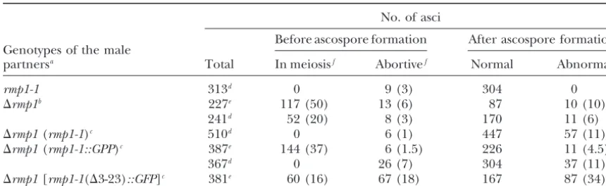
Sexual defects observed in perithecia issued from crosses implicating different alleles ofrmp1
No. of asci
Before ascospore formation After ascospore formation Genotypes of the male
partnersa Total In meiosisf Abortivef Normal Abnormalg
rmp1-1 313d 0 9 (3) 304 0
⌬rmp1b 227e 117 (50) 13 (6) 87 10 (10)
241d 52 (20) 8 (3) 170 11 (6)
⌬rmp1(rmp1-1)c 510d 0 6 (1) 447 57 (11)
⌬rmp1(rmp1-1::GPP)c 387e 144 (37) 6 (1.5) 226 11 (4.5)
367d 0 26 (7) 304 37 (11)
⌬rmp1[rmp1-1(⌬3-23)::GFP]c 381e 60 (16) 67 (18) 167 87 (34) aIn all crosses, the female partner was a wild-type rmp1-2 mat⫹ strain. Delay in ascospore ejection from perithecia was observed in the second and the fifth crosses.
bThe⌬rmp1male gametes are issued from the heterocaryotic strain⌬rmp1 mat⫺leu1⫹/rmp1-2 mat⫹leu1-1. cThe transgenes are SS2, RGFP2, and NRGFP2 for (rmp1-1), (rmp1-1::GPP), and [rmp1-1(⌬3-23)::GFP], respectively (see Table 2).
dThe contents of perithecia were examined the fifth day after fertilization.
eThe contents of perithecia were examined the fourth day after fertilization.
fThe numbers in parentheses give the percentage with respect to the total of asci.
gThe numbers in parentheses give the percentage of asci with abnormal ascospores among sporulated asci.
DISCUSSION are reported in Table 4 and lead to the following
re-marks. Abortive asci are present in all crosses including The P. anserinaRMP1 protein exhibits several note-a wild-type cross (rmp1-1⫻ rmp1-2). They represent a worthy features with three intertwined facets. First, the small percentage of total asci except when the ⌬rmp1 function(s) of RMP1 is unknown and its putative homo-nucleus contains the rmp1-1 (⌬3-23)::GFP construct: logs are, to date, found only in filamentous ascomycetes. here, nearly 20% of asci are abortive. Asci containing Second, RMP1 is developmentally regulated: the RMP1-abnormal ascospores are found in all crosses except in GFP fusion protein can be undetectable, cytosolic, and/ the wild-type control. In all cases, the abnormalities in or mitochondrial, depending on the cell type and the shape and number of ascospores correlate with an
ab-developmental stage. Third, RMP1 is essential but its normal distribution of nuclei (data not shown). This
mTP is dispensable. defect is always associated with the presence of a⌬rmp1
rmp1encodes a protein whose putative homologs can
nucleus in the crosses, whatever the transgenic
se-be found only in filamentous ascomycetes: RMP1 is a quence. However, while in all mutant crosses the
per-large protein that lacks recognizable motifs. This ham-centage of asci with abnormal ascospores represents
pers understanding of its function. Furthermore,rmp1 ⵑ10% of all spored asci, this value attains one-third of
putative homologs have been found only in the genomes the asci when the⌬rmp1nucleus bears thermp1-1(⌬
3-of filamentous (multicellular) ascomycetes. Three hy-23)::GFP construct. Overall, our observations lead to
potheses can account for this situation. First, thermp1 three conclusions. First, defects observed when ⌬rmp1
function could be restricted to these fungi. The Woro-is heterozygous in a cross suggest that the rmp1 gene
nin body is an example of a structure specific to fila-dosage likely plays a role. For instance, the RMP1
pro-mentous ascomycetes; this specialized vesicle occludes tein could be rate limiting for proper ascospore
forma-septal pores when the filaments are damaged, thus tion. Second, although the 5.2-kbSacI fragment (Figure
avoiding cell death by preventing loss of cytoplasm. 1) fully complements⌬rmp1during vegetative growth,
However, this function is not essential for growth: a lack it is unable to complement its sporulation defect. One
of Woronin bodies is not lethal (JeddandChua2000; simple explanation is that a sequence required for the
Tenney et al. 2000). A systematic search for essential full expression of rmp1 during sexual reproduction is
genes has been undertaken inA. fumigatus(Fironet al. lacking in this fragment. Finally and noteworthy, our
2002;Fironandd’Enfert 2002), but present reports results show thatrmp1-1(⌬3-23)::GFP, whose vegetative
have not yet revealed if some are specific to filamentous features are recessive (see above), acts as a dominant
ascomycetes. In fact, to our knowledge, essential genes negative allele during sexual reproduction. In other
characterized in this evolutionary lineage are either in-words, the extent of defects is higher in a cross
heterozy-volved in general, basic functions common to all organ-gous for this allele than in crosses heterozyorgan-gous for
isms (e.g., translation) or shared by the entire fungal
⌬rmp1. Thus, it seems that a RMP1 protein devoid of
ascomycetes (yeasts) and in basidiomycetes. For instance, genes inS. cerevisiae(reviewed inContamineandPicard 2000).
inactivation of the gene encoding the catalytic subunit
Although all the pieces of the puzzle are still not in of glucan synthase is lethal not only in A. fumigatus
place to explain the evolutionary position ofrmp1, an (Fironet al.2002) but also inS. cerevisiae(Mazuret al.
unsettling observation favors the third model. When the 1995) and inCryptococcus neoformans(Thompsonet al.
putativeH. capsulatumhomolog of RMP1 was used to 1999). This gene is indeed specific to fungi for which
question a general nonredundant database, a significant
-(1-3) glucan is an essential component of the cell wall.
alignment (BLAST E value of 3⫻10⫺11) was found with
With this viewpoint, rmp1 could be the first essential
a hypothetical protein of S. pombe, SPAP8A3.14C. In gene specific to multicellular ascomycetes.
contrast, no putative homologs were found when the Second, thermp1function could be widely distributed
same database was searched for RMP1 and its other but, in other evolutionary lineages, ensured by a
nonho-fungal counterparts, using an E cutoff value of 1⫻10⫺4.
mologous gene encoding a structurally different
pro-TheS. pombeprotein has a weak similarity toS. cerevisiae tein; this implies thatrmp1and the other putative gene
Sls1p, with an E value of 3⫻10⫺7. Sls1p is a
mitochon-have no common ancestor. This situation is illustrated
drial membrane protein required for respiration ( Rou-by the thyA/thyX genes, which both encode a protein
illardet al.1996). It was recently proposed that this with thymidylate synthase activity. However, the two
pro-protein may play a key role in modulating the translation teins lack any sequence similarity and are not
structur-efficiency of mitochondrial mRNAs (Bryanet al.2002). ally related. With a few exceptions,thyAandthyXhave
To date, we do not favor the idea that RMP1 and Sls1p mutually exclusive phylogenetic patterns (
Myllykal-might be homologous. Furthermore, the similarity be-lioet al.2002).
tween the S. cerevisiae and S. pombe proteins appears A third model also makes sense: the rmp1 function
questionable (Z value: 9). However, one cannot exclude could be widely distributed, but the corresponding gene
that the H. capsulatum homolog of RMP1 and the evolves so rapidly that recognition of its homologs in
SPAP8A.14C sequence ofS. pombemight bridge the gap distant species would be impaired. Such a hypothesis is
between unicellular and multicellular ascomycetes in supported by the weak similarities observed between
the case of a rapidly evolving gene. To elucidate this point, RMP1 and its putative homologs in filamentous
ascomy-an understascomy-anding of the function(s) of both RMP1 ascomy-and cetes: they fall between 39 and 25% identity, and this
SPAP8A3.14C is required. is dependent on the phylogeny. In contrast, when 163
Expression of rmp1 and subcellular localization of
P. anserinaputative coding sequences (located in the two
RMP1 are developmentally regulated: In addition to regions surrounding the centromere of chromosome V)
sequence analyses and comparisons, another approach are compared with their putative homologs inN. crassa,
to the function of rmp1was the study of its expression the percentages of identity are centered on 60–70%,
throughout the life cycle and the localization of its prod-with nearly 90% of the proteins exhibiting⬎40%
iden-uct. Our work clearly shows thatrmp1expression is sub-tity (Silaret al.2003). In addition, seven nuclear genes
ject to spatial and temporal controls. This is true during encoding proteins with well-known mitochondrial
func-both the vegetative and sexual cycles. In the vegetative tions have been characterized inP. anserina. The
2002 and references therein). This is a good illustration changes in protein conformation leading to an import-incompetent state. In the case of fumarase, cotransla-of how the expression pattern cotransla-of a gene may indicate
a specific or a general function. However, only a genetic tional import of the precursor could follow two routes, one leading to mitochondrial localization after import analysis determined the precise role offzo(Halesand
Fuller 1997). Similarly, in S. cerevisiae, some nuclear completion and the other to a release of the protein back into the cytosol (Knoxet al.1998). With respect to the genes with known roles in mitochondrial function are
up- or downregulated during sporulation (e.g., Chuet major adenylate kinase, its dual location is explained by a competition between folding (cytosolic location) al.1998; see also theSaccharomycesGenome Database)
but the functional reasons for their expression pattern and import (Strobelet al. 2002). One striking point with RMP1, which makes it a unique case, is that its subcel-remain mostly unknown. With respect tormp1, the
regu-lation of expression seen at the protein level is especially lular location varies according to cell type and develop-ment. If the viewpoints proposed inS. cerevisiaeare applied complex. The reason that RMP1 is undetectable when
the strain resumes growth, as well as in croziers, asci, to RMP1, one could assume that cellular components or factors might differentially influence the ratio of import-and mature ascospores, is puzzling. Although RMP1
might be dispensable in croziers and asci, our data competentvs.import-incompetent forms of RMP1, for instance, by post-translational modifications. In any clearly demonstrate that it is essential for ascospore
ger-mination and protoplast regeneration. Two assump- case, although the extraordinary developmental and cel-lular patterns of RMP1 do not shed light on its function, tions can explain this paradox. First, the proteinper se
may be needed at these stages. This implies that very they do provide an exciting model for further studies.
RMP1 is an essential protein, in which mTP is
dispens-low amounts of RMP1 (undetectable by GFP
fluores-cence) are sufficient to ensure its essential function. able: A third way to shed light onrmp1 function was careful examination of the phenotypic properties of the Second, RMP1 may seem dispensable at certain steps
because it might have an enzymatic activity whose prod- four alleles available. In addition to the two natural alleles, rmp1-1 and rmp1-2, two new alleles were con-uct accumulated during the preceding stages, i.e., in
the female organs before and after fertilization, in the structed: ⌬rmp1, which is a complete deletion of the gene, and rmp1-1(⌬3-23), which carries a deletion of stationary phase, and during ascospore maturation. This
hypothetical product would ensure RMP1 function at codons 3–23 and thus encodes a RMP1 protein without its mTP. In comparison withrmp1-2,rmp1-1is probably subsequent stages. To explain the absence (or very low
levels) of RMP1 at these critical stages, one can hypothe- the fully functional allele. This conclusion is based mainly on the fact that rmp1-2 shares phenotypic fea-size a feedback control: high amounts of the RMP1
product would cause repression ofrmp1until dilution tures withrmp1-1(⌬3-23). In aAS1-4context, both lead to a very long life span. In a AS1⫹ background, the of this product would permit derepression.
In addition to its complex regulation pattern, another defects ofrmp1-2are modest compared to those of rmp1-1(⌬3-23). In the first case, the strains lack aerial hyphae remarkable feature of RMP1 is its localization in the
cytosolic compartment, in mitochondria, or in both, and display a slightly reduced growth rate at 37⬚. In the second case, these defects are also seen at 27⬚and the depending on the cell type. Proteins encoded by a single
gene and exhibiting both mitochondrial and cytosolic strains are heat sensitive. In addition,AS1⫹strains bear-ing the rmp1-1(⌬3-23) allele exhibit life spans twice locations have been described in other organisms.
How-ever, in most cases, the two protein forms correspond those of the reference strains, and they show a few giant mitochondria at 27⬚ and 37⬚. Finally, ⌬rmp1 is lethal. to two different translation products in which the mTP
is present or absent, due to alternative sites for initiation Therefore, one can conclude that rmp1is an essential gene but that absence (or a very low amount) of RMP1 of transcription, alternative splicing, or alternative sites
for initiation of translation. Yeast fumarase (Knox et in the mitochondria is compatible with viability, at least below 37⬚. Interestingly, some properties ofrmp1-1(⌬ 3-al.1998; Sass et al. 2001) and major adenylate kinase
(Strobelet al.2002) belong to a second class of pro- 23) are reminiscent of those previously observed inP. anserinawhen the mitochondrial metabolism is altered. teins, whose dual location is ensured by a single
transla-tion product. RMP1 probably belongs to this class. This For instance, a mutation inPaTOM70, encoding a pro-tein implicated in the import of propro-teins from the cyto-assumption is supported by the fact that armp1-1allele
bearing a frameshift mutation between the first two sol into the mitochondria, leads to reduced formation of aerial hyphae, heat sensitivity, and striking increases ATGs of the ORF is unable to complement⌬rmp1
lethal-ity, whereasrmp1-1(⌬3-23), which encodes a mTP-trun- in life spans of AS1-4 and AS1⫹ strains. In addition, strains bearing this mutation exhibit a few giant mito-cated protein, complements the null allele. If there were
two translation products, initiated at these two ATGs, chondria (Jamet-Vierny et al. 1997; Contamine and Picard1998).
the frameshift mutant should exhibit the properties of
rmp1-1(⌬3-23). In yeast, two mechanisms have been pro- The simplest hypothesis, with respect to the data re-ported above, is that RMP1 ensures the same essential posed to explain the dual (cytosolic/mitochondrial)