Glucocorticoid receptor antagonizes EGFR function
to regulate eyelid development
ANA SANCHIS
#, PILAR BAYO
#, LISA M. SEVILLA and PALOMA PÉREZ*
Instituto de Biomedicina de Valencia, Consejo Superior de Investigaciones Científicas (IBV-CSIC), Valencia, Spain
ABSTRACT The glucocorticoid receptor (GR) plays a crucial role in epidermal morphogenesis during embryonic development, as demonstrated by analyzing genetically modified mouse models of GR gain- and loss-of-function. Eyelid formation constitutes a useful model to study epithelial development, as it requires coordinated regulation of keratinocyte proliferation, apop-tosis and migration. We have analyzed this biological process in GR-/- embryos during ontogeny. Our data demonstrate that GR deficiency results in delayed and impaired eyelid closure, as illustrated by increased keratinocyte proliferation and apoptosis along with impaired differentia-tion in GR-/- eyelid epithelial cells. These defects are due, at least in part, to the lack of antagonism between GR and epidermal growth factor receptor (EGFR) signaling, causing sustained activation of the MAPK/AP-1 pathway and the upregulation of keratin K6 at embryonic stage E18.5. Additionally, we demonstrate that GR regulates epithelial cell migration in vitro by interfering with EGFR-mediated signaling. Overall, GR/EGFR antagonism appears as a major mechanism regulating ocular epithelial development.
KEY WORDS: glucocorticoid receptor, EGFR, genetically modified mice, epithelial cell, eye development
Introduction
It is known that the glucocorticoid receptor (GR) plays a crucial role during embryogenesis, since this ligand-activated transcrip-tion factor is required for maturatranscrip-tion of vital organs such as the lung, heart, kidney, gut and epidermis (reviewed in Wintermantel et al., 2004; Revollo and Cidlowski, 2009). GR exerts its biological effects through two different mechanisms that involve DNA bind-ing-dependent and -independent actions, which can be geneti-cally separated and are commonly refered to as transactivation and transrepression functions. In fact, knock-out GR-/- mice die perinatally whereas mice carrying a point mutation which abro-gates the dimerization- dependent DNA binding of GR (GRdim/dim) are viable (Cole et al., 1995; Reichardt et al., 1998). This distinc-tion, however, is not so clear-cut since GR monomers are also able to bind certain gene promoters and thus, regulate gene transcription (Adams et al., 2003; Rogatsky et al., 2003): In the last years, we have demonstrated that GR is a key player in epithelial development by analyzing genetically modified mouse models of GR gain- and loss-of-function (Pérez et al., 2001; Cascallana et al., 2003; Cascallana et al., 2005; Donet et al.,
BIOLOGY
www.intjdevbiol.com*Address correspondence to: Paloma Pérez. Instituto de Biomedicina de Valencia, Consejo Superior de Investigaciones Científicas (IBV-CSIC). Jaime Roig 11, E-46010 Valencia, Spain. Fax: +34-96-369-0800. e-mail: [email protected]
#Note: Both authors contributed equally to this paper.
Accepted: 31 May 2010. Final author corrected PDF published online: 26 November 2010.
ISSN: Online 1696-3547, Print 0214-6282 © 2010 UBC Press
Printed in Spain
Abbreviations used in this paper: EGFR, epidermal growth factor receptor; GC, glucocorticoid hormone; GR, glucocorticoid receptor.
2008; Bayo et al., 2008). We have recently reported that only the epidermis of GR-/- but not GRdim/dim embryos shows major defects, suggesting that dimerization dependent DNA binding by GR is dispensible for epidermal development (Bayo et al., 2008). In addition, we have shown that transgenic mice with keratinocyte-targeted overexpression of GR (K5-GR mice) featured numerous epithelial defects including epidermal defects and an eyelid open-ing at birth phenotype (EOB). Overexpression of GR transrepression function in keratinocytes (K5-GR-TR mice) also elicited epithelial alterations that partially overlapped with those found in K5-GR mice (Cascallana et al., 2005; Donet et al., 2008). Remarkably, K5-GR-TR mice featured an EOB phenotype iden-tical to K5-GR mice, indicating that the transrepression function of the GR is sufficient to cause these epithelial ocular anomalies (Donet et al., 2008).
spatio-tempo-ral coordination of cellular proliferation, migration, differentiation and apoptosis. In the mouse embryo, eyelid formation starts at midgestation, whensmall invaginations of the surface ectoderm begin to grow (E12.5), become distinguishable as protruding edges (E14.5-E15.5) and gradually cover the corneal surface until they fuse at E16.5. At E18.5, eyelids are unequivocally closed and start to separate postnatally (P3-P5) until they reopen around P12 (Kaufman and Bard, 1999). In this work, we analyzed the consequences of GR functional inactivation in ocular develop-ment by using GR-/- mice.’Our data show that GR is required to modulate eyelid epithelial morphogenesis through regulation of keratinocyte proliferation, apoptosis, differentiation and migra-tion. It is known that GR suppresses keratin expression of either mitotically-active basal keratinocytes (K5 and K14) or migration-associated keratins K6 and K16. The mechanisms involve binding of GR to AP-1 and interaction of four GR monomers with k6 promoter, respectively (Radoja et al., 2000; De Bosscher and Haegeman, 2009). Accordingly, K5 and K6 expression was al-tered in GR-/- epithelia. These actions were mediated, at least in part, through interference with EGFR signaling, the loss of which
results in increased EGFR and MAPK/AP-1 activation and sus-tained expression of the keratin K6. In vitro wound healing assays demonstrated that constitutive GR overexpression in keratinocytes drastically impairs cell migration and interferes with EGFR func-tion suggesting that lack of cross-talk between GR and EGFR pathway is one of the mechanisms causing delayed eyelid closure in GR-/- embryos.
Results
We have anayzed eyelid closure in GR-/- embryos as in vivo model to address the impact of GR loss-of-function in epithelial morphogenesis. Macroscopically, GR-/- embryos showed open eyelids at embryonic days E16.5 and E18.5. A detailed histo-pathological analysis of wt and GR-/- littermates at distinct timepoints showed severe anomalies in the eyelid formation of GR-deficient mice throughout development (Fig. 1A, compare a-d ana-d a´-a-d´). In a wt a-developing mouse, eyelia-d closure is normally completed in 24h, coinciding with the E15.5-E16.5 transition (Fig. 1A; a, b-d; asterisks indicate fused eyelids in GR+/+). In contrast,
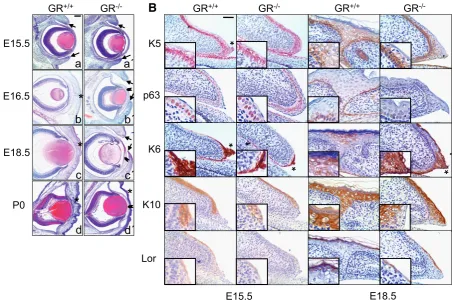
Fig. 1. Delayed and impaired eyelid closure in glucocorticoid receptor (GR) null mice. (A) Histopatholgical analysis of wt and GR-/- embryos at
distinct developmental timepoints. Eyelid closure is normally completed at E15.5-E16.5 transition in wt embryos and eyelids remain fused until postnatal age (a-d). In contrast, GR-/- embryos exhibited unfused eyelids at E16.5 and E18.5 (a´-c´, arrows) along with an abnormal corneal stroma with
increased cellularity (b´,c´, arrowheads). GR-/- newborn mice showed closed eyelids with abnormal epithelia and atypical corneal stroma (d´, asterisk
and arrowhead, respectively). Bar: 200 m. (B) Abnormal morphogenesis of eyelid epithelial cells in GR-/- early (E15.5) and late (E18.5) embryos.
Immunostaining for K5, K6, p63, K10 and loricrin was performed using specific antibodies. Note that both K5 and p63 expression stained two-to three suprabasal layers of eyelid epithelia in GR-/- embryos as compared to more restricted labeling at the fused eyelids of GR+/+ individuals (K5 and p63;
see inset). Increased K6 expression at the eyelids tips of E15.5 (asterisks) was observed in wt mice. Elevated levels of K6 were detected in the unfused GR-/- eyelids at E18.5, as compared with restricted K6 expression at closed eyelids of GR+/+ mice. Abnormal K10 and loricrin staining was apparent
in GR-/- eyelid epidermis relative to GR+/+ mice (K10 and loricrin; see inset). Bar: 100 m.
K10
Lor
p63
K5
K6
E16.5
E18.5
E15.5
P0
GR
-/-GR
+/+*
*
*
*
a
a´
b
b´
c
c´
d
d´
E18.5
E15.5
GR
-/-GR
+/+GR
+/+GR
-/-*
*
*
*
GR-/- embryos revealed unfused eyelids at E16.5 and even at E18.5 (Fig. 1A; b´, c´, arrows) along with an abnormal corneal stroma with increased cellularity (Fig. 1A; b´, c´, arrowheads). The eyelids of GR-/- newborn mice were closed but the epithelia appeared abnormally enlarged and undifferentiated (Fig. 1A; compare d and d´) and an atypical corneal stroma persisted. Penetrance of the reported ocular phenotype in GR-/- mice was 89% (n=43), with either one or two eyes affected (54.55% and 45.45%, respectively).
As an attempt to understand how loss of GR is causing these defects, we analyzed the expression of markers of keratinocyte proliferation, migration and differentiation in the eyelid epithelia of early (E15.5) and late (E18.5) embryos by immunostaining (Fig. 1B). In wt embryos, K5 expression was apparent in all epithelia basal cells of the eyelids, cornea and conjunctiva (Fig. 1B, K5). In GR-/- littermates, labeling of these epithelial cell layers was similar, although additional K5 expression was detected suprabasally in the eyelid epithelia (Fig. 1B, K5, see inset). The epithelial-specific marker p63 was detected in keratinocytes of GR+/+ and GR-/- embryo eyelids and cornea. However, p63 immunostaining at the leading edge of early and late GR-/- embryo eyelids was more suprabasal as compared to the more restricted p63 labeling at wt eyelid tips (Fig. 1B, p63).
In E15.5 wt mice, K6 was strongly expressed at the eyelids tips, defining the migrating cells at the leading edge of this epithelium (Fig. 1B, K6, asterisk). The conjuctival epithelium was also K6-positive (not shown). Notably, at E18.5, where cell migration is no longer required since eyelids are already fused, K6 immunostaining was restricted to the closed eyelids (Fig. 1B). In contrast, GR -/-embryos stained weakly for K6 at the eyelid borders at E15.5 whereas an abnormally strong K6 signal was detected at E18.5, both at the tip of unfused GR-/- eyelids and in conjuctival epithe-lium (Fig. 1B, K6, asterisk and data not shown). The observed K6 expression pattern indicates a delay in the ocular epithelia devel-opment of GR-/- mice.
Previous reports described that eyelid fusion precedes epithe-lial differentiation, since positive K10 staining was detected in epithelial cells of wt embryos only after eyelid closure (Zhang et al., 2005). We examined K10 and loricrin expression and found that in E15.5 wt embryos, both markers were present in suprabasal eyelid epithelia although absent at the leading edge. In E18.5 wt embryos, only suprabasal cells of the fused eyelids stained positive for both markers. In contrast, reduced staining of K10 and loricrin was apparent in GR-/- eyelid epidermis of early embryos whereas abnormal expression of these proteins persisted in the unfused eyelids from GR-/- E18.5 embryos (Fig. 1B, K10 and loricrin).
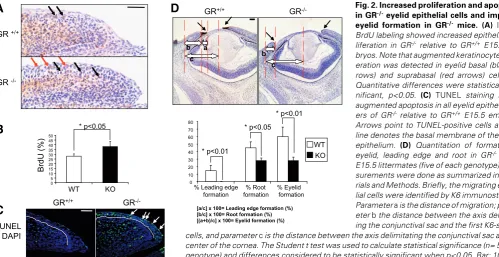
We further assessed altered proliferation in GR-/- eyelid epithe-lial cells by measuring in vivo BrdU incorporation in GR-/-vs wt E15.5 embryos, and found qualitative and quantitative differ-ences among them (Fig. 2A, B). In wt embryos, most BrdU-positive keratinocytes were detected in the basal cell of the eyelid epithelia (black arrows). In contrast, augmented keratinocyte proliferation was detected in basal (black arrows) and suprabasal (red arrows) cells of GR-/- eyelids. When comparing the prolifera-tion of eyelid basal cells, differences were found statistically significant (Fig. 2A, 38.1% vs 28%, respectively, p<0.05). At later developmental stages (E18.5), we could not detect increased proliferation of the eyelid basal keratinocytes of GR-/- relative to wt (data not shown). However, we found BrdU-positive nuclei in the suprabasal layers of E18.5GR-/- eyelids, as occurred in E15.5 GR-deficient embryos. Additionally, we detected augmented apopto-sis in all eyelid epithelial layers of GR-deficient embryos by TUNEL staining, in contrast with scarce apoptotic cells in wt littermates (Fig. 2B, arrows). Overall, our results demonstrate that GR is required for proper proliferation, apoptosis, migration and differentiation of the eyelid epithelial cells of GR-/- embryos.
To further characterize the alterations inGR-/- eyelid closure, we quantitated several parameters to estimate the percentage of formation of the eyelid, leading edge and root, following the
GR
-/-[a/c] x 100= Leading edge formation (%) [b/c] x 100= Root formation (%) [(a+b)/c] x 100= Eyelid formation (%) 0
A
Fig. 2. Increased proliferation and apoptosisin GR-/- eyelid epithelial cells and impaired
eyelid formation in GR-/- mice. (A) In vivo BrdU labeling showed increased epithelial pro-liferation in GR-/- relative to GR+/+ E15.5
em-bryos. Note that augmented keratinocyte prolif-eration was detected in eyelid basal (black ar-rows) and suprabasal (red arar-rows) cells. (B)
Quantitative differences were statistically sig-nificant, p<0.05. (C) TUNEL staining shows augmented apoptosis in all eyelid epithelial lay-ers of GR-/- relative to GR+/+ E15.5 embryos.
Arrows point to TUNEL-positive cells and the line denotes the basal membrane of the eyelid epithelium. (D) Quantitation of formation of eyelid, leading edge and root in GR-/- vs wt
methodology described by Mine and co-workers (Mine et al., 2005). For these measurements, we performed K6 immunostaining to compare eyelid closure in GR-/- and wt E15.5 littermates, using K6-positive cells to define the migrating epithelial cells (Fig. 2C). In wt embryos, extension of both leading edge and the root was already evident at E15.5 with estimated percentage of formation of 14.5% and 45%, respectively (Fig. 2C). In sharp contrast, the leading edge of GR null mice was not formed at this stage and it was not apparent until E18.5 (Fig. 1B and data not shown). In addition, root formation was reduced almost two-fold, as com-pared to wt embryos. These determinations allowed us to quan-titate the overall completion in GR-/- eyelid formation at E15.5 as of 28% relative to 60% in wt (Fig. 2C).
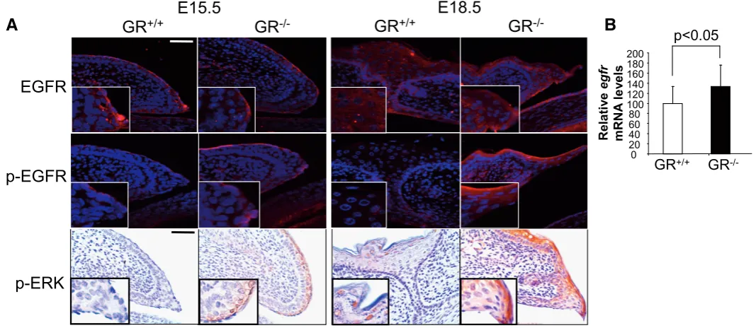
It is well known that the epidermal growth factor (EGF) and its receptor (EGFR) play a crucial role in epithelial development (Zenz et al., 2003; Xia and Karin, 2004). The antagonism between GR and EGFR signaling has been demonstrated in different pathophysiological processes. However, this cross-talk has not been examined in eyelid formation. In wt embryos, EGFR was detected at the tip of eyelid epithelial cells around E15.5 but decreased at E18.5, when eyelid closure had completed. In contrast, EGFR protein expression was evident at E15.5 in GR-/ - embryos and remained abnormally high at E18.5, as compared to wt (Fig. 3A, EGFR). The observed elevation in total EGFR levels correlated with increased phosphorylated (p-)EGFR immunostaining in GR-/- open eyelids at E18.5 (Fig. 3A, p-EGFR). To ascertain whether GR could also regulate EGFR at the transcriptional level, we examined the skin of E18.5 GR-/- em-bryos as compared to wt littermates by quantitative RT-PCR (Fig.
3B). Our data demonstrated increased egfr mRNA levels in GR -/- embryos, thus indicating additional mechanisms of GR/EGFR biological antagonism.
We next examined whether increased EGFR signaling would cause augmented ERK phosphorylation in the developing eyelids of GR-/-embryos (Fig. 3C, p-ERK). Despite unchanged total ERK levels in wt and GR-/-embryos (not shown), we detected p-ERK at E15.5 only in GR-deficient eyelids. In E18.5 wt embryos, p-ERK was restricted to the granular layer of closed eyelids whereas high levels were still detected at the eyelid tip of GR-/- embryos. Overall, our results suggest that proper (and transient) regulation of EGFR signaling and its downstream effectors, such as ERK and K6, are required during ocular epithelia development. Abnormally consti-tutive activation and/or expression of these proteins, as in GR-deficient embryos, likely results in impaired eyelid closure.
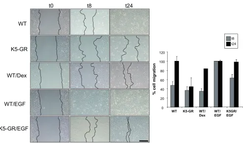
The formation of the mouse eyelid during the embryogenesis is similar to the wound healing process since it requires coordi-nated proliferation, migration and differentiation of keratinocytes (reviewed in Martin and Parkhurst, 2004). We thus examined the role of GR and its interference with EGFR function in epithelial cell migration by in vitro wound healing assays using primary culture keratinocytes (MPKs). Since cell confluence is necessary for these experiments, and given that GR-/- MPKs exhibit abnormal cell growth and apoptosis (Bayo et al., 2008), we evaluated MPKs isolated from K5-GR transgenic mice, in which GR is constitu-tively active (Pérez et al., 2001). This system allows us to evaluate the role of GR in keratinocyte migration without adding exogenous ligands, which can elicit different actions depending on dosing and kinetics. Fig. 4 summarizes three independent wound scratch
Fig. 3. Glucocorticoid receptor (GR) interferes with EGFR-mediated signaling during eyelid development. (A) Immunofluorescences were performed in GR-/- and GR+/+ embryos at E15.5 and E18.5 with specific antibodies against total EGFR and phosphorylated EGFR (p-EGFR). EGFR
expression was slightly induced in GR-/- relative to GR+/+ E18.5 embryos. Moreover, increased p-EGFR immunostaining was also detected in GR-/- E15.5
and E18.5 eyes. (B) Transcript levels of egfr were examined in skin of GR-/- and GR+/+ E18.5 embryos by quantitative RT-PCR. Asterisk denote that
differences in four individuals of each genotype were statistically significant; (student´s t test, p<0.05). (C) Immunostaining in GR-/- and GR+/+ embryos
at E15.5 and E18.5 for phosphorylated (p)-ERK. p-ERK was detected at E15.5 only in GR-deficient eyelids and the phosphorylation was sustained until E18.5, in contrast to wt embryos, where p-ERK was restricted to the granular layer of closed eyelids. Bar: 100 m.
Rel
a
tiv
e
egf
r
mRN
A
le
v
e
ls
0 20 40 60 80 100 120 140 160 180 200
GR
+/+GR
-/-p<0.05
p-ERK
p-EGFR
E18.5
E15.5
GR
-/-GR
+/+EGFR
GR
-/-GR
+/+experiments that elicited consistent results. In wt cells, keratinocyte migration was 47.3% at 8h after scratching (t8) and was com-pleted at 24h. In contrast, K5-GR MPKs exhibited 33.5% migra-tion at t8, a marked delay that was still very prononunced at t24 (44.2%). For comparative purposes, we evaluated the response of wt MPKs to the GC-analogue Dex and EGF (Fig. 4). As previously described (Lee et al., 2005), Dex delayed and EGF accelerated wt keratinocyte migration at t8 (35.2% and 100%, respectively). At t24, retarded migration elicited by Dex treatment was approximately 83.6%. Note that constitutive GR overexpres-sion in K5-GR MPKs elicited effects similar to Dex treatment in wt cells. The retarded migration of K5-GR MPKs was only slightly potentiated by Dex treatment (data not shown). K5-GR keratinocytes also responded to EGF although with delayed kinetics as compared to wt cells (64.7% and 97.3% at t8 and t24, respectively).
Discussion
During mammalian embryogenesis, the surface ectoderm gives rise to the corneal and conjunctival epithelia and the epidermis of the eyelid. Eyelid formation takes place during mouse embryonic days E15.5 to E16.5 and requires the proliferation and migration of epithelial cells to cover the ocular surface, acting as a protective barrier for normal eye development (Xia and Karin, 2004). This process is similar to epidermal formation which results in the
acquisition of a competent barrier necessary to protect the organ-ism from environmental damage (Segre, 2006). Our recent work has shown that GR is required for epidermal formation since its absence results in an immature epidermal barrier (Bayo et al., 2008). Analogously, histological evaluation of GR-/- embryos showed delayed and impaired eyelid closure, which took around 72-96h to complete instead of 24h, as occurs in wt mice (Fig. 1). Delayed progression in total eyelid formation was around 50% in GR-/- E15.5 embryos and approximately 78.3% in E18.5 individu-als relative to their wt counterparts (Fig. 2 and data not shown). The fact that GR modulates proliferation and apoptosis of eyelid epithelial cells (Fig. 2 A,B) adds to our previous findings in epidermis showing that this nuclear receptor regulates these processes in a cell-autonomous manner (Bayo et al., 2008) and highlights a general role of GR in epithelial morphogenesis. Our data also illustrate that these processes need to be temporally coordinated in order to complete proper eyelid formation. We found increased expression of markers of keratinocyte prolifera-tion (K5, p63) in the eyelid epithelial cells of GR-/- embryos relative to wt, at a time where these markers should be restricted to the basal cell layer and a few suprabasal cells at the site of fusion (Fig. 1). We also observed incomplete differentiation of epithelial cells in GR-/- embryos (Fig. 1, K10 and loricrin), most likely due to the absence of eyelid fusion, which should precede terminal differen-tiation (Zhang et al., 2005).
Our previous studies reported an EOB phenotype in K5-GR t8 t24
%
cell
m
igr
a
ti
on
0 20 40 60 80 100 120
WT K5-GR WT/ Dex
WT/ EGF
K5GR/ EGF
t8
t0
t24
WT/EGF
K5-GR
WT/Dex
K5-GR/EGF
WT
and K5-GR-TR embryos that was indeed the most consistent epithelial defect found in both transgenic mouse models (Cascallana et al., 2003; Donet et al., 2008). In addition, our data demonstrated that the transrepression function of the GR is sufficient to cause these epithelial anomalies (Donet et al., 2008). It was indeed surprising to find open eyelids in GR gain- and loss-of-function mouse models although, undoubtedly, it further sup-ports a key role for this transcription factor in ocular epithelial morphogenesis. Since GR-/- embryos exhibit increased corticos-terone levels due to a feedback mechanism mediated by the hypothalamus-pituitary-adrenal axis (Cole et al., 1995 and our unpublished data), it is possible that elevated hormone levels may be acting through the mineralocorticoid receptor (MR) in GR -/-ocular epithelial cells and thus mimick increased GC-signaling, even in the absence of GR. Supporting this hypothesis, mice overexpressing the MR in keratinocytes (K5-MR) showed an EOB phenotype virtually identical to that of K5-GR and K5-GR-TR embryos (Sainte Marie et al., 2007). The similarities between K5-GR and K5-MR mouse models extend to epidermal defects, including abnormal hair follicle formation and hypoplastic epider-mis (Sainte Marie et al., 2007). GR and MR recognize the same hexameric DNA response elements and, theoretically, could transactivate the same gene promoters (Kumar and Litwack, 2009). A subset of epithelial-specific genes may be regulated in common by both nuclear receptors. In this regard, our data show that GR represses egfr transcription in skin (Fig. 3B). However, MR has been reported to upregulate egfr mRNA expression in other cell types (Grossman et al., 2007).
GR exerts many of its biological actions through interactions with distinct signaling pathways, including MAPK/AP-1 and NF-kappaB (reviewed in De Bosscher and Haegeman, 2009). Two main pathways have been involved in eyelid formation, the MEKK1/JNK/c-Jun pathway which transductes TGF- and activin signals and the TGF-/EGFR/ERK pathway; both result in AP-1 activation (revised in Xia and Karin, 2004). EGFR plays a key role in eye morphogenesis and, accordingly, disruption of the EGFR locus resulted in an EOB phenotype as well as immature devel-opment of skin epithelial cells, teeth, lung and gastrointestinal tract (reviewed in Sibilia et al., 2007). We analyzed whether EGFR was abnormally expressed and/or activated in GR-/- embryos, and also investigated alterations in its downstream effector ERK. The expression pattern of both phosphorylated EGFR and ERK dem-onstrated sustained activation in GR-/- E18.5 embryos, a timepoint at which these proteins should be normally restricted to the more differentiated epithelial cells at the fused eyelids (Fig. 3). Sus-tained expression of ERK and K6 in the eyelids of GR-/- E18.5 mice is consistent with previous reports in vitro showing that EGF-signaling can induce the expression of K6 through AP-1 sites in its promoter (Lee et al., 2005).
The process of eyelid closure parallels skin wound healing, which also requires proliferation and migration to complete re-epithelization across the wound (reviewed in Martin and Parkhurst, 2004; Barrientos et al., 2008). The expression of the migration-associated keratin K6 increases during wound healing in adult skin and is required for normal re-epithelialization (Wong and Coulombe, 2003; Wojcik et al., 2000). Activated GR can inhibit the expression of specific keratins through several mechanisms involving transcriptional and non-transcriptional events (Radoja et al., 2000). GCs were shown to cause cytoskeleton remodeling
by repressing K6/K16 expression and thus, inhibiting keratinocyte migration and causing deregulated growth and differentiation (Stojadinovic et al., 2005). These GR actions are relevant during the wound healing process, whereby K6 expression in suprabasal keratinocytes at the wound´s edge is repressed once the epider-mis covers the wound. This occurs through antagonism between GR and EGFR and involves activation of -catenin and c-myc and blockade of EGF effects through the formation of a complex consisting in four GR monomers, -catenin and coactivator-associated-arginine-methyltransferase-1 (Lee et al., 2005; Stojadinovic et al., 2005).
So far, the in vivo cross-talk between GR/EGFR in eyelid formation had not been previously investigated. Our data in vivo show that GR is a master regulator required for the spatio-temporal control of the EGFR/MAPK/AP-1 signaling at the lead-ing edge of eyelid keratinocytes and suggest that this interference is required for normal eyelid development.
Materials and Methods
Animals
All mice were handled in accordance with the current Spanish and European normative which governs research with animals (Real Decreto 1201/2005, B.O.E. #252, 10 of October, 2005 and Convenio Europeo 1-2-3 del 18/3/1986).
GR-/- and K5-GR mice have been previously reported (Cole et al.,
1995; Pérez et al., 2001). GR+/- hemizygous mice (B6D2/F1) intercrosses
were programmed to obtain GR-/-, GR+/- and GR+/+ mice. Embryos were
obtained by cesarean derivation at the indicated day post-conception (dpc; the morning of the day that the vaginal plug was seen was considered as day 0.5 pc.). For histopathological evaluation, we analyzed embryos of different timepoints from GR-/-, GR+/- and GR+/+ genotypes
(n=43). For preparation of mouse primary keratinocytes (MPKs) and wound scratch assays, skin from K5-GR mice and wt newborn littermates was excised and processed (n=38).
Antibodies
The antibodies used included rabbit polyclonal antibodies against keratin K5 (PRB-160P), K6 (PRB-169P), K10 (PRB-159P) and loricrin (PRB-145P) from Covance (Babco, Berkeley, CA). Antibodies against p63 (sc-404), p-c-jun (sc-822) and EGFR (sc-03) were from Santa Cruz Biotechnology, Inc., (Santa Cruz, CA) and EGFR (53A5), anti-p-ERK (Thr202/Tyr204) (#4376) and p-JNK (Thr183/Tyr185) (# 9251) were purchased from Cell Signaling (Cell Signaling Technology Inc., Beverley, MA). Secondary biotin-conjugated anti-rabbit or anti-mouse antibodies were from Jackson ImmunoResearch (Jackson ImmunoResearch Labo-ratories, Inc. West Grove, PA).
Histological and Immunohistochemical analysis
In vivo epithelial BrdU labeling
Epithelial cell proliferation was measured by i.p. injection of BrdU (130
g/g of body weight, Roche) into pregnant female mice 1 h before sacrifice. BrdU incorporation was detected by immunohistochemistry of paraffin-embedded sections using a mouse BrdU monoclonal anti-body (biotest, Roche) followed by hematoxylin counterstaining. The number of BrdU-positive cells and the number of total cells was deter-mined per 200 m of interfollicular epithelium in each section. Experi-ments were performed at least in five individuals of each genotype and differences were assessed by using the t test, with statistical significance when p < 0.05.
Analysis of apoptosis in tissue sections
To detect individual apoptotic cells in paraffin-embedded tissue sec-tions, the In situ Cell Death Detection kit (Roche) was used, following manufacturer´s recommendation. Paraffin sections immersed in 0.1 M citrate buffer, pH 6 were microwave-irradiated for 5 min, and then rinsed with PBS prior to the TUNEL reaction. Four 15.5 dpc embryos of each genotype were examined.
Measurements of eyelid formation, leading edge formation and root formation
We have quantitated the differences in eyelid formation of GR-/-vs wt
E15.5 littermates, following the methodology described by Mine et al. These measurements were done in slides that were stained with K6, in order to delimitate the migrating epitheliall cells as K6-positive cells. This migration distance was considered as parameter a; the distance between the axis delimitating the conjunctival sac and the first K6-stained cells was denominated as b; the distance between the axis delimitating the conjunc-tival sac and the center of the cornea was denominated as c. The percentage of root formation was calculated as [b/c] x 100; percentage of leading edge formation was calculated as [a/c] x 100, and the percentage of eyelid formation was [(a+b)/c] x 100. Calculations were done in five individuals of each genotype and differences considered to be statistically significant when p<0.05.
MPK isolation, culture and wound scratch
MPK isolation was performed as previously described (Bayo et al., 2008). Briefly, skin was peeled off, incubated in 0.25% trypsin to separate the epidermis from the dermis and homogenized. MPKs were pooled (using at least two mice per point) and 106 cells were plated into one 35
mm diameter collagen coated petri dish (BD Biosciences) and cultured at 37C in standard medium. After 24 h, the medium was replaced with complete low calcium medium and cells were grown until confluency. The composition of standard medium was: Essential modified Eagle´s me-dium EMEM (BioWhitakker, Inc., Walkersville, MD), supplemented with 4% fetal calf serum (FCS, BioWhitakker, Inc.) plus 0.6 mM CaCl2 and antibiotics. To prepare low-calcium medium, FCS was depleted of diva-lent cations by treatment with Chelex deionizing resin (BioRad, Hempstead, UK) and supplemented with CaCl2 to a final concentration of approxi-mately 0.05 mM. EGF (Sigma, St. Louis, MO) (10 ng/ml) and antibiotics were added to growth medium.
For wound scratch assays, MPKs were incubated in EMEM/1% FBS O/N, and then treated with mitomycin (10g/ml) for 1h. Next, cells were wounded with a yellow tip, treated as indicated and cell migration followed up for 8-24h. Experiments were performed in triplicate and mean value
SD estimated. Vehicle, Dexamethasone (Dex, Sigma, St. Louis, MO, 100 nM) or EGF (25 ng/ml) were added for the indicated times to confluent wt MPKs.
For each wound scratch experiment, the surface area that remained uncovered by the cells for each time-point and condition was quantitated (Adobe Photoshop 8.1.0). These measurements were expressed as a percentage of distance coverage by cells moving into the scratch wound area 8 h and 24 h after wounding. Six images were analyzed per condition and time-point; then, averages and standard deviations were calculated.
RNA preparation and quantitative RT-PCR
Total RNA was isolated from back skin of GR-/- and control littermates
(four animals of each genotype) by using Trizol reagent (Invitrogen, Molecular Probes, Eugene, Oregon), following manufacturer´s recom-mendations. Reverse transcription was performed by using 1 g of RNA and oligo-dT (Fermentas Inc., Burlington, Canada) followed by qPCR using specific oligonucleotides for egfr
Forward, 5´-CAAAGTGATGTCTGGAGCTAT-3‘;
Reverse, 5´CTTGCTGGGATTCCATCATAAG-3‘. Technical triplicates were performed and mean value SD estimated.
Acknowledgements
This work was supported by grant SAF2008-00540 of the Ministerio de Ciencia e Innovación from the Spanish government and Fundación Ramón Areces (050507070007). LMS is recipiente of a contract JAE-Doc co-funded by European Funds. We are grateful to Prof. Günther Schütz for providing us with GR-/- mice and ackowledge Fátima Riveiro for
histological work.
References
ADAMS, M., MEIJER, O.C., WANG, J., BHARGAVA, A. and PEARCE, D. (2003). Homodimerization of the glucocorticoid receptor is not essential for response
element binding: activation of the phenylethanolamine N-methyltransferase
gene by dimerization-defective mutants. Mol Endocrinol 17: 2583-2592.
BARRIENTOS, S., STOJADINOVIC, O., GOLINKO, M.S., BREM, H. and
TOMIC-CANIC, M. (2008). Growth factors and cytokines in wound healing. Wound
Repair Regen 16: 585-601.
BAYO, P., SANCHIS, A., BRAVO, A., CASCALLANA, J.L., BUDER, K., TUCKERMANN, J., SCHÜTZ, G. and PÉREZ, P. (2008). Glucocorticoid
recep-tor is required for skin barrier competence. Endocrinology 149: 1377-1388.
CASCALLANA, J.L., BRAVO, A., PAGE, A., BUDUNOVA, I., SLAGA, T.J., JORCANO, J.L., and PÉREZ, P. (2003). Disruption of eyelid and cornea
development by targeted overexpression of the glucocorticoid receptor. Int J
Dev Biol 47: 59-64.
CASCALLANA, J.L., BRAVO, A., DONET, E., LEIS, H., JORCANO, J.L., and PÉREZ, P. (2005) Ectoderm-targeted overexpression of the glucocorticoid
receptor induces hypohidrotic ectodermal dysplasia Endocrinology 146:
2629-2638.
COLE, T.J, BLENDY, A.P., MONAGHAN, K., SCHMID, W., AGUZZI, A., FANTUZZI, G., HUMMLER, E., UNSICKER, K. and SCHÜTZ, G. (1995). Targeted disrup-tion of the glucocorticoid receptor gene blocks adrenergic chromaffin cell
development and severely retards lung maturation. Genes Dev 9: 1608-1621.
DE BOSSCHER, K. and HAEGEMAN, G. (2009). Minireview: latest perspectives on
antiinflammatory actions of glucocorticoids. Mol Endocrinol 23: 281-289.
DONET, E., BOSCH, P., SANCHIS, A., BAYO, P., RAMÍREZ, A., CASCALLANA, J.L., BRAVO, A. and PÉREZ, P. (2008). Transrepression function of the glucocorticoid receptor regulates eyelid development and keratinocyte
prolif-eration but is not sufficient to prevent skin chronic inflammation Mol
Endocrinol-ogy 22: 799- 812
GROSSMANN, C., KRUG, A.W., FREUDINGER, R., MILDENBERGER, S., VOELKER, K. and GEKLE M. (2007). Aldosterone-induced EGFR expression: interaction between the human mineralocorticoid receptor and the human
EGFR promoter. Am J Physiol Endocrinol Metab 292: 1790-1800.
KAUFMAN, M.H. and BARD, J.B.L. (1999). The anatomical basis of mouse development. San Diego, Academic Press.
KUMAR, R. and LITWACK, G. (2009). Structural and functional relationships of the
steroid hormone receptors’ N-terminal transactivation domain. Steroids 74:
877-883.
LEE, B., VOUTHOUNIS, C., STOJADINOVIC, O., BREM, H., IM, M. AND TOMIC-CANIC, M. (2005). From an Enhanceosome to a Repressosome: Molecular Antagonism between Glucocorticoids and EGF Leads to inhibition of Wound
Healing. J Mol Biol 345: 1083-1097.
MARTIN, P. and PARKHURST, S.M. (2004). Parallels between tissue repair and
MINE, N., IWAMOTO, R. and MEKADA, E. (2005). HB-EGF promotes epithelial cell
migration in eyelid development. Development 132: 4317-4326.
PEREZ, P., PAGE, A., BRAVO, A., DEL RÍO, M., GIMÉNEZ-CONTI, I., BUDUNOVA, I., SLAGA, T.J. and JORCANO, J.L. (2001). Altered skin development and impaired proliferative and inflammatory responses in transgenic mice
overexpressing the glucocorticoid receptor. FASEB J 15: 2030-2032.
RADOJA, N., KOMINE, M., JHO, S.H., BLUMENBERG, M. and TOMIC-CANIC, M. (2000). Novel mechanism of steroid action in skin through glucocorticoid
receptor monomers. Mol Cell Biol 20: 4328-4339.
REICHARDT, H.M., KAESTNER, K.H., TUCKERMANN, J., KRETZ, O., WESSELY, O., BOCK, R., GASS, P., SCHMID, W., HERRLICH, P., ANGEL, P. and SCHÜTZ, G. (1998). DNA binding of the glucocorticoid receptor is not essential
for survival. Cell 93: 531-541.
REVOLLO, J.R. and CIDLOWSKI, J.A. (2009). Mechanisms Generating Diversity
in Glucocorticoid Receptor Signaling. Ann NY Acad Sci 1179: 167-178.
ROGATSKY, I., WANG, J.C., DERYNCK, M.K., NONAKA, D.F., KHODABAKHSH, D.B., HAGG, C.M., DARIMONT, B.D., GARABEDIAN, M.J. and YAMAMOTO, K.R. (2003). Target-specific utilization of transcriptional regulatory surfaces by
the glucocorticoid receptor. Proc Natl Acad Sci USA 100: 13845-13850.
SAINTE MARIE, Y., TOULON, A., PAUS, R., MAUBEC, E., CHERFA, A., GROSSIN, M., DESCAMPS, V., CLEMESSY, M., GASC, J.M., PEUCHMAUR, M., GLICK, A., FARMAN, N. and JAISSER, F. (2007). Targeted skin overexpression of the mineralocorticoid receptor in mice causes epidermal atrophy, premature skin
barrier formation, eye abnormalities, and alopecia. Am J Pathol 171: 846-860.
SEGRE, J.A. (2006). Epidermal barrier formation and recovery in skin disorders. J
Clin Invest 116: 1150-1158.
SIBILIA, M., KROISMAYR, R., LICHTENBERGER, B.M., NATARAJAN, A., HECKING, M. and HOLCMANN, M. (2007). The epidermal growth factor
receptor: from development to tumorigenesis. Differentiation 75: 770-787.
STOJADINOVIC, O., BREM, H., VOUTHOUNIS, C., LEE, B., FALLON, J., STALLCUP, M., MERCHANT, A., GALIANO, R.D. and TOMIC-CANIC, M. (2005). Molecular Pathogenesis of Chronic Wounds. The Role of b-Catenin and
c-myc in the Inhibition of Epithelialization and Wound Healing. Am J Pathol 167:
59-69.
WINTERMANTEL, T.M., BERGER, S., GREINER, E.F. and SCHÜTZ, G. (2004).
Genetic dissection of corticosteroid receptor function in mice. Horm Metab Res
36: 387-391.
WOJCIK, S.M., BUNDMAN, D.S. and ROOP, D.R. (2000). Delayed Wound Healing
in Keratin 6a Knockout Mice. Mol Cell Biol 20: 5248-5255.
WONG, P. and COULOMBE, P.A. (2003). Loss of keratin 6 (K6) proteins reveals a
function for intermediate filaments during wound repair. J Cell Biol 163:
327-337.
XIA, Y. and KARIN, M. (2004). The control of cell motility and epithelial
morphogen-esis by Jun kinases. Trends Cell Biol 14: 94-101.
ZENZ, R., SCHEUCH, H., MARTIN, P., FRANK, C., EFERL, R., KENNER, L., SIBILIA, M. and WAGNER, E.F. (2003). c-Jun Regulates Eyelid Closure and
Skin Tumor Development through EGFR Signaling. Dev Cell 4: 879-889.
ZHANG, H., HARA, M., SEKI, K., FUKUDA, K and NISHIDA, T. (2005). Eyelid Fusion and Epithelial Differentiation at the Ocular Surface During Mouse
5 yr ISI Impact Factor (2008) = 3.271
Further Related Reading, published previously in the
Int. J. Dev. Biol.
See our recent Special Issue Placenta edited by Joan S. Hunt and Kent L. Thornburg at: http://www.ijdb.ehu.es/web/contents.php?vol=54&issue=2-3
Corneal development associated with eyelid opening
James D. Zieske
Int. J. Dev. Biol. (2004) 48: 903-911
Analysis of mouse eye development with chimeras and mosaics
J. Martin Collinson, Robert E. Hill and John D. West Int. J. Dev. Biol. (2004) 48: 793-804
Disruption of eyelid and cornea development by targeted overexpression of the glucocorticoid receptor
José Luis Cascallana, Ana Bravo, Angustias Page, Irina Budunova, Thomas J Slaga, José L Jorcano and Paloma Pérez
Int. J. Dev. Biol. (2003) 47: 59-64
PPAR expression and function during vertebrate development
Liliane Michalik, Béatrice Desvergne, Christine Dreyer, Mathilde Gavillet, Ricardo N Laurini and Walter Wahli
Int. J. Dev. Biol. (2002) 46: 105-114
Role of the retinoic acid receptor beta (RARbeta) during mouse development
N B Ghyselinck, V Dupé, A Dierich, N Messaddeq, J M Garnier, C Rochette-Egly, P Chambon and M Mark