International Journal of Nanomedicine
Dove
press
O r I g I N A L r e s e A r c h
open access to scientific and medical research
Open Access Full Text Article
evaluating the effects of crystallinity in new
biocompatible polyester nanocarriers on drug
release behavior
Vassilios Karavelidis1,2
evangelos Karavas2
Dimitrios giliopoulos1
sofia Papadimitriou1
Dimitrios Bikiaris1
1Laboratory of Polymer chemistry
and Technology, chemistry Department, Aristotle University of Thessaloniki, Thessaloniki,
2Pharmathen sA, Pharmaceutical
Industry, Pallini Attikis, Attiki, greece
correspondence: Dimitrios Bikiaris Laboratory of Polymer chemistry and Technology, chemistry Department, Aristotle University of Thessaloniki, 541 24 Thessaloniki, greece Tel +30 23 1099 7812 Fax +30 23 1099 7769 email [email protected]
Abstract: Four new polyesters based on 1,3-propanediol and different aliphatic dicarboxylic acids were used to prepare ropinirole HCl-loaded nanoparticles. The novelty of this study lies in the use of polyesters with similar melting points but different degrees of crystallinity, varying from 29.8% to 67.5%, as drug nanocarriers. Based on their toxicity to human umbilical vein endothelial cells, these aliphatic polyesters were found to have cytotoxicity similar to that of polylactic acid and so may be considered as prominent drug nanocarriers. Drug encapsulation in polyesters was performed via an emulsification/solvent evaporation method. The mean particle size of drug-loaded nanoparticles was 164–228 nm, and the drug loading content was 16%–23%. Wide angle X-ray diffraction patterns showed that ropinirole HCl existed in an amorphous state within the nanoparticle polymer matrices. Drug release diagrams revealed a burst effect for ropinirole HCl in the first 6 hours, probably due to release of drug located on the nanoparticle surface, followed by slower release. The degree of crystallinity of the host polymer matrix seemed to be an important parameter, because higher drug release rates were observed in poly-esters with a low degree of crystallinity.
Keywords: biocompatible polyesters, nanoparticles, ropinirole, release, crystallinity
Introduction
Today’s pharmaceutical formulation demands require that drug delivery systems are precise in their control of drug distribution and, preferably, respond directly to the local pathological environment in order to achieve a dynamic and beneficial interaction with the host pathology or physiology.1,2 The development of new biodegradable and
biocompatible polymers capable of releasing physically incorporated therapeutic agents with well defined kinetics is a subject that has been receiving an increasing amount of attention.3,4 Aliphatic polyesters, due to their favorable features of
bio-degradability and biocompatibility, are one of the most important classes of synthetic biodegradable polymers and are nowadays available commercially in a variety of types.4,5 These polyesters are appropriate for medical and biomedical applications,
including drug delivery systems, when prepared as drug-loaded nanoparticles or solid dispersions.
A significant effort has been made to develop appropriate nanocarriers for drug delivery because nanotechnology offers a suitable means of delivering small molecular weight drugs, as well as macromolecules, such as proteins, peptides or genes, by either localized or targeted delivery to the tissue of interest.6–8 Nanotechnology focuses on
incorporating therapeutic agents into biocompatible nanocomposites, such as nano-particles, nanocapsules, micellar systems, and conjugates, using different materials
International Journal of Nanomedicine downloaded from https://www.dovepress.com/ by 118.70.13.36 on 23-Aug-2020
For personal use only.
Number of times this article has been viewed
This article was published in the following Dove Press journal: International Journal of Nanomedicine
Dovepress
Karavelidis et al
and techniques.9,10 These systems in general can be used to
provide targeted delivery of drugs, to improve oral bioavail-ability, to sustain drug effects in target tissue, to solubilize drugs for intravascular delivery, and to improve the stability of therapeutic agents against enzymatic degradation (nucle-ases and prote(nucle-ases), especially of proteins, peptides, and nucleic acids.11–13 Furthermore, targeted nanocarriers reduce
drug toxicity, provide more efficient drug distribution and overcome resistance offered by the physiological barriers in the body because efficient delivery of drug to various parts of the body is directly affected by particle size. Nanocarriers also hold promise for delivery of biotech drugs to the ana-tomic extremities of the body.14
However, nanocarriers have some limitations. They are difficult to handle, store, and administer because of their susceptibility to aggregation. They are also unsuitable for less potent drugs, and can gain access to unintended environ-ments with harmful consequences.14 Furthermore, release
rates from some polymer nanoparticles are very low. The determination of drug release behavior from polymer nanoparticles has been reported by many research groups to be a rather complicated process.15 Drug release can be
affected by many factors, including polymer degradation, molecular weight, the thermal properties of the polymer, as well as the binding affinity between the drug and the polymer matrix, the capability of the polymer to incorporate a high amount of drug, size of the nanoparticles, and the hydrophi-licity or hydrophobicity of the drug.16–18
One of the major questions that arises for these kinds of polyesters when used as drug nanocarriers is whether crystal-linity affects the release profile of the model drug. The novelty of this study lies upon the investigation of polyesters that have similar melting points but different degrees of crystal-linity. In our previous work, we studied polyesters with simi-lar crystallinity but different melting points varying from 46.7°C to 166.4°C and their effects on nanoparticle charac-teristics. In the present study, we synthesized polyesters of different crystallinity but with almost the same melting point in an attempt to study how crystallinity affects the charac-teristic release profile of these nanocarriers, excluding the parameter of melting point. To our knowledge, no other research team until now has referred to or performed any studies related to the drug release rate in terms of the degree of crystallinity in a series of aliphatic polyester nanocarriers with similar melting points.
Taking into consideration all the above factors, the aim of the present study was to evaluate how the degree of crys-tallinity of different aliphatic polyesters affects the release
profile of ropinirole HCl, a model hydrophilic drug, from loaded nanoparticles. Four aliphatic polyesters, ie, poly(propylene azelate) (PPAz), poly(propylene pimalate) (PPPim), poly(propylene glutarate) (PPGlu), and poly(propylene adipate) (PPAd), with similar melting points and different crystallinity were synthesized, characterized, and used as drug nanocarriers for the encapsulation of rop-inirole HCl. Roprop-inirole HCl (4-[2-(dipropylamino)ethyl]-1,3- dihydro-2H-indol-2-one) is a nonergoline dopamine D2- receptor agonist with antiparkinsonian properties.
Materials and methods
Materials
Adipic acid (99%), glutaric acid (99%), pimelic acid (99%), and azelaic acid (99%) were purchased from Aldrich Chemical Company (St Louis, MO). 1,3-propanediol (CAS 504-63-2, purity . 99.7%) was kindly supplied by Du Pont de Nemours and Company (Pembroke Pines, FL). Analytical grade tetrabutyl titanate was purchased from Aldrich and used as a catalyst. Polyphosphoric acid, used as a heat stabilizer, was supplied by Fluka (Milwaukee, WI). Ropinirole HCl was purchased from Ragactives (Valladolid, Spain) as a white to pale greenish-yellow powder with a melting range of 241°C–245°C, a solubility of 133 mg/mL in water, and a molecular weight of 296.84 (260.38 as the free base). All other materials and solvents used were of analytical grade.
synthesis of polyesters
Synthesis of aliphatic polyesters was performed following the two-stage melt polycondensation method (esterification and polycondensation) in a glass batch reactor.19 In brief,
appropriate amounts of diacid and 1,3-propanediol in a molar ratio of 1:1.1 and the catalyst tetrabutyl titanate (3 × 10⁻⁴ mol
tetrabutyl titanate/mol diacid) were put into the reaction tube of a polycondensation apparatus. The apparatus with the reagents was evacuated several times and filled with argon in order to remove all oxygen. The reaction mixture was heated to 190°C in an argon atmosphere and stirred at a constant speed (500 rpm). This first step (esterification) was considered to be complete after collection of almost all the theoretical amount of H2O, which was removed from the reaction mixture by distillation and collected in a graduated cylinder. In the second polycondensation step, polyphos-phoric acid was added (5 × 10⁻⁴ mol polyphosphoric acid/mol
diacid) and a vacuum (5.0 Pa) was applied slowly over about 30 minutes, to avoid excessive foaming and to minimize oligomer sublimation. The temperature during this time was
International Journal of Nanomedicine downloaded from https://www.dovepress.com/ by 118.70.13.36 on 23-Aug-2020
Dovepress Biocompatible polyester nanocarriers
slowly increased to 230°C, and the stirring speed was also increased to 720 rpm. Polycondensation continued for about 60 minutes for all prepared polyesters. After the end of the polycondensation reaction, the polyesters were easily removed, milled, and washed with methanol. Four polyesters were prepared according to this procedure, ie, PPAd, PPGlu, PPPim, and PPAz.
Polymer characterization
Intrinsic viscosity measurement
Intrinsic viscosity measurements were performed on the iso-lated polymers using an Ubbelohde capillary viscometer at 25°C in chloroform at a solution concentration of 1 wt%.
gel permeation chromatography
Gel permeation chromatography analysis was performed using a Waters 150C gel permeation chromatography column equipped with a differential refractometer as the detector and three Ultrastyragel (103, 104, 105Å) columns in series. CHCl3 was used as the eluent (1 mL/minute) and the mea-surements were performed at 35°C. Calibration was per-formed using polystyrene standards with a narrow molecular weight distribution.
Differential scanning calorimetry
Differential scanning calorimetry was performed on a Perkin-Elmer Pyris Diamond differential scanning calori-meter calibrated with high purity standards. Functioning of the system at low temperatures (down to −65°C) was achieved using a Perkin Elmer Intracooler 2P cooling accessory. Samples weighing 5 ± 0.1 mg were sealed in aluminum pans and scanned using the instrument in a nitrogen atmosphere. A cyclic scanning procedure was followed to record the thermal behavior of each polyester. The procedure involved: heating from 0°C to 40°C above the melting point of each sample at a heating rate of 20°C/minute and holding at this temperature for 2 minutes in order to remove any thermal history of the sample; rapid cooling to −65°C and equilibration; reheating at a heating rate 2.5°C/minute from −65°C to 40°C above the melting temperature and holding for 2 minutes; and final cooling at a rate of 10°C/minute down to −50°C.
Wide angle X-ray diffraction
X-ray diffraction measurements were performed using an automated powder diffractometer Rigaku Mini Flex II with Bragg-Brentano geometry (θ–2θ), using CuKα radiation (λ= 0.154 nm) in the angle 2θ range of 5–60 degrees.
cytotoxicity study of prepared polyesters
Human umbilical vein endothelial cells were grown routinely in RPMI-1640 medium supplemented with 15% fetal bovine serum, 15 mg endothelial cell growth supplement, penicillin 100 U/mL, streptomycin 100 µg/mL, gentamicin 50 µg/mL, and amphotericin B 2.5 µg/mL. Cultures were maintained at 37°C, 5% CO2, and 100% humidity.
The cytotoxicity of the aliphatic polyesters in comparison with biocompatible polylactic acid was evaluated by measur-ing the viability of human umbilical vein endothelial cells in the presence of different concentrations of the polymers. Cell viability was determined by the '3-(4,5-dimethylthiazol-2-yl)-2,5-diphenyl tetrazolium bromide) (MTT) assay. Human umbilical vein endothelial cells were seeded in 24-well plates at a density of 30,000 cells per well in 500 µL cell culture medium. Twenty-four hours after plating, differ-ent amounts of aliphatic polyesters in the form of nanopar-ticles (suspended in culture medium) were added in the wells. After 24 hours of incubation at 37°C, 50 µL of MTT solution (5 mg/mL in phosphate-buffered solution pH 7.4) was added into each well and the plates were incubated at 37°C for 2 hours. The medium was withdrawn and 200 µL of acidified isopropanol (0.33 mL HCl in 100 mL isopropanol) was added to each well and agitated thoroughly to dissolve the crystals formed. The solution was transferred to 96-well plates and immediately read on a microplate reader (Biorad, Hercules, CA) at a wavelength of 490 nm. The experiments were performed in triplicate. Biocompatibility of the poly-mers was expressed as percent cell viability, which was calculated from the ratio of the number of cells treated with the nanoparticles to that of nontreated cells (control).
Preparation of ropinirole hcl-loaded
polyester nanoparticles
Ropinirole HCl nanoencapsulation in polyester matrices was carried out using a water-oil-water (w/o/w) emulsification/ solvent evaporation technique. In brief, 50 mg of the poly-ester was dissolved in 2 mL of dichloromethane and mixed with 0.5 mL of 10 mg/mL ropinirole HCl aqueous stock solution. The mixture was sonicated for 1 minute. Then, 5.5 mL of 12 mM sodium cholate aqueous solution was added and the new mixture was sonicated for 1 minute. Sodium cholate was added to prevent drug particle aggregation during solvent evaporation. As a result, the drug-loaded polymer was dispersed in the form of nanoparticles. The emulsion formed was gently stirred until evaporation of the organic solvent was complete. The nanoparticles were purified by centrifugation (9500 rpm for 20 minutes). The samples were
International Journal of Nanomedicine downloaded from https://www.dovepress.com/ by 118.70.13.36 on 23-Aug-2020
Dovepress
Karavelidis et al
reconstituted with deionized water. Polymer aggregates were removed by filtering the suspension through a 1.2 µm pore size microfilter.
characterization of drug-loaded
nanoparticles
Nanoparticle yield, drug loading, and entrapment efficiency
For determination of drug loading content, 3 mg of the nanoparticles was dissolved in 50 µL dichloromethane and diluted to 1 mL using phosphate buffer (pH 3/ACN 65/35 v/v solution). A clear solution was obtained for high-pressure liquid chromatographic analysis. Quantitative analysis of the synthesized nanoparticles was carried out using a Shimadzu high-pressure liquid chromatographic column (model LC-20 AD). The column used was a Hypersil BDS, 5 µm, 200 × 4.6 mm with a column temperature of 30°C. The mobile phase consisted of phosphate buffer at a flow rate of 1 mL/minute. Concentration determination was performed using high-pressure liquid chromatography-ultraviolet appa-ratus at 250 nm, and was based on a calibration curve previ-ously created by diluting a stock aqueous solution of 1 mg/mL ropinirole HCl to concentrations of 80, 40, 20, 10, 5, 1, and 0.5 µg/mL using the same mobile phase mentioned above. Nanoparticle yield, drug loading, and drug entrapment efficiency were calculated from equations (1) to (3), respectively:
(1)
(2)
(3)
Morphology
The morphology of the prepared nanoparticles was exam-ined by scanning electron microscopy (JMS–840, JEOL Inc, Peabody, MA). The samples were coated with carbon black to avoid any charging under the electron beam. The operating conditions included an accelerating voltage of 20 kV, a probe current of 45 nA, and a counting time of 60 seconds.
Particle size distribution
The particle size distribution of the ropinirole HCl polyester nanoparticles was determined by dynamic light scattering using a Zetasizer (Nano ZS, ZEN3600, Malvern Instruments, Worcestershire, UK) operating with a 532 nm laser. A suitable amount of nanoparticles was dispersed in distilled water, creating a total concentration of 1%, and this was kept at 37°C and agitation at 100 rpm. Particle size was measured at different time intervals after introduction of the sample into the dispersed medium. All measurements were per-formed in triplicate and the results are reported as the mean diameter ± standard deviation.
In vitro drug release studies
The rate of ropinirole HCl release from the nanoparticles was measured in dissolution apparatus (DISTEK 2100B) equipped with an autosampler using the paddle (USP II) method. Each dissolution vessel was loaded with a quantity of nanoparticles corresponding to 2.5 mg of ropinirole HCl. The test was performed at 37°C ± 1°C with a rotation speed of 100 rpm. The dissolution medium was 500 mL of phos-phate buffer (pH 7.4). At predetermined time intervals, samples of 5 mL were withdrawn from the dissolution medium, filtered through 45 µm ultrahigh molecular weight polyethylene filters, and assayed using a high-pressure liquid chromatography-ultraviolet method for the drug at 250 nm. An equal volume of fresh dissolution medium was transferred to the vessel after withdrawal of the sample. All measurements were performed in triplicate.
Results and discussion
Polymer characterization
Synthesis and characterization of polyesters derived from the reaction of 1,3-propanediol and different dicarboxylic acids, as well as their biodegradability, have been discussed analytically in a previous study.20 Our polyester
characteriza-tion results for intrinsic viscosity and molecular weight are presented in Table 1. In brief, both intrinsic viscosity and gel permeation chromatography measurements demonstrated high molecular weight values for the prepared polyesters. However, there were some differences in Mn values, with PPAd and PPGlu having the lower Mn of 14,000 Da and PPAz having a higher Mn of 25,000 Da.
Polyesters exhibit variable thermal behavior according to the number of methylene groups in the repeating unit, as well as the number of monomer units. However, the polyes-ters selected for the present study had similar melting points in the range of 43.3°C–57.1°C. After melting, the samples
Nanoparticles yield
Weight of na articles Weight of polymer
(%)
= nop
aand drug fed initially ×100
Drug loading (%)
= Weight of drug in nanoparticles
Weight of nanopartiicles ×100
Entrapment efficiency
Weight of drug in nanoparticles Weight o
(%)
=
ff drug fed initially ×100
International Journal of Nanomedicine downloaded from https://www.dovepress.com/ by 118.70.13.36 on 23-Aug-2020
Dovepress Biocompatible polyester nanocarriers
were rapidly cooled in the differential scanning calorimetry instrument down to −65°C, and a second scan was performed in the quenched samples to record the glass transition and cold crystallization of the amorphous polyesters. PPAd has the lowest glass transition temperature at −58.8°C while PPGlu has the highest at −45.0°C. However, it can be said that these aliphatic polyesters show increased chain flexibility since their glass transition temperatures are very low. PPAz and PPPim also had very low glass transition temperatures at −46.7°C and −56.9°C, respectively. A slow heating rate of 2.5°C/minute was used in the second scan to record the cold crystallization of the quenched polyesters. However, as can be seen, PPGlu and PPAz could not be crystallized during heating, and only PPAd and PPPim gave a cold crystalliza-tion temperature. The different crystallizacrystalliza-tion rates may also
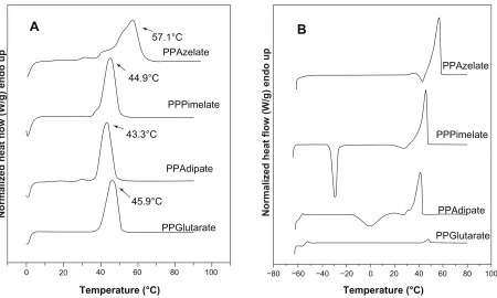
affect the degree of crystallinity of the polyesters. The respec-tive heating scans (first scan and after quenching) are shown in Figure 1.
It was demonstrated in a previous study that the melting points and glass transition temperatures of polyesters may play an important role in their drug release characteristics.21
Polyesters with a high melting point give a slower drug release while those having melting points close to that of the human body have higher drug release rates. Similar behavior was observed for the glass transition temperatures of the polyes-ters. With these results in mind, polyesters with very similar melting points and glass transition temperatures were selected as candidate nanocarriers for the present study. As shown in Table 1, there is no great variation in melting points and glass transition temperatures for the selected aliphatic polyesters which would affect evaluation of the effect of other physical characteristics and, in particular, the effect of degree of crys-tallinity on drug release from these polymeric nanoparticles. From the heat of fusion values measured in the heating traces of the samples as received, which crystallized during 2 months of storage at room temperature before scanning, the degree of crystallinity values can be calculated. PPAz and PPPim show higher heat of fusion values compared with PPGlu and PPAd in Table 1. In order to calculate the degree of crystallinity for each polyester, the heat of fusion of the
Table 1 Intrinsic viscosity [η], average molecular weight (Mn), and thermal properties of the prepared polyesters
[η] (dL/g)
Mn (Da)
Tm (°C)
Tg (°C)
Tcc (°C)
ΔHm (J/g)
PPAd 0.57 14,000 43.3 –58.8 –1.1 48.1
PPglu 0.58 14,000 45.9 –45.0 – 46.0
PPPim 0.70 19,000 44.9 –56.9 –29.6 53.7
PPAz 0.84 25,000 57.1 –46.7 – 65.7
Abbreviations: PPAz, poly(propylene azelate); PPPim, poly(propylene pimalate); PPglu, poly(propylene glutarate); PPAd, poly(propylene adipate).
−80
0 20 40 60 80 100
PPGlutarate PPAdipate
PPPimelate PPAzelate
PPGlutarate PPAdipate PPPimelate
PPAzelate 57.1°C
44.9°C
43.3°C
45.9°C
−60 −40 −20
Temperature (°C) Temperature (°C)
Normalized heat flow (W/g) endo up Normalized heat flow (W/g) endo up
0 20 40 60 80 100
A
B
Figure 1 Differential scanning calorimetry thermograms of the studied aliphatic polyesters. (A) First scan of polyesters as received and kept at room temperature for 2 months, and (B) a second scan of polyesters after quenching.
Abbreviations: PPAz, poly(propylene azelate); PPPim, poly(propylene pimalate); PPglu, poly(propylene glutarate); PPAd, poly(propylene adipate).
International Journal of Nanomedicine downloaded from https://www.dovepress.com/ by 118.70.13.36 on 23-Aug-2020
Dovepress
Karavelidis et al
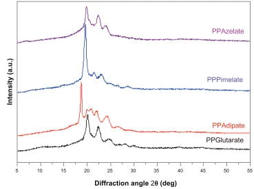
perfect crystal (100% crystal material) should be known, but is not. For this reason, the degree of crystallinity of all the polyesters studied was calculated using wide angle X-ray diffraction patterns (Figure 2).
Although there are some differences in the intensities of the peaks, the wide angle X-ray diffraction patterns are similar for PPAz, PPPim, and PPGlu, because the character-istic diffractions peaks appear at about the same diffraction angles of 2θ. The unit cells of the polymers in this group are probably similar, and some progressive changes in the dimen-sions occur with increasing length of the repeating unit of dicarboxylic acid, given that propylene glycol is the same in all polyesters. However, for PPAd, which has the highest degree of crystallinity, a double peak is obtained at 2θ 19.9 and 20.8, which is not recorded for the other polyesters. The crystallinity values were calculated from wide angle X-ray diffraction patterns using the relative areas under the crystal-line peaks (Ac ) and the amorphous background (Aa ) using equation 3, according to Lu and Hay,22 and are summarized
in Table 2.
X A
A
c am
c = +
−
1
1
(4)
Crystallinity values were calculated using a special pro-gram on the diffractometer and are summarized in Table 2.
As can be seen, PPAd has the highest degree of crystallinity at 67.5%, while PPAz has the lowest at 29.8%. PPPim and PPGlu give intermediate values of 43.9% and 55.4%, respectively. The difference between the aliphatic polyesters in this respect is considerable, and is expected to play the most important role in drug release.
In vitro cytotoxicity of aliphatic
polyesters
Aliphatic polyesters such as polycaprolactone and poly-lactide are extensively used as drug carriers because they are biocompatible materials. The polyesters used in the present study are aliphatic and similar to polycaprolactone and polylactide. However, their in vitro biocompatibility needs to be evaluated in order for these compounds to be considered as potential drug nanocarriers. Use of similar aliphatic polyesters derived from 1,3 propanediol and carboxylic acids as nanocarriers has been previously stud-ied and reported.23–25
These polyesters have been used previously for the preparation of a solid dispersion by melt mixing, while some of their copolymers have been effective for nanoencapsula-tion and sustained release of nimodipine.25 Figure 3
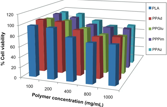
demon-strates that all the polyesters studied had low toxicity in human umbilical vein endothelial cells, with appreciable
5 10 15 20 25 30
Diffraction angle 2θ (deg)
PPAzelate
PPPimelate
PPAdipate
PPGlutarate
Intensity (a.u.)
35 40 45 50 55
Figure 2 Wide angle X-ray diffraction patterns of the studied aliphatic polyesters.
Abbreviations: PPAz, poly(propylene azelate); PPPim, poly(propylene pimalate); PPglu, poly(propylene glutarate); PPAd, poly(propylene adipate).
International Journal of Nanomedicine downloaded from https://www.dovepress.com/ by 118.70.13.36 on 23-Aug-2020
Dovepress Biocompatible polyester nanocarriers
cytotoxicity (more than 20% reduction of cell viability) being observed only after exposing the cells to high nanoparticle concentrations, ie, higher than 800 µg/mL. Based on their toxicity to human umbilical vein endothelial cells, the bio-compatibility of polyesters was comparable with that of polylactide, which is a polymer of high biocompatibility and has a wide variety of biomedical applications.26 The results
of our present study indicate that these polyesters may be considered as novel nanocarriers.
Nanoparticle characterization
The polyesters prepared were used for drug encapsulation in the form of nanoparticles. The main nanoparticle char-acteristics, such as nanoparticle yield, drug loading, and encapsulation efficiency, as well as nanoparticle size, are presented in Table 3. Drug loading content for all the polyesters varied between 16% and 23%, and entrapment efficiency was relatively high. PPGlu had the highest entrapment efficiency (61%) while PPAd had the lowest (49%). Drug loading content and entrapment efficiency
values were quite satisfactory, considering that ropinirole HCl is a highly water-soluble drug and is rather difficult to encapsulate into these hydrophobic aliphatic polyesters. Similar data were also obtained in previous studies using an emulsification solvent evaporation method for drug encapsulation in aliphatic polyesters with a polymer matrix.21,25 Several factors may affect drug loading content
and encapsulation efficiency, including affinity of the loaded drug for the preformed polymer, hydrophobicity of the polymer matrix, drug solubility in water, and drug-drug interaction, ie, its ability to self-aggregate.27,28 However,
in the present study, the polyesters showed no trends with regard to drug loading and entrapment efficiency, so it may be stated that differences in degree of crystallinity may not play an important role in polyesters.
Nanoparticle size is an important parameter because it can affect drug release, physical stability, and uptake by cells. Nanoparticle yield and size distribution may be affected by a number of parameters, including stirring rate, type and amount of dispersing agent, viscosity of organic and aqueous
Table 2 Diffraction angles 2θ for poly(propylene alkanedicarboxylate) crystals
Polyester Diffraction angle 2θ (degrees) Degree of crystallinity (%)
PPAz 19.90 22.36 23.98 29.8
PPPim 19.64 21.45 23.09 25.06 26.43 28.67 43.9
PPglu 20.04 24.70 28.07 30.00 39.62 44.72 55.4
PPAd 18.71 19.90 20.80 22.09 24.14 26.65 67.5
Abbreviations: PPAz, poly(propylene azelate); PPPim, poly(propylene pimalate); PPglu, poly(propylene glutarate); PPAd, poly(propylene adipate).
100 0
20 40 60 80 100 120
400 Polymer con
centration (mg/mL) 200
800
1000
PLA
% Cell viability
PPAd
PPGlu
PPPim
PPAz
Figure 3 human umbilical vein endothelial cell viability after incubation for 24 hours with different polymers and different polymer concentrations.
Abbreviations: PPAz, poly(propylene azelate); PPPim, poly(propylene pimalate); PPglu, poly(propylene glutarate); PPAd, poly(propylene adipate); PLA, polylactic acid.
International Journal of Nanomedicine downloaded from https://www.dovepress.com/ by 118.70.13.36 on 23-Aug-2020
Dovepress
Karavelidis et al
phases, and temperature,29 as well as encapsulation efficiency
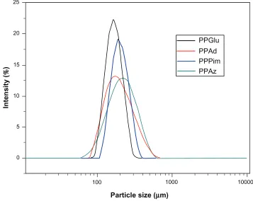
and drug loading. The size (Table 3) and distribution ( Figure 4) of the nanoparticles was measured by light scat-tering. It is obvious that all the nanoparticles in this study showed a unimodal size distribution. The mean nanoparticle diameter for the polyesters varied from a minimum of 164 nm for PPGlu to a maximum of 228 nm for PPAz. Similarly small particle sizes were also observed in a previous study using the same kind of aliphatic polyesters for encapsulation of ropinirole HCl.21 Furthermore, as has been previously
reported for copolymer of lactic acid/glycolic acid nanopar-ticles, an increase in molecular weight leads to an increase in nanoparticle size.30 This may explain the nanoparticle size
for PPAz and PPPim, because both materials have a higher molecular weight (Table 1). However, it should be mentioned that the polydispersity of the nanoparticles was high in all cases, and so their mean particle size should not have a sig-nificant effect on drug release.
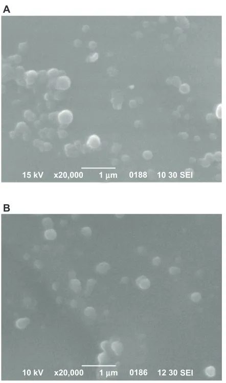
Scanning electron micrographs of samples of the nano-particles are shown in Figure 5, and establish that the drug-loaded nanoparticles have a discrete spherical shape with different sizes ranging from 150 nm to 250 nm. These results are in accordance with results obtained from dynamic light scattering measurements, but with some small differences due to the different methods used. Similar results have been previously reported elsewhere.31
Wide angle X-ray diffraction was used to identify the physical state of the drug incorporated into the polymeric nanoparticles, and the patterns of pure ropinirole HCl and drug-loaded nanoparticles are presented in Figure 6. Crystalline ropinirole HCl showed a large number of sharp diffraction peaks, whereas these characteristic peaks were not detected for the drug-loaded nanoparticles, in which only the characteristic diffraction peaks of aliphatic polyesters were recorded. Thus, it can be stated that ropinirole HCl was dispersed in an amorphous state in all the semicrystalline polymer matrices. As can be seen, the patterns of the
Table 3 Nanoparticle yield, drug loading content, and entrapment efficiency of the polyesters
Polyester Nanoparticle yield (%)
Drug loading content (%)
Entrapment efficiency (%)
Particle size (nm)
PPAz 36 ± 6 19 ± 4 57 ± 7 228 ± 14
PPPim 28 ± 10 18 ± 3 52 ± 5 192 ± 12
PPglu 30 ± 12 23 ± 4 61 ± 7 164 ± 12
PPAd 31 ± 8 16 ± 3 49 ± 5 175 ± 15
Note: All measurements were performed in triplicate.
Abbreviations: PPAz, poly(propylene azelate); PPPim, poly(propylene pimalate); PPglu, poly(propylene glutarate); PPAd, poly(propylene adipate).
1000
PPGlu PPAd PPPim PPAz
Particle size (µm)
Intensity
(
%
)
100 0
5 10 15 20 25
10000
Figure 4 Particle size distribution of ropinirole hcl-loaded nanoparticles.
Abbreviations: PPAz, poly(propylene azelate); PPPim, poly(propylene pimalate); PPglu, poly(propylene glutarate); PPAd, poly(propylene adipate).
International Journal of Nanomedicine downloaded from https://www.dovepress.com/ by 118.70.13.36 on 23-Aug-2020
Dovepress Biocompatible polyester nanocarriers
A
B
15 kV x20,000 1 µm 0188 10 30 SEI
10 kV x20,000 1 µm 0186 12 30 SEI
Figure 5 scanning electron micrographs of ropinirole hcl-loaded nanoparticles with the studied polyesters. (A) PPAz; (B) PPPim.
Abbreviations: PPAz, poly(propylene azelate); PPPim, poly(propylene pimalate).
nanoparticles are identical to that of the pure polyesters, indicating that nanoencapsulation of ropinirole HCl into polyester nanoparticles does change their crystalline form. The degree of crystallinity in the nanoparticles was calculated from these patterns. In the case of PPAz, crystallinity was about 29.5%, 42.6% in PPPim, 52.9% in PPGlu, and 62.6% in PPAd. These are very close to those reported for the pure aliphatic polyesters, but slightly smaller values were detected in all the nanoparticles. This is probably due to the wide angle X-ray diffraction patterns being recorded just after preparation of the nanoparticles and not enough time being available to reach a higher degree of crystallinity.
In vitro drug release
Aliphatic polyesters are appropriate vehicles for encapsulat-ing all kinds of active pharmaceutical encapsulat-ingredients, whether hydrophilic or hydrophobic. It is possible to control drug
release using an appropriate kind of aliphatic polyester. Furthermore, using the same polyester and changing its physical characteristics, such as its molecular weight, it is possible to modify drug release in order to achieve the desired drug release behavior.30 A lot of other parameters can also
affect drug release from nanoparticles, such as the polymer degradation rate, the binding affinity between the polymer and the drug, the capability of the polymer to incorporate high amounts of drug, nanoparticle size, and drug hydrophilicity.16,32 In a previous study, release of ropinirole
HCl from polyester nanoparticles with similar crystallinity and different melting points was examined, and it was veri-fied that melting point plays a critical role in drug dissolution behavior.21 In fact, polyesters with melting points near human
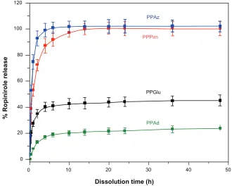
body temperature show enhanced drug release. In the present work, we studied how drug release is affected when aliphatic polyesters with similar melting points but different degrees of crystallinity were used as nanomatrices. Figure 7 shows the ropinirole HCl release profiles for the prepared nanopar-ticles. In all cases, a burst release was observed during the early stages of dissolution (up to 6–8 hours), followed by a phase of relatively slow drug release (until 24 hours). The main factor contributing to the fast release of ropinirole HCl from the nanoparticles into the aqueous dissolution medium is that ropinirole HCl is a hydrophilic drug with high solubil-ity. It has been reported in the literature that most drug-loaded nanoparticle formulations show a biphasic release pattern in which an initial burst is followed by slow release rates from the internal phase of nanoparticles.33 In the present study, a
characteristic burst effect was also detected, which appears probably due to the fraction of drug located on (or close to) the surface of the nanoparticles,33 as well as the high
hydro-philicity and high solubility of ropinirole HCl. Since the first w/o emulsion of the double emulsion method used here to encapsulate the hydrophilic ropinirole in the nanoparticles was not stabilized with a surfactant, migration of the hydro-philic drug to the external aqueous phase during the second emulsification step (and nanoparticle hardening process) might have occurred. As a result, most of the hydrophilic drug molecules that remained in the nanoparticles (ie, the drug fraction not lost to the external aqueous phase) may be located close to the surface, with only a small portion parti-tioned deep into the hydrophobic core, as has been reported previously.34 However, in this case, all nanoparticles should
have similar behavior. Therefore, factors in addition to drug location are also responsible for such behavior. Of course, the high release rate of ropinirole HCl from nanoparticles at initial dissolution should be attributed to the high drug
International Journal of Nanomedicine downloaded from https://www.dovepress.com/ by 118.70.13.36 on 23-Aug-2020
Dovepress
Karavelidis et al
0 0 20 40 60 80 100 120
10 20
Dissolution time (h)
% Ropinirole releas
e
30 PPAd PPGlu PPPim
PPAz
40 50
Figure 7 Release profiles for ropinirole HCl from polyester nanoparticles.
Abbreviations: PPAz, poly(propylene azelate); PPPim, poly(propylene pimalate); PPglu, poly(propylene glutarate); PPAd, poly(propylene adipate).
20
Diffraction angle 2θ (deg)
PPAz/ropinirole
PPPim/ropinirole
PPGlu/ropinirole
Ropinirole
PPAd/ropinirole
Intensity (a.u.)
40
Figure 6 Wide angle X-ray diffraction patterns for ropinirole hcl and ropinirole hcl-loaded nanoparticles.
Abbreviations: PPAz, poly(propylene azelate); PPPim, poly(propylene pimalate); PPglu, poly(propylene glutarate); PPAd, poly(propylene adipate).
solubility and the low mean nanoparticle diameter size, enabling drug diffusion to take place easily.
Comparing the release rates of ropinirole HCl from the different aliphatic polyesters, it can be seen that the release rates are higher for PPAz and PPPim and lower for PPGlu
and PPAd. Furthermore, it is also observed that release of ropinirole HCl from PPAz and PPPim reaches 100%, while for PPGlu and PPAd it is 40% and 20%, respectively. Taking into consideration the fact that the hydrolysis rate for the studied aliphatic polyesters at 37°C is less than 2.4% in the
International Journal of Nanomedicine downloaded from https://www.dovepress.com/ by 118.70.13.36 on 23-Aug-2020
Dovepress Biocompatible polyester nanocarriers
absence of enzymes,35 it is believed that the drug release is
not caused by erosion of the polymer matrices, since the degradation of these nanoparticles appears to be limited. It is our strong impression that the profile of ropinirole HCl release from nanoparticles is mainly due to diffusion. In this situation, the mean size of the nanoparticles could play a key role in the release profile, as reported above, and the release rate should be higher from nanoparticles of small size. If this was the main factor affecting drug release, then PPGlu should have the highest release rate because it has the lowest mean particle size, while PPAz should have the lowest release rate. However, from Figure 7, such a pattern of release behavior was not observed. Nevertheless, in our study, we cannot conclude that the effect of particle size is negligible. Furthermore, our data for particle size distribution (Figure 4) show relatively high polydispersity values, so it can be said that the actual sizes of the different batches are very close and overlapping may be expected. The zeta potential and possibly particle surface charge could also have a contribu-tory effect. However, the types of polyesters used in this study were very hydrophobic materials with a negative zeta potential (−15 to −18). This is due to the existence of hydroxyl and mainly carboxyl end groups in the prepared polyesters. Thus, it is to be expected that the nanoparticles we prepared would behave in a similar way.
A factor that might also play a key role in the release profile of ropinirole HCl is the polymer structure and, as a result, molecular weight, crystallinity, melting point, and glass transition temperature, which can affect the release of the drug from the polymer matrix to the dissolution medium.33 High crystallinity could lead to formation of a
microchannel structure, and at the same time the large sur-face area of the polymer matrix could cause the drug to be released easily from the nanoparticles.36 However, in the
present study, it was verified that, using polyesters with a similar melting point and glass transition temperature but different degrees of crystallinity, the release rate of ropini-role HCl is lower from polyesters with a higher degree of crystallinity. This could also be verified for PPAz nanopar-ticles, in which the polyester has a high molecular weight, melting point, and glass transition temperature, compared with the other polyesters. However, the release rate of rop-inirole HCl is higher from all other nanoparticles. This is because PPAz has the lowest degree of crystallinity (29.5%). Furthermore, PPAd, which has a low molecular weight and the lowest glass transition temperature, has the slowest drug release due to its high degree of crystallinity. This is in agreement with other studies reporting that high crystallinity
of the matrix may have a negative influence on the release rate of a drug, because the lamellae may act as a barrier during drug diffusion.18,37,38 Bigger and more perfectly
shaped crystalline lamellae should reduce overall release of the drug. This is probably the reason for higher release rates in the case of PPAz and PPPim, which show a low degree of crystallinity, ie, 29.5% and 42.6% respectively. In con-trast, very low release rates were obtained for PPAd and PPGlu, which have a higher degree of crystallinity at 62.6% and 51.9%, respectively. Diffusion of the active pharma-ceutical ingredient through the amorphous matrix seems to be easier due to the higher mobility of the macromolecular chains of the polyester in the amorphous state, and thus easier penetration of water through them and, consequently, a faster release rate for ropinirole is obtained.
Conclusion
We prepared polyesters with high molecular weight, similar glass transition temperatures and melting points, but very different degrees of crystallinity, ranging from 29.8% to 67.5%. All the polyesters demonstrated low cytotoxicity, so may be used as drug carriers for nanoencapsulation of a freely soluble drug like ropinirole HCl. Drug loading, entrapment efficiency, and particle size of the prepared nanoparticles may be considered satisfactory, keeping in mind that a highly soluble drug was used, while in all cases the drug was encap-sulated in an amorphous form. The rate of ropinirole HCl release was affected by drug solubility, as well as the degree of crystallinity of the polyesters used. Thus, the release rate is higher in polyesters having a low degree of crystallinity, due to higher macromolecular chain mobility.
Acknowledgment
This work was funded by the General Secretariat of Research and Technology of Greece in the framework of the PABET-NE project.
Disclosure
The authors have no conflicts of interest to report in this work.
References
1. Pillai O, Panchagnula R. Polymers in drug delivery. Curr Opin Chem Biol. 2001;5:447–451.
2. Lin YQ, You HB. Polymer architecture and drug delivery. Pharm Res. 2006;23:1–30.
3. Lakschmi SN, Cato TL. Biodegradable polymers as biomaterials. Prog
Polym Sci. 2007;32:762–798.
4. Chasin M, Langer R, editors. Biodegradable Polymers as Drug Delivery
Systems. New York, NY: Marcel Dekker Inc; 1990.
International Journal of Nanomedicine downloaded from https://www.dovepress.com/ by 118.70.13.36 on 23-Aug-2020
International Journal of Nanomedicine
Publish your work in this journal
Submit your manuscript here: http://www.dovepress.com/international-journal-of-nanomedicine-journal
The International Journal of Nanomedicine is an international, peer-reviewed journal focusing on the application of nanotechnology in diagnostics, therapeutics, and drug delivery systems throughout the biomedical field. This journal is indexed on PubMed Central, MedLine, CAS, SciSearch®, Current Contents®/Clinical Medicine, Journal
Citation Reports/Science Edition, EMBase, Scopus and the Elsevier Bibliographic databases. The manuscript management system is completely online and includes a very quick and fair peer-review system, which is all easy to use. Visit http://www.dovepress.com/ testimonials.php to read real quotes from published authors.
Dovepress
Dove
press
Karavelidis et al
5. Mohaned F, Van der Walle CF. Engineering biodegradable polyester particles with specific drug targeting and drug release properties.
J Pharm Sci. 2008;97:71–87.
6. Moghimi SM, Hunter AC, Murray JC. Long-circulating and target-specific nanoparticles: Theory to practice. Pharmacol Rev. 2001;53: 283–318.
7. Emerich D, Thanos CG. The pinpoint promise of nanoparticle-based drug delivery and molecular diagnosis. Biomol Eng. 2006;23: 171–184.
8. Des Rieux A, Fievez V, Garinot M, Schneider YJ, Preat V. Nanoparticles as potential oral delivery systems of proteins and vaccines: a mechanistic approach. J Control Release. 2006;116:1–27.
9. Vauthier C, Bouchemal K. Methods for the preparation and manufacture of polymeric nanoparticles. Pharm Res. 2009;26:1025–1058. 10. Rao JP, Geckeler KE. Polymer nanoparticles: preparation techniques
and size control parameters. Prog Polym Sci. 2011;36:887–913. 11. Panyam J, Labhasetwar V. Biodegradable nanoparticles for drug and
gene delivery to cells and tissue. Adv Drug Deliv Rev. 2003;55: 329–347.
12. Couvreur P, Vauthier C. Nanotechnology: intelligent design to treat complex disease. Pharm Res. 2006;23:1417–1450.
13. Torchilin VP. Micellar nanocarriers: pharmaceutical perspectives.
Pharm Res. 2007;24:1–16.
14. Rawat M, Singh D, Saraf S. Nanotechcarriers: Promising vehicle for bioactive drugs. Biol Pharm Bull. 2006;29:1790–1798.
15. Ge H, Hu Y, Jiang, X, et al. Preparation, characterization, and drug release behaviors of drug nimodipine-loaded poly(ε -caprolactone)-poly(ethylene oxide)-poly(ε-caprolactone) amphiphilic triblock copo-lymer micelles. J Pharm Sci. 2002;91:1463–1473.
16. Gref R, Minamitake Y, Peracchia MT, Trubetskoy V, Torchilin V, Langer R. Biodegradable long-circulating polymeric nanospheres.
Science. 1994;263:1600–1603.
17. Shin IG, Kim SY, Lee YM, Cho CS, Sung YK. Methoxy poly(ethylene glycol)/ε-caprolactone amphiphilic block copolymeric micelle contain-ing indomethacin. I. Preparation and characterization. J Control Release. 1998;51:1–11.
18. Jeong JC, Lee J, Cho K. Effects of crystalline microstructure on drug release behavior of poly(ε-caprolactone) microspheres. J Control
Release. 2003;92:249–258.
19. Papageorgiou GZ, Bikiaris DN. Crystallization and melting behavior of three biodegradable poly(alkylene succinates). A comparative study.
Polymer. 2005;46:12081–12092.
20. Bikiaris DN, Papageorgiou GZ, Giliopoulos DJ, Stergiou CA. Correlation between chemical and solid-state structures and enzymatic hydrolysis in novel biodegradable polyesters. The case of poly(propylene alkanedicarboxylate)s. Macromol Biosci. 2008;8:728–740.
21. Karavelidis V, Giliopoulos D, Karavas E, Bikiaris D. Nanoencapsula-tion of a water soluble drug in biocompatible polyesters. Effect of polyesters melting point and glass transition temperature on drug release behavior. Eur J Pharm Sci. 2010;41:636–643.
22. Lu XF, Hay JN. Isothermal crystallization kinetics and melting behav-iour of poly(ethylene terephthalate). Polymer. 2001;42:9423–9431. 23. Bikiaris D, Karavelidis V, Karavas E. Effectiveness of various drug
carriers in controlled release formulations of raloxifene HCl prepared by melt mixing. Curr Drug Deliv. 2009;6:425–436.
24. Bikiaris D, Karavelidis V, Karavas E. Novel biodegradable polyesters. synthesis and application as drug carriers for the preparation of ralox-ifene HCl loaded nanoparticles. Molecules. 2009;14:2410–2430. 25. Bikiaris DN, Papadimitriou S, Papageorgiou GZ, Kanaze FI,
Georgarakis M. Nanoencapsulation of nimodipine in novel biocompat-ible poly(propylene-co-butylene succinate) aliphatic copolyesters for sustained release. J Nanomaterials. 2009; art. no. 716242.
26. Athanasiou KA, Niederauer GG, Agrawal CM. Sterilization, toxicity, biocompatibility and clinical applications of polylactic acid/ polyglycolic acid copolymers. Biomaterials. 1996;17:93–102.
27. Niwa T, Takeuchi H, Hino T, Kunou N, Kawashima Y. Preparations of biodegradable nanospheres of water-soluble and insoluble drugs with D,L-lactide/glycolide copolymer by a novel spontaneous emulsification solvent diffusion method, and the drug release behavior. J Control
Release. 1993;25:89–98.
28. Khoee S, Hassanzadeh S, Goliaie B. Effects of hydrophobic drug-polyesteric core interactions on drug loading and release properties of poly(ethylene glycol)-polyester-poly(ethylene glycol) triblock core-shell nanoparticles. Nanotechnology. 2007;18: art. no. 175602. 29. Pinto Reis C, Neufeld RJ, Ribeiro AJ, Veiga F. Nanoencapsulation I.
Methods for preparation of drug-loaded polymeric nanoparticles.
Nanomedicine. 2006;2:8–21.
30. Mittal G, Sahana DK, Bhardwaj V, Ravi Kumar MN. Estradiol loaded PLGA nanoparticles for oral administration: Effect of polymer molecu-lar weight and copolymer composition on release behavior in vitro and in vivo. J Control Release. 2007;119:77–78.
31. Papadimitriou S, Bikiaris D. Novel self-assembled core-shell nanopar-ticles based on crystalline amorphous moieties of aliphatic copolyesters for efficient controlled drug release. J Control Release. 2009;138: 177–184.
32. Zhang L, Hu Y, Jiang X, Yang C, Lu W, Yang YH. Camptothecin derivative-loaded poly(caprolactone-co-lactide)-b-PEG-b-poly(caprolactone-co-lactide) nanoparticles and their biodistribution in mice. J Control Release. 2004;96:135–148.
33. Ge H, Hu Y, Yang S, Jiang X, Yang C. Preparation, characterization, and drug release behaviors of drug-loaded ε-caprolactone/L-lactide copolymer nanoparticles. J Appl Polym Sci. 2000;75:874–882. 34. Vassiliou AA, Papadimitriou SA, Bikiaris DN, Mattheolabakis G,
Avgoustakis K. Facile synthesis of polyester-PEG triblock copolymers and preparation of amphiphilic nanoparticles as drug carriers. J Control
Release. 2010;148:388–395.
35. Papageorgiou GZ, Bikiaris DN. Synthesis, cocrystallization, and enzy-matic degradation of novel poly(butylene-co-propylene succinate) copolymers. Biomacromolecules. 2007;8:2437–2449.
36. Izumikawa S, Yoshioka S, Aso Y, Takeda, Y. Preparation of poly(l-lactide) microspheres of different crystalline morphology and effect of crystalline morphology on drug release rate. J Control Release. 1991;15:133–140.
37. Miyajima M, Koshika A, Okada JI, Kusai A, Ikeda M. Factors influenc-ing the diffusion-controlled release of papaverine from poly (l-lactic acid) matrix. J Control Release. 1998;56:85–94.
38. Miyajima M, Koshika A, Okada JI, Ikeda M. Effect of polymer/basic drug interactions on the two-stage diffusion-controlled release from a poly(-lactic acid) matrix. J Control Release. 1999;61:295–304.
International Journal of Nanomedicine downloaded from https://www.dovepress.com/ by 118.70.13.36 on 23-Aug-2020