M I C R O R E P O R T
Open Access
Differential sensitivity of three forms of
hippocampal synaptic potentiation to
depotentiation
Pojeong Park
1,2,3,4, Thomas M. Sanderson
1,3,4, Zuner A. Bortolotto
4, John Georgiou
3, Min Zhuo
1,2,
Bong-Kiun Kaang
1and Graham L. Collingridge
1,2,3,4*Abstract
Theta-burst stimulation (TBS) induces short-term potentiation (STP) plus two types of transcriptionally-independent forms of long-term potentiation (LTP), termed LTP1 and LTP2. We have compared the susceptibility of these three types of synaptic plasticity to depotentiation, induced by low frequency stimulation (LFS; 2 Hz for 10 min) at the Schaffer collateral-commissural pathway in area CA1 of adult rat hippocampal slices. In interleaved experiments, STP and LTP were induced by three episodes of either compressed or spaced TBS (cTBS or sTBS). LFS had a more
pronounced effect on the LTP induced by the cTBS. One traditional interpretation of these results is a difference in the time-dependent immunity against depotentiation. We suggest an alternative explanation: LFS rapidly reverses STP to reveal a slowly developing LTP. The cTBS protocol induces LTP1 that is moderately sensitive to depotentiation. The sTBS induces an additional component of LTP (LTP2) that is resistant to depotentiation.
Keywords:Long-term potentiation, Depotentiation, Hippocampus
Main text
N-methyl-D-aspartate receptor (NMDAR)-dependent syn-aptic potentiation is not a unitary process, but it can be di-vided into several temporally and mechanistically distinct components. Following a brief period of high-frequency stimulation followed by regular test pulses, there is a decaying phase, known as short-term potentiation (STP), and a persistent phase known as long-term po-tentiation (LTP). LTP can also be further subdivided into two components (termed LTP1 and LTP2) based on its sensitivity to inhibitors of protein kinase A (PKA), protein synthesis and calcium-permeable AMPA receptors (CP-AMPARs) (e.g., [1]). A single or compressed burst of high frequency stimulation, such as theta burst stimulation (TBS), induces LTP1 which is independent of these factors. Multiple spaced stimuli, with an inter-episode interval in the order of minutes, induces LTP of similar magnitude
but a substantial component (i.e., LTP2) is sensitive to in-hibitors of PKA, protein synthesis and CP-AMPARs. In the present study, we compared in interleaved experi-ments, LTP induced by a compressed (cTBS) and a spaced (sTBS) induction protocol, where the only difference was in the inter-episode interval (10 s vs. 10 min; Fig.1a). Both protocols induced a decaying STP that stabilized into an LTP of similar magnitude (Additional file1: Figure S1a).
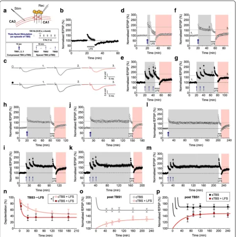
The persistence of LTP can be disrupted by low-fre-quency stimulation (LFS) by a phenomenon known as depotentiation (e.g., [2–10]). In the absence of prior LTP, LFS (2 Hz for 10 min) induced a transient depres-sion of synaptic responses that recovered to baseline within 30 min (n= 8; Fig.1b). In contrast, when delivered after the induction of LTP, LFS invariably induced depo-tentiation of the synaptic response (Fig.1c-n). We varied the timing between the induction of LTP and the delivery of LFS from between 5 and 180 min and quantified the level of depotentiation, 30 min from the end of the LFS train (Fig.1d-n).
When LFS was delivered 5 min after the end of cTBS (Fig. 1d) or sTBS (Fig. 1e) the level of depotentiation was similar (82 ± 8% and 72 ± 10%, respectively). With a
© The Author(s). 2019Open AccessThis article is distributed under the terms of the Creative Commons Attribution 4.0 International License (http://creativecommons.org/licenses/by/4.0/), which permits unrestricted use, distribution, and reproduction in any medium, provided you give appropriate credit to the original author(s) and the source, provide a link to the Creative Commons license, and indicate if changes were made. The Creative Commons Public Domain Dedication waiver (http://creativecommons.org/publicdomain/zero/1.0/) applies to the data made available in this article, unless otherwise stated.
* Correspondence:[email protected]
1
Department of Biological Sciences and Brain and Cognitive Sciences, College of Natural Sciences, Seoul National University, Seoul 151-746, Korea
2Department of Physiology, Faculty of Medicine, University of Toronto, 1
30 min interval the level of depotentiation decreased to 69 ± 10% and 34 ± 9%, respectively (*p< 0.05). From 60 min onwards, the level of depotentiation described by a
single exponential regression plateaued at 46 and 36%, re-spectively (p> 0.05). Comparison of the two plots revealed a significant difference over the time-course of the
Fig. 1Distinct depotentiation of LTP induced by compressed versus spaced theta burst stimulation (TBS).aSchematic for extracellular recordings at Schaffer collateral-commissural CA1 synapses from adult rat hippocampus. The induction protocols for compressed (c) and spaced (s) TBS are summarized below.bThe effects of LFS (2 Hz for 10 min) indicated by the hatched grey bar on naïve (i.e., unconditioned) synapses (n= 8).c
Representative traces for baseline, LTP and depotentiation from experimentsfandg(timing indicated by numbers).d-mThe effects of LFS on cTBS- and sTBS-induced LTP after 5 min (dande;n= 11 and 6), 30 min (fandg;n= 11 and 10), 60 min (handi;n= 11 and 10), 120 min (jandk;
experiment (*p< 0.05; Fig. 1n). We also plotted the depo-tentiation data relative to the second (Additional file1: Fig-ure S1g) and first (Additional file 1: Figure S1h) bout of TBS. Although there was still a trend for a difference in the levels of depotentiation, this was not statistically significant.
An alternative way of interpreting these data is that LFS completely depotentiates STP, an hypothesis compatible with the extreme activity-dependence of decay of STP [11]. Consequently, what is observed is the gradual development of LTP. Consistent with this interpretation, previous work has shown that, in response to high frequency stimulation, LTP comprises an initial presynaptic change, that cor-responds in time with STP, and a gradually developing postsynaptic change, as detected by alterations in sensiti-vity to the activation of AMPA receptors [12]. We per-formed paired-pulse experiments that confirmed there is a slowly decaying presynaptic STP in response to both cTBS and sTBS (Additional file 1: Figure S1b-f ). To compare the overall development of potentiation after LFS, we plot-ted the size of the fEPSP as a function of time after the ini-tial TBS. LTP developed slowly in response to both cTBS (Fig.1o) and sTBS (Fig.1p), but was considerably larger in the latter case. Time-matched comparisons of the extent of LTP, with and without LFS, revealed a statistically sig-nificant susceptibility of LTP to LFS for cTBS but not for sTBS (Additional file1: Figure S1j-k). Therefore, these re-sults can be interpreted by the co-existence of STP, that is highly sensitive to depotentiation, plus a time-invariant (over the course of these experiments) depotentiation of LTP1. The depotentiation of LTP induced by sTBS can be largely, if not exclusively, explained by the LTP1 compo-nent of the response, which comprises ~ 30–40% of the sTBS-induced LTP [13]. Therefore, LTP2 is highly resistant to depotentiation.
Synaptic potentiation at CA1 synapses is often considered as a unitary phenomenon. However, at least three mechanistically-distinct forms of NMDA receptor-dependent synaptic potentiation co-exist during the first few hours following its induction [1]. High fre-quency transmission (e.g., TBS) induces an initial STP, that is mediated by an increase in neurotransmitter release probability, followed by stable LTP, that is mediated, at least in part, by postsynaptic alterations. Our new paired-pulse facilitation data, presented in Additional file1: Figure S1b-f, is consistent with this distinction. By com-paring the ability of LFS to affect synaptic potentiation in-duced by cTBS and sTBS over a 3 h time interval we can propose the following: First, that STP, induced by either cTBS or sTBS, is the most sensitive form of synaptic plas-ticity to the effects of LFS. This sensitivity can be ex-plained by the extreme activity-dependent decay of STP [11]. Second, that LTP1 is more sensitive than LTP2 to depotentiation. In this regard, it should be noted that at least a portion of the depotentiation of the LTP induced
by a sTBS can be attributed to depotentiation of the LTP1 that is invariably induced along with LTP2. The most straightforward interpretation of our results is that STP is rapidly and fully depotentiated by LFS, LTP1 displays a moderate, potentially time-independent depotentiation and LTP2 is insensitive to depotentiation.
Our findings are consistent with the notion that resis-tance against depotentiation requires protein synthesis [9]. In other words, LTP2 is more resistant to depotentiation because of the nature of the potentiation induced, which likely involves morphological changes. In contrast, LTP1, which probably involves primarily post-translational modifications affecting AMPARs at the synapse [1], is more readily reversible. Indeed, both alterations in the single-channel conductance properties and the number of AMPARs at synapses can be rapidly and fully re-versed by depotentiation [10]. In conclusion, three forms of synaptic potentiation co-exist but can be clearly distin-guished on the basis of their sensitivity to depotentiation.
Materials and methods Hippocampal slice preparation
Experiments were performed as described previously [13–15]. In brief, transverse hippocampal slices (400μm) were prepared from male Sprague-Dawley rats (10–12 weeks of age). Animals were anesthetized with isoflurane and sacrificed by decapitation in accordance with Korea and UK animal legislation. The brain was then removed and placed in ice-cold slicing solution that contained (mM): 124 NaCl, 3 KCl, 26 NaHCO3, 1.25 NaH2PO4, 10
MgSO4, 10 D-glucose and 1 CaCl2, saturated with 95%
O2and 5% CO2. The hippocampi were rapidly isolated
from the brain and sliced using a vibratome (Leica, VT1000S) while maintained in the slicing solution. The CA3 region was surgically removed to suppress the up-stream neuronal excitability, and the slices were trans-ferred to an incubation chamber that contained the recording solution (artificial cerebrospinal fluid, ACSF; mM): 124 NaCl, 3 KCl, 26 NaHCO3, 1.25 NaH2PO4, 2
MgSO4, 10 D-glucose and 2 CaCl2 (oxygenated with
95% O2and 5% CO2). Slices were allowed to recover at
32–34 °C for 30 min, and then maintained at 26–28 °C for a minimum of 1 h before recordings were made.
Field excitatory postsynaptic potential (fEPSP) recordings
board at a sampling rate of 20 kHz. Recordings were mon-itored and analyzed using WinLTP. Each experiment was conducted on slices from separate animals, so the n-value reflects both the number of slices and animals used.
Schaffer collateral-commissural fibres were stimulated to obtain the evoked synaptic responses, each at a constant frequency of 0.033 Hz. Following a stable base-line period of at least 20 min, LTP was induced using theta-burst stimulation (TBS) delivered at the same basal stimulus intensity. An episode of TBS comprises 5 bursts at 5 Hz, with each burst composed of 5 pulses at 100 Hz. For compressed (c) TBS induction of LTP, three TBS episodes were delivered with an episode inter-val (IEI) of 10 s. For spaced (s) TBS, the same number of episodes were given with an IEI of 10 min. Representa-tive sample traces are an average of 5 consecuRepresenta-tive responses, collected from typical experiments (stimulus artifacts were blanked for clarity).
To measure the reversibility of LTP (i.e., depotentia-tion), low-frequency stimulation (LFS; 2 Hz for 10 min) was given at various time points (5, 30, 60, 120 and 180 min) following cTBS and sTBS. Often two-input experi-ments were performed to ensure recording stability. Since we found no difference between one and two-input studies, these results were pooled. In order to quantify depotentiation (%), the last 60 s of responses of LTP (pre-LFS) and the 60 s of responses obtained 30 min after the end of LFS were compared. If LFS had no effect on the response size then depotentiation would be con-sidered to be 0% whereas if LFS restored the synaptic response to the pre-TBS baseline then depotentiation would be considered to be 100%. The actual values fell between these ranges.
Statistical analysis
Data are presented as mean ± SEM (standard error of the mean). Responses were normalised to the baseline prior to LTP induction. Statistical significance was assessed using ANOVA with Bonferroni’s correction; the level of significance is denoted as follows: *p< 0.05, **p < 0.01 and ***p< 0.001. Regression curves are single ex-ponentials and compared by extra sum-of-squares F-test. In Fig.1o-p, regressions and time-matched data plots for LTP were obtained from Fig. 1l-m (except for the 220 and 240 min data points that were derived from Additional file2: Figure S2).
Additional files
Additional file 1: Figure S1.Differential sensitivity of STP, LTP1 and
LTP2 to depotentiation. (a) Pooled data (n= 6) showing the effects on synaptic potentiation of a sTBS on one input followed 30 min later by a cTBS on a second independent input. Paired-pulse stimulation (50 ms intervals) was delivered throughout the experiments. (b) A plot of the
paired-pulse ratio for the sTBS input. (c) Plot of paired-pulse ratio for the cTBS input. Note that there is a reduction in the paired-pulse ratio during STP, but not after LTP had stabilized. (d-f) Plot of the paired-pulse ratio as a function of time following either first (TBS1), second (TBS2) or the last TBS (TBS3). (g-h) Extent of depotentiation for cTBS and sTBS, plotted as a function of the time between TBS2 and LFS (g) and TBS1 and LFS (h). (i) Plot of the time course of synaptic potentiation induced by cTBS versus sTBS either in the presence or absence of LFS (pooled plots from Fig.1o-p). (j) Individual data and statistical analysis showing the ability of LFS to effectively depotentiate cTBS-evoked syn-aptic potentiation at time-matched points. (k) In contrast, LFS had no signifi-cant effect when LTP, induced by sTBS, had reached a plateau. ***p< 0.001; **p< 0.01; *p< 0.05 versus control. (TIFF 1040 kb)
Additional file 2: Figure S2.Additional LTP experiments. (a) Pooled
data (n= 7) showing the effects of cTBS monitored during the course of 5 h. (b) The same experiments except for sTBS used for LTP induction (n= 6). (TIFF 454 kb)
Abbreviations
AMPAR:α-amino-3-hydroxy-5-methyl-4-isoxazolepropionate receptor; cTBS: Compressed TBS; LFS: Low-frequency stimulation; LTP: Long-term potentiation; NMDAR: N-methyl-D-aspartate receptor; PKA: Protein kinase A; sTBS: Spaced TBS; STP: Short-term potentiation; TBS: Theta-burst stimulation
Acknowledgements
We thank the BKK and GLC lab members for their productive discussion.
Funding
This work was supported by the Brain Canada Foundation through the Canada Brain Research Fund, with the financial support of Health Canada. Additional support was provided by the MRC, ERC, Canadian Institute for Health Research (CIHR) Foundation grant (G.L.C), the EJLB-CIHR Michael Smith Chair in Neurosciences and Mental Health, Canada Research Chair, and CIHR operating grants (CIHR66975 and 84256) (M.Z.) and the National Honor Scientist Program of the National Research Foundation funded by the Korea government (2012R1A3A1050385) (B-K.K).
Availability of data and materials
The datasets used and/or analysed during the current study are available from the corresponding author on reasonable request.
Authors’contributions
PP performed the experiments, analysis and co-wrote and prepared the manuscript, TMS, ZAB, JG, MZ and BKK contributed to the manuscript, and GLC designed the studies and co-wrote the manuscript. All authors read and approved the final manuscript.
Ethics approval
All experiments were conducted in accordance with the policies and regulations for the care and use of laboratory animals as approved by the Institutional Animal Care and Use Committee of Seoul National University and the UK Home Office.
Consent for publication
Not applicable.
Competing interests
The authors declare that they have no competing interests.
Publisher’s Note
Springer Nature remains neutral with regard to jurisdictional claims in published maps and institutional affiliations.
Author details
1Department of Biological Sciences and Brain and Cognitive Sciences,
College of Natural Sciences, Seoul National University, Seoul 151-746, Korea.
2
Department of Physiology, Faculty of Medicine, University of Toronto, 1 King’s College Circle, Toronto, Ontario M5S 1A8, Canada.
3Lunenfeld-Tanenbaum Research Institute, Mount Sinai Hospital, Toronto,
Physiology and Pharmacology and Neuroscience, University of Bristol, Dorothy Hodgkin Building, Whitson Street, Bristol BS1 3NY, UK.
Received: 27 November 2018 Accepted: 18 March 2019
References
1. Bliss TVP, Collingridge GL, Morris RGM, Reymann KG. Long-term potentiation in the hippocampus: discovery, mechanisms and function. Neuroforum. 2018;24:A103–20.
2. Stäubli U, Lynch G. Stable depression of potentiated synaptic responses in the hippocampus with 1-5 Hz stimulation. Brain Res. 1990;513:113–8. 3. Bashir ZI, Collingridge GL. An investigation of depotentiation of long-term
potentiation in the CA1 region of the hippocampus. Exp Brain Res. 1994; 100:437–43.
4. O'Dell TJ, Kandel ER. Low-frequency stimulation erases LTP through an NMDA receptor-mediated activation of protein phosphatases. Learn Mem. 1994;1:129–39.
5. Doyle CA, Cullen WK, Rowan MJ, Anwyl R. Low-frequency stimulation induces homosynaptic depotentiation but not long-term depression of synaptic transmission in the adult anaesthetized and awake rat hippocampus in vivo. Neuroscience. 1997;77:75–85.
6. Xu L, Anwyl R, Rowan MJ. Spatial exploration induces a persistent reversal of long-term potentiation in rat hippocampus. Nature. 1998;394:891–4. 7. Stäubli U, Scafidi J. Time-dependent reversal of long-term potentiation in
area CA1 of the freely moving rat induced by theta pulse stimulation. J Neurosci. 1999;19:8712–9.
8. Abraham WC. How long will long-term potentiation last? Philos Trans R Soc Lond Ser B Biol Sci. 2003;358:735–44.
9. Woo NH, Nguyen PV. Protein synthesis is required for synaptic immunity to depotentiation. J Neurosci. 2003;23:1125–32.
10. Lüthi A, Wikström MA, Palmer MJ, Matthews P, Benke TA, Isaac JT, et al. Bi-directional modulation of AMPA receptor unitary conductance by synaptic activity. BMC Neurosci. 2004;5:44.
11. Volianskis A, Jensen MS. Transient and sustained types of long-term potentiation in the CA1 area of the rat hippocampus. J Physiol. 2003;550: 459–92.
12. Davies SN, Lester RAJ, Reymann KG, Collingridge GL. Temporally distinct pre- and post-synaptic mechanisms maintain long-term potentiation. Nature. 1989;338:500–3.
13. Park P, Sanderson TM, Amici M, Choi S-L, Bortolotto ZA, Zhuo M, et al. Calcium-permeable AMPA receptors mediate the induction of the protein kinase A-dependent component of long-term potentiation in the Hippocampus. J Neurosci. 2016;36:622–31.
14. Choi J-H, Park P, Baek G-C, Sim S-E, Kang SJ, Lee Y, et al. Effects of PI3Kγ overexpression in the hippocampus on synaptic plasticity and spatial learning. Mol Brain. 2014;7:78.