Open Access
Research article
Surgical physiology of inguinal hernia repair - a study of 200 cases
Mohan P Desarda*
1,2
Address: 1Department of Surgery, Poona Hospital & Research Centre, Pune-411030, India and 2Department of Surgery, Kamala Nehru General Hospital, Pune-411011, India
Email: Mohan P Desarda* - [email protected] * Corresponding author
Inguinal hernia repairphysiologyinguinal canalprotective mechanism
Abstract
Background: Current inguinal hernia operations are generally based on anatomical considerations. Failures of such operations are due to lack of consideration of physiological aspects. Many patients with inguinal hernia are cured as a result of current techniques of operation, though factors that are said to prevent hernia formation are not restored. Therefore, the surgical physiology of inguinal canal needs to be reconsidered.
Methods: A retrospective study is describer of 200 patients operated on for inguinal hernia under local anaesthesia by the author's technique of inguinal hernia repair.
Results: The posterior wall of the inguinal canal was weak and without dynamic movement in all patients. Strong aponeurotic extensions were absent in the posterior wall. The muscle arch movement was lost or diminished in all patients. The movement of the muscle arch improved after it was sutured to the upper border of a strip of the external oblique aponeurosis (EOA). The newly formed posterior wall was kept physiologically dynamic by the additional muscle strength provided by external oblique muscle to the weakened muscles of the muscle arch.
Conclusions: A physiologically dynamic and strong posterior inguinal wall, and the shielding and compression action of the muscles and aponeuroses around the inguinal canal are important factors that prevent hernia formation or hernia recurrence after repair. In addition, the squeezing and plugging action of the cremasteric muscle and binding effect of the strong cremasteric fascia, also play an important role in the prevention of hernia.
Background
Inguinal hernia repair still remains a problem because of the 1) high recurrence rates seen in the hands of the junior surgeons, 2) risky dissection of the inguinal floor in the Bassini/Shouldice repair and 3) infection and chronic groin pain following mesh repair. The successful manage-ment of any problem depends on the understanding of its patho-physiology. In this context, some questions related
to the physiology of the inguinal canal or factors that pre-vent herniation still exist. Lateral and cephalad displace-ment of the internal ring beneath the transversus abdominis muscle and approximation of the crura results in a shutter mechanism at the internal ring.[1] When the arcuate fibers of the internal oblique and transversus ab-dominis muscle contract, they straighten out and move closer to the inguinal ligament (shutter mechanism at the Published: 16 April 2003
BMC Surgery 2003, 3:2
Received: 16 November 2002 Accepted: 16 April 2003
This article is available from: http://www.biomedcentral.com/1471-2482/3/2
BMC Surgery 2003, 3 http://www.biomedcentral.com/1471-2482/3/2
inguinal canal).[2,3] This opposite movement (upward & downward) of the same muscle needs proper explanation. The term "obliquity of the inguinal canal" is not a perfect description since the spermatic cord is lying throughout its course on the transversalis fascia. Repeated acts of cry-ing, thereby increasing the intra-abdominal pressure do not increase the incidence of hernia in new born babies inspite of the almost absent "obliquity of the inguinal ca-nal" or "shutter mechanism". Similarly, every individual with a high arch or a patent processus vaginalis does not develop hernia.[4] Factors that are said to prevent hernia-tion are not restored in the tradihernia-tional techniques of in-guinal hernia repair and yet 70–98% of patients are cured. Then what are the additional factors that play a role in the prevention of hernia after surgery?
The author conducted this study in 200 patients who were operated by his technique under local anaesthesia, and observed the changes in the physiology of structures in and around the inguinal canal, before and after repair of the inguinal hernia. The author described for the first time, a new method of inguinal hernia repair based on physiological principles.
Methods
247 inguinal canals in 212 patients were opened under lo-cal anaesthesia between January 1995 to December 2001. From among these, the author conducted a retrospective study of 200 patients between the age group of 20–60 years, with normal or average nourishment. Malnour-ished and obese patients were excluded. 116 patients had indirect and 84 patients had direct inguinal hernia.
60–80 ml. of 1% xylocaine local anesthetic agent was used without adrenaline. No sedation was used before or dur-ing surgery. Repair was done by suturdur-ing an undetached strip of the external oblique aponeurosis as described in the author's technique of hernia repair.[5] General obser-vations were made and recorded about anatomical struc-tures forming the canal, and their movements during surgery both before and after repair by asking each patient to cough on the operation table with maximum force. Electric stimulation was used only for the cremasteric muscle, which did not respond to cough in any of the pa-tients. Movements of the posterior wall, external oblique, internal oblique and cremasteric muscle were recorded in three grades as no movement, poor movement, or good movement. Density of the cremasteric or interparietal fas-cia was judged as loose or firm by the force required sev-ering this fascia by blunt dissection.
Operative technique
Skin and fascia are incised through a regular oblique in-guinal incision to expose the external oblique aponeuro-sis. The thin, filmy fascial layer covering it is kept
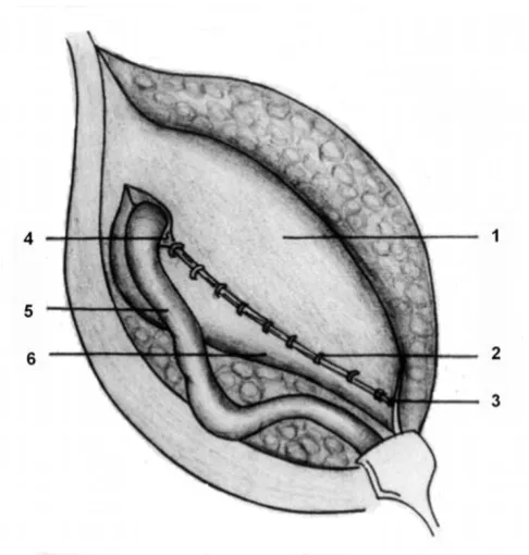
undisturbed as far as possible and an assessment made about the strength of it and its thinned-out portion. The thinned out portion is usually seen at the top of the hernia swelling, extending and fanning out to the lower crux of the superficial ring. The external oblique is cut in line with the upper crux of the superficial ring, which leaves the thinned out portion in the lower leaf so a good strip can be taken from the upper leaf. The external oblique, which is thinned out as a result of aging or long standing large hernias, can also be used for repair if it is able to hold the interrupted sutures. The cremasteric muscle is incised for the herniotomy and the spermatic cord together with the cremasteric muscle is separated from the inguinal floor. The sac is excised in all cases except in small direct hernias where it is inverted. The medial leaf of the external ob-lique aponeurosis is sutured with the inguinal ligament from the pubic tubercle to the abdominal ring using 1/0 monofilament polyamide (Ethilon) or polypropylene (Prolene) interrupted sutures. The first two sutures are tak-en in the anterior rectus sheath where it joins the external oblique aponeurosis. The last suture is taken so as to nar-row the abdominal ring sufficiently without constricting the spermatic cord (Figure 1). Each suture is passed first through the inguinal ligament, then the transversalis fas-cia, and then the external oblique. The index finger of the left hand is used to protect the femoral vessels and retract the cord structures laterally while taking lateral sutures.
A splitting incision is made in this sutured medial leaf, partially separating a strip with a width equivalent to the gap between the muscle arch and the inguinal ligament but not more than 2 cms. This splitting incision is extend-ed mextend-edially up to the pubic symphisis and laterally 1–2 cms beyond the abdominal ring. The medial insertion and lateral continuation of this strip is kept intact. A strip of the external oblique, is now available, the lower border of which is already sutured to the inguinal ligament. The up-per free border of the strip is now sutured to the internal oblique or conjoined muscle lying close to it with 1/0 monofilament polyamide or polypropylene interrupted sutures throughout its length (Figure 2). The aponeurotic portion of the internal oblique muscle is used for suturing to this strip wherever and whenever possible to avoid ten-sion, otherwise, it is not a must for the success of the op-eration. This will result in the strip of the external oblique being placed behind the cord to form a new posterior wall of the inguinal canal.
sutured to the newly formed medial leaf of the external oblique in front of the cord, as usual, again using 1/0 monofilament polyamide or polypropylene interrupted sutures. Undermining of the newly formed medial leaf on both of its surfaces facilitate its approximation to the lat-eral leaf. The first stitch is taken between the latlat-eral corner of the splitting incision and lateral leaf of the external ob-lique. This is followed by closure of the superficial fascia and the skin as usual.
Results
The external oblique aponeurosis was fairly strong and dy-namic and the posterior inguinal wall was weak and a-dy-namic in all patients. Aponeurotic extensions from the transversus abdominis aponeurotic arch were absent in the posterior wall in all patients. The absent aponeurotic extensions in the posterior wall were replaced with an aponeurotic strip of the EOA to give a strong and physio-logically dynamic posterior wall in all patients. In the au-thor's opinion, this is the main factor that prevents recurrence of hernia after surgery.
Mild to moderate movement was seen in the muscle arch in 24% of the patients. This was a local contraction and it did not appear to straighten and move the arch closer to the inguinal ligament. There was no movement seen in the remaining 76% of patients. Arching fibers of the inter-nal oblique muscle showed loss of tone failing to give an-terior protection to the internal inguinal ring in all the operated patients. Good movement or improved move-ment of the muscle arch, after it received an anchorage to the inguinal ligament through a strip of the external ob-lique aponeurosis (EOA) was seen after the repair in all patients. (Table no.1)
The role of the EOA in anterior-posterior compression of the inguinal canal to prevent herniation is restored by pro-viding a strong and physiologically dynamic posterior wall. The cremasteric muscle did not show any movement on coughing in all patients. However, after electrical stim-ulation the muscle contracted and a pulling effect on the spermatic cord was observed. The interparietal fascia (cre-masteric fascia) was loose in all patients. The binding Figure 1
The medial leaf of the external oblique aponeurosis is sutured to the Inguinal ligament. 1) Medial leaf; 2) Inter-rupted sutures taken to suture the medial leaf to the inguinal ligament; 3) Pubic tubercle; 4) Abdominal ring; 5) Spermatic cord; and 6) Lateral leaf.
Figure 2
BMC Surgery 2003, 3 http://www.biomedcentral.com/1471-2482/3/2
effect of cremasteric fascia between the muscle arch and the incurved part of the inguinal ligament is lost in all pa-tients. This lost anchorage due to loose cremasteric fascia is provided in the author's technique by suturing the strip of EOA between the muscle arch and the inguinal ligament.
Discussion
Inguinal structures, as living entities that move and func-tion cannot really be appreciated either by dissecfunc-tion on cadavers or operations on patients under general or spinal anaesthesia.[6] Live demonstration of movements of the posterior wall and the musculo-aponeurotic structures around the inguinal canal during the acts of internal ab-dominal 'blows' (raised intra abab-dominal pressure) is so far not possible. Imaging of the inguinal canal in patients, with or without hernia, on sonography machines is also not satisfactory because of the small size of the canal and the fact that all structures are seen in black and white.
Many operations developed to date deal only with the anatom-ical aspects of the repair. Any failure in these operations is be-cause the physiological aspects have not been considered while developing a new operating technique. The author has devel-oped a new technique [5] of pure tissue repair of any type of inguinal hernia without a mesh, based on the concept of constructing a strong and physiologically dynamic pos-terior wall to the inguinal canal with the help of the exter-nal oblique muscle and its aponeurosis. A strip of the external oblique aponeurosis gives replacement to the ab-sent aponeurotic extensions in the posterior wall, making it strong, and the additional strength of the external ob-lique muscle to the weakened internal obob-lique and the transversus abdominis muscle keeps it physiologically dynamic. The first recorded observation of dynamic activ-ity in the internal ring of a living nonanaesthetized hu-man being was done in only one patient by Tobin et al.[7] Many such studies of dynamic activity in the internal ring have been done in dogs.[8] Peacock EE [9] states that little is known about the muscular activity in the internal ring because conventional repair of groin hernia does not ade-quately expose the normal muscle fibers. Preperitoneal ex-posure is, of course, performed in an anaesthetized patient; consequently little information has been
ac-quired about the function of a sphincter muscle, even though the internal ring can be visualized clearly. The au-thor has dissected 247 inguinal canals during operations under local anaesthesia and found that in a majority of patients, the muscles around the inguinal canal were weak, with loss of tone and movements, and therefore, failed to provide protection against herniation. The poste-rior wall of the inguinal canal (transversalis fascia) was also weak, containing only adipose tissue, without any aponeurotic extensions. Naturally, the posterior wall of the inguinal canal was not strong and physiologically dynamic.
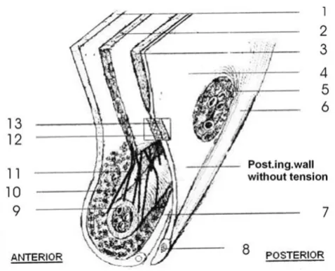
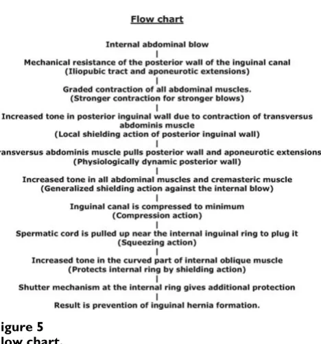
There are constant internal blows on the abdominal wall from inside during every act of coughing, sneezing, strain-ing etc. All the abdominal muscles contract to form a shield against the force of such internal abdominal blows. Phulchound's musculopectineus opening in the abdomi-nal wall near the groin is a weak area because it does not have the cover of all abdominal muscles. Forceful contrac-tion of the strong diaphragmatic muscle, in every act of coughing, straining etc. increases the burden on this weak area, over and above the effect of gravitational force. In-spite of this, not every individual suffers from inguinal hernia because of the protective mechanism, which oper-ates to prevent the inguinal herniation in the normal indi-viduals. Following protective mechanism works in such situations. (Figure 3, Figure 4, and Figure 5)
Strong and physiologically dynamic posterior inguinal wall The posterior inguinal wall is composed of condensed transversalis fascia along with the aponeurotic extensions from the transversus abdominis aponeurotic arch (Figure 3 inset). The condensed fibro-collagenous tissue and aponeurotic extensions give mechanical strength to the posterior inguinal wall to resist internal abdominal blows. The strength of the posterior inguinal wall is directly relat-ed to the number of aponeurotic fibers it contains and there is great variation in normal anatomy. In 47 % of in-dividuals, the endo-abdominal fascia of the posterior wall gets full cover from the aponeurotic extensions. However, nearly 53 % of individuals have negligible or nil aponeurotic extensions.[10,11] If aponeurotic extensions are absent, then the transversalis fascia alone cannot resist Table 1: Table shows movements of the structures before and after repair
STRUCTURE NO MOVEMENT MILD MOVEMENT GOOD MOVEMENT
the internal blows for a long period and herniation oc-curs. Therefore, any new approach to inguinal hernia re-pair must consider replacing this element with any other suitable structure or material. Secondly, the posterior in-guinal wall and accompanying aponeurotic extensions are physiologically active and dynamic. Muscular contraction of the transversus abdominis pulls this posterior wall and the aponeurotic extensions upward and laterally creating tension in it (Figure 4). Such wall with tension resists internal abdominal blows better than a wall without ten-sion. This tension in the posterior wall is created in grada-tion as per the force of contracgrada-tion of the muscles. And the force of contraction of the muscle changes as per the force of the internal abdominal blow. This is important physio-logical phenomenon. The posterior inguinal wall should be described as an independent entity, playing an impor-tant role in the prevention of hernia formation, not only because of its mechanical strength but also because of its dynamic nature. Such a physiologically dynamic posterior wall is constructed in the author's technique of repair.
Action of musculo-aponeurotic structures around the in-guinal canal
The action of the muscular and aponeurotic structures around the inguinal canal plays an equally important role
in preventing the hernia formation. Various combinations come in to play whenever there is an internal abdominal 'blow' : – (SCS action = shielding action + compression ac-tion + squeezing acac-tion)
Shielding action
Contraction of a muscle increases the tone of the muscle to guard against the 'blows'. This is the "shielding action". Increased tone in the curved fibers of the internal oblique muscle by contraction gives protection to the internal in-guinal ring. Increased tone in the aponeurotic extensions by contraction of transversus abdominis muscle gives dy-namic protection to the posterior wall (Figure 4). The cremasteric muscle, an offshoot of the internal oblique muscle, is seen occupying the gap between the muscle arch and the incurved part of the inguinal ligament. Contraction and increased tone in this muscle gives direct protection in front of the posterior wall. Aponeurosis of the external oblique muscle gives generalized shielding protection in front of the inguinal canal without disturb-ing the exit of the spermatic cord at the level of the super-ficial inguinal ring.
Figure 3
Section of inguinal canal at rest. 1) External oblique aponeurosis, 2) Internal oblique muscle, 3) Transversus abdominis muscle, 4) Endo abdominal fascia, 5) Internal inguinal ring, 6) Iliopubic tract, 7) Inguinal ligament, 8) Pubic symphisis, 9) Spermatic cord, 10) Interparietal connective tis-sue (cremasteric fascia), 11) cremasteric muscle, 12) Apone-urotic layer of posterior inguinal wall, 13) Fascial layer of posterior inguinal wall
Figure 4
BMC Surgery 2003, 3 http://www.biomedcentral.com/1471-2482/3/2
Compression action
Contraction of the external oblique muscle (aponeurosis) and the curved part of the internal oblique muscle com-presses the inguinal canal against the posterior inguinal wall (anterior-posterior compression, Figure 4). This is possible only if the posterior wall is also strong and physiologi-cally dynamic. Superior-inferior compression of the in-guinal canal takes place by movement of both the muscle arch and the incurved part of the inguinal ligament (supe-rior-inferior compression). The inguinal canal gets compressed to a minimum space, thereby preventing her-nia formation.
Squeezing action
The cremasteric muscle plays an important role by virtue of the arrangement of its muscle fibers. Contraction of crossing fibers around the spermatic cord squeezes the spermatic cord to reduce open or loose spaces in side the cord to minimum and brings all the contents near the in-ternal ring together to plug it. This squeezing action pre-vents entry of any organ into the internal ring and the cord, thereby preventing hernia formation.
Strength of the interparietal connective tissue
The spaces between the muscle arch, cremasteric muscle, incurved part of the inguinal ligament and the posterior inguinal wall is filled with interparietal fibrocollagenous tissue (cremasteric fascia). The importance of the strength of this fascia in preventing herniation is not properly
em-phasized in literature. This fascia binds all those structures together for their optimal performance and to give protection in front of the posterior inguinal wall. If this tissue is strong then the lowermost bundles of the internal oblique muscle contracts better because of some anchor-age it receives through those structures. This is elegantly seen in the author's technique of inguinal hernia opera-tion. It was observed in all operated cases that the internal oblique muscle, which did not show any contraction on asking the patient to cough on the operation table, showed improved contraction after it was sutured to the upper border of the strip of the external oblique aponeu-rosis. This was obviously due to the new anchorage that this muscle received in this procedure. The cremasteric muscle is an offshoot of the internal oblique muscle and this fascia keeps them strongly together as one muscle for optimal performance and protection. It is probable, as stated by Read [12] that some metabolic changes in the collagen metabolism in this area result in loss of strength of this connective tissue that loosens those structures or muscles from each other resulting in proportionate loss of their performance and protection.
Ideally, movements of the muscles and aponeurosis or tension in the strip should have been measured objective-ly with some device as described by Peiper et al, Chirurg 1998;69(19),1077E81. Since a measuring device was not available, this author could not measure tissue move-ments accurately. However, naked eye observations of such movements were felt to be adequate enough to draw conclusions.
Conclusions
1. Anterior-posterior compression of the inguinal canal is lost due to the loose and adynamic posterior inguinal wall. Providing a strong and physiologically dynamic posterior inguinal wall is essential to restore this anterior-posterior compression.
2. Local contraction of the muscle arch is essential to pre-vent herniation. Muscle arch contractions showed im-provement after it receives new anchorage.
3. Contraction of the cremasteric muscle provides an ad-ditional guarding effect in front of the posterior inguinal wall and squeezes and pulls up the spermatic cord to plug the internal ring.
4. The strong cremasteric fascia between the muscle arch and the incurved part of the inguinal ligament keeps mus-cles dynamic by giving anchorage. New anchorage is pro-vided by suturing a strip of the EOA between the inguinal ligament and the muscle arch in the author's technique. Figure 5
Publish with BioMed Central and every scientist can read your work free of charge
"BioMed Central will be the most significant development for disseminating the results of biomedical researc h in our lifetime."
Sir Paul Nurse, Cancer Research UK
Your research papers will be:
available free of charge to the entire biomedical community
peer reviewed and published immediately upon acceptance
cited in PubMed and archived on PubMed Central
yours — you keep the copyright
Submit your manuscript here:
http://www.biomedcentral.com/info/publishing_adv.asp
BioMedcentral 5. The author's technique is physiologically sound
be-cause: A) Absent aponeurotic extensions in the posterior wall are replaced with an aponeurotic structure. B) Addi-tional muscle strength of the external oblique muscle helps the weakened muscle arch to keep the newly formed posterior wall physiologically dynamic. C) Contractions of the muscle arch are improved.
6. Similar study conducted with a device that measures tis-sue movements and tensions accurately, can provide con-clusive evidence to support the statements made in this article. No shutter mechanism is restored at the internal ring or the inguinal canal level or no efforts are made to bring down the highly placed muscle arch, if present. But still, the results of the operation are excellent with virtual-ly zero recurrence rate observed by the author in his series wherein 106 patients were followed up for more than 10 years [5]. Providing a strong and physiologically dynamic posterior wall should be the principle to be observed in any type of inguinal hernia repair.
Competing Interests
None declared.Additional material
References
1. Lytle WJ The internal inguinal ringBr J Surg 1945, 32:441 2. Hammond TE The etiology of indirect inguinal hernia Lancet
1923, 204:1206
3. Keith A On the origin and nature of herniaBr J Surg 1923, 11:455 4. Murray RW The saccular theory of herniaBr Med J 1907, 2:1385 5. Desarda MP New method of inguinal hernia repair-A new
solutionANZ J Surg 2001, 71:241-44
6. Griffith CA Inguinal hernia: an anatomic-surgical correlation
Surg Cl North Am 1959, 39:531
7. Tobin GR, Clark DS and Peacock EE Jr A neuromuscular basis for development of indirect inguinal hernia Arch Surg 1976, 111:464
8. MacGregor WW The demonstration of a true internal inguinal sphincter and its etiologic role in herniaSurg Gynaecol Obstet
1929, 49:510
9. Peacock EE Jr and Madden JW Studies on the biology and treat-ment of recurrent inguinal hernia. II. Morphological changes
Ann Surg 1974, 179:567
10. Anson BJ and McVay CB Surgical anatomy Philadelphia, WB Saunders 1971, 1:461-532
11. Anson BJ, Morgan EH and McVay CB Surgical anatomy of the in-guinal region based upon a study of 500 body-halvesSurg Gy-naecol Obstet 1960, 111:707
12. Read RC A review: the role of protease – antiprotease imbal-ance in the pathogenesis ofherniation and abdominal aortic aneurysm in certain smokersPostgraduate General Surgery 1992, 4:161-165
Pre-publication history
The pre-publication history for this paper can be accessed here:
http://www.biomedcentral.com/1471-2482/3/2/prepub
Additional File 1
'Dr.Desarda's hernia repair', a media file, showing video of a live operation.
Click here for file