Themed Section: Science and Technology
SARCOPENIA: Immunological Perspective and Efficacy of
Exercise Interventions
Dr. Ashish Mathur1, Dr. Amit Gupta*2
1School of Physiotherapy, PP Savani University, Kosamba, Surat, Gujarat, India
2School of Sciences (Biotechnology/Microbiology/Environmental Science), PP Savani University, Kosamba, Surat, Gujarat, India
ABSTRACT
Sarcopenia (Greek word “sarx” or flesh; penia or loss), syndrome is a progressive as well as ongoing and generalized loss of skeletal muscle mass and strength along with the risk of adverse outcome such as physical disability, poor quality of life and death. Various studies were conducted related to Sarcopenia and believed that signs of this disease kindred with ageing are an unpreventable process. In this regard, immunological and physiological interventions should be taken pertaining to slow down the ageing process. In this review article, we discuss about correlation of Sarcopenia and understanding its mechanism related to immunological perspective and physiotherapy interventions specifically in relation to loss of muscle mass and strength. In addition, we also understanding the mechanism of this disease associated with ageing process.
Key words: Sarcopenia; skeletal muscle; ageing; disease
I.
INTRODUCTION
Sarcopenia, is defined as an age-related involuntary loss of muscle mass along with its function which generally leads to weakness and ultimately showing greater risk in older adults with respect to functional disability [1, 2]. Now a day, sarcopenia cases increases dramatically with the increase in older adult population and also associated with various diseases i.e. obesity, type II diabetes etc. As per the literature, more than 70 % of population especially older people (age group 65-75 years) suffered from sarcopenia [3, 4]. In other words, this disease i.e. sarcopenia is associated with ageing process. In this disease, loss of muscle mass along with its strength is still reported in patients and also not be able to perform daily functions. These type of observations are observed in older patients and these are the hallmark signs of this disease [1-4].
the body), total body bone densitometry and anthropometric measures [4-9]. Out of these, densitometry is often used because it provides assessment of the body composition, bone mass, lean mass and total fat mass whereas anthropometric measures (proposed by Ashwell), including waist-to-hip ratio, have also been used to assess sarcopenia. Inspite of this techniques, Dual-energy X-ray absorptiometry (DXA) is considered to be one of the gold-standard technique in terms of accuracy, low cost and radiation and simplicity for assessing and analysing sarcopenia in single/whole body region with respect to body composition at the molecular level; quantify fat/lean mass including bone mineral content. Recently, this technique is considered as a reference assessment in muscle mass evaluation [4-9].
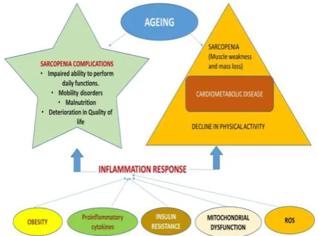
Figure 1. Complications and inflammation response in Sarcopenia
Immunological perspectives- Pathogenesis of
sarcopenia points out to the fact that, this disease is generally associated with various intrinsic and
extrinsic factors including accumulation of
proinflammatory cytokines and oxidative stress along with mitochondrial dysfunction, insulin resistance and ageing-related loss of anabolic hormones and motor neuron end plates [5]. In this regard, most familiar example in sarcopenia is rheumatoid arthritis where significant reduction or decline in muscle
strength and is generally caused by muscle protein wasting and loss of functionality [10].
Major important factors i.e. lifestyle, neurological,
hormonal, nutritional, and immunological
determinants are responsible for causing Sarcopenia. Out of these, various immunological conditions that are applied which may leads to muscle wasting and also involved in different pathways of intracellular signalling which may trigger programmed cell death (apoptosis); increased protein degradation through autophagy; calcium-dependent proteases (calpains and caspases); proteasome system; decreased satellite cell activation, responsible for muscle regeneration [11-15]. In other words, skeletal muscle loss is due to improper balance of protein metabolism i.e. protein degradation and protein synthesis. In skeletal muscle, protein degradation is through coordinated network of various signalling pathways which is activated or suppressed by various hormones including cytokines and therefore, catabolism is stimulated or initiated through various proinflammatory cytokines including glucocorticoids, and reactive oxygen species (ROS) [11-15].
One of the examples is observed in case of elderly
healthy people showed systemic low-grade
inflammation whereas the levels of C-reactive protein (CRP), interleukin (IL)-1β, IL-6 and tumor necrosis factor (TNF)-α are totally related to severe muscle wasting and cachexia because these inflammatory cytokines are involved in the muscle catabolic processes that are associated with inflammation [16-18]. Numerous studies were conducted with muscle hypertrophy and was associated with reduction of CRP and TNF-α. In this regard, these findings indicate that inflammatory markers and cytokines may be potential biomarkers for sarcopenia and useful as
criteria when developing interventions and
In contrast, pain is also considered under age-related factors that may contributes to the progression of sarcopenia. Increased in the level of cytokines especially IL-6 and TNF-α levels were detected by assessing local tissue in adults with low back pain. As per the literature, one of the cytokines i.e. IL-1β production is primarily initiated by TNF-α, which induces IL-6 production. IL1β is known for its muscle-catabolic properties and has a central role in common with the degenerative processes of ageing by acting in the degradation of cartilage in joint inflammation. In short, sarcopenia disease is directly linked with these cytokines (IL-1, IL-6 and TNF-α) and showed some enhancement but it reduced muscle mass and strength between high levels of cytokines and reduction of muscle mass and strength [19-23].
One of the studies were conducted and suggested that cytokine IL-1 activate TGF-β-activated kinase (TAK)-1 in vitro and then blocked human myoblasts and finally differentiate into myotubes. Somehow this cytokine also involved in sarcopenia by activating TAK-1. In addition, various clinical studies were conducted related to these cytokines (IL-1, IL-6 and TNF-α) and inflammatory markers (CRP) in sarcopenia [19-23]
Sarcopenia and protein degradation
Generally, muscle size depends on fibre with respect to number (set during development) and size (continuously adapts during the entire lifetime). So these fibres are usually dependent on activity, nutrition, diseases and ageing. In the last decade, myostatin emerged as a key maintenance regulator of muscle mass. Myostatin (also called as growth differentiation factor 8, GDF-8) belongs to TGF-β superfamily expressed in skeletal muscle and also called as myokine, protein produced and released through myocytes and is generally act on muscle cells, autocrine function to inhibit myogenesis: muscle cell growth and differentiation [24-27]. In muscles,
strategy for assessing sarcopenia and is one of the potent negative regulator of muscle growth. In other words, myostatin controls the metabolism rate in order to support the immune system. Numerous work is done related to this myostatin protein, if there is any infection in the body, muscle growth is compromised because of the increased secretion of myostatin. This infection is generally leads to inflammation that automatically triggers the increase of this protein in the blood. Muscle cells should not able to grow or multiply during these conditions priority only to the immune system because of the organism rather than muscle growth. In this regard, amino acids are used in immune system processes and not as building blocks during inflammation. In short, too much of this muscle growth suppressing this protein can ruin a well build physique. Interestingly, myostatin expression is also observed as well as reported in several human diseases and most of it is directly associated with skeletal muscle wasting, such as cancer cachexia, AIDS, and heart failure. In addition to its antisarcopenic effects, increasing skeletal muscle mass through myostatin inhibition may also considered as a promising strategy for the treatment of type 2 diabetes and related metabolic disease [24-28].
In sarcopenia, one of the major observations i.e. loss of skeletal muscle fiber number along with change in the cross-sectional area of the remaining fibers [1-5]. Lot of studies were conducted related to skeletal muscle protein and explaining some of the sudden changes in total muscle mass which includes
a) lacking of regular physical exercise b) reorganization of protein metabolism
c) Alterations in the endocrine levels (decrease in growth hormone (GH) and testosterone and an increase in cortisol and cytokines)
Several physiological stimuli have been demonstrated to affect either protein synthesis (for repair) or protein degradation (apoptosis). In this pathway, it showed the presence of both extracellular as well as intracellular regulators. In this pathway, no single mechanism (decreased amino acid quantity; hormonal balance etc.) is solely accountable for sarcopenia [2-8].
With reference to muscle mass, synthesis of protein is almost equal to the rate of degradation [6, 8, 29]. The degradation of this protein and ultimately it is present in the form of amino acids that will combine with dietary components and showing some difference in the utilization of amino acids during synthesis as well as degradation. In this regard, there is some imbalance were reported during protein synthesis and degradation e.g. young adults reported 30% whole body protein turnover whereas elderly only 20%. In other words, sarcopenia only happens due to advancing age so there is inadequate dietary component of amino acids. So, these oxidized proteins were increased enormously with the passage of time and may not be removed efficiently by the proteolysis system (ubiquitination and lysosomal degradation) resulting in the accumulation of lipofuscin and cross-linked proteins [24-29].
Efficacy of Exercises
Regular optimal Exercises plan in supervision of a physiotherapist is very useful as it increases muscle strength, enhances functional reserve capacity and also reduce body fat. Supervision of a Physiotherapist is very important for ensuring high exercise compliance and constant motivation in older people. If exercises are not being done regularly then it will lead to decreasing cardiovascular function and at the same time aerobic capacity will also diminish, which in turn leads to increased perception of exercise effort. There is significant association between sarcopenia and muscle power; numerous researches shows that high-intensity resistance training program can improve muscle strength and improve overall
functional independence. Thus, Physiotherapy has important role in the prevention of sarcopenia by optimal prescription of strength and power training program in older population. This can be done effectively by creating social awareness and constant motivation. Following are the various factors which are contributing for causing sarcopenia i.e.
Though diet along with exercise should reduce the rate of muscle and strength loss; even active seniors will experience decline in muscle function.
People with chronic illnesses and activity limitations caused by conditions like sarcopenia have more physician visits and fill more prescriptions than those individuals with no activity limitations, all of which presents a greater burden on our health care system
A loss in muscle mass is related to metabolic problems such as insulin resistance, type 2 diabetes and obesity. Although age‐related muscle loss is inevitable, therapies and interventions that can halt or reverse these effects hold great promise and are a realistic possibility.
One of the chief requirement of successful physical therapy of management of sarcopenia is patients motivation for the exercise program. Creating awareness and associating improvements due to exercises with functional aspects of daily life is necessary for successful management of sarcopenia by a physical therapist. Physical therapists can play a vital role in the prevention of sarcopenia through creating awareness and careful selection of power and strength training regimen in older population. An exercise program consists of high progressive resistance exercises of the hip and knee extensors for frail individuals. Hip and Knee Extensors muscle groups are selected because of their critical role in functional activities such as preventing falls, and regaining/ maintaining balance. Training program includes exercise sessions done 3 days/week for a total of 10-12 weeks and each session lasting 30- 45 minutes.
II.
CONCLUSION
Even though muscle loss related to age is inevitable, physical therapies, optimal nutrition and other interventions can minimize, stall or reverse these debilitating effects caused by sarcopenia. Maintaining activities of daily living and independence of ageing population by promoting appropriate exercises and physical activities can effectively contribute to quality of life of older people and thus reduce burden on our health care system.
III.
REFERENCES
[1]. Castillo EM, Goodman-Gruen D,
Kritz-Silverstein D, Morton DJ, Wingard DL, Barrett-Connor E. Sarcopenia in elderly men and women-The Rancho Bernardo Study. Am J Prev Med 2003; 25: 226-231
[2]. Volpi E, Nazemi R, Fujita S. Muscle tissue changes with ageing. Curr Opin Nutr Metab Care 2004; 7(4):405-410.
[3]. Morley JE, Anker SD, Von Haehling S.
sarcopenia: facts, numbers, and epidemiology— update 2014. Journal of Cachexia, Sarcopenia and Muscle. 2014; 5(4):253-259.
[4]. Fan J, Kou X, Jia S, Yang X, Yang Y, Chen N. Autophagy as a potential target for sarcopenia. J Cellular Physiology 2016; N231 (7):1450-1459. [5]. Sayer AA, Robinson SM, Patel HP, Shavlakadze
T, Cooper C, Grounds MD. New horizons in the pathogenesis, diagnosis and management of sarcopenia. Age Ageing 2013; 42(2):145-150. [6]. Cruz-Jentoft AJ, Baeyens JP, Bauer JM, Boirie Y,
Cederholm T, Landi F et al. Sarcopenia: European consensus on definition and diagnosis: Report of the European Working Group on Sarcopenia in Older People. Age Ageing. 2010; 39(4):412-423.
[7]. Fielding RA, Vellas B, Evans WJ, Bhasin S, Morley JE, Newman AB, et al. Sarcopenia: an undiagnosed condition in older adults. Current consensus definition: prevalence, etiology, and consequences. International working group on sarcopenia. J Am Med Dir Assoc. 2011; 12(4):249-256.
[8]. Rubbieri G, Mossello E, Bari MD. Techniques for the diagnosis of sarcopenia. Clin Cases Miner Bone Metab 2014; 11(3): 181-184.
[9]. Mijnarends DM, Meijers JM, Halfens RJ, ter Borg S, Luiking YC, Verlaan S, et al. Validity and reliability of tools to measure muscle mass,
strength, and physical performance in
community-dwelling older people: a systematic review. J Am Med Dir Assoc 2013; 14(3):170-178.
[10]. Ngeuleu A, Allali F, Medrare L, Madhi A, Rkain H, Hajjaj-Hassouni N. Sarcopenia in rheumatoid arthritis: prevalence, influence of disease activity and associated factors. Rheumatol Int 2017; 37(6):1015-1020.
[12]. White JR, Confides AL, Moore-Reed S, Hoch JM, Dupont-Versteegden EE. Regrowth after skeletal muscle atrophy is impaired in aged rats, despite similar responses in signalling pathways. Experimental Gerontology 2015; 64:17-32. [13]. Kaasik P, Umnova M, Pehme A et al. Ageing
and dexamethasone associated sarcopenia: peculiarities of regeneration. Journal of Steroid Biochemistry and Molecular Biology. 2007; 105 (1-5): 85-90.
[14]. Howard C, Ferrucci L, Sun K et al. Oxidative protein damage is associated with poor grip strength among older women living in the community. Journal of Applied Physiology. 2007; 103(1):17-20.
[15]. Trendelenburg AU, Meyer A, Jacobi C, Feige JN, Glass DJ. TAK-1/p38/nNFkB signalling inhibits myoblast differentiation by increasing levels of Activin A. Skelet Muscle 2012; 2:3.
[16]. Chung HY, Sung B, Jung KJ, Zou Y, Yu BP. The molecular inflammatory process in ageing. Antioxid Redox Signal 2006; 8: 572e81.
[17]. Zhang JM, An J. Cytokines, inflammation, and pain. Int Anesthesiol Clin 2007; 45: 27-37. [18]. Chung HY, Cesari M, Anton S, Marzetti E,
Giovannini S, Seo AY et al. Molecular inflammation: underpinnings of ageing and age related diseases. Ageing Res Rev 2009; 8: 8-30. [19]. Luo G, Hershko DD, Robb BW, Wray CJ,
Hasselgren PO. IL1beta stimulates IL-6 production in cultured skeletal muscle cells through activation of MAP kinase signalling pathway and NF-kappa B. Am J Physiol Regul Integr Comp Physiol 2003; 284: R1249- 254. [20]. Reid MB, Li YP. Tumor necrosis factor-alpha
and muscle wasting: a cellular perspective. Respir Res 2011; 2: 269-272.
[21]. Lang CH, Frost RA. Role of growth hormone, insulin-like growth factor-I, and insulin-like growth factor binding proteins in the catabolic response to injury and infection. Curr Opin Clin Nutr Metab Care 2002; 5:271- 279.
[22]. Wang DT, Yin Y, Yang YJ, Lv PJ, Shi Y, Lu L, et al. Resveratrol prevents TNF-alpha-induced
muscle atrophy via regulation of
Akt/mTOR/FoxO1 signalling in C2C12
myotubes. Int Immunopharmacol 2014; 19: 206-213.
[23]. Schaap LA, Pluijm SM, Deeg DJ, Harris TB, Kritchevsky SB, Newman AB, et al. Higher inflammatory marker levels in older persons: associations with 5-year change in muscle mass and muscle strength. J Gerontol A Biol Sci Med Sci 2009; 64: 1183-1189.
[24]. Yarasheski KE, Bhasin S, Sinha-Hikim I, Pak-Loduca J, Gonzalez-Cadavid NF: Serum myostatin-immunoreactive protein is increased in 60- to 92-year-old women and men with muscle wasting. J Nutr Health Ageing 2002; 6:343-348.
[25]. Lee SJ, McPherron AC: Regulation of myostatin activity and muscle growth. Proc Natl Acad Sci USA 2001; 98: 9306-9311.
[26]. Lee SJ: Regulation of muscle mass by myostatin. Annu Rev Cell Dev Biol 2004; 20:61-86.
[27]. Lebrasseur NK: Building muscle, browning fat and preventing obesity by inhibiting myostatin. Diabetologia 2012; 55:13-17.
[28]. Hittel DS, Berggren JR, Shearer J, Boyle K, Houmard JA: Increased secretion and expression of myostatin in skeletal muscle from extremely obese women. Diabetes 2009; 58:30-38.
[29]. Sartori R, Milan G, Patron M, Mammucari C,