EUKARYOTICCELL, Feb. 2007, p. 182–197 Vol. 6, No. 2 1535-9778/07/$08.00⫹0 doi:10.1128/EC.00364-06
Copyright © 2007, American Society for Microbiology. All Rights Reserved.
The Anaphase-Promoting Complex/Cyclosome Is Required for
Anaphase Progression in Multinucleated
Ashbya gossypii
Cells
䌤
†
Amy S. Gladfelter,* Nicoleta Sustreanu, A. Katrin Hungerbuehler, Sylvia Voegeli,
Virginie Galati, and Peter Philippsen
University of Basel Biozentrum, Molecular Microbiology, Klingelbergstrasse 0/70, 4056 Basel, Switzerland
Received 17 November 2006/Accepted 28 November 2006
Regulated protein degradation is essential for eukaryotic cell cycle progression. The anaphase-promoting complex/cyclosome (APC/C) is responsible for the protein destruction required for the initiation of anaphase and the exit from mitosis, including the degradation of securin and B-type cyclins. We initiated a study of the
APC/C in the multinucleated, filamentous ascomyceteAshbya gossypiito understand the mechanisms
under-lying the asynchronous mitosis observed in these cells. These experiments were motivated by previous work which demonstrated that the mitotic cyclin AgClb1/2p persists through anaphase, suggesting that the APC/C
may not be required for the division cycle in A. gossypii. We have now found that the predicted APC/C
components AgCdc23p and AgDoc1p and the targeting factors AgCdc20p and AgCdh1p are essential for growth and nuclear division. Mutants lacking any of these factors arrest as germlings with nuclei blocked in mitosis. A likely substrate of the APC/C is the securin homologue AgPds1p, which is present in all nuclei in hyphae except those in anaphase. The destruction box sequence of AgPds1p is required for this timed disappearance. To investigate how the APC/C may function to degrade AgPds1p in only the subset of anaphase nuclei, we localized components and targeting subunits of the APC/C. Remarkably, AgCdc23p, AgDoc1p, and AgCdc16p were found in all nuclei in all cell cycle stages, as were the APC/C targeting factors AgCdc20p and AgCdh1p. These data suggest that the AgAPC/C may be constitutively active across the cell cycle and that proteolysis in these multinucleated cells may be regulated at the level of substrates rather than by the APC/C itself.
Regulated protein degradation ensures linear order in the cell division cycle. Timely protein degradation prevents pre-mature passage through future stages of the cycle and limits slippage back to previous stages once they are completed. A series of positive and negative feedback loops connect cyclin-dependent kinase (CDK)/cyclin complexes to the machinery that targets specific proteins for degradation by the proteo-some. This web of interactions culminating in protein destruc-tion gives accuracy to cell division (27, 32, 37).
The anaphase-promoting complex/cyclosome (APC/C) is one of several multisubunit protein complexes that add a polyubiquitin modification to specific protein substrates, which leads to their termination in the proteosome (33, 37). The components of the APC/C include the enzymatic machinery for attaching ubiquitin as well as factors for substrate docking and recognition. There are at least 13 conserved subunits to the eukaryotic APC/C which coordinate the tagging of proteins for destruction, although the exact means by which they func-tion and assemble together as a complex is not yet known (35). In yeast cells, the APC/C is present throughout the cell cycle
but its activity peaks in M through G1 to promote anaphase
entry and mitotic exit and facilitate prereplication complex formation (17).
How does the APC/C recognize specific substrates destined for the proteosome? Two conserved targeting/adaptor factors, Cdc20p and Cdh1p, which contain WD40 repeats are respon-sible for linking the APC/C to specific substrates (42, 43, 50). InSaccharomyces cerevisiae, these two factors seem to prefer-entially interact with distinct sets of target proteins and func-tion in different windows of time and are themselves controlled by different mechanisms (17). ScCdc20p functions early in mi-tosis by marking the S-phase cyclin ScClb5p and a fraction of the pools of ScClb2p for destruction. The anaphase inhibitor ScPds1p (securin) is another target of ScCdc20p, and ScPds1p degradation leads to the loss of sister chromatid cohesion and anaphase (11, 18, 30, 41, 44, 45, 49, 51, 56). ScPds1p degrada-tion also helps to set off the FEAR/MEN signaling cascade that leads to the activation of ScCdh1p, which will then cover the
bulk of APC/C targeting through G1and early S phase,
includ-ing markinclud-ing the mitotic cyclin ScClb2p for degradation (21, 43). Cdc20p is itself degraded by the APC/C via Cdh1p in many systems, whereas Cdh1p persists across the cell cycle but its activity is limited from S to late M by inhibitory phosphoryla-tion delivered by CDK/cyclin complexes (23, 57). An addiphosphoryla-tional conserved factor called Doc1p functions to stabilize substrate-APC/C interactions by associating with substrates, but it re-mains associated with the APC/C throughout the cell cycle (7, 8, 16, 36).
Most targets of the APC/C have a short N-terminal motif called the destruction box (D box) and/or a KEN box, both of which facilitate, though are not essential in all cases,
identifi-* Corresponding author. Present address: Department of Biology, Gilman Hall, Dartmouth College, Hanover, NH 03755. Phone: (603) 646-1346. Fax: (603) 646-8706. E-mail: Amy.Gladfelter@dartmouth .edu.
† Supplemental material for this article may be found at http://ec .asm.org/.
䌤Published ahead of print on 8 December 2006.
182
on September 8, 2020 by guest
http://ec.asm.org/
cation by Cdc20/Cdh1 proteins (5, 18, 38). Additionally, the D-box motif contributes to the assembly and stability of the substrate-APC/C-Cdh1 ternary complex (6, 28). Regulated degradation of both the B-type cyclins and securin was shown to be the only essential role of the APC/C for accurate and orderly progression in budding yeast (47, 48).
In addition to uninucleated yeasts, some components of the APC/C have also been studied in several filamentous fungi and genes encoding components of the APC/C, BimA and BimE
(blocked in mitosis), were identified nearly 30 years ago by
genetic screens inAspergillus nidulans(31). Subsequent work
identified these gene products as negative cell cycle regulators and central components of the APC/C, with BimA homologous to Cdc27/APC3 and BimE homologous to APC1. The APC/C inA. nidulansfunctions both in exit from mitosis and during interphase, where it regulates the stability of NimA, a kinase that promotes mitosis (22, 34, 55). A Cdh1p homologue, Cru1,
has also been characterized inUstilago maydis, where it was
shown to target the degradation of B-type cyclins and control
the length of G1(9). Interestingly, Cru1 expression is regulated
by cyclic AMP and mutants lacking Cru1 are defective in pathogenicity. Little is known about how the APC/C may be spatially regulated in large, multinucleated cells. The APC/C changes localization and exists in functionally distinct
subcom-plexes in Drosophila melanogaster syncytial embryos; thus,
there is some limited data to suggest that the APC/C can be uniquely regulated in space in multinucleated cells (20). Spa-tial control of the APC/C has not yet been explored in multinu-cleated, filamentous fungal cells.
We have begun to investigate how the APC/C, Cdc20p, and Cdh1p function in the filamentous, multinucleated ascomycete
Ashbya gossypii.A. gossypiiis evolutionarily related to the
bud-ding yeastS. cerevisiaebut has a notably different growth and
nuclear division cycle (12). Nuclei in A. gossypii cells divide
asynchronously so that a mitotic nucleus divides in the same cytoplasm beside neighbors in different stages. Furthermore, the mitotic cyclin homologue AgClb1/2p appears to persist across all nuclear division cycle stages rather than be com-pletely destroyed at the exit of mitosis, as is common in most eukaryotic systems (14). We hypothesized that in a syncytial
and asynchronous system such asA. gossypii, other forms of
negative cell cycle regulation may be favored over protein destruction. In these cells, proteins are continually being ex-pressed from adjacent nuclei in different nuclear cycle stages, and we speculated that it would therefore be challenging for degradation in a single nucleus to match the influx of new proteins from multiple neighbors. Thus, in this paper, we set out to analyze how the APC/C and associated factors function within this asynchronous nuclear division cycle. Is the APC/C essential for nuclear progression? Is the APC/C spatially reg-ulated to be active or present in nuclei of a particular cell cycle stage?
To address these questions, we have identified and charac-terized how the homologues of core APC/C factors, the tar-geting subunits, and the potential APC substrate AgPds1p
function in the asynchronousA. gossypiinuclear cycle. We have
found that despite the lack of AgClb1/2p oscillation in these cells, both the APC/C and the targeting factors AgCdh1p and
AgCdc20p are essential inA. gossypiiand mutants arrest with
a clear, nearly uniform block in mitosis. The predicted APC
substrate AgPds1p was also examined and found to be an essential gene, unlike in budding yeast cells. AgPds1p was absent in anaphase nuclei, and its D box was required for this disappearance. This result raised the question of how the APC/C may be locally activated to degrade AgPds1p in only a subset of nuclei in these cells. Thus, we have also evaluated the spatial and temporal appearances of the APC/C in multinucle-ated hyphae.
MATERIALS AND METHODS
Strains, media, plasmids, DNA manipulations, and transformation.TheA. gossypiireference strain used for all experiments is a derivative of the wild-type strain (ATCC 10895) in which the AgLEU2and AgTHR4genes were deleted (3, 30a). Growth media, culturing conditions, spore isolations, and transformation protocols are described in references 4 and 52. Transformants were selected on
Ashbyafull medium (AFM) containing either Geneticin-G418 (200g/ml) or CloNAT (50g/ml). AllA. gossypiistrains are listed in Table 1. The diploidS. cerevisiaestrain DHD5 (ura3/ura3) was used for tagging and in-frame deletions ofA. gossypii genes cloned in autonomously replicating centromere vectors pRS415 or pRS416 (see Table 2). The modified plasmids were verified by DNA sequencing and then used, cleaved or noncleaved, for transformation ofA. gossypii. These and other plasmids are listed in Table 2. PCR was performed with
Taqpolymerase from Roche (Basel, Switzerland), and restriction enzymes were obtained from New England Biolabs or Roche. Oligonucleotides were synthe-sized at MWG (Ebersberg, Germany) or Microsynth (Balgach, Switzerland) and are listed in Table 3. Nocodazole (Sigma) was dissolved in dimethyl sulfoxide (3 mg/ml) and used at a final concentration of 15g/ml to block nuclear cycles.
Construction of gene deletion mutants. A. gossypii open reading frames (ORFs) were replaced by cassettes carrying either the dominant markerGEN3
or CloNAT. The cassettes were obtained by PCR amplification of the selection markers from pGEN3 (52) or from pUC19NATPS (kindly provided by Dominic Hoepfner), using pairs of 65- to 76-nucleotide primers named S1 and S2 plus the
TABLE 1. Ashbya gossypiistrains used in this studya
Strain Genotype Source or
reference
Reference strain
Agleu2⌬thr4⌬ 3
NSN01 AgCDC23 leu2⌬thr4⌬/cdc23::NAT1 leu2⌬thr4⌬
This study
NSG01 AgCDC20 leu2⌬thr4⌬/cdc20::GEN3 leu2⌬thr4⌬
This study
NSN02 AgDOC1 leu2⌬thr4⌬/doc1::NAT1 leu2⌬thr4⌬
This study
NSG02 AgCDH1 leu2⌬thr4⌬/cdh1::GEN3 leu2⌬thr4⌬
This study
NSG03 AgPDS1 leu2⌬thr4⌬/pds1::GEN3 leu2⌬thr4⌬
This study
NSG09 AgPDS1-6HA-GEN3 leu2⌬thr4⌬ This study NSG10 AgPDS1 leu2⌬thr4⌬(pAgpds1⌬
db-6HA-GEN3)
This study
NSG11 AgDOC1-9myc-GEN3 leu2⌬thr4⌬ This study GVS2 AgCDC16 leu2⌬thr4⌬(pAg
CDC16-13myc-GEN3)
This study
GVS3 AgDOC1 leu2⌬thr4⌬(pAg DOC1-13myc-GEN3)
This study
GVS4 AgCDC23 leu2⌬thr4⌬(pAgCDC23 -13myc-GEN3)
This study
NSG12 AgCDH1 leu2⌬thr4⌬(p GEN3-prom-GFP-AgCDH1)
This study
NSG13 AgCDC20 leu2⌬thr4⌬(p GEN3-prom-GFP-AgCDC20)
This study
aAll NSN, NSG, and GVS strains are derived from the reference strain.
Individual nuclei are haploid, and most strains are heterokaryotic (mixture of nontransformed and transformed nuclei). The genotypes of the two different types of nuclei are separated by a slash. A plasmid name in parentheses indicates a replicating plasmid which is maintained under selection pressure.
on September 8, 2020 by guest
http://ec.asm.org/
gene name for amplification ofGEN3or NS1 and NS2 plus the gene name for amplification of the CloNAT marker (Table 3). The 20 to 22 lowercase letters of the primer sequences have homology to either end of the selection marker, and the 45 to 56 uppercase letters of the primers have homology to the sequences immediately upstream of the start codon or downstream of the stop codon, respectively. The PCR products were directly transformed intoA. gossypiiby electroporation. Heterokaryon strains that have a mixture of wild-type and mu-tant nuclei were selected as primary transformants on plus-G418 (or AFM-plus-CloNAT) plates. Correct ORF targeting was verified by PCR using the primers G1 and G4 (plus gene name) binding upstream and downstream of the deleted ORF, respectively, and the primers G2.1 (GEN) and G3 (GEN) binding inside the GEN marker or V2PDC1P and V3PDC1T binding inside the CloNAT marker. Spores from three independent heterokaryon mycelia each were incu-bated overnight under selective conditions and analyzed by microscopy.
Genomic DNA clones in replicating vectors. Plasmids carrying the genes AgCDC23 (pAG6955), AgDOC1 (pAG1393), AgCDH1 (pAG7019), and AgPDS1(pAG14820) were obtained from the genomic DNA pRS416 library used for sequencing of theA. gossypiigenome (12). These plasmids can auton-omously replicate inS. cerevisiaeandA. gossypii. A genomic clone of AgCDC20
in pRS416 (pNS01) was constructed by PCR amplification of genomic DNA, including 800 bp upstream and 400 bp downstream of its ORF by using the primers Fw-CDC20-cl and Rev-CDC20-cl, respectively. The PCR product was phosphorylated with T4 polynucleotide kinase and cloned into the SmaI site of pRS416. The pNS01 plasmid was verified by sequencing. For cloning of AgCDC16(pGVS1) DNA from bacterial artificial chromosome, clone bAG1591,
TABLE 2. Plasmids used in this study
Plasmid Vector Important sequence features Source or reference
pGEN3 GEN3 52
pRS416 pUC19 ScURA3 CEN ARS 46 pRS415 pUC19 ScLEU2 CEN ARS 46
pAGT146 pUC19 13myc-ScURA3term-GEN3 A. Kaufmann pG3-9Myc pUC19 9myc-KlIPP1term-GEN3 A. Kaufmann pK16Bni-GFP pUC19 GEN3-promAgBNI1-GFP M. Ko¨hli
pUC19NATP pUC19 CloNAT D. Hoepfner
pAGT145 pUC19 GEN3-6HA A. Kaufmann
pAG7019 pRS416 AgCDH1 12
pAG14820 pRS416 AgPDS1 12
pAG6955 pRS416 AgCDC23 12
pAG1393 pRS416 AgDOC1 12
pNS01 pRS416 AgCDC20 This study
pGVS1 pRS415 AgCDC16 This study
pGVS2 pRS415 AgCDC16-13myc-GEN3 This study pGVS3 pAG1393 AgDOC1-13myc-GEN3 This study pGVS4 pAG6955 AgCDC23-13myc-GEN3 This study pNS07 pAG14820 Agpds1⌬db-6HA-GEN3 This study pNS09 pAG14820 Agpds1⌬db This study pNS015 pRS416 GEN3-prom-GFP-AgCDH1 This study pNS016 pRS416 GEN3-prom-GFP-AgCDC20 This study
TABLE 3. Oligonucleotide primers used in this study
Oligonucleotide Sequencea Oligonucleotide Sequencea
CDH1-S1 TTGAAGTGTGCTTCGGGGCTAGGTGAATAGAAAGTCAG URA3term-rev CGAGATTCCCGGGTAATAAC
GAGGAACTgctagggataacagggtaat M13 for CCAGGGTTTTCCCAGTCACGA
CDH1-S2 TAACCAATGTATGCCGGCTGGGTTATAGTTAGGAGTATA CDC16-rev1 (I2) ATCACTCGACGCTGCTCCTG
TTATATAaggcatgcaagcttagatct CDC23-NS1 CATCGCATTGCGGTAGTTCTCGTCCAGCTGCCC
CDH1-G1 TGGACCGCCATTGTACATAGC CTGCTGCTCGCTCAGCAGGTCCccggggatcctctag
CDH1-G4 GTTACATACCGCCTGCAAACTG agtcg
CDH1-I GGTCGCCAAACAGCTCATTC CDC23-NS2 TGAGTAACCACTAGTATTCATCCTAAACTCTTTC
NtS1GFP-CDH1 CGTATTATCCATAAATGGGCTCCCAGTACCCTCggatcctctag TATTTACTTTTAAAGCTACCTctgcagccaaacagtgttcc
tgtttaaacc CDC23-G1 TGCAGCTTGCTCTTCGCCACGTCT
NtS2GFP-CDH1 CATGTATATTTTGATCAGTGACATTTGAAGTGTGCTTCGa CDC23-G4 TAGCCAGAATATGTGCGGTG
ccatgattacgccaagcttgc CDC23-I3 CGCCGTGCGGTAGATATCAA
Fw-CDC20-cl CGTTCACTAGAGCCGGGAGTTTCACCCTGGCACAA F1-13myc CDC23 GCAATAGTTATTGCCAGGGAATGCAGGAAACG
Rev-CDC20-cl CGTTCACTAGACGTGCCGCAGCCATTGCACTAC AATGGAATATCAAaaaacgacggccagtgaattcg
CDC20-S1 GGGCATCTTCGCTGTGAAAACGCACTGTGGAGCGGTCTG F2-13myc CDC23 TATATAGTTAGTAAATGAGTAACCACTAGTATT
GCACAGAGGCGAGGATCgctagggataacagggtaat CATCCTAAACTCcatgattacgccaagcttgc
CDC20-S2 GCACGTTATTAAATGTCGTTAGATACTGCTGAGAGTTTTT CDC23-fw1 (I3) GTATGAGCGAGTCCAGGATG
AGTTTATATGCGTaggcatgcaagcttagatct G3.3 (GEN3) ATGTTGGACGAGTCGGAATC
CDC20-G1 GCTGGAGCGTCGCATCTTAC M13 rev TCACACAGGAAACAGCTATGA
CDC20-G4 ATGGCCCTCCCTCTTACAGG DOC1-NS1 TGAAAAGAGATAAGTAGACCGCGCGCGCAGCC
CDC20-I CTTCACCGCCTCCTCCTTG CAGCCGTCCTCGCCCACGCAGCAccggggatcctct
NtS1GFP-CDC20 TCGACCTGCTTGCACTGAGCCGCGGCTCCTTCGTGTCCAC agagtcg
ggatcctctagtgtttaaacc DOC1-NS2 GCAATATTTATACATAACTATACACGAAATCTC
NtS2GFP-CDC20 ACCAGTTTGGCTAGTGCAGGGAGAGATTCTCGTTGCAacc AGTTCATCGCTAGCTAGCctgcagccaaacagtgttcc
atgattacgccaagcttgc DOC1-G1 TATGCCAAGACATCGACATATATAG
PDS1-S1 CCTCTGAACAGTTCCAACGGTATTCAAAGTAGGCTTTCA DOC1-G4 CGACGCTACGACGTGTAGCA
GGCTGATAgctagggataacagggtaat DOC1-I3 ACGTCGATATCACCTCGCTC
PDS1-S2 GTACATAGTATGCATGCGTCGTATTAGGCTTTATATTAC F1-13myc DOC1 GAAGGGTTCCACAGCATTGAGTTCCTCTCCCA
AAGCACCCaggcatgcaagcttagatct GTCCCGGATACGAaaaacgacggccagtgaattcg
PDS1-G1 GCGGCAAATGAAACGCAATACC F2-13myc DOC1 ATCCGGATCCAAAAAAGCAATATTTATACATA
PDS1-G4 AGACAGAGGACCCCAGCAACAC ACTATACACGAAAcatgattacgccaagcttgc
PDS1-I TTCGGGACATGCGGTAGCTC DOC1-rev1 (I3) CTTCTCGCTCTTCGTCGATG
PDS1-Bdb CGAAGACACCCTGCGACTGCGTGCTATTCCTGCCGGCC GS1-9myc DOC1 TCCACAGCATTGAGTTCCTCTCCCAGTCCCGG
AAG TACGAaaaacgacggccagtgaattcg
PDS1-Cdb CAGTCGCAGGGTGTCTTCGGGCTGAAGACAAGC GS2-9myc DOC1 AGCAATATTTATACATAACTATACACGAAATCT
GS1-PDS1-HA GTGAGGGATTGGATTCTAAGGACCTACATTCTCTATTAG accatgattacgccaagcttgc
AC aaaacgacggccagtgaattcg SEQ-GCYFO CAAGAGTGCCATGCCCGAA
GS2-PDS1-HA GTCGTGGCTAAATACATAACTTGTACATAGTATGCAT SEQ-REV CTGCAGGTCGACTCTAGAG
GCGT accatgattacgccaagcttgc G3.2 (GEN3) CTCCAACTCGGCACTATTTTA
Fw-CDC16-cl ATCCGTGTGCGTCTGCTTCC G3 (GEN3) TCGCAGACCGATACCAGGATC
Rev-CDC16-cl AAGTAACTGCAGAAGGGCGCCAGCAAGAAG G2.1 (GEN3) TGCCTCCAGCATAGTCGAAG
F1-13myc CDC16 GCAGGGCAGTACGTCATCTGATGAGGGTGATTCCATGG V2PDC1P GAACAAACCCAAATCTGATTGCAAGGAGAGTG
ATATAGAGaaaacgacggccagtgaattcg AAAGAGCCTT
F2-13myc CDC16 CATATGTTTCAATTATGTATGTGGCTTCTATTAACGTAA V3PDC1T GACCAGACAAGAAGTTGCCGACAGTCTGTT
CCTAAATcatgattacgccaagcttgc GAATTGGCCTG
CDC16-fw2 (I3) AACCACGGAAATCGAGCTAC
aLowercase letters are regions of homology to the cassette containing a selectable marker.
184 GLADFELTER ET AL. EUKARYOT. CELL
on September 8, 2020 by guest
http://ec.asm.org/
used in the genome project, was amplified with primers Fw-CDC16-cl and Rev-CDC16-cl; the first one binds upstream of a genomic PstI site, and the second primer contains a PstI site. The 3.2-kb PstI fragment was first cloned into PstI-digested pBSII SK⫹(Stratagene) with an in-frame inactivation of the ScaI site and, after amplification inEscherichia coli, into PstI-digested pRS415. The cloned AgCDC16gene (pGVS1) was verified by sequencing.
Construction of tagged ORFs for fluorescence microscopy.To generateA. gossypiistrains expressing myc-tagged APC/C components, the primer pair F1-13myc and F2-F1-13myc plus the gene name (Table 3) was used to generate PCR products. The 3⬘ends of these primers have sequence homology to either end of the 13myc-ScURA3term-GEN3 cassette in plasmid pAGT146, kindly provided by Andreas Kaufmann. The 5⬘ends have sequence homology to the end of the targeted ORF immediately upstream and several nucleotides downstream of the stop codon, respectively. The PCR products containing the 13-myc-GEN3 cas-sette were cotransformed into the yeast DHD5 strain with the plasmids pGVS1, pAG1393, and pAG6955 to obtain plasmids pGVS2 (AgCDC16-13myc), pGVS3 (AgDOC1-13myc), and pGVS4 (AgCDC23-13myc), respectively. The plasmids were amplified inE. coli, verified by sequencing, and transformed intoA. gossypii
to generate the strains GVS2, GVS3, and GVS4, respectively. TheA. gossypii
myc-tagged transformants were selected on Geneticin-G418 and then verified by PCR using the primers listed in Table 3 with the F1-13myc and F2-13myc primers for each gene construct. For construction of strain NSG11 carrying a genomic AgDOC1-9myc-GEN3fusion allele, the primers GS1-9myc DOC1 and GS2-9myc DOC1 were used for amplification of the GS2-9myc-KlIPP1term-GEN3 cassette in pG3-9Myc, kindly provided by Andreas Kaufmann. After cotransformation of the PCR product with pAG1393 (AgDOC1) into yeast DHD5 and verification of correct targeting, the plasmid with the AgDOC1-9myc-GEN3allele was ampli-fied inE. coliand cleaved to isolate a fragment with long flanking sequence homology to the genomic AgDOC1locus for one-step gene replacement. Geneticin-G418-resistant primary transformants were sporulated to isolate homokaryotic transformants. Strain NSG11 did not lose the resistance marker after 6 days of nonselective growth and carried a correct gene replacement, as shown by PCR verification using the primer pair DOC1-I3/SEQ-REV and DOC1-G4/G3.2 (GEN) as well as by hybridization of an AgDOC1and a Myc probe to cleaved genomic DNA.
Tagging of AgCdh1p and AgCdc20p by direct PCR targeting at the C terminus was not successful, most likely because epitope fusions to the highly conserved carboxy end of both proteins interfered with protein function. For tagging of the N terminus, a GEN3-AgBni1promoter-GFP cassette (kindly provided by Michael Ko¨hli) was targeted to the start codon of the cloned genes. For AgCDH1, the PCR primers NtS1GFP-CDH1 and NtS2GFP-CDH1 were used and, for AgCDC20, the primers NtS1GFP-CDC20 and NtS2GFP-CDC20. PCR products were cotransformed into the DHD5 strain together with the plasmid pNS01 (AgCDC20) or pAG7019 (AgCDH1). Plasmids with 5⬘green fluorescent protein (GFP) gene fusions were rescued and verified by enzymatic digestions, KpnI/XmnI forGFP-CDH1(pNS015) and MluI/XbaI forGFP-CDC20(pNS016).
A. gossypiiwas transformed with pNS015 and pNS016, generating the strains NSG12 and NSG13, respectively. The heterokaryotic transformants were verified by PCR using the primer pairs SEQ-GCYFO/CDH1-I (for NSG12) and SEQ-GCYFO/ CDC20-I (for NSG13).
Construction of the AgPDS1destruction box deletion and the AgPDS1-HA
fusion.Thepds1⌬dbmutant allele was made using an overlap PCR approach which deleted 30 base pairs (124 to 153) in the ORF of AgPDS1. To construct the plasmid pAgpds1⌬db, a first PCR with the primer pair PDS1-G1/PDS1-Bdb and the cloned AgPDS1gene (pAG14820) as the template produced a 478-bp frag-ment named product A. The second PCR with the primer pair PDS1-I/PDS1-Cdb and the same template produced a 434-bp product named product B. The primers PDS1-G1 and PDS1-I have homology a few hundred base pairs up-stream and downup-stream, respectively, of the D box. The primer PDS1-Bdb has homology immediately upstream and downstream of the D-box coding sequence but lacks 10 codons of the D box. The PDS1-Cdb primer binds immediately downstream of the D-box coding sequence. The overlapping region of the prod-ucts A and B was used in a third PCR as the template to produce an 889-bp-long fragment that has an in-frame deletion of the D box. This gel-purified PCR product was cotransformed into the DHD5 yeast strain together with MluI-digested pAG14820 DNA. MluI cuts only in the D-box nucleotide region of AgPDS1in pAG14820. The gap-repaired plasmid containing the deleted D box was selected based on the plasmid-carryingURA3marker, confirmed by sequenc-ing the complete insert on both strands, and was named pNS09 (Agpds1⌬db).
In the next step, the 3⬘end of the deletion allele in this plasmid was fused with a sequence coding for the hemagglutinin (HA) epitope. The 6HA-GEN3 cassette of pAGT145, kindly provided by Andreas Kaufmann, was amplified using the primers GS1-PDS1-HA and GS2-PDS1-HA. These primers have homology at
their 5⬘end to the AgPDS1gene (upstream and downstream of the stop codon, respectively) and at their 3⬘end to the6HA-GEN3marker. The PCR fragment was cotransformed with pNS09 into the DHD5 yeast strain, generating the plasmid pNS07 (Agpds1⌬db-6HA-GEN3). Verification of this construct was done by enzymatic digestion with SspI, by PCR verification using the primer pair G3.3 (GEN3)/PDS1-G4, and by DNA sequencing.
A. gossypiiwas transformed with either nondigested plasmid pNS07 or NheI/ BssSI-cleaved plasmid, which generates a DNA fragment with long regions of homology to the genomic AgPDS1locus to replace the wild-type allele by the HA-tagged destruction box deletion allele. In both cases, viable Geneticin-G418-resistant transformants were obtained. The nondigested plasmid can replicate in
A. gossypiiand can be maintained under selective conditions, indicating that expression of the destruction box deletion allele is not lethal in the presence of the wild-type allele. However, these transformants did not sporulate. One veri-fied strain, NSG10, was selected for immunofluorescence. Two types of Geneti-cin-G418-resistant transformants were obtained with the linearized fragment. Transformants of the first type grew and sporulated like the wild type allowing the isolation of homokaryotic transformants, which maintained the resistance marker after 6 days of nonselective growth. These sporulating homokaryon strains were examined by PCR using oligonucleotides PDS1-I and PDS1-G1, followed by enzymatic digestion with MluI, a site that is lost if the D box is deleted. In all cases, the wild-type and not the deletion allele was present, indicating a dominant-negative effect of the deletion allele. One strain, NSG09, was rigorously tested with PCR verification and DNA hybridizations and showed at its AgPDS1locus a wild-type allele fused in frame to6HA-GEN3. The end of the transforming linear DNA fragment carrying the destruction box deletion was most likely degraded prior to or during gene replacement. The second type of heterokaryotic transformants obtained with the linear fragment did not sporu-late, thus preventing the isolation of homokaryotic strains. Verifications by PCR of primary transformants with the primer pairs PDS1-G4/G 3.2 (GEN3) and Seq-Rev/PDS1-Cdb did not give conclusive results, and these transformants were not further investigated.
Immunofluorescence, Hoechst staining, and microscopy.A. gossypiicells were processed for immunofluorescence as described for yeast cells (40), with slight modifications. Young mycelia containing approximately 75 to 100 nuclei were fixed for 1.0 h in 3.7% formaldehyde (Fluka) and digested in 1.0 mg/ml Zymo-lyase (Seikagaku Corporation) plus 1% beta-mercaptoethanol (Sigma) for 30 to 45 min before antibody incubation. Anti-myc, anti-HA, or anti-GFP and tubulin stainings were done sequentially so as to limit cross-reactivity, beginning with mouse anti-HA (Covance), mouse anti-myc (Santa Cruz Biotech), or rabbit anti-GFP (Molecular Probes) at 1/50, then Alexa 488 anti-mouse (Molecular Probes) or Alexa 488 anti-rabbit at 1/200, then rat antitubulin (YOL34; Serotec) at 1/50, and finally Alexa 568 anti-rat (Molecular Probes) at 1/200 with Hoechst (Molecular Probes) dye to visualize nuclei at 5g/ml. Antibody dilutions and washes were performed with phosphate-buffered saline plus 1.0 mg/ml immuno-globulin G-free bovine serum albumin (Sigma).
The microscope used for all fixed cell images (immunofluorescence and Hoechst stainings with cells mounted in standard fluorescent mounting medium containing 1 mg/mlp-phenylenediamine in 90% glycerol) was essentially as described by Hoepfner et al. (19) and consisted of an Axioplan 2 imaging microscope (Carl Zeiss) with a Plan Neofluar 100⫻Ph3 numerical-aperture 1.3 objective. It was equipped with a 75 W XBO and a 100 W HBO illumination source controlled by a MAC2000 shutter and filter wheel system (Ludl Electron-ics). The camera was a TE/CCD-1000PB back-illuminated cooled charge-coupled-device camera (Princeton Instruments). The following filter sets for different fluorophores were used: no. 10 for Alexa 488 and no. 20 for Rhoda-mine-Alexa 568 (Carl Zeiss) and no. 41018 for GFP (Chroma Technology, Brattleboro, VT). The excitation intensity was controlled with different neutral density filters (Chroma Technology). MetaMorph 4.6r9 software (Universal Im-aging) controlled the microscope, camera, and Ludl controller and was used for processing images. All images presented here are maximum projections of⬎20⫻
0.5-m step images acquired along thezaxis to ensure that all hyphae were observed.
Bioinformatic analysis.Multiple protein alignments were performed with se-quences fromS. cerevisiae(15),A. gossypii(12),Saccharomyces bayanus, Saccha-romyces paradoxus,Saccharomyces mikatae,Saccharomyces kudriavzevii, Saccha-romyces castellii(10, 26),Kluyveromyces waltii(25), andKluyveromyces lactis(13). These sequences can be retrieved from theSaccharomycesGenome Database maintained at Stanford University (http://genome-www.stanford.edu/Saccharomyces/). Clustal W software (http://www.ebi.ac.uk/clustalw/) was used to align the protein sequences. To search for APC domains and motifs (WD repeats, C box, D box, and KEN box) Swiss-Prot (http://www.expasy.ch/sprot) was used as a primary source of information for theS. cerevisiaeorthologues. This facilitated visual
on September 8, 2020 by guest
http://ec.asm.org/
screening to find known and new motifs in the multiple alignments. We gained confirmative and also additional information by separately aligning orthologues from some species with nonduplicated genomes (A. gossypii,K. waltii, andK. lactis) and those from other species with duplicated genomes.
RESULTS
The APC/C and the targeting proteins Cdh1p and Cdc20p
are present and key domains are conserved inA. gossypii.To
begin to evaluate the function of the APC/C inA. gossypii, we
searched for homologues of the targeting subunits Cdh1p and
Cdc20p as well as the core components of the APC/C in theA.
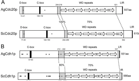
gossypiigenome (Fig. 1 and Table 4). We identified 14 possible
APC/C subunits inA. gossypiiwith amino acid homology
rang-ing from 22% to 53% identity compared toS. cerevisiae, with 8
of the subunits with less than 38% identity. The A. gossypii
APC/C and coactivator genes are present in syntenic positions in the genome compared to their positions in the yeast ge-nome, supporting that these are functional homologues of the APC/C despite the low identity to yeast proteins. The targeting factors AgCdh1p and AgCdc20p were somewhat more similar to the yeast homologues, with identity levels of 66% and 59%,
TABLE 4. Comparison of APC/C core factors and coactivator subunits fromS. cerevisiaeandA. gossypiia
Core factor or subunit
% Identity
Protein length (aa)
A. gossypii S. cerevisiae
Apc1 35 1,670 1,748
Apc2 33 710 853
Apc4 22 716 652
Apc5 24 667 685
Apc9 32 257 265
Apc11 53 148 165
Cdc16 53 708 840
Cdc23 49 614 626
Cdc26 25 119 124
Cdc27 48 657 758
Doc1 45 250 283
Mnd2 25 263 368
Swm1 22 146 170
Cdh1 66 522 566
Cdc20 59 558 610
Ama1 47 573 562
aData from http://www.yeastgenome.org and reference 35. aa, amino acids.
FIG. 1. Comparison of homologous domains and sequence features in APC/C cofactors fromA. gossypiiandS. cerevisiae. (A) AgCdc20p (AFL014C) and ScCdc20p (YGL116W) are 59% identical on the amino acid level. The positions of a D box, a C box, and seven WD repeats are shown for each protein, and notably, a stretch of 15 identical amino acids (aa) precedes the WD repeats. Asterisks mark potential CDK phosphorylation sites (S20, S136, and S493 in AgCdc20p and S24, S173, S439, and S534 in ScCdc20p). These sites are conserved in eight yeast species andA. gossypiiexcept S439 (in the fifth WD repeat). In all nine Cdc20p orthologues, the last amino acid is arginine preceded by two hydrophobic amino acids. (B) AgCdh1p (AFL007C) and ScCdh1p (YGL003C) are 66% identical, with the highest homology in the WD repeat region and the preceding 21 amino acids. All nine analyzed Cdh1 orthologues terminate with an arginine preceded by a hydrophobic amino acid. The eight asterisks in AgCdh1p mark CDK phosphoryation consensus sites (T12, S16, S44, T144, T161, S212, S224, and S421), all of which are conserved in eight yeast orthologues. In ScCdh1p, the homologous sites are phosphorylated with the possible exceptions of S42 and S227 (16a, 57). The three additional consensus CDK sites in ScCdh1p (S169, T173, and S418) are most likely not phosphorylated. From the 11 non-CDK phosphorylation sites identified in ScCdh1p by those authors, only 5 (S38, S172, S193, S225, and S556) are conserved inA. gossypiiand other yeast Cdh1 sequences. LIR, LR, and IR refer to amino acids.
186 GLADFELTER ET AL. EUKARYOT. CELL
on September 8, 2020 by guest
http://ec.asm.org/
respectively, and the genes were also in syntenic positions. In
most cases, the predictedA. gossypiiAPC/C protein was 5 to
15% shorter in length than theS. cerevisiaeprotein.
Both AgCdc23p and AgDoc1p, the two core subunits we have analyzed below, have conserved sequence features sup-porting their function in the APC/C. In AgCdc23p, nine out of nine tetratricopeptide repeats, which are important for associ-ations between APC/C core subunits, are conserved compared to those in the budding yeast homologue (29). AgDoc1p clearly has a “DOC domain” conserved which is found in various proteins associated with ubiquitin protein ligase activity (16). The conservation of domains in the targeting subunits AgCdc20p and AgCdh1p is summarized in Fig. 1. These pro-teins contain seven WD repeat motifs which mediate protein-protein interactions and thus interact with substrates and spe-cifically D boxes (6, 28). Homology within these repeats is high
between the A. gossypii and S. cerevisiae homologues (73 to
83% identity for Cdh1p and 66 to 90% identity for Cdc20p) and the orthologues of seven other yeast species. The 15 amino acids preceding the WD repeat region and the last three amino acids of all orthologues are even more highly conserved. Ad-ditionally, a D box is present in AgCdc20p and three CDK phosphorylation consensus sites (Fig. 1A) which are conserved in all known yeast orthologues. All ScCdh1p regulatory CDK phosphorylation sites are conserved in AgCdh1p and in other yeast orthologues (see legend to Fig. 1B). Furthermore, both AgCdh1p and AgCdc20p have C boxes (conserved in Cdc20 family members), which are regions thought to link these tar-geting factors to the APC/C (39, 43). When combined, it
ap-pears as ifA. gossypiicells have the basic subunits of the APC/C
to direct regulated protein degradation. However, given the low identity between most of the factors and the differences between uninucleated cell division and multinucleated, asyn-chronous nuclear division, it is likely that some aspects of APC/C function or regulation have diverged since budding
yeast andA. gossypiishared a common ancestor.
Cells lacking APC/C core subunits and targeting subunits
arrest in late stages of mitosis.Mutants were generated with
deletions in components of the APC/C and evaluated for de-fects in cell cycle progression. All mutants created by gene
targeting in A. gossypii are initially made as heterokaryons
because only a subset of nuclei in the multinucleated hyphae is transformed. The heterokaryons produce uninucleated spores carrying either the wild-type or the mutated allele. Spores can be germinated under selection to produce homokaryons in which all nuclei contain the mutation; however, these spores may also contain small amounts of wild-type protein packaged from the heterokaryon. This pool of “maternal” protein may sustain some growth in the presence of an ultimately deleteri-ous deletion. If the APC/C plays a role in degrading factors
required for promoting anaphase and mitotic exit inA. gossypii,
an arrest or delay in mitosis would be expected in mutant cells lacking the APC/C components.
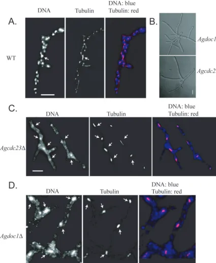
Deletion of either AgCDC23 or AgDOC1 led to a “late
lethal” terminal phenotype in which cells reached the size of small mycelia with 4 to 7 nuclei (which in wild-type cells would have 10 to 30 nuclei) but could not develop further (Fig. 2 and Table 5). Nuclear density was low compared to that of the wild type, and nuclei in these mutant strains were often fragmented
and had elongated mitotic spindles (⬎80% of nuclei compared
to 13% in the wild type;n⫽150 nuclei). We determined the
nuclear division stage of nuclei in these cells by measuring the length of mitotic spindles and comparing them to wild-type spindle lengths in normally proliferating cells. A spindle length
of more than 2.8m was considered anaphase, based on
pre-vious work looking at the nuclear dynamics and kinetics of anaphase by time-lapse microscopy (2). In wild-type cells, 42% of nuclei with a mitotic spindle were in anaphase and the
anaphase spindle was 4.8⫾0.41m long (mean⫾ standard
deviation), with lengths ranging from 2.8 to 9.5m (46
ana-phase spindles, 110 mitotic spindles). In contrast, the anaana-phase
spindle length for Agcdc23mutants at the terminal growth
stage was 3.9⫾0.26m (n⫽112) and for Agdoc1mutants
3.8⫾0.36m (n⫽58). However, the overall proportions of
nuclei in anaphase were similar in both mutants compared
to that of the wild type (243 total spindles for Agcdc23and
134 for Agdoc1). The shortened average anaphase spindle
length suggests that the nuclei in these mutants can enter anaphase, but spindles fail to completely elongate and nu-clei are challenged to exit mitosis (Table 5). Based on these
phenotypes, we predict that theA. gossypii APC/C does in
fact regulate nuclear division and that there must be some factor(s) that has to be degraded by the APC/C for comple-tion of mitosis in these multinucleated cells.
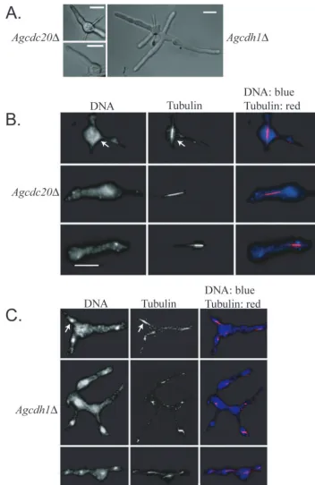
AgCDH1 and AgCDC20were then deleted, and null
mu-tants were characterized to determine whether they may func-tion to target substrates to the APC/C. Both of these APC targeting factors were also essential, and mutants stopped growing as young germlings, with clear defects in nuclear
di-vision (Fig. 3 and Table 5). Agcdc20cells arrest with only one
or two germ tubes containing fragmented nuclei and a single elongated spindle (90% of nuclei with a spindle compared to
13% of those in the wild type;n⫽150) and with an anaphase
spindle length of 3.8⫾0.43m (41 spindles) (Table 5). Only
35% of mitotic nuclei were in anaphase compared to 42% of those in the wild type, suggesting a possible, slight delay in entering anaphase (117 total mitotic nuclei). Furthermore, the
maximum spindle length measured was only 5.9m compared
to 9.5m in the wild type. The length and appearance of the
spindles suggest that the nuclei are challenged in progressing through anaphase and exiting mitosis in the absence of AgCdc20p (Fig. 3A and B and Table 5).
Agcdh1mutants grow somewhat larger than Agcdc20cells
and form small germlings with several branches (Fig. 3A). Like
those in Agcdc20mutants, however, nuclei in Agcdh1cells are
fragmented and blocked predominantly in mitosis (Fig. 3C and Table 5). Compared to wild-type cells, where only 13% of
nuclei have mitotic spindles, 58% of Agcdh1mutant spindles
appeared mitotic (⬎150 nuclei) and of these only 33% were in
anaphase compared to 42% in the wild type. Agcdh1⌬ cells
had an anaphase spindle length of 3.6⫾0.40m and a
max-imum length of 5.0 m (based on 47 spindles) (Table 5),
suggesting that, as with the Agcdc20mutants, there may be a
problem entering and progressing through anaphase in cells
lacking AgCdh1p. Most germlings of Agcdh1had one to four
spindles, but cells could be found with up to seven spindles, suggesting that at least two to three rounds of nuclear division can occur prior to the lethal arrest; however, it is possible that these rounds of division are promoted by “maternal” stores of
on September 8, 2020 by guest
http://ec.asm.org/
AgCdh1p from the germinating spore. It is also conceivable
that Agcdh1 mutants have elevated levels of AgCdc20p that
can partially compensate, since in other systems Cdh1p homo-logues actually target Cdc20p for degradation (21). Thus, AgCdh1p, AgCdc20p, and the predicted APC/C components AgCdc23p and AgDoc1p contribute to normal nuclear division
inA. gossypiicells and, based on the length of spindles at arrest, promote progression through anaphase. Given that the levels of the mitotic cyclin AgClb1/2p appear quite stable across the nuclear division cycle, these data suggest that factors other than this cyclin must be targeted for degradation by the APC/C to progress through mitosis.
FIG. 2. Cells lacking AgCDC23and AgDOC1are inviable and arrest in mitosis. (A) Spores from wild-type (WT)A. gossypiicells were grown for 15 h at 30°C and processed for antitubulin immunofluorescence and DNA staining. (B) Brightfield images depicting “late lethal” arrest point as small mycelia from Agdoc1⌬and Agcdc23⌬mutants. (C and D) Spores from Agcdc23⌬heterokaryons (RNSN01) and Agdoc1⌬heterokaryons (RNSN02), respectively, were grown under selection for 15 h at 30°C and processed for antitubulin immunofluorescence and DNA staining. Arrows point to examples of mitotic nuclei. Bar, 10m.
188 GLADFELTER ET AL. EUKARYOT. CELL
on September 8, 2020 by guest
http://ec.asm.org/
AgPds1p, a securin homologue, is involved in nuclear
pro-gression inA. gossypii. In addition to the mitotic cyclin, the
APC/C is essential inS. cerevisiaefor the degradation of one
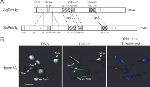
other factor, the securin protein Pds1p. ScPds1p degradation, which is mediated primarily by ScCdc20p, triggers timely ana-phase entry once all of the chromosomes are properly attached and aligned on the spindle, and this function appears to be conserved throughout eukaryotes. Securins limit the activity of separases, which dissolve sister chromatid cohesion for ana-phase entry, and degradation of securins at the metaana-phase/ anaphase transition in turn liberates separase (33). The genes for AgPds1p and ScPds1p are in a syntenic position, but these homologues are only 29% identical and AgPds1p is almost 80 amino acids shorter in length, due mainly to two missing stretches of amino acids flanking the potential D box (Fig. 4A). It does, however, have a predicted D box and KEN box,
sug-gesting that it could be degraded by the APC/C inA. gossypii
(Fig. 4A). Despite the low sequence identity, does AgPds1p function as a mediator of anaphase entry in these multinucle-ated cells?
Deletion of AgPDS1 demonstrated that it is an essential
gene and that it is required for normal nuclear cycle
progres-sion inA. gossypii. Agpds1deletion mutants arrested growth as
germlings with generally a single elongated spindle (79% of
mitotic nuclei;n⬎150) (Fig. 4B and Table 5). This is a more
severe phenotype than that of Scpds1null mutant cells, which
are viable but temperature sensitive and at elevated tempera-tures attempt to enter additional rounds of the cell cycle with-out chromosome segregation (53, 54). Nuclei were frequently
fragmented in Agpds1mutants (often only one spindle end
showed DNA staining), and over 60% of spindles appeared to be in anaphase compared to only 42% in the wild type. The
anaphase spindle length was 4.2⫾0.37m, and the maximal
length observed was 9.5m (based on 56 spindles) (Fig. 4B
and Table 5). The proportion of anaphase nuclei combined
with the spindle length suggests that Agpds1deletion mutants
may prematurely enter anaphase, potentially before proper chromosome alignment, and then progress through to late
anaphase/telophase but are challenged in exiting mitosis and in initiating the next round of division. Thus, the AgPds1p ho-mologue is clearly involved in nuclear division and could be an
essential substrate for theA. gossypiiAPC/C.
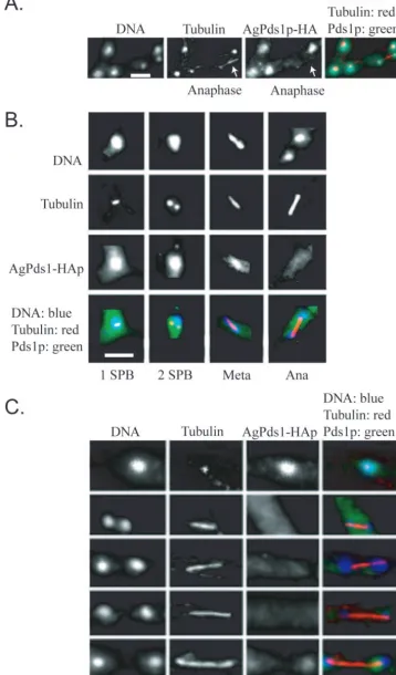
AgPds1p levels vary depending upon the stage of the nuclear
division cycle.If AgPds1p is in fact a target of the APC/C inA.
gossypii, we predicted that AgPds1p would be present in early
stages (S/G2/early M) of the cell cycle, when sister chromatid
cohesion must be maintained, but absent in late stages, as nuclei progress through anaphase and exit of mitosis. To assay when AgPds1p was present, a strain expressing an HA-tagged protein from the endogenous promoter was analyzed by im-munofluorescence. AgPds1p-6HA was present as a diffuse nu-clear signal in nuclei with a single spindle pole body (SPB), duplicated SPBs, and metaphase spindles (Fig. 5A and B) (SPBs can be detected when microtubules are visualized by immunofluorescence). It colocalized with the spindle in a frac-tion of metaphase nuclei but was also still observed in the nucleoplasm, and then levels dropped substantially in ana-phase nuclei. Some faint HA signal was present in 10% of anaphase nuclei, and the amount of AgPds1p-6HA detected increased as spindles elongated (Fig. 5C). When levels of HA-tagged protein were assayed in lysates from mycelia where
nuclei were synchronized in G2/M by nocodazole and then
released, the level of AgPds1p-6HA similarly diminished but was not eliminated completely when 50% of nuclei were in anaphase (data not shown). Thus, the majority of AgPds1p must be degraded at the metaphase-to-anaphase transition (potentially, the pool bound to the separase homologue AgEsp1p), but some limited protein can still be observed in 10% of anaphase nuclei.
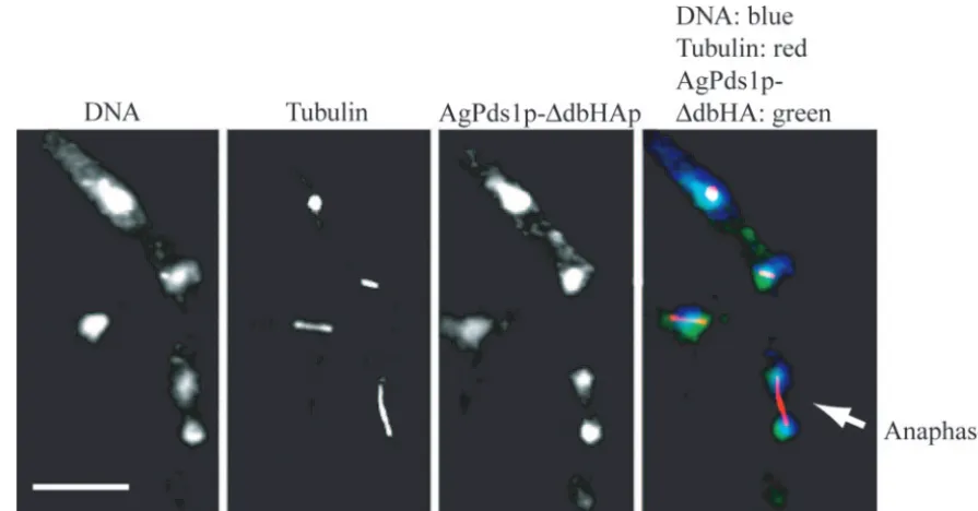
To test whether the disappearance of AgPds1p in anaphase is essential and whether it is likely mediated by the APC/C, we
constructed Agpds1mutants lacking the destruction box motif.
The plasmid-coded HA-tagged destruction box deletion allele Agpds1⌬db-6HA (strain NSG10) expresses a securin which lacks the predicted destruction box motif TRMPLASKDRN (Fig. 4). To evaluate whether this deletion of the destruction
TABLE 5. Summary of terminal phenotypes ofA. gossypiiwild-type and homokaryotic deletion mutants
Straina No. of nuclei in 80% of the myceliab
Maximum no. of nuclei in a myceliumb
% of nuclei with a mitotic
spindle
% of nuclei with one or two spindle poles
Total no. of spindles measured No. of spindles in metaphasec No. of spindles in anaphasec Anaphase spindle length (m)c
Range Mean⫾SD
WT 15–30 70 13 87 110 64 46 2.8–9.5 4.83⫾0.41
cdc23⌬mutantd 4–7 10 81 19 243 131 112 2.8–9.0 3.9⫾0.26
doc1⌬mutantd 4–7 9 83 17 134 86 58 2.8–7.7 3.8⫾0.36
cdh1⌬mutant 1–4 7 58 42 142 95 47 2.8–5.0 3.6⫾0.40
cdc20⌬mutant 1–2 3 90 10 117 66 41 2.8–5.9 3.8⫾0.43
pds1⌬mutant 1–2 3 79 21 92 36 56 2.8–9.5 4.2⫾0.37
a
Spores were inoculated in AFM (plus CloNAT for thecdc23⌬anddoc1⌬deletion mutants and plus G418 for thecdh1⌬,cdc20⌬, andpds1⌬deletion mutants). After growth for 14 h at 30°C, mycelia were washed and processed for immunofluorescence with antitubulin antibody. Fifty independent mycelia were analyzed for the wild type (WT), and more than 100 mycelia were analyzed for each mutant strain.
b
Nuclei were visualized with Hoechst dye. Many nuclei, however, appeared to be fragmented in deletion mutant cells, and in these cases, the number of nuclei per mycelium was determined based on the number of spindles and spindle poles observed by tubulin immunofluorescence.
c
Spindles with lengths between 1.5 and 2.8m are considered metaphase spindles. Spindles longer then 2.8m are considered anaphase spindles. The length measurements include the spindle pole bodies, so metaphase spindles are slightly longer than the diameter of nuclei, which is 2.23⫾0.21m (2).
d
The mycelia produced from mutants generated with the CloNAT resistance cassette had two classes of phenotypes. Seventy percent of the mycelia reached a terminal phenotype, with a high frequency of anaphase spindles, and these measurements are presented in this table. Thirty percent stopped growing, with a high percentage of G1nuclei as observed in the wild type. These mycelia very likely originated from wild-type spores in the primary heterokaryotic transformants, which carry
relatively high concentrations of the CloNAT resistance protein. This protein packaged in the spores will support limited resistance to and growth in the presence of the drug, even in the absence of the gene encoding this product. Such background was not observed in mutants selected with G418.
on September 8, 2020 by guest
http://ec.asm.org/
box stabilized AgPds1p during the nuclear cycle,
plasmid-de-rived AgPds1⌬db-6HAp was localized along with tubulin. The
mutant protein could be readily seen in nuclei of all division stages, including anaphase (Fig. 6), in contrast to the wild-type
AgPds1p, which was essentially absent in anaphase (Fig. 5B).
Additionally, in regions of mycelia where AgPds1⌬dbp was
clearly observed in anaphase nuclei, there were many aber-rantly large nuclei with mitotic spindles, suggesting that
stabi-FIG. 3. Cells lacking AgCDC20or AgCDH1are inviable and arrest in mitosis. (A) Brightfield images depicting arrest as uni- or bipolar germlings for Agcdc20⌬and small mycelia for Agcdh1⌬. (B) Spores from Agcdc20⌬heterokaryons (NSG01) were incubated under selection for 15 h at 30°C and processed for tubulin and DNA staining. After this incubation time, single mitotic spindles are still present in the arrested germlings but nuclei appear fragmented. (C) Spores from Agcdh1⌬heterokaryons (NSG02) were incubated under selection for 15 h prior to tubulin and DNA staining. The arrow points to an example of a mitotic spindle. Bar, 10m.
190 GLADFELTER ET AL. EUKARYOT. CELL
on September 8, 2020 by guest
http://ec.asm.org/
lized AgPds1p leads to a mitotic delay and/or defects. It is
likely that AgPds1p is an important target of the APC/C inA.
gossypiiand that its stability may contribute to the lethality of Agcdc20, Agcdh1, and/or APC/C deletion mutants. As out-lined in Materials and Methods, it was not possible to isolate a
homokaryotic strain in which the wild-type AgPDS1allele was
replaced by the destruction box deletion allele. Only when both alleles are simultaneously expressed can the multinucleated mycelia grow, but such coexpression inhibits sporulation. This
strongly suggests that the AgPds1⌬dbp allele is a
dominant-negative allele in terms of sporulation and that AgPds1p levels must be carefully regulated in this process.
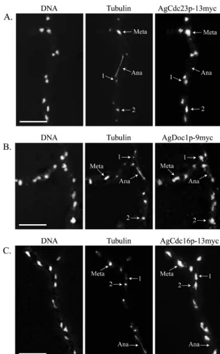
APC/C subunits and targeting factors are nuclear in all
stages of the cell cycle.AgPds1p is at least one factor that is
likely to be degraded for normal nuclear division inA. gossypii,
and this degradation seems to be limited to the metaphase-to-anaphase transition. Given that nuclei are asynchronously
di-viding in A. gossypii, this raises the question as to how the
APC/C may be activated only in the subset of nuclei at the meta-anaphase boundary and presumably inactive in neighbor-ing nuclei that are in other stages. It is conceivable that the APC/C and/or its targeting factors AgCdh1 and AgCdc20 may vary in their localization through the different periods of the nuclear cycle. The predicted core APC/C subunits AgCdc23p,
AgDoc1p, and AgCdc16p were found in all nuclei of all nu-clear division cycle stages when localized by immunofluores-cence (Fig. 7). This suggests that the APC/C is present in all nuclei and specifically activated in anaphase, leading to the prediction that the targeting factors AgCdc20p and AgCdh1p would fluctuate in their localization across the cell cycle. Re-markably, however, GFP-AgCdh1p and GFP-AgCdc20p were also found to be nuclear and present in all cell cycle stages
when expressed from the AgBNI1promoter and localized by
immunofluorescence using an anti-GFP antibody (Fig. 8). This antibody did not produce any nonspecific nuclear staining in untagged strains (see Fig. S1 in the supplemental material). We speculate then that any cell cycle-specific substrate degra-dation must be due to the regulation of APC/C activity, which either impacts its ability to interact with potential substrates or alters how factors within the APC/C itself associate, but not the presence or absence of factors in certain nuclei.
DISCUSSION
With these experiments, we have begun to evaluate the role of protein degradation during the nuclear cycle in multinucle-atedA. gossypiicells. We found that components of the APC/C and the activator proteins AgCdh1p and AgCdc20p are
re-FIG. 4. (A) Comparison between AgPds1p (AGR083W) and ScPds1p (YDR113C). The percent identity between the two proteins is 29%. N-terminal protein destruction motifs (KEN box and D box) are noted in addition to three conserved regions in the middle of Pds1p, one highly enriched in negatively charged amino acids (aa) and one enriched for proline, all of which are conserved in yeast species. A major difference between Pds1p orthologues ofSaccharomycesspecies andA. gossypiiis the absence of 39 amino acids upstream and 20 amino acids downstream of the D box. Pds1 orthologues fromK. waltiiandK. lactisalso lack blocks of amino acids of similar sizes in this region. The asterisks in ScPds1p mark the positions of five CDK consensus phosphorylation sites (T27, S71, S277, S292, and T304); only two (T27 and S292) are conserved in AgPds1p (T23 and S213) and in seven other yeast orthologues. (B) Spores from Agpds1⌬heterokaryons (NSG03) were incubated under selection for 18 h at 30°C and processed for DNA and tubulin staining. Cells arrested as germlings with one germ tube, most of which carried one nucleus, arrested in either metaphase (Meta) or anaphase (Ana) (arrows denote different stage nuclei). Bar, 10m.
on September 8, 2020 by guest
http://ec.asm.org/
FIG. 5. AgPds1p is present in nuclei but diminishes in anaphase. Spores from a strain expressing AgPds1p-HA (NSG09) were grown under selective conditions and then processed for immunofluorescence. (A) Segment of a branched hypha showing four nonmitotic nuclei and one anaphase nucleus (arrow). Only the nonmitotic nuclei contain AgPds1p-HA. (B) AgPds1p-HA localization in different nuclear cycle stages showing its presence in nuclei with one or two spindle pole bodies and in metaphase (Meta) nuclei but not in anaphase (Ana) nuclei. (C) Hyphae were released from a nocodazole block to follow AgPds1p-HA through anaphase in a large number of nuclei. Weak AgPds1p-HA signals are observed only in maximally extended spindles (bottom panels). Bar, 5m.
192
on September 8, 2020 by guest
http://ec.asm.org/
quired for growth and nuclear cycle progression. This study was initiated because of the previous observation that the
mi-totic cyclin AgClb1/2p does not appear to be degraded inA.
gossypii (14). Here, we have demonstrated that there is an essential role for APC/C-mediated degradation and that there are other important substrates of the APC/C that must be
eliminated from A. gossypii nuclei in a timely manner.
AgPds1p, a securin homologue, is one factor whose levels must be precisely controlled such that both deletion and stabiliza-tion of AgPds1p lead to strong phenotypes. Given that mitosis
is asynchronous inA. gossypii, the APC/C must be regulated in
a nuclear autonomous manner so that its activation is limited in time and space. We discuss here ways in which the APC/C in A. gossypii may have evolved to adapt to a syncytial and asynchronous division cycle.
All of the APC/C mutants that we generated showed specific defects in mitosis and impacted the progression through ana-phase and/or exit from mitosis. Our data suggest that one key substrate of the APC/C is AgPds1p. We first characterized the function of wild-type AgPds1p and then evaluated its possible regulation by the APC/C by mutating its destruction box. In budding yeast, ScPds1p has a paradoxical role of binding and inhibiting ScEsp1p, the separase that dissolves sister chromatid cohesion for anaphase, and also promoting the nuclear local-ization of ScEsp1p. Remarkably, yeast cells lacking Pds1p are viable but restricted for growth at high temperatures, presum-ably because ScEsp1p can enter the nucleus through an alter-native route to trigger dissolution of cohesion (1, 24).
One possible reason Agpds1deletion mutants fail to grow is
that AgEsp1p lacks such an alternate route, so it may never properly be localized to nuclei. In addition to impairing chro-matid separation, this could lead to a failure in the activation of AgCdh1p and AgSic1p, both of which may respond to a
signal originating from active AgEsp1p to activate the AgCdc14 early-anaphase release network (FEAR). Con-versely, AgEsp1p may be overactive due to the loss of AgPds1p, and this leads to a lack of tension across a spindle, blocking paths that promote anaphase independent of AgPds1p. Future study of the behavior of AgEsp1p should distinguish between these possibilities and lend insight into
how anaphase entry may be regulated inA. gossypii.
Just as the loss of AgPds1p is deleterious, timely degradation of this protein is also likely required for normal nuclear
divi-sion. Agpds1⌬db homokaryon mutants were not recovered,
and this allele was dominant in heterokaryon cells, suggesting that the levels of AgPds1p must be tightly regulated. Unregu-lated and stable AgPds1p could act to prevent activation of AgCdh1p by dephosphorylation (through failure to activate AgEsp1p and the FEAR network). It is not possible with the
tools currently available inA. gossypii to determine whether
AgPds1p degradation is important only for sporulation or whether it is also essential in the vegetative cycle and the likely cause of lethality of some of the APC/C mutants. Regions of
heterokaryon hyphae with high levels of AgPds1⌬dbp show an
increase in metaphase nuclei, suggesting that at least some degradation of AgPds1p is important for normal progression. If, in fact, future experiments show that AgPds1p degradation is not essential, then other essential targets of the APC/C remain to be identified and possible targets to test are the S-phase cyclins AgClb5/6p and AgClb3/4p.
We would predict based on the presence of WD motifs that AgPds1p is targeted for degradation by AgCdc20p and/or
AgCdh1p. Notably, both AgCDH1and AgCDC20are essential
genes in contrast to yeast, where ScCdh1p and ScCdc20 are partially redundant and thus nonessential. Why are these tar-geting factors unable to functionally complement each other in
FIG. 6. AgPds1⌬dbp persists in anaphase nuclei. Hyphae containing a plasmid expressing AgPds1⌬db-6HA (NSG10) were scraped from a selective plate, vortexed in liquid medium to break apart the mycelia, grown under selection for 15 h, and processed for tubulin and HA epitope staining. The arrow highlights one anaphase nucleus with nondegraded AgPds1⌬db-6HA. Bar, 10m.
on September 8, 2020 by guest
http://ec.asm.org/
A. gossypii? Lack of redundancy between proteins is often explained by differences in either the time of expression or the subcellular localization; however, both AgCdh1p and AgCdc20p, as well as the core APC/C, coexist in space and time. This lack of complementation could likely be ex-plained by significant substrate specificity differences be-tween these factors. There may be less redundancy in a
multinucleated nuclear division cycle than in a uninucleated cell division cycle because the presence of many nuclei in one cytoplasm may essentially act as a buffer, where some mistakes in division are tolerated in the system because many other nuclei are there to compensate.
Our characterization of the APC/C, its activators, and a substrate points to a role for regulated protein degradation in
FIG. 7. The predictedA. gossypiiAPC/C subunits AgCdc23p, AgDoc1p, and AgCdc16p localize to nuclei in all stages of the nuclear division cycle. (A) AgCdc23p-13myc (GVS4), (B) AgDoc1p-9myc (NSG11), and (C) AgCdc16p-13myc (GVS2) cells were grown for 16 h at 30°C under selective conditions and processed for anti-myc and antitubulin immunofluorescence. In AgDOC1p-13myc (GVS3), a signal was also observed for all nuclear cycle stages. Arrows highlight nuclei in different stages of the division cycle, where “1” indicates a nucleus with a single SPB and “2” indicates a nucleus with duplicated SPBs. Meta, metaphase; Ana, anaphase. Bar, 10m.
194 GLADFELTER ET AL. EUKARYOT. CELL
on September 8, 2020 by guest
http://ec.asm.org/
mitotic progression inA. gossypii, despite the lack of oscillation in the pool of mitotic cyclins in this system. We proposed previously that AgClb1/2p degradation was replaced with di-rect inhibition by AgSic1p (14). We thought that because new cyclin protein is continually supplied to nuclei from the cyto-plasm, this influx may be challenging to continually eliminate by proteolysis. The data we present here, however, show that
there is still some role inA. gossypiifor regulated proteolysis of
at least some factors, including AgPds1p. InDrosophila
em-bryos, there is precedent for spatially and functionally distinct pools of the APC/C and Cdh1/Cdc20 that help to regulate when and what substrates are degraded (20). In contrast, the core factors of the APC/C examined here are uniformly
present in all nuclei inA. gossypii, but presumably, the whole
complex is not constitutively active. This raises the question of how APC/C proteolysis activity is spatially and temporally
lim-ited to only mitotic/G1 nuclei in these multinucleated and
asynchronous cells.
The ubiquitous localization of the APC/C suggests that the control of degradation is regulated at the level of the substrate rather than by the APC/C itself. This could be, such as is observed in the SCF degradation system in budding yeast, through cell cycle-regulated modification, such as phosphory-lation of the substrate. Thus, the APC/C will always be ready and potentially active but substrates themselves will dictate their timely destruction. Modifications to proteins which trig-ger their destruction would likely be nuclear limited given the
asynchronous pattern of division. Modification events could be regulated by pathways such as the spindle assembly checkpoint and thus be generated as a result of chromosome attachment or tension on the spindle and may therefore remain limited to within nuclei. If this type of a nuclear intrinsic event leads to the marking of a protein for recognition by the APC/C, then
this may ensure that a neighboring nucleus in S or G2does not
prematurely degrade a given substrate. Evaluation of the func-tion and localizafunc-tion of proteins in the spindle assembly check-point, such as Mad2 and those proteins thought to communi-cate tension status in spindles such as Ipl/Aurora kinase and kinetochore proteins, would be a first approach to testing whether these components play a normal role in marking the timely degradation of proteins for anaphase progression.
Fu-ture study of the APC/C inA. gossypiishould yield insights into
how protein degradation can be limited in time and space in large, multinucleated hyphal cells.
ACKNOWLEDGMENTS
We thank A. Kaufmann, D. Hoepfner, and M. Koehli for plasmids, Rachel Shakked for her assistance in constructing two deletion mu-tants, and Philipp Knechtle for advice and helpful discussions.
This work was supported by a grant from the Swiss National Science Foundation (3100A0-100734) to P.P. and A.S.G., by the Roche Foun-dation, and by a National Science Foundation postdoctoral fellowship to A.S.G.
FIG. 8. APC activators AgCdh1p and AgCdc20p localize to all nuclei independent of the nuclear cycle stage. (A) Immunostained hyphae expressing N-terminally GFP-labeled AgCdh1p (NSG12). (B) Immunostained hyphae expressing N-terminally GFP-labeled AgCdc20p (NSG13). All nuclei, including anaphase nuclei (arrows), show anti-GFP immunofluorescence. The nuclei of nontransformed hyphae were not stained (see
Fig. S1 in the supplemental material). Bar, 10m.