Volume No.02, Issue No. 11, November 2014 ISSN (online): 2348 – 7550
SUBCUTANEOUS VEIN DETECTION USING
EMBEDDED LINUX ON ARM
Vidya Priyadarshini .P
1,Prof. Leelavathi .G
2, Dr. Siva S. Yellampalli
31,2,3
Department of VLSI Design and Embedded Systems, VTU Extension Centre,
UTL Technologies, Bangalore, (India)
ABSTRACT
The uses of technology in the field of medicine have benefitted the lives of people in many ways. These advanced technologies allow specific parts of the body to be studied in great detail which in turn have helped the doctors to accurately diagnose and treat several problems. Locating the veins for the purpose of intravenous drug delivery is sometimes a problem in case of infants, obese and dark toned people. Though many devices have come up in the markets which resolve the above problem by providing high definition images, the main problem lies in the cost. A low cost vein detection system that uses a VGA camera for capturing the images is proposed in this paper. The captured images are interfaced to the ARM 9 Single Board Computer (SBC). These images are lower in contrast so, histogram equalization technique is used to enhance the contrast of the images and finally the enhanced images is displayed on the LCD of the SBC. The programs for capturing images, enhancing the contrast are written in Open CV. These programs are cross compiled so that they are compatible with the ARM architecture. The reduction in the cost is achieved by using open source software's such as Open CV and Qt frameworks.
Keywords
—
Intravenous drug delivery, high definition images, VGA camera, ARM 9, Single board computer, histogram equalization, LCD, Open CV, Qt frameworksI.
INTRODUCTION
Vein puncture, the process of obtaining intravenous (IV) access is an everyday invasive procedure in medical
settings. At times even trained nurses and doctors find it difficult to exactly locate the blood veins on the first
hit. Especially in infants and children, recurrent attempts to insert a needle to gain access to vein causes anxiety,
pain and distress and elevate the risk of damaging the vein causing infiltration of the surrounding area and
subsequent possibility of a catheter-related, hospital-acquired bloodstream infection. It is also necessary to know
the exact position of the veins during blood transfusions.
Presently there are many devices like AccuVein AV300, AV400, VascuLuminator, Vein Viewer that cater to
the need of vein detection. These devices provide high definition images but at the same time are very
expensive. Therefore, a low-cost, portable, compact and efficient vein detection system is the need of the hour.
II.
CONCEPT
OF
VEIN
IMAGING
There are many technologies currently being used for obtaining images like x-rays, infrared, ultra scan , ct-scan,
Doppler study and MRI. X-rays and infrared techniques are cost effective compared to other techniques
mentioned above. X-rays follow a invasive technique i.e. the region is exposed to X-rays and the rays penetrate
Volume No.02, Issue No. 11, November 2014 ISSN (online): 2348 – 7550
Infrared technique requires no exposure to harmful rays, is contactless, non-invasive and safer compared to X -rays. There are two types of infrared techniques : Near infrared (NIR) and Far infrared (FIR) . Far-Infrared
method has difficulties in capturing the vein images in the palm and wrist. It is more suitable for capturing the
large veins at the back of the hand . Far - Infrared is sensitive to ambient conditions and does not provide stable
image quality. Near-Infrared imaging produces good quality images even in the back of the hand, wrist and palm.
It is more tolerant to changes in environmental and body conditions but has problems of disruption due to skin
features such as hairs and line patterns. In order to understand the NIR technique better, it is necessary to first
understand the manner in which light propagates through the biological tissues.
2.1 Light Absorption
The range of frequencies covered by the radiations in infrared region of the electromagnetic spectrum(~ 300THz
-300GHz) is comparable to the natural frequencies at which atoms or molecules will vibrate in the absence of an
applied field. Thus when infrared radiation is incident on a system of matter , resonance will occur around the
natural frequencies where energy is transferred from the incident field to the system and its amplitude of
vibration is greatly increased. Although the life time of the excited state is around 10-7 to 10-8 seconds , the
atoms or molecules will usually lose their energy by colliding with one another within 10-12 seconds, thereby
raising the kinetic energy of the other particles involved in the collisions. Hence the energy associated with the
incident field is most often dissipated as heat within the medium. This process is known as absorption. The
overall effect of absorption is reduction in intensity of the light beam traversing the medium. The important
substances that are generally considered to dominate the absorption of light within the visible region are
Haemoglobin , Melanin and water. Haemoglobin is a dominant absorber of light in the dermis. Haemoglobin
consists of the protein globin bound to form four haem groups. Each haem group contains an iron atom at the
centre of a ring like structure. An iron atom in the ferrous (Fe2+) form will bind physically to an oxygen
molecule to become oxygenated. The haemoglobin in the oxygenated state is known as oxyhaemoglobin (HbO2)
and the haemoglobin with no oxygen molecules attached i.e in the deoxygenated state is known as
deoxyhaemoglobin (Hb). In the arteries 90 -95 % of the haemoglobin is oxygenated and in the veins more than
Volume No.02, Issue No. 11, November 2014 ISSN (online): 2348 – 7550
Water is the most abundant chemical substance in the human body accounting for 60 - 80 % of total body mass.Because of its high concentration in most biological tissue, water is considered to be one of the most important
chromophores in the tissue spectroscopy measurements.
The absorption spectrum of water reveals a relatively low absorption region between 200 and 900 nm. Above
900nm the absorption coefficient increases rapidly. The region of low absorption acts as a 'window ' of
transparency or optical window in the tissues. Very little light can penetrate into the tissues outside this region.
The difference in the absorption coefficients lies in the oxygenation of the blood vessels . The veins carry the
deoxygenated blood due to which they absorb the radiation completely and the arteries which carry the oxidized
blood become almost transparent.
2.2 Light Scattering
Scattering describes a change in the direction, polarization or phase of light and is commonly portrayed as either
a surface effect such as reflection or refraction or as an interaction with a small region whose optical properties
differ from its surroundings. It is estimated that around 4% to 7% of visible light is reflected from the surface of
the skin , independent of wavelength and skin color. The remaining light is refracted as it passes from light into
the skin. The interaction of light with human tissue is shown in Figure 2.
III. PROPOSED FLOW OF THE WHOLE SCHEME
The image is first captured by an USB camera followed by the visibility check of the veins. The check is made
by mere observation. If the vein is visible, then the image is displayed on the LCD screen of the SBC without
undergoing any enhancement procedures. If the vein is not visible, then it is converted to gray scale. The
conversion to gray scale is done because it is an important requisite in situations where contrast enhancement
techniques are employed. The images are subjected to histogram equalization for contrast enhancement. The
Volume No.02, Issue No. 11, November 2014 ISSN (online): 2348 – 7550
IV. HARDWARE IMPLEMENTATION
The hardware required for the implementation of the vein detection system are:
ARM 9 Single Board Computer (SBC) which is capable of supporting huge libraries and Array LCD.
Microsoft VX -1000 USB camera.
LED shoot light to focus the light on region to be detected.
Host Computer with 80GB Hard disk and 1GB RAM.
4.1 Camera
The camera plays a very crucial role in the subcutaneous vein detection system. The selection criteria of the
camera are based on the following aspects:
The camera should not be bulky.
The camera should be economical.
It should have a USB interface.
The camera's resolution should not be less than 640x480.
Volume No.02, Issue No. 11, November 2014 ISSN (online): 2348 – 7550
Based upon these factors, a USB camera, Microsoft's VX-1000 has been chosen for capturing vein images.The set up consists of the host machine, Mini 2440 Single Board Computer (target), lighting system and USB
camera. The enhanced image is displayed on the SBC.
V. SOFTWARE IMPLEMENTATION
To manage the overall functioning of the vein detecting system , starting from powering on the board , enabling
the camera, capturing the image, performing histogram equalization on the images and displaying the images on
the monitor, an extensive software support is required to execute and manage all these operations
simultaneously.
Volume No.02, Issue No. 11, November 2014 ISSN (online): 2348 – 7550
A toolchain is a collection of utilities that help in converting High level language like C, C++ , ADA tomachine level code that can be executed by the microprocessor. The GNU toolchain plays an important role in
the development of Linux and software for embedded systems.
The development environment on the host machine must be first setup before starting the development for the
SBC. Installing Ubuntu LTS as the development platform is the first step in setting up the host.
The kernel acts like an abstraction between the real hardware and the user space applications. The linux kernel
version 2.6. is used for the development and the kernel is modified accordingly.
Cross compilation is the act of compiling code for one computer system (often known as the target) on a different system, called the host. It's a very useful technique specially when the target system is too small to host
the compiler and all relevant files.
Before building the root filesystem for mini board, there are set of image processing libraries (OpenCV) and
GUI libraries(Qt) which are to be cross-compiled and put into the mini FS directory structure.
The root file system contains all the required libraries and the user-applications arranged in a hierarchical
manner. The root file system can also either be located on the ROM/Flash memory mostly as a YAFFS2, JFFS2
or a CRAMFS type file system.
5.2 Histogram Equalization
A histogram is a graphical representation of the distribution of data. Histograms provides a global description
information about the appearance of the image and its properties. The histogram of a digital image with gray
values r0 , r1 , r2 ...r L-1 is given by a discrete function :
p r ( r k ) = n k / N , 0 ≤ r k ≤ 1 and k = 0 , 1 , 2 ....L - 1....(1)
where r k - normalized intensity value.
L - no. of gray levels in the image.
n k - no. of pixels with gray level r k.
N - total number of pixels.
Volume No.02, Issue No. 11, November 2014 ISSN (online): 2348 – 7550
Histogram equalization is a powerful point processing enhancement technique that seeks to optimize thecontrast of an image at all points. Histogram equalization seeks to improve the image contrast by flattening or
equalizing, the histogram of an image.
VI. RESULTS
Initially, the images that were captured from the USB camera were subjected to histogram equalization. The
images before and after subjecting to equalization and their corresponding histograms are shown below:
It can be clearly observed that image in figure 8 is darker compared to the image in figure 9. This infers that the
contrast of the image is enhanced after subjecting to equalization procedure. The narrow peak in the histogram
which is indicated by the circle shows darker portions of the image. In the darker regions, the pixels are clustered
together or unevenly distributed. It can also be observed that the narrow peaks that were visible in the histogram
of figure 8 is not seen in the histogram of figure 9. In other words, the pixels of the image are uniformly spread
across the entire image. The final output is obtained on the LCD display of the SBC. Images of the fist and wrist
are captured from dark toned , medium toned and obese persons.The circle in the figures indicates the vein .
Volume No.02, Issue No. 11, November 2014 ISSN (online): 2348 – 7550
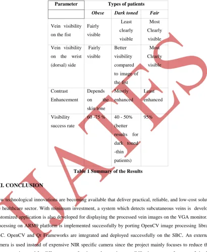
The testing of the system carried out on 4 patients for each of the three cases : obese , dark toned and fair
complexioned.
Obese : 3 out of 4 persons veins were visible clearly, which gives a success rate of 75%. However the
Volume No.02, Issue No. 11, November 2014 ISSN (online): 2348 – 7550
Dark toned : 2 out of 4 persons veins were visible which gives 50% success rate. The percentage might reducefurther if the patient is obese apart from being dark toned.
Fair complexioned : The veins of all the four test subjects were visible clearly. Only slight variation can be
seen in extreme cases where the patient is highly obese.The results are summarized in table 1 shown below :
Parameter Types of patients
Obese Dark toned Fair
Vein visibility
on the fist
Fairly visible Least clearly visible Most Clearly visible
Vein visibility
on the wrist
(dorsal) side Fairly visible Better visibility compared
to image of
the fist Most Clearly visible Contrast Enhancement Depends
on the
skin tone Mostly enhanced Least enhanced Visibility success rate
60 -75 % 40 - 50%
(better
results for
dark toned
-thin
patients)
95%
Table 1 Summary of the Results
VII. CONCLUSION
New technological innovations are becoming available that deliver practical, reliable, and low-cost solutions to
the healthcare sector. With minimum investment, a system which detects subcutaneous veins is developed. A
customized application is also developed for displaying the processed vein images on the VGA monitor. Image
processing on ARM9 platform is implemented successfully by porting OpenCV image processing libraries on
SBC. OpenCV and Qt Frameworks are integrated and deployed successfully on the SBC. An external USB
camera is used instead of expensive NIR specific camera since the project mainly focuses to reduce the cost.
The system is tested for different types of patients whose vein accessibility is a reason of concern. It is observed that the veins are very clearly visible for fair complexioned (both lean and obese) patients. The vein visibility of
a lean, dark toned person is better than that of a dark toned obese person. The clarity can be improved by
employing NIR specific camera in which case there is a trade - off between clarity and cost. The use of Open
Source tools have proven to be effective and reliable for development. The extensive use of open source
software packages and tools have helped in making the system low cost to a large extent. The number of cases
Volume No.02, Issue No. 11, November 2014 ISSN (online): 2348 – 7550
REFERENCES
[1] Tom Lister, Philip A. Wright, Paul H. Chappell , “Optical properties of human skin,” J. Biomed. Opt, vol. 17(9), pp. 1 - 12, September 2012.
[2] A. N. Bashkatov, E. A. Genina, V. I. Kochubey, and V. V. Tuchin, “Optical properties of human skin,
subcutaneous and mucous tissues in the wavelength range from 400 to 2000 nm,” J. Physics D: Applied Physics, vol. 38, no. 15, pp. 2543 – 2555, 2005
[3] S. Crisan, I. G. Tarnovan, and T. E. Crişan, “A low cost vein detection system using near infrared
radiation,” in Proc. IEEE Sensors Applications Symposium (SAS '07), pp. 1 - 6 , February 2007
[4] Wang Lingyu, Graham Leedham, "Near- and far- infrared imaging for vein pattern biometrics," Proc. IEEE International Conference on Advanced Video and Signal Based Surveillance, vol. 0, p. 52, 2006
[5] Yeganeh . H , Ziaei . A , Rezaie .A , " A novel approach for contrast enhancement based on Histogram Equalization," 2008 International Conference on Computer and Communication Engineering (ICCCE), pp. 256 - 260, 2008
[6] Mini 2440 friendly arm [Online]. Available : " http://www.friendlyarm.net/ ".
[7] Karim Yaghmour , Building Embedded Linux Systems, 1st ed. O’Reilly & Associates Inc., 2003.
[8] Gary Bradski , Adrian Kaehler, Learning OpenCV, 1st ed. O’Reilly Media, Inc., 2008.