R E S E A R C H
Open Access
Cardiopulmonary exercise testing early after stroke
using feedback-controlled robotics-assisted treadmill
exercise: test-retest reliability and repeatability
Oliver Stoller
1,2,3*, Eling D de Bruin
2,4,5, Matthias Schindelholz
1,3, Corina Schuster-Amft
1,3, Rob A de Bie
2,5and Kenneth J Hunt
1,3Abstract
Background:Exercise capacity is seriously reduced after stroke. While cardiopulmonary assessment and intervention strategies have been validated for the mildly and moderately impaired populations post-stroke, there is a lack of effective concepts for stroke survivors suffering from severe motor limitations. This study investigated the test-retest reliability and repeatability of cardiopulmonary exercise testing (CPET) using feedback-controlled robotics-assisted treadmill exercise (FC-RATE) in severely motor impaired individuals early after stroke.
Methods:20 subjects (age 44–84 years, <6 month post-stroke) with severe motor limitations (Functional Ambulatory Classification 0–2) were selected for consecutive constant load testing (CLT) and incremental exercise testing (IET) within a powered exoskeleton, synchronised with a treadmill and a body weight support system. A manual human-in-the-loop feedback system was used to guide individual work rate levels. Outcome variables focussed on standard cardiopulmonary performance parameters. Relative and absolute test-retest reliability were assessed by intraclass correlation coefficients (ICC), standard error of the measurement (SEM), and minimal detectable change (MDC). Mean difference, limits of agreement, and coefficient of variation (CoV) were estimated to assess repeatability. Results:Peak performance parameters during IET yielded good to excellent relative reliability: absolute peak oxygen uptake (ICC =0.82), relative peak oxygen uptake (ICC =0.72), peak work rate (ICC =0.91), peak heart rate (ICC =0.80), absolute gas exchange threshold (ICC =0.91), relative gas exchange threshold (ICC =0.88), oxygen cost of work (ICC =0.87), oxygen pulse at peak oxygen uptake (ICC =0.92), ventilation rate versus carbon dioxide output slope (ICC =0.78). For these variables, SEM was 4-13%, MDC 12-36%, and CoV 0.10-0.36. CLT revealed high mean differences and insufficient test-retest reliability for all variables studied.
Conclusions:This study presents first evidence on reliability and repeatability for CPET in severely motor impaired individuals early after stroke using a feedback-controlled robotics-assisted treadmill. The results demonstrate good to excellent test-retest reliability and appropriate repeatability for the most important peak cardiopulmonary performance parameters. These findings have important implications for the design and implementation of cardiovascular exercise interventions in severely impaired populations. Future research needs to develop advanced control strategies to enable the true limit of functional exercise capacity to be reached and to further assess test-retest reliability and repeatability in larger samples.
Keywords:Stroke rehabilitation, Subacute, Severe motor impairment, Robotics-assisted gait training, Aerobic capacity, Treadmill exercise
* Correspondence:[email protected]
1Department of Engineering and Information Technology, Institute for
Rehabilitation and Performance Technology, Bern University of Applied Sciences, Burgdorf, Switzerland
2
Department of Epidemiology, Maastricht University and Caphri Research School, Maastricht, The Netherlands
Full list of author information is available at the end of the article
Background
Exercise capacity and activity status have become well-established predictors of cardiovascular and overall mortal-ity, both of which are seriously reduced after stroke [1,2]. It has been shown that peak oxygen uptake (VO2peak) is ap-proximately 50% lower compared to normative values of healthy adults 30 days post-stroke [3,4]. Despite extensive inpatient rehabilitation procedures and spontaneous recov-ery of cardiovascular fitness, the exercise capacity of stroke survivors entering the chronic phase remains below rec-ommended levels [5]. The rapid deterioration of fitness not only predisposes to secondary medical complications, but also restricts the degree to which individuals can partici-pate in rehabilitation routines and limits the ability of the individual to perform functional activities independently [6]. Therefore, research into cardiovascular exercise train-ing in the early stages after stroke has been highlighted as a priority [7,8]. Effective assessment and intervention strat-egies are needed to assess, monitor, and improve cardio-vascular fitness early after stroke.
Current research has investigated several modalities for cardiopulmonary exercise testing (CPET) in subacute stroke (6 days-6 months post-stroke) [3,4,9-13] and in chronic stroke (>6 months post-stroke) [14-19] using treadmill exercise [14-16], body weight supported tread-mill exercise [3], leg cycle ergometry [4,9-11,15,17,18], and combined upper- and lower-limb ergometry [12,19]. The most common concepts, i.e. treadmill exercise and leg cycle ergometry, are primarily designed for individ-uals with mild to moderate motor impairment, because limited motor control (non-ambulatory status, limited trunk control), poor postural control, and poor coordin-ation of the affected limbs may restrict severely impaired individuals from performing on these devices. As a result, most studies focussing on exercise capacity after stroke have excluded individuals not able to walk independently and those presenting with low levels of motor function.
A potential option to overcome severe motor restric-tions is the introduction of combined upper- and lower-limb ergometry [12,19,20]. Current study results demon-strated feasibility and validity, and emphasised the fact that an all-extremity exercise protocol might decrease early onset of lower limb fatigue which leads to better estimates of exercise capacity due to the incorporation of more muscle mass. However, this approach does not embody the concept of repetitive task-specific exercise during the early stages of stroke recovery and might be not appropriate for implementation into early rehabilita-tion phases [21,22]. Considering the relatively short intervention window during subacute stroke rehabilita-tion and the current recommendarehabilita-tions for cardiovascu-lar exercise training after stroke [8], novel approaches should incorporate task-specific activities such as walking or stair climbing. The combination of motor function
training and cardiovascular exercise might have the poten-tial to positively influence overall therapy outcomes and to prevent or mitigate the loss of exercise capacity in the early stages after stroke onset [23].
A promising approach to overcome motor limitations while facilitating task-specific activity and cardiovascular stress is body weight supported treadmill training. Initial research has shown that gait symmetry improved with increasing body weight support (BWS) [24]. However, during walking with BWS of more than 15%, vertical ground reaction forces and functional activity of anti-gravity muscles decreased, which led to substantially lower oxygen uptake levels during body weight supported tread-mill training compared to conventional treadtread-mill exercise [25,26]. Because severely impaired stroke survivors need considerable physical support during walking with low body weight support, the application of robotics-assisted treadmill exercise (RATE) might be of relevance in this context. A powered exoskeleton for the lower extremities, synchronised with a treadmill and BWS, provides active support during the gait trajectory that enables progressive body weight loading for individuals with severe motor restrictions.
Recent research on exercise intensity during RATE has shown substantial increases in cardiopulmonary perform-ance parameters after stroke [27,28], and spinal cord in-jury [29], including complete tetraplegia [30]. However, oxygen uptake levels were below that of overground walk-ing, recommended cardiovascular training intensities could not be achieved [31], and conventional control strat-egies such as the modulation of walking speed, BWS, and guidance force had only a minor influence on exercise in-tensity [27,28,31]. There is a need for voluntary effort dur-ing walkdur-ing within an exoskeleton to provoke substantial cardiovascular stress comparable to conventional tread-mill exercise [32]. Therefore, novel protocols have been developed to control and direct active participation during RATE with the specific aim of provoking cardiorespiratory responses [33-38]. This incorporates biofeedback mecha-nisms allowing the control of exercise intensity through the guidance of the individual’s voluntary effort. The ap-proach presented here provides control of exercise in-tensity during RATE by biofeedback and voluntary adaptation of the hip and knee forces by the subject. A first clinical study in non-ambulatory stroke survivors in the subacute phase revealed that feedback-controlled RATE (FC-RATE) can be used to implement CPET [39]. Results yielded acceptable cardiopulmonary per-formance parameters following standardized CPET pro-tocols. Thus, this approach might have the potential to assess exercise capacity and guide cardiovascular exer-cise in stroke survivors with severe motor limitations. This needs to be formally investigated for clinical feasi-bility, test-retest reliability and repeatability.
Stolleret al. Journal of NeuroEngineering and Rehabilitation2014,11:145 Page 2 of 13
The aims of this study were: (1) to assess the clinical feasibility of FC-RATE for CPET in severely motor im-paired individuals early after stroke, (2) to examine the ability of the concept to meet standard cardiopulmonary criteria for maximal exercise capacity, and (3) to assess the test-retest reliability and the repeatability of the approach.
Methods Participants
20 first-ever stroke inpatients were recruited at a neurological rehabilitation clinic in the north-western part of Switzerland (Reha Rheinfelden) and screened according to the selection criteria. Subjects were then presented to the responsible ward physician and a cardiologist to confirm eligibility. Inclusion criteria were: (1) clinical diagnosis of initial stroke (ischemic or haemorrhagic), (2) <20 weeks after stroke onset, (3) age >18 years, (4) Functional Ambulation Classification (FAC) of <3, (5) ability to understand the procedures and provide informed consent. Subjects were excluded if they had (1) cardiac contraindications for exercise testing according to the American College of Sports Medicine (ACSM) [40], (2) contraindications for RATE ac-cording to guidelines from the manufacturer (Hocoma AG, Volketswil, Switzerland), (3) concurrent neurological disease (e.g. Multiple Sclerosis, Parkinson’s Disease, etc.), (4) concurrent pulmonary disease (e.g. COPD, etc.), (5) his-tory of dementia.
Recorded characteristics included gender, age, body mass index, diagnosis, affected body side, time post-stroke, medications, comorbidities, FAC [41] and func-tional independence using the Extended Barthel Index [42]. All subjects were informed about risks and benefits, and gave signed informed consent. The Ethics Review Committee of the Swiss canton of Aargau approved the study (Reference No: 2012/051).
Technical implementation
The Lokomat system (Hocoma AG, Volketswil, Switzerland) was used to implement FC-RATE. The powered exo-skeleton provides control of both legs using DC mo-tors, synchronised with an integrated treadmill (h/p/ cosmos sports & medical GmbH, Traunstein, Germany) and a motor-driven BWS system with real time feed-back control for precise body weight unloading (Lokolift, Hocoma AG). The total mechanical work rate exerted on the exoskeleton by the subject was computed from the force, moment arm and velocity data at the four active joints (hips and knees). The active mechanical work rate (Pmech), applied by the subject’s effort was estimated by subtracting the passive mechanical work rate (work rate necessary to move the subject passively within the exo-skeleton) from the total mechanical work rate. A manual human-in-the-loop feedback system was implemented to
control the subject’s active work rate. Pmech was pro-jected onto a screen at the front of the treadmill together with a target mechanical work rate (P*mech). The subject was instructed to vary the forces applied on the exoskel-eton by volitional muscle activity and to keep the mea-sured and visualized active work rate as close as possible to the target (Figure 1).
Experimental protocol
At study entry, all included subjects completed a famil-iarisation session with the FC-RATE concept, which started by qualified and experienced physiotherapists adjusting the Lokomat system to provide a physiological gait pattern and to ensure that the subjects could walk comfortably. Then, an initial test of decreasing BWS continuously by 5% per minute was implemented to de-fine the minimal possible BWS level. There was strict adherence to physiological gait pattern criteria through visual observation: (1) heel strike (physiological knee ex-tension), (2) no foot dragging during the swing phase, and (3) active weight-bearing during the stance phase (physiological knee extension) [43]. After the first adjust-ments, subjects were asked to perform a short constant load exercise test for 5 min (P*mech =20 W) to explain the approach and practice with the feedback-control structure. Finally, the safety procedures for potential ad-verse events were explained in detail.
After a break of at least 24 h, subjects then completed repeated constant load testing (CLT) and incremental exercise testing (IET) on separate days, with 48–72 h be-tween the trials. All sessions were controlled for time of day. Subjects were instructed to avoid additional strenu-ous activity during participation in the study and not to consume food, alcohol, nicotine or caffeine at least 3 h prior to testing.
Subjects were asked at the beginning of the first CLT and IET to increase their maximal voluntary effort dur-ing RATE within 30 s to define the maximal work rate (Pmax) for the subsequent tests. Walking cadence was fixed at 60 steps/min and individual BWS was consistent for all sessions. An experienced examiner performed all tests. There was close adherence to established models for exercise testing according to the ACSM guidelines [40].
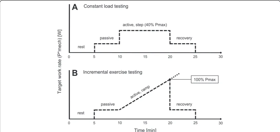
IET was based on progressive ramp exercise and sepa-rated into 4 phases: (1) rest - subjects stood on the treadmill for 5 min with 0% BWS, (2) passive phase -subjects walked passively with their individual BWS for 5 min, (3) active phase - subjects actively contributed to the walking by pushing forward within the exoskeleton during the swing phase of each leg to reach the target work rate, (4) recovery - subjects walked passively with their individual BWS for 5 min. The progressive ramp
(active phase) was defined as a continuous slope aiming to the reach predefined Pmax in 10 min (Figure 2B).
Both test protocols followed strict termination criteria for CPET including: (1) abnormal blood pressure responses, i.e. hypertensive (systolic >210 mmHg/diastolic >115 mmHg) when exercising at high work rate, or hypotensive re-sponses (decrease of >10 mmHg) despite an increase in work rate, (2) individual work rate below target work rate for 60 s, (3) peak heart rate within 10 beats per
Figure 1Feedback-controlled robotics-assisted treadmill exercise.Hip and knee joint forces and angles are measured in real time to allow calculation of the mechanical work rate (Pmech, solid line) and projection onto a screen in front of the subject. Individual target work rate profiles (P*mech, dashed line) are used to guide exercise intensity during robotics-assisted walking. The passive mechanical work rate (Ppassive) is evaluated before every session and subtracted from Pmech. Legend: Praw = raw mechanical work rate,Μi= moments of force,ωi= angular velocity,
Ptotal = total mechanical work rate.
Figure 2Exercise testing protocols.Schematic representation of constant load testing, CLT,(A)and incremental exercise testing, IET,(B)using feedback-controlled robotics-assisted treadmill exercise. The dashed line represents the target work rate (P*mech). The slope during incremental exercise testing was estimated such that the predefined work rate maximum (Pmax) was reached at 10 min during the active phase. When individual termination criteria were met the incremental phase was ended and P*mech set back to the passive level (recovery).
Stolleret al. Journal of NeuroEngineering and Rehabilitation2014,11:145 Page 4 of 13
minute of the age-predicted heart rate maximum [44], where the formula was adjusted down to 70% of heart rate maximum for subjects on beta-blocker medications [45], (d) pain or discomfort. Subjects rated their per-ceived exertion using the Borg rating of perper-ceived exer-tion scale (RPE) (6 = no exerexer-tion at all, 20 = maximal exertion) [46].
Several risk management strategies were implemented to ensure subjects’safety: (1) clearly defined eligibility criteria to include medically stable subjects only, (2) screening by cardiologists to exclude subjects with potential risk factors (i.e. abnormalities in resting ECG, history of any cardiac/ cardiovascular disease, uncontrolled metabolic disease), (3) continuous blood pressure and heart rate monitoring during exercise testing, (4) presence of resuscitation-trained assistants, (5) opportunity to call the emergency medical re-suscitation team in the clinic, and (6) presence of personnel trained to release the subject within 60 s from the exoskel-eton. Detailed information on FC-RATE-based CPET can be found elsewhere [39,47].
Outcomes
Measured cardiopulmonary performance parameters were: oxygen uptake (VO2), carbon dioxide output (VCO2), ven-tilation rate (VE), respiratory rate (Rf), and heart rate (HR). These were recorded by a breath-by-breath cardiorespira-tory monitoring system (MetaMax 3B, Cortex Biophysik, Leipzig, Germany), including a heart rate belt (T31, Polar Electro, Kempele, Finland) and a receiver board (HRMI, Sparkfun, Boulder, USA). Pmech was calculated using the exoskeleton geometry and interaction forces, and angular signals, which were available in real time from a custom interface unit.
For CLT, outcome variables were speed of oxygen uptake kinetics (time constant τ), oxygen cost of passive walking (Δrest vs. passive walking), oxygen cost of active walking (Δ passive walking vs. active walking), and accuracy of work rate tracking (RMSEP). IET focused on peak perform-ance parameters for oxygen uptake (VO2peak), time to VO2peak (tVO2peak), work rate (Ppeak), ventilation rate (VEpeak), respiratory rate (Rfpeak), heart rate (HRpeak), and respiratory exchange ratio (RERpeak). In addition, gas exchange threshold (GET), oxygen cost of work (ΔVO2/ ΔP), O2pulse at VO2peak (O2pulse), VEversus VCO2slope (ΔVE/ΔVCO2), and RMSEPwere evaluated.
Data processing
Raw breath-by-breath data were processed using a zero phase shift moving average filter over 15 breaths [48]. For CLT, the time constant for the oxygen uptake kinet-ics (τ) was calculated using a non-linear least-squares al-gorithm to fit the data as described in the following mono-exponential equation: VO2(t) = VO2(b) +ΔVO2 (1‐e‐(t‐Td)/τ), t > 0, with VO2(b) = oxygen uptake at
baseline, ΔVO2= step increase in oxygen uptake, Td = time delay of 20 s corresponding to the cardio-dynamic phase of the response, and τ= time constant [49]. Steady-state was defined by excluding the first 2 minutes and last minute of each phase, i.e. steady-state calcula-tions were done using data from the 3rd–4thminute of a given phase. Cost of passive walking was defined as the difference between rest and passive steady-state values, whereas cost of active walking was estimated from the difference between passive and active steady-states. For IET, peak cardiopulmonary response variables were defined as the maximal values in the final 30 s dur-ing the incremental phase. Criteria for maximal aerobic capacity were (1) plateau in oxygen uptake, (2) respira-tory exchange ratio (RER)≥1.15, and (3) peak heart rate within 10 beats per minute of the age-predicted heart rate maximum (adjusted for subjects on beta-blocker medications) [40]. The identification of a plateau or re-duction in VO2was performed by plotting the slope and 95% confidence interval (CI) of the VO2-Pmech slope by least-squares linear regression analysis, where the pres-ence of data points that fell below and outside the ex-trapolated 95% CI were taken as evidence of plateauing or levelling-off behaviour [50]. The GET was estimated using the v-slope method, where the anaerobic threshold is identified as the deflection point of the VO2-VCO2 relationship [51]. The accuracy of work rate tracking (RMSEP) was expressed by the root mean square error between Pmech and P*mech. Data processing was per-formed using MATLAB (Version R2010a, MathWorks, Natick MA, USA) and LabVIEW (Version 2009, National Instruments, Austin TX, USA).
Statistical analysis
Descriptive statistics were calculated for all outcome variables. Due to the small sample size, Wilcoxon-tests were applied to exclude significant practice effects. Test-retest reliability was quantified using intraclass correlation coefficients (ICC3,1) with 95% CI. The ICC provides an estimate of the relative reliability of meas-urement when the population under study is heteroge-neous [52]. ICC results of 0.60-0.74 were considered as
NY, USA) and MATLAB (Version R2010a, MathWorks, Natick MA, USA).
Results
General observations
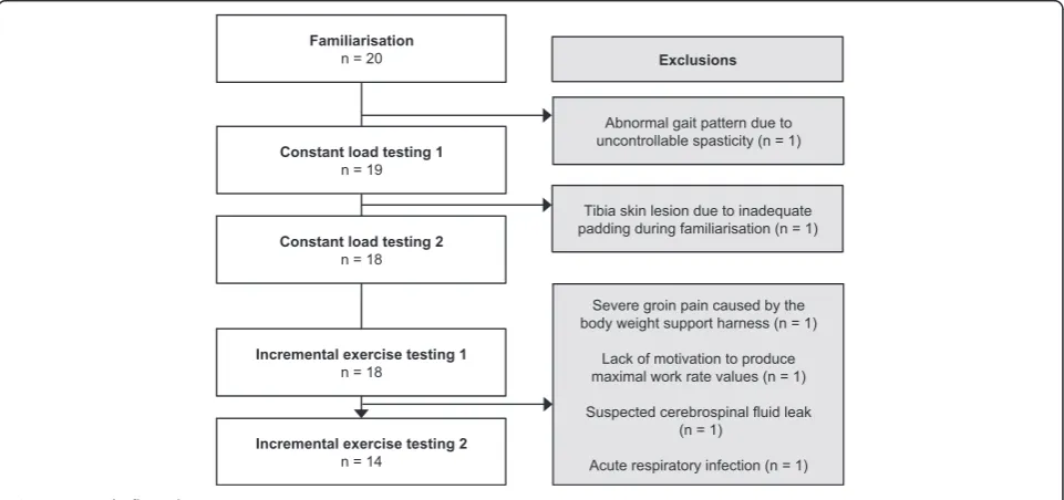
Of the 20 subjects enrolled in the study, 1 subject showed an abnormal gait pattern due to uncontrollable spasticity during familiarisation and 1 subject developed a tibia skin lesion due to inadequate padding of the exoskeleton, which led to withdrawal from the study (Figure 3). Further, 4 withdrawals after the first IET occurred due to groin pain, lack of motivation, suspected cerebrospinal fluid leak, and acute respiratory infection. Thus, 18 subjects (90%) performed the two CLTs and, of these, 14 (70%) also performed the two IETs. All subjects presented with severe motor impairments and were non-ambulatory (FAC range 0–2). BWS ranged between 46-77% and walking speed was set at 60 steps/minute (0.47-0.67 m/s). The subject characteristics are summarized in Table 1.
All subjects successfully completed the predefined CPET protocol (rest, passive, active, recovery). During CLT, 3 subjects stopped the active walking phase after 5 minutes due to generalized fatigue and continued with the recovery phase. All other CLT conditions were per-formed according to the plan. 13 subjects completed both IETs without symptomatic responses requiring ter-mination per safety criteria; the reason for test termin-ation was in every case the inability to reach P*mech due to generalized and/or leg fatigue. The examiner stopped 2 consecutive IET sessions in 1 subject due to high blood pressure responses according to the safety criteria (2 adverse events); however, no serious adverse events occurred during testing. Mean IET duration was
23.2 ± 2.6 min (active phase: 8.2 ± 2.6 min). RPE at peak performance was 14.8 ± 1.9. RMSEPvalues were <10 W.
Exercise capacity
For CLT, time constants of oxygen uptake kinetics (τ) could not be evaluated due to continuous disturbances of VO2 in the transition phases (rest/passive walking, passive walking/active walking). Cost of passive walking (Δrest vs. passive walking) was (mean ± standard devi-ation): VO2=184.2 ± 124.0 mL/min, HR =1.4 ± 6.4 beats/min, and cost of active walking (Δ passive walk-ing vs. active walkwalk-ing) was: VO2=45.7 ± 56.6 mL/min, HR =2.3 ± 3.1 beats/min. RMSEP during CLT was 5.5 ± 5.5 W. For IET, peak performance parameters were: absolute VO2peak =1280.9 ± 564.8 mL/min, relative VO2peak =15.5 ± 4.9 mL/min/kg (51.6 ± 20.5% of predicted VO2max [56]), tVO2peak =8.2 ± 2.6 min, Ppeak =53.9 ± 33.8 W, VEpeak =41.3 ± 18.6 L/min, Rfpeak =36.1 ± 8.3 1/min, HRpeak =126.0 ± 19.5 beats/ min (84.3 ± 12.2% of age-predicted heart rate maximum [44]), and RERpeak =0.92 ± 0.09. Absolute GET was at a VO2 of 878.9 ± 316.6 mL/min and relative GET at 11.0 ± 3.1 mL/min/kg, which was GET% =72.6 ± 12.2% of VO2peak. ΔVO2/ΔP was 20.1 ± 14.4 mL/W, O2pulse was 10.2 ± 4.1 mL/beat, and ΔVE/ΔVCO2 was 36.3 ± 7.3 L. RMSEPduring IET was 8.7 ± 9.0 W.
With respect to the 3 criteria for maximal aerobic cap-acity, 2 subjects (14%) showed a plateau in VO2 at the end of IET, 1 subject (7%) achieved an RER value≥1.15, and 5 subjects (36%) reached peak heart rate within 10 beats per minute of the age-predicted heart rate max-imum, where 2 of these subjects had an adjusted heart rate due to beta-blockers. Thus, 57% of the subjects
Figure 3Study flow chart.
Stolleret al. Journal of NeuroEngineering and Rehabilitation2014,11:145 Page 6 of 13
achieved at least 1 of the 3 criteria for maximal exercise capacity.
Test-retest reliability and repeatability
Table 2 shows mean values, test-retest reliability and re-peatability results of the repeated CLT and IET trials. No practice effects could be detected; trials were not sig-nificantly different. Outcome variables for CLT yielded high MD between tests and insufficient test-retest reli-ability and repeatreli-ability throughout. For IET, good to excellent relative reliability was found for absolute VO2peak (ICC =0.82), relative VO2peak (ICC =0.72), Ppeak (ICC =0.91), HRpeak (ICC =0.80), absolute GET (ICC =0.91), relative GET (ICC =0.88),ΔVO2/ΔP (ICC =0.87), O2pulse (ICC =0.92), and ΔVE/ΔVCO2 (ICC =0.78). SEM were between 4-13% and MDC ranged from 12-36%. MD ± SDdiff of the outcome variables that were analysed for relative reliability were: absolute VO2peak =45.5 ± 353.7 mL/min, relative VO2peak =1.0 ± 3.8 mL/min/kg, Ppeak =2.4 ± 15.0 W, HRpeak =3.6 ± 12.6 beats/min, absolute GET =67.3 ± 124.2 mL/min, relative GET =0.2 ± 1.6 mL/min/kg, ΔVO2/ΔP =2.7 ± 7.2 mL/min/W, O2pulse =0.1 ± 1.7 mL/beat, ΔVE/ ΔVCO2=0.5 ± 5.1 L. CoV for peak cardiopulmonary performance parameters ranged from 0.10-0.44. Bland– Altman plots for the major outcome variables visualize the differences between tests (Additional files 1, 2).
Discussion
This is the first study to evaluate the test-retest reliabil-ity and repeatabilreliabil-ity of FC-RATE for assessment of
exercise capacity early after severe stroke. The aims were: (1) to assess the clinical feasibility of FC-RATE for CPET in severely motor impaired individuals early after stroke, (2) to examine the ability of the concept to meet standard cardiopulmonary criteria for maximal exercise capacity, and (3) to assess the test-retest reli-ability and the repeatreli-ability of the approach.
General observations
Despite rigorous exclusion criteria, only 90% of the sam-ple comsam-pleted both CLTs and 70% comsam-pleted both IETs. Of the 6 subjects who dropped out during the study, only 2 were due to reasons based on uncontrollable fac-tors such as cerebrospinal fluid leak and acute respira-tory infection. The remaining 4 dropouts were caused by controllable factors such as abnormal gait pattern, tibia skin lesion, severe groin pain and lack of motivation. Skin lesions and severe groin pain due to inappropriate padding are preventable by extended familiarisation and padding procedures, whereas abnormal gait patterns due to spasticity and lack of motivation are difficult factors to control. Advanced control strategies might provide solutions for abnormal gait patterns and virtual reality approaches might facilitate motivation in the near fu-ture. Nevertheless, dropout rates were comparable with previous CPET studies in subacute stroke [3,9-11].
The guidance of work rate for FC-RATE-based CPET was successful. RMSEP values were below 10 W, which can be seen as acceptable based on previous pilot study results [39]. The approach presented here used work rate values of both legs together and does not consider
Table 1 Subject characteristics
Constant load testing Incremental exercise testing
(n =18) (n =14)
Men/women 11/7 9/5
Type of stroke: ischemic/haemorrhagic 12/6 11/3
Hemiparetic side: right/left 9/9 8/6
Time post-stroke [d] 49±31(14–139) 43±25(14–92)
Age [y] 61±11(44–84) 61±12(44–84)
BMI [kg/m2] 27±5(19
–38) 28±6(19–38)
FAC (0–5) 1.1±0.8(0–2) 0.9±0.8(0–2)
EBI (0–64) 43±9(27–56) 42±9(27–55)
Medications: beta-blockers/ACE inhibitors/both 6/11/4 4/11/4
Comorbidities: Hypertension/Dyslipidemia/Adipositas/Diabetes mellitus 9/6/3/3 8/6/3/3
BWS [%] 59±9(46–77) 60±9(46–77)
Walking speed [m/s] 0.57±0.05(0.47-0.67) 0.56±0.05(0.47-0.64)
RPE (6–20) 13±2(6–17) 15±2(11–18)
Pmax [W] 38.4±23.0(8.5-77.2) 57.1±33.1(11.3-127.7)
Values are given in numbers (n) or mean ± standard deviation (range).
Abbreviations: BMIBody mass index,FACFunctional Ambulation Classification,EBIExtended Barthel Index,ACE inhibitorsAngiotensin-converting enzyme inhibitors,
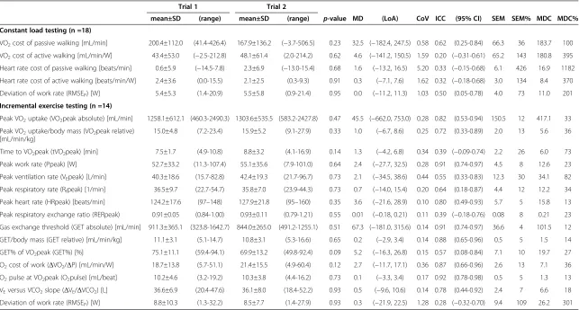
Table 2 Test-retest reliability and repeatability of feedback-controlled robotics-assisted treadmill exercise based cardiopulmonary exercise testing
Trial 1 Trial 2
mean±SD (range) mean±SD (range) p-value MD (LoA) CoV ICC (95% CI) SEM SEM% MDC MDC%
Constant load testing (n =18)
VO2cost of passive walking [mL/min] 200.4±112.0 (41.4-426.4) 167.9±136.2 (−3.7-506.5) 0.23 32.5 (−182.4, 247.5) 0.58 0.62 (0.25-0.84) 66.3 36 183.7 100
VO2cost of active walking [mL/min/W] 43.4±53.0 (−2.5-212.8) 48.1±61.4 (2.0-214.2) 0.62 4.6 (−141.2, 150.5) 1.59 0.20 (−0.31-0.61) 65.2 143 180.8 395
Heart rate cost of passive walking [beats/min] 0.6±5.9 (−14.5-7.8) 2.3±6.9 (−13.0-15.4) 0.68 1.6 (−13.2, 16.5) 5.20 0.33 (−0.15-0.68) 6.1 426 16.9 1182
Heart rate cost of active walking [beats/min/W] 2.4±3.6 (0.0-15.5) 2.1±2.5 (0.3-9.3) 0.91 0.3 (−7.1, 7.6) 1.62 0.32 (−0.18-0.68) 3.0 134 8.4 370
Deviation of work rate (RMSEP) [W] 5.4±5.3 (1.4-20.9) 5.5±5.8 (0.9-21.4) 0.95 0.0 (−11.2, 11.3) 1.03 0.50 (0.05-0.78) 4.0 73 11.0 201
Incremental exercise testing (n =14)
Peak VO2uptake (VO2peak absolute) [mL/min] 1258.1±612.1 (460.3-2490.3) 1303.6±535.5 (583.2-2427.8) 0.47 45.5 (−662.0, 753.0) 0.28 0.82 (0.53-0.94) 150.5 12 417.1 33
Peak VO2uptake/body mass (VO2peak relative)
[mL/min/kg]
15.0±4.8 (7.2-23.4) 15.9±5.2 (9.1-27.9) 0.33 1.0 (−6.7, 8.6) 0.25 0.72 (0.33-0.89) 2.0 13 5.6 36
Time to VO2peak (tVO2peak) [min] 7.5±1.7 (4.9-10.8) 8.8±3.2 (4.1-16.9) 0.14 1.3 (−4.2, 6.8) 0.34 0.39 (−0.09-0.74) 2.2 26 6.0 73
Peak work rate (Ppeak) [W] 52.7±33.2 (11.3-107.4) 55.1±35.6 (7.9-101.0) 0.64 2.4 (−27.7, 32.5) 0.28 0.91 (0.74-0.97) 4.5 8 12.6 23
Peak ventilation rate (VEpeak) [L/min] 40.3±18.6 (15.7-82.8) 42.4±19.3 (21.7-96.7) 0.73 2.1 (−34.5, 38.6) 0.44 0.55 (0.33-0.83) 12.3 30 34.1 82
Peak respiratory rate (Rfpeak) [1/min] 36.5±9.7 (22.7-54.7) 35.8±7.0 (23.9-44.3) 0.73 0.7 (−14.0, 15.4) 0.20 0.64 (0.18-0.87) 4.4 12 12.2 34
Peak heart rate (HRpeak) [beats/min] 124.2±17.6 (97–148) 127.9±21.8 (95–160) 0.35 3.6 (−21.6, 28.9) 0.10 0.80 (0.49-0.93) 5.7 5 15.8 13
Peak respiratory exchange ratio (RERpeak) 0.91±0.05 (0.84-1.00) 0.93±0.11 (0.79-1.21) 0.55 0.01 (−0.18, 0.21) 0.11 0.39 (−0.18-0.76) 0.08 8 0.21 23
Gas exchange threshold (GET absolute) [mL/min] 911.3±365.1 (323.8-1642.7) 844.0±265.0 (491.2-1255.1) 0.51 67.3 (−181.0, 315.6) 0.14 0.91 (0.74-0.97) 36.6 4 101.5 12
GET/body mass (GET relative) [mL/min/kg] 11.1±3.1 (5.1-14.7) 10.8±3.1 (5.3-16.6) 0.65 0.2 (−2.9, 3.4) 0.14 0.88 (0.65-0.96) 0.5 5 1.5 14
GET% of VO2peak (GET%) [%] 75.1±11.1 (59.4-94.1) 69.9±13.2 (49.8-92.4) 0.09 5.2 (−16.3, 26.8) 0.15 0.57 (0.08-0.84) 7.1 10 19.7 27
O2cost of work (ΔVO2/ΔP) [mL/min/W] 18.7±13.8 (5.7-51.1) 21.4±15.5 (4.9-60.4) 0.12 2.7 (−11.7, 17.1) 0.36 0.87 (0.66-0.96) 2.6 13 7.1 36
O2pulse at VO2peak (O2pulse) [mL/beat] 10.2±4.6 (3.2-19.2) 10.3±3.8 (4.4-16.2) 0.73 0.1 (−3.3, 3.4) 0.17 0.92 (0.78-0.98) 0.5 5 1.3 13
VEversus VCO2slope (ΔVE/ΔVCO2) [L] 36.6±6.9 (20.4-47.6) 36.1±8.0 (18.4-52.2) 0.93 0.5 (−9.6, 10.6) 0.14 0.78 (0.44-0.92) 2.4 7 6.6 18
Deviation of work rate (RMSEP) [W] 8.8±10.3 (1.3-32.2) 8.5±7.7 (1.4-27.9) 0.93 0.3 (−21.9, 22.5) 1.28 0.28 (−0.32-0.70) 9.4 109 26.2 301
Abbreviations:SDStandard deviation,MDMean difference,LoALimits of agreement,CoVCoefficients of variation,ICCIntraclass correlation coefficient,
CIConfidence interval,SEMStandard error of the measurement,MDCMinimal detectable change.
Stoller
et
al.
Journal
of
NeuroEng
ineering
and
Rehabilitatio
n
2014,
11
:145
Page
8
o
f
1
3
http://ww
w.jneuroengr
ehab.com/co
ntent/11/1/14
the severe hemiplegia of the included subjects. As a re-sult, subjects generally tend to exercise using the un-affected side more dominantly, which led to deviations from the predefined physiological gait pattern. The pow-ered exoskeleton allowed the subjects to remain in an acceptable movement trajectory during FC-RATE. While the approach presented here aimed to recruit as much muscle mass as possible to provoke peak exercise cap-acity, the imbalance of muscular activation during FC-RATE might be relevant when applying the method in longitudinal training interventions, because continuous imbalance in the gait cycle might facilitate unwanted compensation patterns.
Overall, the findings present a promising method for CPET in severely motor impaired individuals after stroke, but important factors such as appropriate padding and force interactions between subject and robot must be well controlled to gain improvements towards clinical feasibility.
Exercise capacity
For CLT, the difficulty of estimating the time constants for VO2 uptake kinetics (τ) in the transitions from rest to passive walking and passive walking to active walking was due to the inherent noisiness of the breath-by-breath data and the consequent poor signal-to-noise ra-tio. The approach presented here seems not appropriate to provide consistent CLT outcome values for the transi-tion phases. Sudden onset of changes in BWS and walk-ing pattern seem to have a strong impact on individual performance levels during conventional RATE that re-stricts valid assessment of VO2uptake kinetics (τ) values. Cost of passive walking was comparable with previous studies using an identical setup [27,31], whereas cost of active walking was considerably higher than previous re-sults [39]. This difference might be caused by the inclu-sion of non-ambulatory subjects showing severe motor impairments (FAC 2.3 vs. FAC 1.1) in the present study.
With relative VO2peak values of 15.5 ± 4.9 mL/min/kg (51.6% of predicted VO2max), the results confirm that exercise capacity is seriously reduced within this group of severely motor impaired stroke survivors. Peak per-formance parameters during IET found in this study were slightly higher compared to previous trials using leg cycle ergometry, body weight supported treadmill training, and combined upper- and lower-limb ergome-try [3,4,9-12]. This finding might be due to the introduc-tion of treadmill exercise based CPET that has previously confirmed higher VO2peak values compared to leg cycle ergometry protocols [57]. Furthermore, the feedback-control approach presented in this study might have recruited additional muscle mass that provoked higher peak values compared to body weight supported treadmill training. Considering the inclusion of individuals with serious motor impairments and the comparable peak
cardiovascular performance parameters, this study opens new perspectives regarding assessment of exercise capacity early after severe stroke.
The GET values observed in the current study (%GET =72.6 ± 12.2% of VO2peak) were in the upper range compared to sedentary healthy individuals (50-76% of VO2peak) [58], providing additional evidence of com-promised exercise capacity in this population.ΔVO2/ΔP was higher compared to leg cycle ergometry and conven-tional treadmill exercise meaning that subjects required more oxygen for a given work rate level; this may be explained by a substantial amount of unaccounted work performed during the test [59-61]. Further research seems indicated to explore the impact onΔVO2/ΔP while walk-ing within different robotics-assisted systems.
Although most of the subjects appear to have performed in the submaximal range, the estimation of the work rate slope using MWC-W was shown to be successful. The ap-proach implemented was able to reach peak performance within 8–12 minutes (tVO2peak =8.2 ± 2.6 min) during IET by defining the walking cadence at 60 steps/min while increasing the target work rate profile. This is an important finding for further research regarding the initial estimation of target work rate profiles for CPET in severely motor impaired populations.
Finally, considering the peak performance results and the low frequency of achieved criteria for VO2max of this study, in combination with previous study results on peak exercise capacity in subacute stroke, we hypothe-sise that the guidelines postulated for healthy popula-tions may not be realistic for determination of true exercise capacity in the early stages after stroke [64-66].
Test-retest reliability and repeatability
Most studies that examined test-retest reliability using CPET after stroke reported excellent relative reliability, whereas only Tang et al. revealed fair to good associa-tions between trials [10,14,15,17,18]. The present study using a novel robotics-assisted treadmill-based method for assessment of exercise capacity revealed good to ex-cellent relative reliability for the major peak cardiopul-monary performance parameters. There is only limited evidence so far on absolute reliability for CPET early after stroke. Compared to a previous study in chronic stroke using leg cycle ergometry that has shown SEM for relative VO2peak of 1 mL/min/kg (6%), our approach presented higher SEM values (2 mL/min/kg (13%)) [15]. Likewise, a previous study that used semi-recumbent leg cycle ergometry in subacute stroke with cognitive im-pairments yielded considerably lower MDC values (e.g. relative VO2peak: 0.97 mL/min/kg (4%) vs. 5.6 mL/min/kg (36%)) compared to our study [18].
While studies in healthy subjects and individuals with cardiac or respiratory disease have revealed high repeatability (CoV <0.10) [64,67-69], the present study yielded considerably higher CoV for the major cardio-pulmonary parameters (absolute VO2peak =0.28, relative VO2peak =0.25, Ppeak =0.28, HRpeak =0.10, absolute GET =0.14, relative GET =0.14, ΔVO2/ΔP =0.36, O2pulse =0.17,ΔVE/ΔVCO2=0.14).
There was visual suspicion of heteroscedasticity for ab-solute VO2peak and Ppeak (Additional file 1); however, logarithmic transformation of the data (Additional file 2) did not change the relevant outcome variables, and thus appeared irrelevant fur further consideration. We hy-pothesise that the increase in bias along with higher work rate values is caused by large day-to-day variability in strength and/or coordinative capabilities. Although general factors that may contribute to variability in
test-retest situations such as disease severity, patient instruc-tion, time of day, testing procedure, and equipment, were well controlled in the protocol presented here, the novel approach might have led to additional confounding factors that could have influenced test-retest reliability and repeatability. A major factor was the high coordinative demand of the concept. Subjects not only had to walk (or pedal, as in earlier cycle ergometry studies); the challenge was to produce additional forces in the walking direction, where the exoskeleton restricted the movement. The re-sults clearly indicated that the major reason for test ter-mination was the inability to maintain P*mech, which led to the assumption that muscular and/or coordinative fatigue was the reason for test termination. Therefore, variation might be reinforced by day-to-day variability (normally ±3% [64]) and influenced by whether the test was maximal or not. This hypothesis is supported by the low RERpeak values reported in this study. More sophisticated strategies are required to reduce the load on the neuromuscular system while increasing cardio-vascular stress during FC-RATE. This will possibly lead to a better approximation of true exercise capacity early after stroke and might improve the reliability and the repeatability of FC-RATE based CPET.
The approach presented here seems suitable for com-parison of groups of stroke individuals or for assessment of group intervention effects in future studies, considering the range of between-group improvement in VO2peak of 12.6-34.8% [70-73] and Ppeak of 23.4-176.9% [72-74] after cardiovascular exercise in subacute stroke. Whether the absolute reliability and the repeatability reported are adequate to identify effectiveness of intervention pro-grammes to improve exercise capacity should be part of future studies including larger sample sizes.
Limitations
The major limitation of the current study is the small sample size, which may render the results underpow-ered. A sample size of at least 50 is generally seen as ad-equate for the assessment of the agreement parameter, based on a general guideline by Altman [75]. Consider-ing the experimental approach of the method and the difficulty of implementing and performing CPET in the early stages after severe stroke, our sample of 20 subjects at onset was a realistic group size to evaluate first esti-mates from a clinical perspective.
The conventional sequencing of the test situations (CLT, CLT, IET, IET) might have led to practice effects. The severely impaired and early-post-stroke status of the individuals included in this experimental approach may justify this order to progressively increase the exercise intensity over time to control potential risks.
The present study protocol did not include ECG mon-itoring for reasons of practicability, which influenced the
Stolleret al. Journal of NeuroEngineering and Rehabilitation2014,11:145 Page 10 of 13
study sample by excluding individuals with cardiac risk factors for CPET. While around 75% of stroke survivors present some degree of cardiovascular disease [76], the sample of this study might not be representative. Never-theless, there was an uncontrolled risk for cardiac events due to the absence of ECG despite the adherence to strict exclusion criteria for cardiovascular disease.
While the study protocol strictly controlled time of day for CPET, the tests were performed within 48 h or 72 h due to practical reasons. This time difference might have affected the recovery phase of the subjects, thus in-fluencing the results.
Conclusion
This study presents first evidence on reliability and repeat-ability for CPET in severely motor impaired individuals early after stroke using a feedback-controlled robotics-assisted treadmill. The results demonstrate good to excel-lent test-retest reliability and appropriate repeatability for the most important peak cardiopulmonary performance parameters. These findings have important implications for the design and implementation of cardiovascular exer-cise interventions in severely impaired populations. Future research needs to develop advanced control strategies to enable the true limit of functional exercise capacity to be reached and to further assess test-retest reliability and re-peatability in larger samples.
Additional files
Additional file 1:Bland-Altman plots.The difference between trial 2 (T2) and trial 1 (T1) is plotted against the mean of T1 and T2 for the major outcome variables.
Additional file 2:Bland-Altman plots (logarithmically transformed). The difference between trial 2 (T2) and trial 1 (T1) is plotted against the mean of T1 and T2 for the major outcome variables.
Abbreviations
ACSM:American College of Sports Medicine; BWS: Body weight support; CI: Confidence interval; CLT: Constant load testing; CoV: Coefficient of variation; CPET: Cardiopulmonary exercise testing; FAC: Functional ambulation classification; FC-RATE: Feedback-controlled robotics-assisted treadmill exercise; GET: Gas exchange threshold; HR: Heart rate; ICC: Intraclass correlation coefficient; IET: Incremental exercise testing; LoA: Limits of agreement; MD: Mean difference; MDC: Minimal detectable change; O2pulse: O2pulse at
VO2peak; Pmax: Maximal work rate; Pmech: Work rate; P*mech: Target work
rate; RATE: Robotics-assisted treadmill exercise; RER: Respiratory exchange ratio; Rf: Respiratory rate; RMSEP: Accuracy of work rate tracking (root-mean-square
error); RPE: Rating of perceived exertion; SDdiff: Standard deviation of the difference; SEM: Standard error of the measurement; VCO2: Carbon dioxide
output; VE: Ventilation rate; VO2: Oxygen uptake; VO2peak: Peak oxygen uptake; ΔVE/ΔVCO2: VE versus VCO2slope;ΔVO2/ΔP: Oxygen cost of work.
Competing interests
The authors declare that they have no competing interests.
Authors’contributions
OS, EDB, RDB and KH were responsible for the design and the methodology of the study. OS and CSA prepared and obtained the ethical approval. MS was responsible for the technical development and maintenance and supported
data processing. OS screened the subjects, performed the tests, processed and analysed the data and wrote the manuscript. EDB, RDB and KH supervised the process and provided expertise. EDB, MS, CSA, RDB and KH critically revised the manuscript. All authors read and approved the final manuscript.
Acknowledgements
The authors would like to acknowledge Prof. Dr. T. Ettlin, Dr. N. Urscheler, Dr. B. Spoendlin, Dr. A. Rohner , and Dr. M. Kummer for clinical support and medical advice, H. Rosemeyer, N. Springinsfeld, and D. Vosseler for assistance during recruitment and exercise testing, and Dr. J. Wandel and Dr. D. Bättig for statistical support.
Author details
1Department of Engineering and Information Technology, Institute for
Rehabilitation and Performance Technology, Bern University of Applied Sciences, Burgdorf, Switzerland.2Department of Epidemiology, Maastricht
University and Caphri Research School, Maastricht, The Netherlands.
3Research Department, Reha Rheinfelden, Rheinfelden, Switzerland. 4Department of Health Sciences and Technology, Institute of Human
Movement Sciences and Sport, ETH Zurich, Zurich, Switzerland.5Centre for
Evidence Based Physiotherapy, Maastricht University, Maastricht, The Netherlands.
Received: 4 June 2014 Accepted: 3 October 2014 Published: 11 October 2014
References
1. Myers J, Prakash M, Froelicher V, Do D, Partington S, Atwood JE:Exercise capacity and mortality among men referred for exercise testing.N Engl J Med2002,346(11):793–801.
2. Paffenbarger RS Jr, Hyde RT, Wing AL, Lee IM, Jung DL, Kampert JB:The association of changes in physical-activity level and other lifestyle characteristics with mortality among men.N Engl J Med1993, 328(8):538–545.
3. MacKay-Lyons MJ, Makrides L:Exercise capacity early after stroke.Arch Phys Med Rehabil2002,83(12):1697–1702.
4. Kelly JO, Kilbreath SL, Davis GM, Zeman B, Raymond J:Cardiorespiratory fitness and walking ability in subacute stroke patients.Arch Phys Med Rehabil2003,84(12):1780–1785.
5. MacKay-Lyons MJ, Makrides L:Longitudinal changes in exercise capacity after stroke.Arch Phys Med Rehabil2004,85(10):1608–1612.
6. Roth EJ:Heart disease in patients with stroke. Part 2: Impact and implications for rehabilitation.Arch Phys Med Rehabil1994,75(1):94–101. 7. Saunders DH, Greig CA, Young A, Mead GE:Physical fitness training for
patients with stroke an updated review.Stroke2010,41(3):E160–E161. 8. Billinger SA, Arena R, Bernhardt J, Eng JJ, Franklin BA, Johnson CM,
MacKay-Lyons M, Macko RF, Mead GE, Roth EJ, Shaughnessy M, Tang A, American Heart Association Stroke, Council and Council on, Cardiovascular and Stroke, Nursing and Council on, Lifestyle and Cardiometabolic, Health and Council on, Epidemiology and Prevention and Council on Clinical, Cardiology: Physical activity and exercise recommendations for stroke survivors: a statement for healthcare professionals from the American Heart Association/American Stroke Association.Stroke2014,45(8):2532–2553. 9. Chen JK, Chen TW, Chen CH, Huang MH:Preliminary study of exercise capacity
in post-acute stroke survivors.Kaohsiung J Med Sci2010,26(4):175–181. 10. Tang A, Sibley KM, Thomas SG, McIlroy WE, Brooks D:Maximal exercise test
results in subacute stroke.Arch Phys Med Rehabil2006,87(8):1100–1105. 11. Yates JS, Studenski S, Gollub S, Whitman R, Perera S, Lai SM, Duncan PW: Bicycle ergometry in subacute-stroke survivors: feasibility, safety, and exercise performance.J Aging Phys Act2004,12(1):64–74.
12. Hill DC, Ethans KA, MacLeod DA, Harrison ER, Matheson JE:Exercise stress testing in subacute stroke patients using a combined upper- and lower-limb ergometer.Arch Phys Med Rehabil2005,86(9):1860–1866. 13. Baert I, Daly D, Dejaeger E, Vanroy C, Vanlandewijck Y, Feys H:Evolution of
cardiorespiratory fitness after stroke: a 1-year follow-up study, influence of pre-stroke patients’characteristics and stroke-related factors. Cerebrovasc Dis2011,31:189.
15. Eng JJ, Dawson AS, Chu KS:Submaximal exercise in persons with stroke: test-retest reliability and concurrent validity with maximal oxygen consumption.Arch Phys Med Rehabil2004,85(1):113–118.
16. Macko RF, Katzel LI, Yataco A, Tretter LD, DeSouza CA, Dengel DR, Smith GV, Silver KH:Low-velocity graded treadmill stress testing in hemiparetic stroke patients.Stroke1997,28(5):988–992.
17. Lennon OC, Denis RS, Grace N, Blake C:Feasibility, criterion validity and retest reliability of exercise testing using the Astrand-rhyming test protocol with an adaptive ergometer in stroke patients.Disabil Rehabil 2012,34(14):1149–1156.
18. Olivier C, Dore J, Blanchet S, Brooks D, Richards CL, Martel G, Robitaille NM, Maltais DB:Maximal cardiorespiratory fitness testing in individuals with chronic stroke with cognitive impairment: practice test effects and test-retest reliability.Arch Phys Med Rehabil2013,94(11):2277–2282. 19. Billinger SA, Tseng BY, Kluding PM:Modified total-body recumbent
stepper exercise test for assessing peak oxygen consumption in people with chronic stroke.Phys Ther2008,88(10):1188–1195.
20. Durstine LJ, Moore G, Painter P, Roberts S:ACSM’s Exercise Management for Persons With Chronic Diseases and Disabilities.3rd edition. Champaign, IL: Human Kinetics; 2009.
21. French B, Thomas LH, Leathley MJ, Sutton CJ, McAdam J, Forster A, Langhorne P, Price CIM, Walker A, Watkins CL:Repetitive task training for improving functional ability after stroke.Cochrane Database Syst Rev2007, (4). 22. Langhorne P, Coupar F, Pollock A:Motor recovery after stroke: a
systematic review.Lancet Neurol2009,8(8):741–754.
23. Mackay-Lyons M, McDonald A, Matheson J, Eskes G, Klus MA:Dual effects of body-weight supported treadmill training on cardiovascular fitness and walking ability early after stroke: a randomized controlled trial. Neurorehabil Neural Repair2013,27(7):644–653.
24. Hesse S, Bertelt C, Jahnke MT, Schaffrin A, Baake P, Malezic M, Mauritz KH: Treadmill training with partial body weight support compared with physiotherapy in nonambulatory hemiparetic patients.Stroke1995, 26(6):976–981.
25. Danielsson A, Sunnerhagen KS:Oxygen consumption during treadmill walking with and without body weight support in patients with hemiparesis after stroke and in healthy subjects.Arch Phys Med Rehabil 2000,81(7):953–957.
26. MacKay M, Makrides L, Speth S:Effect of 15% body weight support on exercise capacity of adults without impairments.Phys Ther2001, 81(11):1790–1800.
27. Krewer C, Müller F, Husemann B, Heller S, Quintern J, Koenig E:The influence of different Lokomat walking conditions on the energy expenditure of hemiparetic patients and healthy subjects.Gait Posture 2007,26(3):372–377.
28. Stoller O, de Bruin ED, Schindelholz M, Schuster C, de Bie RA, Hunt KJ: Evaluation of exercise capacity after severe stroke using robotics-assisted treadmill exercise: a proof-of-concept study.Technol Health Care 2013,21(2):157–166.
29. Hornby TG, Campbell DD, Zemon DH, Kahn JH:Clinical and quantitative evaluation of robotic-assisted treadmill walking to retrain ambulation after spinal cord injury.Topics in Spinal Cord Injury Rehabilitation2005,11(2):1–17. 30. Nash MS, Jacobs PL, Johnson BM, Field-Fote E:Metabolic and cardiac
responses to robotic-assisted locomotion in motor-complete tetraplegia: a case report.J Spinal Cord Med2004,27(1):78–82.
31. van Nunen MP, Gerrits KH, de Haan A, Janssen TW:Exercise intensity of robot-assisted walking versus overground walking in nonambulatory stroke patients.J Rehabil Res Dev2012,49(10):1537–1546.
32. Israel JF, Campbell DD, Kahn JH, Hornby TG:Metabolic costs and muscle activity patterns during robotic- and therapist-assisted treadmill walking in individuals with incomplete spinal cord injury.Phys Ther2006, 86(11):1466–1478.
33. Hunt KJ, Jack LP, Pennycott A, Perret C, Baumberger M, Kakebeeke TH: Control of work rate-driven exercise facilitates cardiopulmonary training and assessment during robot-assisted gait in incomplete spinal cord injury.Biomedical Signal Processing and Control2008,3(1):19–28. 34. Koenig A, Omlin X, Bergmann J, Zimmerli L, Bolliger M, Muller F, Riener R:
Controlling patient participation during robot-assisted gait training. J Neuroeng Rehabil2011,8:14.
35. Pennycott A, Hunt KJ, Jack LP, Perret C, Kakebeeke TH:Estimation and volitional feedback control of active work rate during robot-assisted gait. Control Eng Pract2008,17(2):322–328.
36. Lam T, Pauhl K, Krassioukov A, Eng JJ:Using robot-applied resistance to augment body-weight-supported treadmill training in an individual with incomplete spinal cord injury.Phys Ther2011,91(1):143–151.
37. Duschau-Wicke A, Caprez A, Riener R:Patient-cooperative control increases active participation of individuals with SCI during robot-aided gait training.J Neuroeng Rehabil2010,7:43.
38. Banz R, Bolliger M, Muller S, Santelli C, Riener R:A method of estimating the degree of active participation during stepping in a driven gait orthosis based on actuator force profile matching.IEEE Trans Neural Syst Rehabil Eng2009,17(1):15–22.
39. Stoller O, Schindelholz M, Bichsel L, Schuster C, de Bie RA, de Bruin ED, Hunt KJ:Feedback-controlled robotics-assisted treadmill exercise to assess and influence aerobic capacity early after stroke: a proof-of-concept study.Disabil Rehabil Assist Technol2013. doi:10.3109/17483107.2013.785038. 40. Thompson WR, Gordon NF, Pescatello LS:American College of Sports
Medicine. Guidelines for Exercise Testing and Prescription.Eight ednth edition. Philadelphia: Lippincott Williams & Wilkins; 2010.
41. Collen FM, Wade DT, Bradshaw CM:Mobility after stroke: reliability of measures of impairment and disability.Int Disabil Stud1990,12(1):6–9. 42. Collin C, Wade DT, Davies S, Horne V:The Barthel ADL index: a reliability
study.Int Disabil Stud1988,10(2):61–63.
43. Perry J:Gait Analysis: Normal and Pathological Function.Thorofare, NJ: SLACK incorporated; 1992.
44. Tanaka H, Monahan KD, Seals DR:Age-predicted maximal heart rate revisited.J Am Coll Cardiol2001,37(1):153–156.
45. Tesch PA:Exercise performance and beta-blockade.Sports Med1985, 2(6):389–412.
46. Borg GA:Perceived exertion.Exerc Sport Sci Rev1974,2:131–153. 47. Schindelholz M, Stoller O, Hunt KJ:A software module for cardiovascular
rehabilitation in robotics-assisted treadmill exercise.Biomed Signal Process Control2014,10:296–307.
48. Robergs RA, Dwyer D, Astorino T:Recommendations for improved data processing from expired gas analysis indirect calorimetry.Sports Med 2010,40(2):95–111.
49. Whipp BJ, Wasserman K:Oxygen-uptake kinetics for various intensities of constant-load work.J Appl Physiol1972,33(3):351–356.
50. Poole DC, Wilkerson DP, Jones AM:Validity of criteria for establishing maximal O2 uptake during ramp exercise tests.Eur J Appl Physiol2008, 102(4):403–410.
51. Beaver WL, Wasserman K, Whipp BJ:A new method for detecting anaerobic threshold by gas-exchange.J Appl Physiol1986, 60(6):2020–2027.
52. Streiner DL:Diagnosing tests: using and misusing diagnostic and screening tests.J Pers Assess2003,81(3):209–219.
53. Streiner DL, Norman GR:Health Measurement Scales: A Practical Guide to Their Development and use.Oxford, NY: Oxford University Press; 2008. 54. Weir JP:Quantifying test-retest reliability using the intraclass correlation
coefficient and the SEM.J Strength Cond Res2005,19(1):231–240. 55. Haley SM, Fragala-Pinkham MA:Interpreting change scores of tests and
measures used in physical therapy.Phys Ther2006,86(5):735–743. 56. Jurca R, Jackson AS, LaMonte MJ, Morrow JR, Blair SN, Wareham NJ, Haskell
WL, van Mechelen W, Church TS, Jakicic JM, Laukkanen R:Assessing cardiorespiratory fitness without performing exercise testing.Am J Prev Med2005,29(3):185–193.
57. McKay GA, Banister EW:A comparison of maximum oxygen uptake determination by bicycle ergometry at various pedaling frequencies and by treadmill running at various speeds.Eur J Appl Physiol Occup Physiol 1976,35(3):191–200.
58. Davis JA, Storer TW, Caiozzo VJ:Prediction of normal values for lactate threshold estimated by gas exchange in men and women.Eur J Appl Physiol Occup Physiol1997,76(2):157–164.
59. Wasserman K, Hansen JE, Sue DY, Stringer WW, Sietsema KE, Sun XG, Whipp BJ:Principles of exercise testing and interpretation: including pathophysiology and clinical applications.Philadelphia, PA: Lippincott Williams & Wilkins; 2011.
60. Hansen JE, Casaburi R, Cooper DM, Wasserman K:Oxygen uptake as related to work rate increment during cycle ergometer exercise.Eur J Appl Physiol Occup Physiol1988,57(2):140–145.
61. Porszasz J, Casaburi R, Somfay A, Woodhouse LJ, Whipp BJ:A treadmill ramp protocol using simultaneous changes in speed and grade.Med Sci Sports Exerc2003,35(9):1596–1603.
Stolleret al. Journal of NeuroEngineering and Rehabilitation2014,11:145 Page 12 of 13
62. Day JR, Rossiter HB, Coats EM, Skasick A, Whipp BJ:The maximally attainable VO2 during exercise in humans: the peak vs. maximum issue. J Appl Physiol2003,95(5):1901–1907.
63. Bruce RA, Cooper MN, Gey GO, Fisher LD, Peterson DR:Variations in responses to maximal exercise in health and in cardiovascular disease. Angiology1973,24(11):691–702.
64. American Thoracic Society, American College of Chest Physicians:ATS/ ACCP statement on cardiopulmonary exercise testing.Am J Respir Crit Care Med2003,167:211–277.
65. Balady GJ, Arena R, Sietsema K, Myers J, Coke L, Fletcher GF, Forman D, Franklin B, Guazzi M, Gulati M, Keteyian SJ, Lavie CJ, MacKo R, Mancini D, Milani RV:Clinician’s guide to cardiopulmonary exercise testing in adults: A scientific statement from the American heart association.Circulation 2010,122(2):191–225.
66. Fletcher GF, Balady GJ, Amsterdam EA, Chaitman B, Eckel R, Fleg J, Froelicher VF, Leon AS, Pina IL, Rodney R, Simons-Morton DG, Williams MA, Bazzarre T:Exercise standards for testing and training - a statement for healthcare professionals from the American Heart Association.Circulation 2001,104(14):1694–1740.
67. Garrard CS, Emmons C:The reproducibility of the respiratory responses to maximum exercise.Respiration1986,49(2):94–100.
68. Nordrehaug JE, Danielsen R, Stangeland L, Rosland GA, Vik-Mo H: Respira-tory gas exchange during treadmill exercise testing: reproducibility and comparison of different exercise protocols.Scand J Clin Lab Invest1991, 51(7):655–658.
69. Barron A, Dhutia N, Mayet J, Hughes AD, Francis DP, Wensel R:Test-retest repeatability of cardiopulmonary exercise test variables in patients with cardiac or respiratory disease.Eur J Prev Cardiol2014,21(4):445–453. 70. da Cunha IT, Lim PA, Qureshy H, Henson H, Monga T, Protas EJ:Gait
outcomes after acute stroke rehabilitation with supported treadmill ambulation training: A randomized controlled pilot study.Arch Phys Med Rehabil2002,83(9):1258–1265.
71. Duncan P, Studenski S, Richards L, Gollub S, Lai SM, Reker D, Perera S, Yates J, Koch V, Rigler S, Johnson D:Randomized clinical trial of therapeutic exercise in subacute stroke.Stroke2003,34(9):2173–2180.
72. Letombe A, Cornille C, Delahaye H, Khaled A, Morice O, Tomaszewski A, Olivier N:Early post-stroke physical conditioning in hemiplegic patients: a preliminary study.Ann Phys Rehabil Med2010,53(10):632–642. 73. Tang A, Sibley KM, Thomas SG, Bayley MT, Richardson D, McIlroy WE, Brooks
D:Effects of an aerobic exercise program on aerobic capacity, spatiotemporal gait parameters, and functional capacity in subacute stroke.Neurorehabil Neural Repair2009,23(4):398–406.
74. Katz-Leurer M, Shochina M, Carmeli E, Friedlander Y:The influence of early aerobic training on the functional capacity in patients with
cerebrovascular accident at the subacute stage.Arch Phys Med Rehabil 2003,84(11):1609–1614.
75. Altman DG:Practical Statistics for Medical Research.CRC Press; 1990. 76. Roth EJ:Heart disease in patients with stroke: incidence, impact, and
implications for rehabilitation. Part 1: Classification and prevalence.Arch Phys Med Rehabil1993,74(7):752–760.
doi:10.1186/1743-0003-11-145
Cite this article as:Stolleret al.:Cardiopulmonary exercise testing early after stroke using feedback-controlled robotics-assisted treadmill exercise: test-retest reliability and repeatability.Journal of NeuroEngineering and Rehabilitation
201411:145.
Submit your next manuscript to BioMed Central and take full advantage of:
• Convenient online submission
• Thorough peer review
• No space constraints or color figure charges
• Immediate publication on acceptance
• Inclusion in PubMed, CAS, Scopus and Google Scholar
• Research which is freely available for redistribution