R E S E A R C H
Open Access
Study of the frequency of
Clostridium difficile tcdA,
tcdB, cdtA
and
cdtB
genes in feces of Calves in
south west of Iran
Abbas Doosti
*and Abbas Mokhtari-Farsani
Abstract
Background:Clostridium difficile(C. difficile) is a gram-positive, toxin-producing bacillus which is an intestinal pathogen in both humans and animals and causes a range of digestive disorders including inflammation of the bowel, abdominal pain, fever and diarrhea.C. difficiletoxins include enterotoxin (Toxin A), cytotoxin (Toxin B) and a binary toxin. Two large protein toxins A and B are encoded by separate genes,tcdAandtcdB. Clostridium difficileinfection (CDI) mainly caused by the activity of the genestcdAandtcdB. The binary toxin is encoded by the genescdtAandcdtB.The binary toxin caused increased adherence of bacteria to intestinal epithelium. The aim of the present study was isolation ofC. difficile from feces of calves, and study of the frequency ofC. difficilevirulence genes.
Methods:150 samples of fresh feces from calves were collected andC. difficilewas isolated from feces of calves using bacterial culture methods. DNA was extracted by a genomic DNA purification kit. Then PCR method was used for definitive diagnosis ofC. difficile. Multiplex PCR method performed for identification oftcdA,tcdB,cdtAandcdtBgenes. In the final stage antimicrobial resistance determining was carried out by standard Bauer-Kirby disk diffusion method. Results:C. difficilewas isolated from 90 samples (60%). ThetcdAwas observed in 8 isolates (8.8%),tcdBin 16 isolates (17.7%),cdtAin 8 isolates (8.8%) andcdtBin 14 isolates (15.5%). Only 1 isolated (1.1%) was containing all four genes tcdA,tcdB,cdtAandcdtB, 2 isolates (2.2%) only had bothtcdAandtcdBgenes, and there was no sample positive only for bothcdtAandcdtB. The highest rate of drug resistance was against clindamycin (100%) and the highest rate of drug sensitivity was against ciprofloxacin (50%).
Conclusion:The results showed high incidence ofC. difficileand also high antibiotic resistance of this bacterium, but frequency of strains containing virulence genes (tcdA,tcdB,cdtAandcdtB) was low.
Keywords:Clostridium difficile, Calves, Toxin A, Toxin B, Binary toxin
Background
Clostridium difficile (C. difficile)is a gram-positive, spore-forming, anaerobic, toxin-producing bacillus, catalase-negative bacterium and is part of the normal gut flora in less than 5% of humans that can infect both humans and animals [1]. C. difficile there at everywhere in the environment and as well as exist as free-living bacteria that it has been found in farm animals, pets, on various surfaces in hospitals and in foods such as vegetables, meats, water and soil [2]. It is transmitted via the fecal– oral route among humans and also is transmitted to
humans from animals and animal products. Clostridium
difficileinfection (CDI) is a global problem that is caused by the ingestion of vegetative organisms and spores, most likely the latter which survive exposure to gastric acidity and germinate in the colon. C. difficile recognized as a major nosocomial pathogen responsible for 15-20% of antibiotic-related diarrhea and is the etiologic agent of perforation of the colon, pseudomembranous colitis or
toxic megacolon and even death in humans [3,4]. C.
difficilealso is an important factor for of enteric disease in other species, including calves (10.2%), cows, ostriches, horses, pigs, rabbits, cats and dogs [5,6]. The symptoms of CDI in both humans and animal include inflammation of the bowel, abdominal pain, fever and diarrhea [7]. * Correspondence:[email protected]
Biotechnology Research Center, Islamic Azad University, Shahrekord Branch, Postal Box: 166, Shahrekord, Iran
Recently, research has suggested that the frequency, relapse, and severity of Clostridium difficile associated diseases (CDAD) are increasing in North America and Europe and also per year 2012, United States health care expenses associated with treating CDAD are estimated about $3.2 billion [8].C. difficiletoxins include toxin A, toxin B and a binary toxin. Toxins A (enterotoxin) and B (cytotoxin) (308 kDa and 270 kDa, respectively), are encoded by two separate genes,tcdAandtcdBwhich are located in a 19.6-kb pathogenicity locus (PaLoc) and are responsible for the symptoms of CDAD and also includes the positive and negative regulatorstcdR andtcdC [9]. They are structur-ally similar to each other, but toxin B is generstructur-ally more ro-bust (~1000 fold) than toxin A [10]. The binary toxin is
encoded by the genes cdtA and cdtB located outside
PaLoc, which these two genes form another operon with
cdtR (cdtR is a positive regulator) [9]. C. difficile toxins directly affect the colon epithelial cells, and immune cells are forced to produce chemokines and cytokines [10]. Standard treatment for CDI in both humans and animals is use of the vancomycin or metronidazole which neither of these antibiotics is not fully effective, but new thera-peutic options include the use of new antibiotics, probio-tics, toxin-absorbing polymer, monoclonal antibodies, fecal treansplant, toxoid vaccines and Intravenous im-munoglobulin (IVIG) [10]. To our knowledge, there are no many studies onC. difficilein feces calves in around the world and also there are no study on C. difficile in feces calves in Iran; thus, the purpose of present study was isolation of C. difficile from feces of calves from Chaharmahal va Bakhtiari province in south west of Iran using bacterial culture methods, and study of the frequency ofC. difficile tcdA, tcdB, cdtAandcdtBgenes using Multiplex PCR method, and also study of anti-biotic resistance in this bacterium.
Methods
Samples collection
In this cross-sectional descriptive study, a total one hun-dred fifty (150) samples of fresh feces from calves with age 3 to 25 days in autumn of 2013 were collected from industrial and indigenous calves farms in Chaharmahal va Bakhtiari province (southwest of Iran). Only one stool sample was collected from each calf. Feces were collected from animals rectum using the new clean gloves and about 3 g of the sample was transferred to a new sterile Tube and were transported to the laboratory immediately in an insulating foam box with ice.
C. difficileCulture
Outset was placed tubes containing 10 ml cooked meat broth medium (Merckoplate, Germany) at 80°C in a water bath for 10 min until the medium be anaerobic. Then it was placed at room temperature until be cooled and about
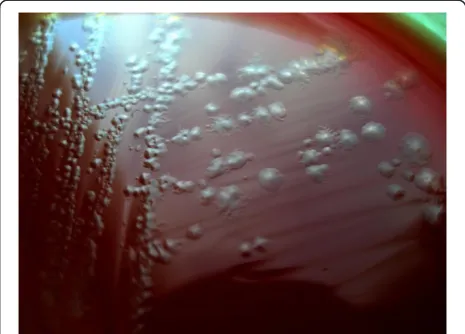
2 g of feces were inoculated into it and was placed at 65°C in a water bath for 10 min in order to select bacterial spores and was incubated in an anaerobic incubator (Finetech, Germany) at 37°C for 7 days. Thereafter, re-moved from the suspension by a sterile loop and was cultured onto blood agar supplemented with 5% sheep blood (Merckoplate, Germany) to linear method and was incubated in an anaerobic incubator at 37°C for 48 h. Identification and isolation ofC. difficilewas based on the characteristic morphology, gram stain and the smell of the colonies. The colonies circular, tarnishes, slightly embossed, white or gray was indicative the pres-ence ofC. difficile(Figure 1). In order to keep the bacteria for late experiments, the plates were wrapped in parafilm and were kept at 4°C.
DNA extraction and PCR
C. difficile was grown on blood agar for 48 h and
approximately 3 colonies were suspended in 100 μL of
sterile ultrapure deionized water and DNA was extracted by a genomic DNA purification kit (CinnaGen Co, Iran) according to the manufacturer’s instructions. The total DNA was measured at 260 nm optical density according to the method described by Sambrook and Russell
(2001). DNA samples were stored at −20°C until they
were used. Polymerase chain reaction (PCR) method was used for definitive diagnosis ofC.difficile. Detection of
C. difficile was performed by amplification with the following primers: C.D-F: 5’-CCTCCTCAAGTACCG
TCATTATC-3’ and C.D-R: 5’-CAAAGGTGAGCCAG
TACAG GA-3’ [Gene Bank: NC_009089 ]. The primers
were design from 16S ribosomal RNA (16S rRNA) gene of
C. difficile using Gene Runner software (Version 3.01) and at the NCBI using the experimental GENINFO BLAST Network Service to assess degree of homology between these primers and other reported sequences and at the end were obtained from CinnaGen Co, Iran.
PCR reactions were performed in a total volume of 25μL in 0.2 ml tubes containing 2 μL of DNA sample,
1μM of each primers, 2 mM MgCl2 , 5μL of 10X PCR
buffer AMS, 200 μM dNTPs and 1 unit of Taq DNA
polymerase (CinnaGen Co, Iran). The PCR assay was performed at 95°C for 5 min and then for 32 cycles of 94°C for 1 min, 61°C for 40 sec, 72°C for 40 sec, and a final extension at 72°C for 5 min, with a final hold at 10°C in a thermal cycler (Mastercycler gradient, Eppendrof, Germany).
Detection of PCR products
The PCR-amplified products (C.D 16S rRNA, 264 bp) were detected in 1.5% ethidium bromide (EtBr)-stained agarose gel electrophoresis. Constant voltage of 84 V for 25 min was used for products separation. The DNA molecular weight marker (100 bp, Fermentas, Germany) was used as a size marker. The gel viewed on UV transil-luminator and photographed were obtained in UVI doc gel documentation systems (UK).
Multiplex PCR for identification oftcdA,tcdB,cdtAand
cdtBgenes
A 4-plex PCR was performed for identification of tcdA,
tcdB, cdtA and cdtB genes. All four primers pairs for multiplex PCR were designed and obtained by using the method listed before (Table 1). The multiplex PCR was run in final reaction volumes of 50 μL containing the following reagents: 2μL of genomic DNA, 200μM each of dATP, dCTP, dGTP and dTTP, 50 pmol of each pri-mer (pripri-mers details are presented in Table 1), 6 mM MgCl2, 50 mM KCl, 10 mM Tris–HCl (pH 8.5) and 5 unit of Taq DNA polymerase (CinnaGen Co., Iran). Re-actions were initiated at 95°C for 5 min, followed by 40 cycles of 94°C for 1 min, 59°C for 1 min, 72°C for 1 min and a final extension step at 72°C for 7 min, with a final hold at 10°C in a thermal cycler (Mastercycler gradient, Eppendrof, Germany). Detection of PCR prod-ucts was performed by using the method listed before.
Antimicrobial drug susceptibility tests
Drug resistance testing using standard Bauer-Kirby disk diffusion method for all samples (positive culture) was performed. A total of 5 antibiotic discs (Padtanteb, Iran) with tetracycline, ciprofloxacin, clindamycin, erythro-mycin and vancoerythro-mycin were used. These antimicrobial agents were chosen on the basis of their importance in treating humans or animals CDI. Diameters of the inhib-ition zones were interpreted based on the NCCLS subcom-mittee’s recommendations [11].
Statistical analysis
All data were analyzed by using MS Excel 2007 and SPSS software (Version 17.SPSS Inc, USA) and p value was calculated using Chi-square and Fisher’s exact tests to find any significant relationship. P value less than 0.05 was considered statistically significant.
Results
The frequency ofC. difficileand virulence genes
In the present study, a total of 150 samples of feces of calves were tested forC. difficile. In order to diagnosis of Table 1 Primers for identification of C.difficile tcdA, tcdB, cdtAandcdtBgenes
Gene target Primer name Sequence (5'–3') Accession Amplicon size (bp)
tcdA tcdA-F tcdA-R AATGATGTTACCTAATGCTCCTTC KC292125 311 bp
AGTAAGTTCCTCCTGCTCCATC
tcdB tcdB-F tcdB-R CCAGCTAATACACTTGATGAAAACC KC292190 432 bp
TTTCTTCACCTTCTTCATTTCCT
cdtA cdtA-F cdtA-R GGAAGCACTATATTAAAGCAGAAGC HQ639678 219 bp
TCTGGGTTAGGATTATTTACTGGAC
cdtB cdtB-F cdtB-R AAAGTTGATGTCTGATTGGGAAG HQ639678 608 bp
TTTGTTGTTGGTGTCACTTTGTA
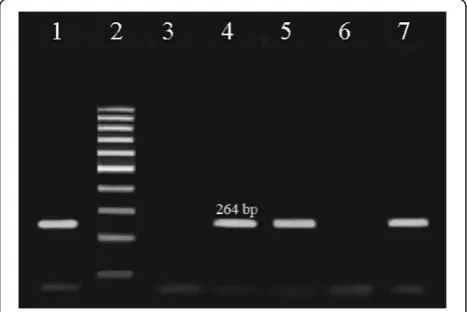
C. difficile, PCR test was carried out. PCR products on 1.5% agarose gel showed 264 bp fragment forC. difficile
(Figure 2). The results obtained revealed that there are 90 samples (60.0%) containingC. difficile, and also it was found that the peak incidence ofC. difficilein calves is in age 16 days (21.3%). A 4-plex PCR was developed for study of the frequency of virulence genes (tcdA,tcdB,cdtA
and cdtB) (Figure 3). Eight isolates (8.8%) possessed the
tcdA gene, sixteen isolates (17.7%) possessed the tcdB
gene, eight isolates (8.8%) possessed the cdtA gene and fourteen isolates (15.5%) possessed the cdtB gene. Two isolates (2.2%) were positive only for bothtcdAand tcdB
genes, three isolates (3.3%) were positive only for both
tcdB and cdtB genes and only one isolated (1.1%) was
containing all four genestcdA, tcdB, cdtAand cdtB, in addition, there was no sample positive only for both
cdtAandcdtB.
Antibiogram profile
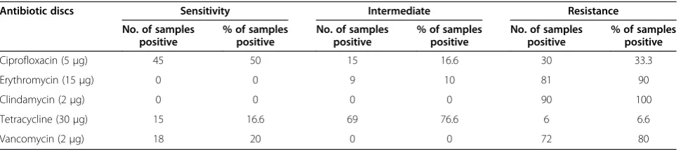
The resistance pattern of C. difficile isolates to 5 anti-microbial agents tested in this study is shown in Table 2.
All isolates (100%; n = 90) were resistant to one or more antimicrobial agent. The highest rate of drug resist-ance was against clindamycin (100%) and erythromycin (90.0%), in addition, the highest rate of drug sensitivity was against ciprofloxacin (50.0%) and vancomycin (20.0%). Three isolates (3.3%) were only resistant to single antibiotic, fifteen isolates (16.6%) were resistant to 2 antibiotics, forty five isolates (50.0%) were resistant to 3 antibiotics and twenty seven isolates (30.0%) showed resistance to 4 or more than 4 antimicrobial agents.
Discussion
C. difficile was recognized as an intestinal pathogen in the late 70th of twentieth century [12]. C. difficile is agent for more than 95% of cases of pseudomembranous colitis and 15–25% of cases of antibiotic-associated diar-rhea [13]. CDAD in humans may have an animal origin, for example, C. difficile is found in market meat, and studies have shown that Pathogenic strains in humans and animals especially calves, are very similar [6]. In recent years, has emerged changes in the epidemiology of CDAD in humans and also CDI is increasing in prevalence, severity and mortality. Development of antibiotic resistance in C. difficile serotypes has been considered as a public health problem throughout the world. It seems that in order to determine the epidemi-ology ofC. difficile- associated disease should studies and more research be done around the world. In present study was observed a relatively high prevalence ofC. difficile
and virulence genes, and also isolates showed high percentage of resistance against antibiotics. The study of Arroyo et al. showed that C. difficile isolated from calves are carrier the genes encoding toxins A and B [14]. In Rodriguez-Palacio et al. investigation in Canada, con-tamination rate withC. difficilein calves with a mean age of 14.2 days, 11.2% was reported, also examinedC. difficile
ribotypes, which 7 of 8 ribotype were common between humans and calves, furthermore, in antibiotic susceptibil-ity testing, all isolates were sensitive to vancomycin, which is somewhat similar to the present study results [5]. In Pirs et al. investigation,C. difficilewas isolated from 1/56 calf samples (1.8%) [6]. The study of Janvilisri et al. showed that only 9 (52.9%) of the 17 calves were positive Figure 3Multiplex-PCR for detection of virulence genes (Lanes
1 and 7 are negative for virulence genes, Lanes 2, 3, 4 and 5 are positive only for one of virulence genes, Lane 6 shows fermentas 100 bp DNA molecular marker, Lane 8 is positive fortcdBand cdtBgenes, Lane 9 is positive for all four genestcdA,tcdB,cdtA andcdtB, and Lane 10 is positive fortcdAandtcdBgenes).
Table 2 Antibiogram profile of the 90C. difficileisolates from feces of calves
Antibiotic discs Sensitivity Intermediate Resistance No. of samples
positive
% of samples positive
No. of samples positive
% of samples positive
No. of samples positive
% of samples positive
Ciprofloxacin (5μg) 45 50 15 16.6 30 33.3
Erythromycin (15μg) 0 0 9 10 81 90
Clindamycin (2μg) 0 0 0 0 90 100
Tetracycline (30μg) 15 16.6 69 76.6 6 6.6
toC. difficileand every 9 positive samples were possessed the binary toxin, which is somewhat similar to the present study results [15]. Hoffer et al. in Switzerland, announced Low occurrence ofC. difficilein stool of calves, that C. difficile was isolated from only one fecal sample of a calf, furthermore, was determined that sample isolated is containing toxin A, toxin B and binary toxin [16]. In Rodriguez et al. investigation in Belgium, prevalence of
C. difficile was reported in calves on the farms 22.2% [17]. Zidaric et al. found that the peak incidence of C. difficilein calves is in age 18 days (16%), also was found that the isolates were resistant to erythromycin, which is similar to the present study results [18].
The above data indicate that in around the world there are different rate of frequency ofC. difficileand virulence genes of this bacterium. Furthermore, with review previ-ous studies will be determined that there are different patterns of antimicrobial resistance ofC. difficileisolated of different parts of the world. Our study and other studies throughout the world show that in recent years prevalence of pathogenic strains and resistant to antimicrobial drugs with speed is on the rise. Considering that, there aren’t large studies on virulence genes of C. difficile isolated from calves and domestic animals, the prevalence of these genes cannot be examined, but considering that amount of pathogenicity of C. difficile in worldwide is rising can be said that the prevalence of pathogenic strains that are containing these genes is on the rise. In the present study it was determined that ciprofloxacin is the best antibiotic for infections treatment caused byC. difficileand although there was a high rate of sensitivity to vancomycin, but resistance rate to this antibiotic was 80%. Nonetheless, examining antibiotic resistance patterns in other studies indicate that vancomycin is currently the best antibiotic for infections treatment caused byC. difficilein calves and other domestic animals, that is inconsistent with the results of our study.
In conclusion, our data show high prevalence of C.
difficilein feces of calves in south west of Iran, that for-tunately frequency of pathogenic strains (containing virulence genes) was low. On the other hand, our studies showed a high percentage of antibiotic resistance in the
C. difficilewhich is a result of the indiscriminate use of antibiotics. Thus, given that many of C. difficile strains are common between humans and domestic animals and given the increasing importance of CDI in public health, we suggest further studies for recognition the epidemiology and detect any changes in resistance pattern, new therapies, restricting use of antimicrobial drugs in human and animals, performing antimicrobial susceptibil-ity tests to select suitable antimicrobial agent, application of recommended dosage of antibiotic and following duration of therapy can help to decrease developing of resistant strains and pathogenic.
Competing interests
The authors declare that they have no competing interests.
Authors’contributions
AD: Study supervision, study concept and design, critical revision of the manuscript for important intellectual content. AM-F: Drafting of the manuscript, critical revision of the manuscript for important intellectual content, carried out the molecular genetic studies, analyzed the data. All authors read and approved the final manuscript.
Acknowledgements
The authors gratefully acknowledge all the staff of Biotechnology Research Center of Islamic Azad University of Shahrekord Branch in southwest Iran for their sincere support.
Received: 21 April 2014 Accepted: 27 May 2014 Published: 5 June 2014
References
1. Hammond EN, Donkor ES:Antibacterial effect of Manuka honey on
Clostridium difficile.BMC Res Notes2013,6:1–5.
2. Rogers MAM, Greene MT, Saint S, Chenoweth CE, Malani PN, Trivedi I, Aronoff DM:Higher Rates ofClostridium difficileInfection among Smokers.Plos one2012,7:e42091.
3. Janvilisri T, Scaria J, Chang YF:Transcriptional profiling ofClostridium difficileand Caco-2 cells during infection.J Infect Dis2010,202:282–290. 4. Hung YP, Tsai PJ, Hung KH, Liu HC, Lee CI, Lin HJ, Wu YH, Wu JJ, Ko WC:
Impact of ToxigenicClostridium difficileColonization and Infection among Hospitalized Adults at a District Hospital in Southern Taiwan.
Plos one2012,7:e42415.
5. Rodriguez-Palacios A, Stämpfli HR, Duffield T, Peregrine AS, Trotz-Williams LA, Arroyo LG, Brazier JS, Weese JS:Clostridium difficilePCR Ribotypes in Calves, Canada.Emerging Infect Dis2006,12:1730–1736.
6. Pirs T, Ocepek M, Rupnik M:Isolation ofClostridium difficilefrom food animals in Slovenia.J Med Microbiol2008,57:790–792.
7. Davies NL, Compson JE, MacKenzie B, O’Dowd VL, Oxbrow AKF, Heads JT, Turner A, Sarkar K, Dugdale SL, Jairaj M, Christodoulou L, Knight DE, Cross AS, Hervé KJ, Tyson KL, Hailu H, Doyle CB, Ellis M, Kriek M, Cox M, Page MJ, Moore AR, Lightwood DJ, Humphreys DP:A Mixture of Functionally Oligoclonal Humanized Monoclonal Antibodies That Neutralize
Clostridium difficile TcdAandTcdBwith High Levels of In Vitro Potency Shows In Vivo Protection in a Hamster Infection Model.Clin Vaccine Immunol2013,20:377–390.
8. Swett R, Cisneros GA, Feig AL:Conformational Analysis ofClostridium difficileToxin B and Its Implications for Substrate Recognition.Plos one
2012,7:e41518.
9. Persson S, Jensen JN, Olsen KEP:Multiplex PCR Method for Detection of
Clostridium difficile tcdA,tcdB,cdtA, andcdtBand Internal In-Frame Deletion of tcdC.J Clin Microbiol2011,49:4299–4300.
10. Sun X, Savidge T, Feng H:The Enterotoxicity ofClostridium difficileToxins.
Toxins2010,2:1848–1880.
11. National Committee for Clinical Laboratory Standards:Performance standards for antimicrobial susceptibility testing. InTwelfth Informational Supplement.Wayne: NCCLS document M100-S12 22, NCCLS; 2002:42. 12. Lemee L, Dhalluin A, Testelin S, Mattrat MA, Maillard K, Lemeland JF, Pons
JL:Multiplex PCR Targeting tpi (Triose Phosphate Isomerase),tcdA(Toxin A), andtcdB(Toxin B) Genes for Toxigenic Culture ofClostridium difficile.
J Clin Microbiol2004,42:5710–5714.
13. Sadeghifard N, Salari MH, Ranjbar R, Ghafouryan S, Raftari M, Abdulamir AS, Fatimah AB, Kazemi B:The clinical and environmental spread and diversity of toxigenicClostridium difficilediarrhea in the region of the Middle East.Reviews in Infect2010,1:180–187.
14. Arroyo LG, Kruth SA, Willey BM, Staempfli HR, Low DE, Weese JS:PCR ribotyping ofClostridium difficileisolates originating from human and animal sources.J Med Microbiol2005,54:163–166.
16. Hoffer E, Haechler H, Frei R, Stephan R:Low occurrence ofClostridium difficilein fecal samples of healthy calves and pigs at slaughter and in minced meat in Switzerland.J Food Prot2010,73:973–975.
17. Rodriguez C, Taminiau B, Van Broeck J, Avesani V, Delmée M, Daube G: Clostridium difficile in young farm animals and slaughter animals in Belgium.J. Anaerobe2012,18:621–625.
18. Zidaric V, Pardon B, Dos Vultos T, Deprez P, Brouwer MS, Roberts AP, Henriques AO, Rupnik M:Different antibiotic resistance and sporulation properties within multiclonalClostridium difficilePCR ribotypes 078, 126, and 033 in a single calf farm.Appl Environ Microbiol2012,78:8515–8522.
doi:10.1186/1476-0711-13-21
Cite this article as:Doosti and Mokhtari-Farsani:Study of the frequency of
Clostridium difficile tcdA, tcdB, cdtAandcdtBgenes in feces of Calves in south west of Iran.Annals of Clinical Microbiology and Antimicrobials201413:21.
Submit your next manuscript to BioMed Central and take full advantage of:
• Convenient online submission
• Thorough peer review
• No space constraints or color figure charges
• Immediate publication on acceptance
• Inclusion in PubMed, CAS, Scopus and Google Scholar
• Research which is freely available for redistribution