R E S E A R C H A R T I C L E
Open Access
Identification of specificity determining residues
in peptide recognition domains using an
information theoretic approach applied to
large-scale binding maps
Kevin Y Yip
1,2, Lukas Utz
3, Simon Sitwell
3, Xihao Hu
2, Sachdev S Sidhu
3,7,8, Benjamin E Turk
4, Mark Gerstein
1,5,6and
Philip M Kim
3,7,8,9*Abstract
Background:Peptide Recognition Domains (PRDs) are commonly found in signaling proteins. They mediate protein-protein interactions by recognizing and binding short motifs in their ligands. Although a great deal is known about PRDs and their interactions, prediction of PRD specificities remains largely an unsolved problem. Results:We present a novel approach to identifying these Specificity Determining Residues (SDRs). Our algorithm generalizes earlier information theoretic approaches to coevolution analysis, to become applicable to this problem. It leverages the growing wealth of binding data between PRDs and large numbers of random peptides, and searches for PRD residues that exhibit strong evolutionary covariation with some positions of the statistical profiles of bound peptides. The calculations involve only information from sequences, and thus can be applied to PRDs without crystal structures. We applied the approach to PDZ, SH3 and kinase domains, and evaluated the results using both residue proximity in co-crystal structures and verified binding specificity maps from mutagenesis studies.
Discussion:Our predictions were found to be strongly correlated with the physical proximity of residues, demonstrating the ability of our approach to detect physical interactions of the binding partners. Some high-scoring pairs were further confirmed to affect binding specificity using previous experimental results. Combining the covariation results also allowed us to predict binding profiles with higher reliability than two other methods that do not explicitly take residue covariation into account.
Conclusions:The general applicability of our approach to the three different domain families demonstrated in this paper suggests its potential in predicting binding targets and assisting the exploration of binding mechanisms.
1 Background
1.1 Protein recognition domains (PRDs) and specificity determining residues (SDRs)
Peptide recognition domains are key elements on pro-teins with many roles in central signaling pathways [1]. PRDs are involved in many diseases and are a focus of drug discovery efforts [2]. Some viruses co-opt host PRDs via mimicry, emphasizing their relevance to
human diseases [3-5]. Representative PRDs include PDZ, SH3 and kinase domains. These domains recog-nize short (usually around seven to ten amino acids long) motifs in their target proteins. Such motifs often lie in unstructured regions [6] and hence resemble pep-tides in their conformational flexibility. In particular, PDZ domains recognize hydrophobic C-terminal tails, SH3 domains recognize proline-rich motifs, and kinase domains bind several different classes of motifs; for instance, basophilic kinases bind arginine-rich motifs. As many PRDs are well behaved in solution, they are ideal model systems for studying protein-protein interactions
* Correspondence: [email protected] 3
Terrence Donnelly Centre for Cellular and Biomolecular Research, University of Toronto, Toronto, Ontario, Canada
Full list of author information is available at the end of the article
and ligand binding specificity. New approaches, namely phage display [7], protein or peptide arrays [8-10] and oriented peptide libraries [11] have greatly facilitated the measurement of specificities and produced an hitherto unimaginable wealth of binding data. Despite these advances, the residues within the domains that deter-mine their binding specificity have only partly been elu-cidated. Such specificity determining residues (SDRs) [12,13] can be identified using dedicated experiments involving site-directed mutagenesis and subsequent measurement of specificity. For example, recent studies have directly tested effects of point mutations in kinases [14] and PDZ domains [7]. However, because of the size of the sequence space to be covered, exhaustive experi-mental search is infeasible. While co-crystal structures of PRDs with the bound ligand are often used to priori-tize residues, identification of SDRs remains a complex problem: on the one hand, close proximity to the ligand does not necessarily implicate a residue in specificity determination. On the other hand, a residue that is far away from the ligand can also affect specificity due to secondary effects on the binding site [13,15]. Hence, computational methods are needed to select and priori-tize the positions to be tested through mutagenesis. Meanwhile, the aforementioned wealth of specificity data offers ample resources to be computationally ana-lyzed for information about SDRs.
Currently, there are two major classes of computational approaches for SDR identification: first, there are struc-ture driven approaches, making use of physical properties from protein structures, such as the hydration site free energies displaced by ligand atoms upon binding [16]. The second class of approaches adopts a statistical approach, and identifies SDRs by looking for sites that are conserved across or within functional groups [17], or more conserved in orthologs than paralogs [12]. Our method also takes on a statistical approach, the systema-tic nature of which enables the potential identification of non-local interactions between residues that are signifi-cant for peptide binding. Due to the lack of large-scale binding data previously, most current statistical methods attempt to detect SDRs based only on conservation sig-nals from multiple sequence alignments of the PRDs. These predictions are noisy as the conservation of a resi-due could be resi-due to roles other than binding specificity determination. Hence, these approaches do not make optimal use of currently available data. Conversely, utiliz-ing the information about the actual bound peptide pro-files from the recent large-scale binding datasets has the potential to boost the power of new predictions. In this paper we demonstrate how these new datasets can be incorporated in the search for SDRs. Previously we explored this idea using a proof-of-principle correlation-based method as part of a larger experimental study [14].
Here, we develop a new approach that is based on a novel formalism of mutual information.
1.2 Traditional and generalized covariation methods
Our approach is based on the notion that SDRs would covary with the binding specificity. The concept of cov-ariation has first been used to study internal residue contacts in proteins [18,19]. It has subsequently been used in the identification of energetically coupled resi-dues [20], coevolution of proteins with their interaction partners [21-25], protein fold [26], functionally or struc-turally coupled residues within a protein [27,28] and protein fusion [29]. The basic idea is to look for two sites in a multiple sequence alignment (MSA), or a pair of MSAs when studying the interaction between two proteins, that exhibit coordinated changes. Such covaria-tion could suggest funccovaria-tional or structural dependency between the two sites, as there exists evolutionary pres-sure against their independent evolution. In the context of SDR identification, the covariation between a residue in the PRD and a residue in the bound peptide could imply its role in mediating the binding.
2 Methods
2.1 Entropy and mutual information
Our method for identifying SDRs is based on an informa-tion theoretic measure called mutual informainforma-tion, which has been used in detecting covarying residues in the tra-ditional setting [27,34]. We first give some general defini-tions here, and then discuss how they are used in the identification of SDRs in the next subsection. Given a dis-crete random variableXwith a distributionp(x) over the domain ofX,D(X), the entropy ofXis defined as:
H(X) =− x∈D(X)
p(x) logp(x),
(1)
where log 0 is defined as 0, the asymptotic value of log(x) towards 0. In the general definition of entropy, the base of the logarithm can be set to any value. In analyzing protein sequences, whereD(X)is the set of 20 amino acid residues, it is convenient to use 20 as the base so that the entropy value is always between zero and one [43].
Similarly, if we have two random variables X and Y with a joint distribution p(x, y), the joint entropy of them is defined as:
H(X,Y) =− x∈D(X)
y∈D(Y)
p(x,y)logp(x,y). (2) Site
Sequence 1 2 3 4 5 6
I V E T V T A
II E K V I K A
III V K T M E A
IV I E Y A K A
Site
Residue 1 2 3 4 5
A 0.00 0.13 0.01 0.00 0.12 C 0.00 0.70 0.01 0.00 0.02 D 0.00 0.01 0.01 0.00 0.21 …
Y 0.95 0.01 0.01 0.00 0.02 …
Site
Residue 1 2 3 4 5
A 0.09 0.00 0.00 0.19 0.00 C 0.02 0.00 0.00 0.01 0.00 D 0.02 0.94 0.00 0.01 0.00 …
Y 0.02 0.00 0.12 0.01 0.96 …
Site
Residue 1 2 3 4 5
A 0.11 0.20 0.10 0.00 0.00 C 0.00 0.00 0.00 0.78 0.00 D 0.01 0.00 0.00 0.00 0.00 …
Y 0.00 0.04 0.00 0.00 0.00 …
Site
Residue 1 2 3 4 5
A 0.00 0.13 0.00 0.18 0.01 C 0.00 0.01 0.91 0.01 0.01 D 0.00 0.07 0.00 0.04 0.01 …
Y 0.98 0.01 0.00 0.01 0.04 …
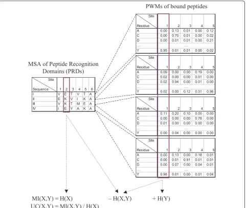
MSA of Peptide Recognition
Domains (PRDs)
PWMs of bound peptides
H(X)
– H(X,Y)
+ H(Y)
MI(X,Y) =
UC(X,Y) = MI(X,Y) / H(X)
Based on the concepts of entropy and joint entropy, the mutual information between two random variables Xand Yis defined as follows:
MI(X,Y) =H(X) +H(Y)− H(X,Y). (3)
2.2 Adapting mutual information to the identification of SDRs
Suppose we are given an MSA A with n rows and m columns. Each row corresponds to a PRD sequence and each column corresponds to a site of the alignment. We useAijto denote the residue at site jof sequencei. The
residues at a site can be viewed as a sample drawn from a distribution of residues specific to the site. LetAjbe a
random variable for the residues at sitej. Using Formula 1, we can calculate the entropy of Ajby replacing p(x)
with the sample distribution, pj(x), defined by the
fre-quency of each residue at the site:
pj(x) = n
i=11(Aij=x)
n , (4)
where1is the indicator function with1(true) = 1and
1(false) = 0.
The entropy can be interpreted as the uncertainty of which residue we would encounter at the site if we ran-domly draw a sequence from the MSA, where uncertainty here is mathematically quantified by the number of bits needed to encode the information on average. A comple-tely conserved site has an entropy of zero, and indeed there is no uncertainty as the residue being drawn must always be the same. A site with equal probability of all 20 residues has the maximum possible entropy of one. In general, a more diverse site has a larger entropy.
Similarly, suppose we are given a set of n aligned PWMsWeach withwsites. Theith PWM represents the peptides bound by theith PRD in the MSA (Figure 1). DenoteWik(y) as the probability of residueyat thekth
site of theith PWM. LetWkbe a random variable for the
residues at sitek. Again, we can calculate the entropy of Wkusing Formula 1 by replacingp(x) with the expected
probability ofyin the different PWMs,pk(y), assuming a
uniform distribution of the observed sequences:
pk(y) = n
i=1Wik(y)
n . (5)
Now, we can compute the joint entropy of sitejin the MSA and sitek in the PWMs using Formula 2, based on the sample distribution ofAjand the probabilities
Wik(y):
pj,k(x,y) = n
i=11(Aij=x)Wij(y)
n . (6)
The joint entropy measures the uncertainty of which two residues we would encounter at MSA site j and PWM site kif we randomly draw a sequence from the MSA and get its corresponding PWM.
Finally, we can compute the mutual information between MSA sitejand PWM sitekusing Formula 3. SinceH(X,Y) is the uncertainty of the two sites that per-sists even when we consider them together, subtracting it fromH(X) + H(Y) gives the uncertainty that is elimi-nated by considering the two sites together. In other words, mutual information measures the information shared by the two sites. A larger mutual information indi-cates a stronger dependency between them. This could indicate a functional or structural relationship between the two sites. For example, it could suggest that the two sites coevolve in the sense that when the residue at the MSA site is changed, binding strength is restored by hav-ing a correspondhav-ing change at the PWM site.
2.3 Handling uneven sequence representation
In many cases, the input MSA for studying residue covar-iation has an uneven representation of sequences from different clades. For example, it is common to have more sequences from model organisms or species that are bet-ter studied, and fewer sequences from other species. As a result, the MSA could contain many sequences that are highly similar, and few that are significantly different. Each of the highly similar sequences contributes little additional information, but still has an equal amount of influence on the calculation of mutual information under the assumption of a uniform distribution of the observed sequences. Consequently, they could mask the novel information from low abundance sequences. To counter-act this effect, we associate weights with the sequences so that the more unique ones receive higher weights. Statis-tically, it is equivalent to placing a prior distribution to the observed sequences if the weights are normalized to take values between zero and one and have a sum of one. Here we assume there is an external procedure for deter-mining the weights. For instance, one possible way is to construct a phylogenetic tree from the sequences in the MSA, and then recursively distribute the total weight to different branches of the tree, so that each sequence in the crowded branches will receive a smaller share of weights [44]. Suppose the procedure assignsaias the
weight of sequencei, we can redefinepj(x),pk(y) andpj,k
(x,y) as follows:
pj(x) = n
i=11(Aij=x)αi n
i=1αi
, (7)
pk(y) = n
i=1αiWik(y) n i=1αi
pj,k(x,y) = n
i=11(Aij=x)αiWik(y) n
i=1αi
. (9)
Entropy, joint-entropy and mutual information can then be calculated using these new definitions of probabilities.
2.4 Handling uneven site conservation
A potential issue of the mutual information measure is that a pair of unrelated sites could have even higher mutual information than a pair of truly covarying sites if the unrelated sites are individually much more diverse than the covarying sites. This is illustrated by the hypothetical example shown in Table 1. For simplicity, suppose the sequences all have equal weights and the base of logarithms is two. Sites 1 and 2 are truly covary-ing. When site 1 changes from the non-polar residue alanine in sequences I and II to the polar residue threo-nine in sequences III and IV, site 2 simultaneously changes from the non-polar residue valine to the polar residue tyrosine. The entropy of each of the two sites is one and the mutual information between them is also one, the maximum possible value given the two indivi-dual entropy values. On the other hand, sites 3 and 4 are random, unrelated sites. The entropy values of them are 2 and 1.5, respectively, and their mutual information is 1.5, which is higher than the mutual information between sites 1 and 2 due to larger entropy values of sites 3 and 4.
To deal with this problem, various kinds of normaliza-tion have been proposed to penalize overly diverse sites [34]. We will focus on the uncertainty coefficient [45], which was found to be one of the best normalized mutual information scores in our preliminary study. For an MSA site Aj and a PWM site Wk, the uncertainty
coefficient is defined as follows:
UC(Aj,Wk) =
MI(Aj,Wk)
H(Aj)
. (10)
We have also tried handling the problem using a sta-tistical test. Specifically, we used mutual information as the test statistic to calculate how unlikely it is to get a
mutual information at least as large as the observed one under the null hypothesis that the two sites are indepen-dent. The distribution of mutual information can be obtained by permuting the residues of a site, or by using a chi-square approximation. It was shown that when n is large, (2 ln 2n)MI(X, Y) tends to have a chi-square distribution with(|D(X)| −1)2(|D(Y)| −1)2degrees of freedom [46,47]. It turns out that the results based on this statistical test were not better than using the simple normalization approach. We thus focus on the use of the uncertainty coefficient measure in handling uneven site conservation in the remaining of this paper.
Table 1 also demonstrates the tradeoff between the mutual information between two sites and their indivi-dual conservation, both of which are indicators of their functional importance. One may try to derive a measure that takes both into account, similar to what the Sequence Harmony method handles both the conserva-tion and similarity of two groups of residues simulta-neously, for the problem of identifying important residues that determine the functional differences of protein subfamilies [48]. We leave the derivation of a new covariation measure to a future study.
2.5 Predicting the PWMs of bound peptides
One important use of the covariation scores is to contri-bute towards predicting the PWMs of bound peptides. This can be done by aggregating the detected covaria-tion signals. Suppose we are given a new PRD sequence (the (n+ 1)th sequence) of the MSA Mwithout the cor-responding PWM of its bound peptides. We would like to predict the PWM based on then + 1 sequences in the MSA and the n known PWMs. We investigate the use of the covariation scores in this problem by compar-ing a prediction method that considers site covariation with two methods that do not.
A simple prediction method that does not take site covariation into account is to perform a weighted aver-aged of the known PWMs, where the weights are based on the similarity of the new PRD sequence and the ori-ginal ones. Specifically, the probability of finding residue yat site kof the bound peptides of the new PRD is pre-dicted by the following formula:
ˆ
W(n+1)k(y) = n
i=1s(i,n+ 1)Wik(y) n
i=1s(i,n+ 1)
, (11)
wheres(i, i’) is a similarity between sequences iandi’ in the MSA, such as their sequence identity:
s(i,i) =
m
j=11(Mij=Mij)
m . (12)
Using the covariation scores, we propose an alterna-tive way to define the similarity function. Each MSA site
Table 1 A hypothetical example MSA that illustrates the problem of uneven site conservation.
Sequence/Site 1 2 3 4
I A V A C
II A V C D
III T Y D E
IV T Y E C
receives a different weight in the calculation, where the weight depends on the covariation score between the site and the target PWM site k. In other words, the similarity scores(i, i’) is replaced with a new scoresk (i,
i’) that is specific tok:
ˆ
W(n+1)k(y) = n
i=1sk(i,n+ 1)Wik(y) n
i=1sk(i,n+ 1)
, (13)
sk(i,i) = m
j=11(Mij=Mij)UC(Aj,Wk) m
j=1UC(Aj,Wk)
, (14)
where the uncertainty coefficient UC(Aj, Wk) is
com-puted based on thenoriginal sequences.
We also investigated if prediction accuracy can be improved by using a more complex model. Specifically, we treated each MSA site as a categorical attribute and trained a regression tree model for each probability value in the PWM. The models were then applied to the new sequence to predict the PWM of its bound pep-tides. We implemented this method using REPTree of the Weka package [49].
To evaluate the effectiveness of the different meth-ods, we performed left-out validation as follows. Each time, we drew a random sample of PRDs to form the testing set. Each sequence in the testing set took turn to take the role of the (n + 1)th sequence. The sequences not included in the sample formed the training set. These sequences were used to compute covariation scores and train prediction models. The procedure was repeated 1,000 times for PDZ and SH3 and 50 times for kinase (due to the long running time) with different random training-testing splits, and the average performance of the trained models on the test-ing sets was recorded. To eliminate near-identical sequences in the training and testing sets, for sequences with 90% or more identity, we kept only one of them in the dataset before making the training-testing splits. As most of the synthetic PDZ sequences (described below) are highly similar, we excluded this dataset from this part of study.
Each predicted PWM was compared to the actual PWM, and a prediction error was computed as the root-mean-square difference between their distributions per site:
e(Wˆ(n+1),W˜(n+1)) =
w
k=1
y[Wˆ(n+1)k(y)− ˜W(n+1)k(y)]2
w , (15)
whereWˆ(n+1)andW˜(n+1)are the predicted and actual PWMs for the bound peptides of the testing sequence, respectively, and the inner summation is taken over all 20 amino acid residues.
3 Results
3.1 Application of the method to PDZ, SH3 and kinase domains
3.1.1 Natural PDZ domains
We obtained 33 class I human and worm PDZ domains from a recent large-scale study on the specificity map of PDZ domains [13]. Class I PDZ domains were defined by two positions on the ligand, with the patternX[T/S] XjCOOH, where X and j represent a residue and a
hydrophobe, respectively. In the same study, a number of SDRs were experimentally determined, allowing us to validate our prediction results. We focused on only one class of domains as the sequences in different classes are difficult to align due to divergence. The pairwise sequence identity ranges from 0.13 to 0.87, with an average of 0.28.
The binding profile of each domain, in the form of a PWM, was obtained from phage display experiments that expressed a random library of C-terminal peptides [13]. The domains were then aligned to form an MSA using ClustalW [50] followed by manual corrections of some obvious errors. A phylogenetic tree of the MSA sequences was constructed using Biopython’s Nexus module [51], and the tree was used to produce sequence weights according to a described algorithm [44]. The uncertainty coefficient between each MSA site and each site of the peptide PWMs was computed. To reduce noise and eliminate highly conserved sites that provide little information about covariation, we considered only sites with no gaps [52] and the most frequent residue occupying no more than half of the total sequence weights. This filtering was also applied to the other domain families described below.
The remaining unfiltered site pairs were then evalu-ated in two ways. First, their uncertainty coefficients were compared to their physical distances in the co-crystal structure 2H2C of ligand-bound human ZO-1 PDZ1 domain [53] in PDB [54]. Although proximity does not necessarily mean functional or structural dependency, it is usually used as a quick check in covar-iation studies [18,27,29,33]. It also provides a complete and unbiased alternative to the more costly experimen-tal validations.
Second, we examined the top-scoring site pairs, and compared them with known SDRs from a mutagenesis study [13] in which ten sites of the ERBB2IP-1 domain were mutated and the corresponding changes of peptide specificity reported. This comparison provides direct evaluation of our SDR identification procedure on the subset of sites that were tested in the assay.
3.1.2 Synthetic PDZ domains
were introduced at the ten sites, resulting in 61 varia-tions of the Erbin domain that are functional in recog-nizing some C-terminal heptapeptides [55]. As with the natural PDZ domains, we compared the uncertainty coefficients with the physical distances in the 2H2C PDB structure. Since the synthetic PDZ domains are 100% conserved at sites other than the ten selected ones, and have a specific set of mutations at the ten sites introduced by the mutagenesis experiments, their MSA exhibits some statistical properties different from those of the natural PDZ domains.
3.1.3 SH3 domains
We obtained 23 yeast SH3 domains and the correspond-ing PWMs of the bound peptides from phage display experiments from a recent study [38]. We aligned the PRDs based on a published structural alignment [15], and aligned the peptide PWMs based on both the gen-eral PxxP pattern and some published alignments [15,56]. The pairwise sequence identity ranges from 0.07 to 0.79, with an average of 0.24.
We applied the same prediction and evaluation proce-dures as in the case of PDZ, except that in this case we did not have large-scale mutagenesis data, and therefore the prediction results were only evaluated against the physical distances calculated from the crystal structure 1N5Z in PDB, which contains the yeast Pex13 SH3 domain bound to a Pex14 peptide [57], and the findings of some previous studies.
3.1.4 Kinase domains
We also obtained 149 serine/threonine protein kinase domains from four species (S. cerevisiae, H. sapiens,S. pombeandD. discoideum) and the PWMs of their cor-responding bound peptides [14]. The MSA was made using MUSCLE [58] followed by some manual correc-tions. The prediction results were evaluated against dis-tances calculated from the crystal structure 1ATP of mouse catalytic subunit of cAMP-dependent protein kinase complexed with Mn-ATP and a peptide inhibitor [59] in PDB, and some findings in previous studies. The pairwise sequence identity ranges from 0.09 to 0.92, with an average of 0.23.
3.2 Covariation score correlates with physical proximity and reconfirms previous findings
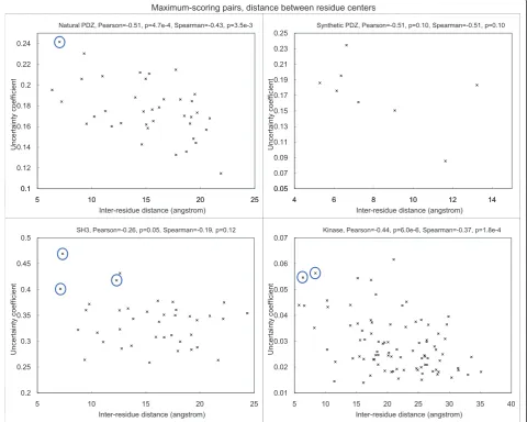
The covariation score between two sites is found to cor-relate negatively with the physical distance between them, regardless of the exact definition of the distance measure [see Additional file 1 Figure S1, Additional file 2 Figure S2 and Additional file 3 Figure S3 for the results] when the distance between residue centers, alpha carbon atoms and closest atoms minus their van der Waals’radii were used, respectively. All P-values were computed using Fisher transformation [60]. For PDZ, we have also compared the correlations based on
several other PDZ structures, and observed similar pat-terns [Additional file 4 Figure S4].
Since low-scoring pairs are more subject to noise, here for each PRD site, we focus on the peptide site that gives the highest uncertainty coefficient with it (Figure 2). The site pairs with the highest covariation scores are listed in Table 2.
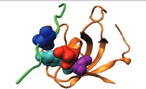
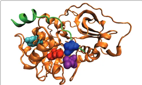
For natural PDZ domains, the highest-scoring pair (circled) is between Leu60 (b3:a1-1, structural nomen-clature from [53]) of the PDZ domain in 2H2C and position -1 of the binding motif on the bound peptide. These residues are in physical contact in the dimer structure (Figure 3, all visualization produced using VMD [61]). Interestingly, it has been reported that the side chain at b3:a1-1 can contribute to the recognition of the -1 position of the motif [53], and in the crystal structure from Shank1, a salt bridge is observed between Asp(b3:a1-1) of the PDZ domain and Arg(-1) of the ligand [62]. Our covariation analysis has thus identified these verified SDRs of the PDZ domains in silico.
For SH3 domains, the highest-scoring pair is between Asn71 of the SH3 domain and Leu7 (P+2 residue) of the ligand in 1N5Z. These residues are in close physical proximity in the crystal structure (Figure 4). Interest-ingly, we found that the MSA residues in the first (Asn71), third (Ile73) and fourth (Tyr72) top-scoring pairs are consecutive in the protein sequence, and two of them are close to Leu7 of the ligand. In a previous study [63], the corresponding residue of Tyr72 on P53BP2, which is an unusual leucine, was hypothesized to cause the protein to bind a peptide very different from its usual ligands. However, mutating the leucine to tyrosine did not affect recognition specificity. As the corresponding residue of Ile73 on P53BP2 is also mutated from the class consensus, and the covariation scores for Asn71 and Ile73 are also high, the three resi-dues may have some combined effects in affecting recognition specificity.
It is also known that the residue corresponding to Glu31 in the RT loop of SH3 domains is a major deter-minant of the identity of the P-3 residue of the ligand [64,65]. Since the P-3 residue does not exist in the 1N5Z structure, it is not included in the correlation plots. However, when we checked the covariation scores, indeed the ligand residue having the highest score with Glu31 is the P-3 residue. This observation illustrates the potential of our method to identify SDRs when struc-tural information is not available.
0.1 0.12 0.14 0.16 0.18 0.2 0.22 0.24
5 10 15 20 25
U
nc
er
tai
nty
c
oef
fic
ient
Natural PDZ, Pearson=-0.51, p=4.7e-4, Spearman=-0.43, p=3.5e-3
0.05 0.07 0.09 0.11 0.13 0.15 0.17 0.19 0.21 0.23 0.25
4 6 8 10 12 14
U
nc
er
tai
nty
c
oef
fic
ient
Synthetic PDZ, Pearson=-0.51, p=0.10, Spearman=-0.51, p=0.10
0.1
5 10 15 20 25
Inter-residue distance (angstrom)
0.05
4 6 8 10 12 14
Inter-residue distance (angstrom)
0.2 0.25 0.3 0.35 0.4 0.45 0.5
5 10 15 20 25
U
nc
er
tai
nty
c
oef
fic
ient
SH3, Pearson=-0.26, p=0.05, Spearman=-0.19, p=0.12
0.01 0.02 0.03 0.04 0.05 0.06 0.07
5 10 15 20 25 30 35 40
U
nc
er
tai
nty
c
oef
fic
ient
Kinase, Pearson=-0.44, p=6.0e-6, Spearman=-0.37, p=1.8e-4 0D[LPXPVFRULQJSDLUVGLVWDQFHEHWZHHQUHVLGXHFHQWHUV
0.2
5 10 15 20 25
Inter-residue distance (angstrom)
0.01
5 10 15 20 25 30 35 40
Inter-residue distance (angstrom)
Figure 2Correlation between covariation score and physical proximity between each PRD site and its highest-scoring peptide PWM position for the four types of PRDs. Selected top-scoring pairs discussed in the text are circled. The correlation for synthetic PDZ was not statistically significant due to the small number of data points involved.
Table 2 Site pairs with the highest covariation scores.
Domain family Ref. PDB Structure rank Natural 2H2C Synthetic PDZ 2H2C
SH3 1N5Z
Kinase 1ATP
PRD Peptide PRD Peptide PRD Peptide PRD Peptide
1 Leu60 Trp119 His88 Thr118 Asn71 Leu7 Tyr229 Ile22
2 Val55 Thr117 Ala37 Thr117 Leu34 Leu7 Leu205 Ile22
3 Ala76 Thr117 Ser39 Thr117 Ile73 Leu7 Leu198 Ile22
4 Pro30 Trp119 Leu60 Thr117 Tyr72 Leu7 Lys189 Ile22
5 Ser98 Trp119 Val92 Thr118 Lys35 Leu7 Phe129 Ile22
6 Ala89 Thr117 Ser57 Thr117 Ser44 Leu7 Glu230 Ile22
7 Gln93 Trp119 Asp58 Thr117 Lys61 Leu7 Glu203 Ile22
8 Asp58 Thr117 Phe34 Thr118 Ile68 Leu7 Ile180 Ile22
9 Ala37 Thr117 Leu20 Leu7 Phe187 Arg19
10 Lys103 Trp119 Ala17 Leu7 Cys199 Ile22
Leu198 and Leu205 were previously found to have hydro-phobic interactions with position +1 of the peptide [66,67]. They are two of the three residues (the third being Pro202, which has the highest covariation score with posi-tion +1 as compared to other peptide posiposi-tions) that form the binding pocket for position +1 of the peptide, and con-tributes to the positioning of a significant portion (-3 to +1 positions) of the bound peptide [67].
We found that the pair between Tyr229 and position +1, besides being the top-scoring pair when uncertainty coefficient was used as the covariation score, was also consistently one of the highest-scoring pairs when cov-ariation was measured by other normalized forms of mutual information. These consistent results made us hypothesize that Tyr229 is also involved in determining binding specificity of the kinase domain, and has long-range coupling with position +1 of the bound peptide. In a previous study, this residue was predicted to be involved in linking nucleotide binding and peptide bind-ing in protein kinases [68]. Interestbind-ingly, in the study Tyr229 and Leu205 are predicted to belong to the same special network (termed the θ-shaped network) of related residues. It thus might be the case that Tyr229 acts through the residues in the network to affect the recognition of the +1 position of the peptide.
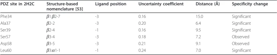
3.3 PDZ predictions are consistent with mutagenesis results
We further validated our predictions with the natural PDZ domains by using a mutagenesis dataset from a domain specificity study [13] as described in the Materi-als and Methods section.
Figure 3Top-scoring residue pair for the natural PDZ domain. The PDZ domain (orange) and the ligand (green) in the biological assembly (dimer) of the PDB structure 2H2C are shown. The top-scoring residue pair between Leu60 (red) on the domain and position -1 of the binding motif on the ligand (blue) are in physical contact.
In our covariation calculation, among the ten mutated sites, four were filtered as they were too con-served (Ile36 [b2-1], Ile38 [b2-3], His88 [a2-1] and Val92 [a2-5]). Interestingly, in the mutagenesis study, indeed no significant changes to the peptide PWM could be observed for Ile36 and Ile38. The high con-servation of them is thus probably caused by a struc-tural or functional role that is independent of peptide binding.
For the remaining six sites, we examined their max-imum-scoring peptide residues. Four out of the six had significant changes of the PWM at the predicted positions in the mutagenesis study (Table 3), includ-ing the top-scorinclud-ing pair among all predictions, between Leu60 and position -1. For the remaining two, changes were also observed, albeit with lower statistical significance.
The predicted pair between Phe34 and position -3 has a distance of 15.0 Å in the PDB structure 2H2C. If the SDRs of different class I PDZ domains are similar, this predicted pair is another example that suggests our cov-ariation method could potentially identify physically dis-tant SDR pairs.
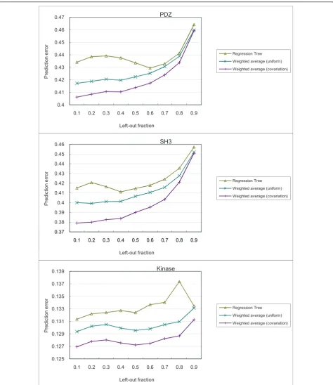
3.4 Covariation scores improve prediction of bound peptide profiles
As described in the Materials and Methods section, we compared three different methods for predicting the profiles of bound peptides. The prediction results are shown in Figure 6.
In general, the prediction error is lower with a smaller fraction of PRDs left out for testing. This is expected, as having more PRDs in the training set allows for more accurate computation of covariation scores and the con-struction of more informative prediction models.
In all cases, the weighted average method using covar-iation scores outperformed regression tree and the weighted average method using uniform scoring. This suggests that the covariation score provided a meaning-ful way to weight usemeaning-ful features (that is, PRD sites) for predicting residue distributions of the bound peptides. Interestingly, while the regression tree method also per-formed feature weighting, in general it perper-formed worse than both weighted average methods. The low perfor-mance could be due to over-fitting, as the regression trees are rich in expressive power while the numbers of PRDs in the datasets are small.
4 Discussion
We present a novel way to use underutilized data. Our method is a valuable tool for exploring the specificity space of PRDs. Moreover, as the amount of specificity data is increasing swiftly, also due to the advent of next-generation sequencing and its applications to phage dis-play [69], our method will prove even more valuable to make optimal use of this kind of data.
We think our covariation approach can be used in conjunction with other methods to improve SDR predic-tions. Since most current approaches are based on force fields and structural methods, our method opens up a new perspective for improvement. As shown in the recent PRD specificity prediction challenge of the DREAM4 competition [70], most likely a combination of structural and statistical methods will be most suc-cessful at predicting specificities.
One limitation of our approach is that it does not consider possible relationships between different residue pairs. As multiple PRD residues could simultaneously interact with a peptide residue and multiple peptide residues could simultaneously interact with a PRD resi-due [71], binding specificity could be more accurately modeled by considering covariation signals between resi-due groups. Furthermore, since a resiresi-due could have an indirect covariation signal with another residue through an intermediate residue [42], performing residue group analysis could help filter out these non-SDR intermedi-ates that have relatively high covariation signals.
Another limitation of our approach is that it fails to identity SDRs that are highly conserved. Indeed we have observed that in PDZ, some highly conserved residues (for examplea2-1) are physically close to the peptide and have been experimentally shown to affect binding specifi-city when mutated [13] [Additional file 5 Figure S5]. On the other hand, some SDRs are not highly conserved, but exhibit strong covariation patterns with peptide residues (for exampleb3:a1-1). Future approaches could improve upon our current method by combining information about conservation with covariation to identity SDRs.
There are also PRD residues that determine binding but not binding specificity in that if they are mutated, the resulting effect on binding cannot be restored by a second mutation. For instance, if a residue is critical to the protein structure, mutating it could seriously affect the stability of the protein, which in turn affects pep-tide binding. These residues are likely to be very con-served, and thus would not be ranked high by our method.
A third limitation of our approach, and more gener-ally of all covariation analysis methods based on a multiple sequence alignment, is the dependency on the alignment quality. Different alignments could give very different results, especially for alignments with many gaps. To cope with this issue, we have used a pub-lished alignment for SH3, and made some manual cor-rections to the PDZ and kinase alignments generated by computer programs. Future approaches should try to minimize the effect of alignment quality on the ana-lysis results.
While we have attempted to predict peptide PWMs, it is also possible to predict interactions given a PRD and a peptide. This problem has recently been studied using some large-scale datasets [9,52]. It would be interesting to study how the concept of covariation can be incorpo-rated into these prediction methods.
Conclusions
We have presented a novel approach that utilizes an as of yet underused source of data. We have shown that the covariation scores are consistent with pre-vious findings from both a large-scale study, and other individual experiments. In addition, we have identified a number of candidate SDRs in a ranked list for future experimental validation. In particular, with the top-scoring pairs from natural PDZ domains and kinase domains both verified in previous work, the SH3 top-scoring pairs are good candidates for testing their roles in determining the binding specificity of SH3 domains.
Table 3 Validation results of our PDZ predictions.
PDZ site in 2H2C Structure-based nomenclature [53]
Ligand position Uncertainty coefficient Distance (Å) Specificity change
Phe34 b1:b2-7 -3 0.16 15.0 Significant
Ala37 b2-2 -3 0.20 6.4 Significant
Ser39 b2-4 -1 0.16 9.5 Significant
Ser57 b3-4 -3 0.18 7.2 Observed
Asp58 b3-5 -3 0.21 9.1 Observed
Leu60 b3:a1-1 -1 0.24 7.0 Significant
0.4 0.41 0.42 0.43 0.44 0.45 0.46 0.47
0.1 0.2 0.3 0.4 0.5 0.6 0.7 0.8 0.9
P
redi
ct
ion error
Left-out fraction
PDZ
Regression Tree Weighted average (uniform) Weighted average (covariation)
0.37 0.38 0.39 0.4 0.41 0.42 0.43 0.44 0.45 0.46
0.1 0.2 0.3 0.4 0.5 0.6 0.7 0.8 0.9
P
redi
ct
ion error
SH3
Regression Tree Weighted average (uniform) Weighted average (covariation)
0.37
0.1 0.2 0.3 0.4 0.5 0.6 0.7 0.8 0.9
Left-out fraction
0.125 0.127 0.129 0.131 0.133 0.135 0.137 0.139
0.1 0.2 0.3 0.4 0.5 0.6 0.7 0.8 0.9
P
redi
ct
ion error
Left-out fraction
Kinase
Regression Tree Weighted average (uniform) Weighted average (covariation)
Additional material
Additional file 1: Correlation between covariation score and physical proximity between each PRD site and each PWM position for the three types of PRDs when distances are computed between residue centers. Figure S1.
Additional file 2: Correlation between covariation score and physical proximity between each PRD site and each PWM position for the three types of PRDs when distances are computed between alpha carbon atoms. Figure S2.
Additional file 3: Correlation between covariation score and physical proximity between each PRD site and each PWM position for the three types of PRDs when distances are computed between the closest atoms minus their van der Waal’s radii. Figure S3
Additional file 4: Correlation between covariation score and physical proximity between each PRD site of a PDZ domain and each PWM position when distances are computed between alpha carbon atoms in four different PDB structures. Figure S4
Additional file 5: Conservation and distance to the closest peptide residue of each PDZ domain site. For each site on the domain, we computed the total sequence weight of the sequences having a particular amino acid at the site. The conservation of the site is defined by the maximum of such total weights normalized by the total sequence weight of all sequences in the MSA. Figure S5.
Abbreviations
MI: mutual information; MSA: multiple sequence alignment; PRDs: peptide recognition domains; PWM: position weight matrix; SCA: statistical coupling analysis; SDRs: specificity determining residues; UC: uncertainty coefficient.
Acknowledgements
We thank Xiaodan Fan for helpful discussions and Nisa Dar for technical assistance. We acknowledge support from the Natural Science and Engineering Research Council, the Ontario Research Fund, the Connaught Foundation, the NIH, the AL Williams Professorship funds, and the Yale University Biomedical High Performance Computing Center.
Author details 1
Department of Molecular Biophysics and Biochemistry, Yale University, New Haven, CT, USA.2Department of Computer Science and Engineering, The Chinese University of Hong Kong, Hong Kong.3Terrence Donnelly Centre for Cellular and Biomolecular Research, University of Toronto, Toronto, Ontario, Canada.4Department of Pharmacology, Yale University, New Haven, CT, USA. 5Program in Computational Biology and Bioinformatics, Yale University, New Haven, CT, USA.6Department of Computer Science, Yale University, New Haven, CT, USA.7Banting and Best Department of Medical Research, University of Toronto, Toronto, Ontario, Canada.8Department of Molecular Genetics, University of Toronto, Toronto, Ontario, Canada.9Department of Computer Science, University of Toronto, Toronto, Ontario, Canada.
Authors’contributions
All authors conceived the project and design. KY, LU, SS and PK prepared the data. KY, LU, SS and XH implemented the algorithms, performed the computational experiments, and analyzed the results. KY, LU, MG and PK wrote the paper. All authors read and approved the document.
Competing interests
The authors declare that they have no competing interests.
Received: 26 July 2011 Accepted: 11 August 2011 Published: 11 August 2011
References
1. Pawson T, Nash P:Assembly of cell regulatory systems through protein interaction domains.Science2003,300(5618):445-452.
2. Pawson T, Linding R:Network medicine.FEBS Lett2008,582:1266-1270.
3. Alto NM, Shao F, Lazar CS, Brost RL, Chua G, Mattoo S, McMahon SA, Ghosh P, Hughes TR, Boone C, Dixon JE:Identification of a bacterial type III effector family with G protein mimicry functions.Cell2006, 124:133-145.
4. Doorbar J:Molecular biology of human papillomavirus infection and cervical cancer.Clin Sci2006,110:525-541.
5. Carducci M, Licata L, Peluso D, Castagnoli L, Cesareni G:Enriching the viral-host interactomes with interactions mediated by SH3 domains.Amino Acids2010,38(5):1541-1547.
6. Lam HYK, Kim PM, Mok J, Tonikian R, Sidhu SS, Turk BE, Snyder M, Gerstein MB:MOTIPS: automated motif analysis for predicting targets of modular protein domains.BMC Bioinformatics2010,11:243.
7. Tonikian R, Zhang Y, Boone C, Sachdev SS:Identifying specificity profiles for peptide recognition modules from phage-displayed peptide libraries. Nat Protocols2007,2(6):1368-1386.
8. Landgraf C, Panni S, Montecchi-Palazzi L, Castagnoli L, Schneider-Mergener J, Volkmer-Engert R, Cesareni G:Protein interaction networks by proteome peptide scanning.PLoS Biol2004,2:e14.
9. Stiffler MA, Chen JR, Grantcharova VP, Lei Y, Fuchs D, Allen JE,
Zaslavskaia LA, MacBeath G:PDZ domain binding selectivity is optimized across the mouse proteome.Science2007,317(5836):364-369.
10. Kaushansky A, Allen JE, Gordus A, Stiffler MA, Karp ES, Chang BH, MacBeath G:Quantifying protein-protein interactions in high throughput using protein domain microarrays.Nat Protocols2010,5(4):773-790. 11. Hutti JE, Jarrell ET, Chang JD, Abbott DW, Storz P, Toker A, Cantley LC,
Turk BE:A rapid method for determining protein kinase phosphorylation specificity.Nat Methods2004,1:27-29.
12. Li L, Shakhnovich EI, Mirny LA:Amino acids determining enzyme-substrate specificity in prokaryotic and eukaryotic protein kinases.Proc Natl Acad Sci USA2003,100(8):4463-4468.
13. Tonikian R, Zhang Y, Sazinsky SL, Currell B, Yeh JH, Reva B, Held HA, Appleton BA, Evangelista M, Wu Y, Xin X, Chan AC, Seshagiri S, Lasky LA, Sander C, Boone C, Bader GD, Sidhu SS:A specificity map for the PDZ domain family.PLoS Biol2008,6:e239.
14. Mok J, Kim PM, Lam HYK, Piccirillo S, Zhou X, Jeschke GR, Sheridan DL, Parker SA, Desai V, Jwa M, Cameroni E, Niu H, Good M, Remenyi A, Ma JLN, Sheu YJ, Sassi HE, Sopko R, Chan CSM, Virgilio CD, Hollingsworth NM, Lim WA, Stern DF, Stillman B, Andrews BJ, Gerstein MB, Snyder M, Turk BE: Deciphering protein kinase specificity through large-scale analysis of yeast phosphorylation site motifs.Sci Signal2010,3:ra12.
15. Cesareni G, Panni S, Nardelli G, Castagnoli L:Can we infer peptide recognition specificity mediated by SH3 domains?FEBS Lett2002, 513:38-44.
16. Beuming T, Farid R, Sherman W:High-energy water sites determine peptide binding affinity and specificity of PDZ domains.Protein Sci2009, 18:1609-1619.
17. Lichtarge O, Bourne HR, Cohen FE:An evolutionary trace method defines binding surfaces common to protein families.J Mol Biol1996, 257:342-358.
18. Göbel U, Sander C, Schneider R, Valencia A:Correlated mutations and residue contacts in proteins.Proteins: Structure, Function, and Bioinformatics 1994,18:309-317.
19. Shindyalov IN, Kolchanov NA, Sander C:Can three-dimensional contacts in protein structures be predicted by analysis of correlated mutations? Protein Eng1994,7(3):349-358.
20. Lockless SW, Ranganathan R:Evolutionarily conserved pathways of energetic connectivity in protein families.Science1999, 286(5438):295-299.
21. Goh CS, Bogan AA, Joachimiak M, Walther D, Cohen FE:Co-evolution of proteins with their interaction partners.J Mol Biol2000,299:283-293. 22. Pazos F, Valencia A:In silico two-hybrid system for the selection of
physically interacting protein pairs.Proteins: Structure, Function, and Bioinformatics2002,47:219-227.
23. Ramani AK, Marcotte EM:Exploiting the co-evolution of interacting proteins to discover interaction specificity.J Mol Biol2003,327:273-284. 24. Pazos F, Ranea JAG, Juan D, Sternberg MJE:Assessing protein co-evolution
in the context of the tree of life assists in the prediction of the interactome.J Mol Biol2005,352(4):1002-1015.
26. Socolich M, Lockless SW, Russ WP, Lee H, Gardner KH, Ranganathan R: Evolutionary information for specifying a protein fold.Nature2005, 437(7058):512-518.
27. Gloor GB, Martin LC, Wahl LM, Dunn SD:Mutual information in protein multiple sequence alignments reveals two classes of coevolving positions.Biochemistry2005,44(19):7156-7165.
28. Fuchs A, Martin-Galiano AJ, Kalman M, Fleishman S, Ben-Tal N, Frishman D: Co-evolving residues in membrane proteins.Bioinformatics2007, 23(24):3312-3319.
29. Halperin I, Wolfson H, Nussinov R:Correlated mutations: advances and limitations. A study on fusion proteins and on the Cohesin-Dockerin families.Proteins: Structure, Function, and Bioinformatics2006,63:832-845. 30. Pazos F, Helmer-Citterich M, Ausiello G, Valencia A:Correlated mutations
contain information about protein-protein interaction.J Mol Biol1997, 271:511-523.
31. Shulman AI, Larson C, Mangelsdorf DJ, Ranganathan R:Structural determinants of allosteric ligand activation in RXR heterodimers.Cell 2004,116:417-429.
32. Pollock DD, Taylor WR, Goldman N:Coevolving protein residues: maximum likelihood identification and relationship to structure.J Mol Biol1999,287:187-198.
33. Dekker JP, Fodor A, Aldrich RW, Yellen G:A perturbation-based method for calculating explicit likelihood of evolutionary co-variance in multiple sequence alignments.Bioinformatics2004,20(10):1565-1572.
34. Martin LC, Gloor GB, Dunn SD, Wahl LM:Using information theory to search for co-evolving residues in proteins.Bioinformatics2005, 21(22):4116-4124.
35. Larson SM, Nardo AAD, Davidson AR:Analysis of covariation in an SH3 domain sequence alignment: applications in tertiary contact prediction and the design of compensating hydrophobic core substitutions.J Mol Biol2000,303:433-4446.
36. Galitsky B:Revealing the set of mutually correlated positions for the protein families of immunoglobulin fold.In Silico Biol2003,3(0022). 37. Dimmic MW, Hubisz MJ, Bustamante CD, Nielsen R:Detecting coevolving
amino acid sites using Bayesian mutational mapping.Bioinformatics2005, 21(Suppl. 1):i126-i135.
38. Tonikian R, Xin X, Toret CP, Gfeller D, Landgraf C, Panni S, Paoluzi S, Castagnoli L, Currell B, Seshagiri S, Yu H, Winsor B, Vidal M, Gerstein MB, Bader GD, Volkmer R, Cesareni G, Drubin DG, Kim PM, Sidhu SS, Boone C: Bayesian modeling of the yeast SH3 domain interactome predicts spatiotemporal dynamics of endocytosis proteins.PLoS Biol2009, 7:21000218.
39. Yip KY, Patel P, Kim PM, Engelman DM, McDermott D, Gerstein M:An integrated system for studying residue coevolution in proteins. Bioinformatics2008,24(2):290-292.
40. Halabi N, Rivoire O, Leibler S, Ranganathan R:Protein sectors: evolutionary units of three-dimensional structure.Cell2009,138:774-786.
41. Weigt M, White RA, Szurmant H, Hoch JA, Hwa T:Identification of direct residue contacts in protein-protein interaction by message passing.Proc Natl Acad Sci USA2009,106:67-72.
42. Burger L, van Nimwegen E:Disentangling direct from indirect co-evolution of residues in protein alignments.PLoS Comput Biol2010,6: e1000633.
43. Cover TM, Thomas JA:Elements of Information Theory.2 edition. New York: Wiley-Interscience; 2006.
44. Gerstein M, Sonnhammer ELL, Chothia C:Volume changes in protein evolution.J Mol Biol1994,236:1067-1078.
45. Press WH, Flannery BP, Teukolsky SA, Vetterling WT:Numerical Recipes: The Art of Scientific ComputingCambridge: Cambridge University Press; 1986. 46. Goebel B, Dawy Z, Hagenauer J, Mueller JC:An approximation to the
distribution of finite sample size mutual information estimates. Proceedings of IEEE International Conference on Communications (ICC’05), Volume 22005, 1102-1106.
47. Aktulga HM, Kontoyiannis I, Lyznik LA, Szpankowski L, Grama AY, Szpankowski W:Identifying statistical dependence in genomic sequence via mutual information estimates.EURASIP J Bioinformatics Syst Biol2007, 2007:14741.
48. Pirovano W, Feenstra KA, Heringa J:Sequence comparison by sequence harmony identifies subtype-specific functional sites.Nucleic Acids Res 2006,34(22):6540-6548.
49. Hall M, Frank E, Holmes G, Pfahringer B, Reutemann P, Witten IH:The WEKA data mining software: an update.SIGKDD Explorations2009, 11:10-18.
50. Thompson JD, Higgins DG, Gibson TJ:CLUSTAL W: improving the sensitivity of progressive multiple sequence alignmnet through sequence weighting, position-specific gap penalties and weight matrix choice.Nucleic Acids Res1994,22(22):4673-4680.
51. Cock PJA, Antao T, Chang JT, Chapman BA, Cox CJ, Dalke A, Friedberg I, Hamelryck T, Kau F, Wilczynski B, de Hoon MJL:Biopython: freely available python tools for computational molecular biology and bioinformatics. Bioinformatics2009,25(11):1422-1423.
52. Chen JR, Chang BH, Allen JE, Stiffler MA, MacBeath G:Predicting PDZ domain-peptide interactions from primary sequences.Nat Biotechnol 2008,26(9):1041-1045.
53. Appleton BA, Zhang Y, Wu P, Yin JP, Hunziker W, Skelton NJ, Sidhu SS, Wiesmann C:Comparative structural analysis of the Erbin PDZ domain and the first PDZ domain of ZO-1. Insights into determinants of PDZ domain specificity.J Biol Chem2006,281(31):22312-22320.
54. Berman HM, Westbrook J, Feng Z, Gilliland G, Bhat TN, Weissig H, Shindyalov IN, Bourne PE:The protein data bank.Nucleic Acids Res2000, 28.
55. Ernst A, Sazinsky SL, Hui S, Currell B, Dharsee M, Seshagiri S, Bader GD, Sidhu SS:Rapid evolution of functional complexity in a domain family. Sci Signal2009,2:ra40.
56. Tong AHY, Drees B, Nardelli G, Bader GD, Brannetti B, Castagnoli L, Evangelista M, Ferracuti S, Nelson B, Paoluzi S, Quondam M, Zucconi A, Hogue CWV, Fields S, Boone C, Cesareni G:A combined experimental and computational strategy to define protein interaction networks for peptide recognition modules.Science2002,295(5553):321-324. 57. Douangamath A, Filipp FV, Klein AT, Barnett P, Zou P, Voorn-Brouwer T,
Vega M, Mayans OM, Sattler M, Distel B, Wilmanns M:Topography for independent binding ofα-helical and PPII-helical ligands to a peroxisomal SH3 domain.Mol Cell2002,10:1007-1017.
58. Edgar RC:MUSCLE: multiple sequence alignment with high accuracy and high throughput.Nucleic Acids Res2004,32(5):1792-1797.
59. Zheng J, Trafny EA, Knighton DR, Xuong NH, Taylor SS, Eyck LFT, Sowadski JM:A refined crystal structure of the catalytic subunit of cAMP-dependent protein kinase complexed with MnATP and a peptide inhibitor.Acta Crystallogr D1993,49:362-365.
60. Fisher RA:On the‘probable error’of a coefficient of correlation deduced from a small sample.Metron1921,1:3-32.
61. Humphrey W, Dalke A, Schulten K:VMD: visual molecular dynamics.J Mol Graph1996,14:33-38.
62. Im YJ, Lee JH, Park SH, Park SJ, Rho SH, Kang GB, Kim E, Eom SH:Crystal structure of the Shank PDZ-ligand complex reveals a class I PDZ interaction and a novel PDZ-PDZ dimerization.J Biol Chem2003, 278(48):48099-48104.
63. Espanel X, Sudol M:Yes-associated protein and p53-binding protein-2 interact through their WW and SH3 domains.J Biol Chem2001, 276(17):14514-14523.
64. Musacchio A, Saraste M, Wilmanns M:High-resolution crystal structures of tyrosine kinase SH3 domains complexed with proline-rich peptides.Nat Struct Mol Biol1994,1(8):546-551.
65. Yu H, Chen JK, Feng S, Dalgarno DC, Brauer AW, Schrelber SL:Structural basis for the binding of proline-rich peptides to SH3 domains.Cell1994, 76:933-945.
66. Smith CM, Radzio-Andzelm E, Akamine P, Taylor SS:The catalytic subunit of cAMP-dependent protein kinase: prototype for an extended network of communication.Prog Biophys Mol Biol1999,71:313-341.
67. Johnson DA, Akamine P, Radzio-Andzelm E, Taylor SS:Dynamics of cAMP-dependent protein kinase.Chem Rev2001,101(8):2243-2270.
69. Gfeller D, Butty F, Wierzbicka M, Verschueren E, Vanhee P, Huang H, Ernst A, Dar N, Stagljar I, Serrano L, Sidhu SS, Bader GD, Kim PM:The multiple-specificity landscape of modular peptide recognition domains.Mol Syst Biol2011,7:484.
70. DREAM4, Challenge 1: Peptide Recognition Domain (PRD) Specificity Prediction. 2009 [http://wiki.c2b2.columbia.edu/dream/index.php/D4c1]. 71. London N, Movshovitz-Attias D, Schueler-Furman O:The structural basis of
peptide-protein binding strategies.Structure2010,18:188-199.
doi:10.1186/1741-7007-9-53
Cite this article as:Yipet al.:Identification of specificity determining
residues in peptide recognition domains using an information theoretic approach applied to large-scale binding maps.BMC Biology20119:53.
Submit your next manuscript to BioMed Central and take full advantage of:
• Convenient online submission
• Thorough peer review
• No space constraints or color figure charges
• Immediate publication on acceptance
• Inclusion in PubMed, CAS, Scopus and Google Scholar
• Research which is freely available for redistribution