1. Introduction
orsal cochlear nucleus neurons represent %#" " to auditory nerve stimulation (Godfrey et al. 1975; Pfeiffer1966; Rhode et al. 1983; Rhode & Smith 1986; Manis 1990; Gold-berg & Brownell 2003; Street & Manis 2007). A fast transient potassium current, I kif , in DCN neurons was ## $ "# -ing patterns (Kanold & Manis 1999). The role of I kif in producing such patterns was proved by a detailed ten-variable physiological model, Km model (Kanold & Manis 2000) and Later on by a reduced version of it, Km-LIF model (Meng & Rinzel 2010). The latter model $ $ # "$"-cal mechanism on a three variable model.
Zahra Daneshparvar 1, Mohammad Reza Daliri 2,*
1. Biomedical Engineering Department, Faculty of Electrical Engineering, Iran University of Science and Technology (IUST), Tehran, Iran. 2 Biomedical Engineering Department and Iran Neural Technology Center, Faculty of Electrical Engineering, Iran University of Science and Technology (IUST), 16846-13114 Tehran, Iran
D
* Corresponding Author:
? &X # $" outward potassium currents in producing temporal aspects of dorsal cochlear Z` "$ & " $ "" pauser and the buildup. This conductance based model is a reduced version of X¦/^$#{_&" # of a detailed physiological model (Kanold & Manis, 2000). For our development & $ " $ "" !! & # # #!$" minimal model outputs in response to a hyperpolarizing stimulus fallowed by a depolarizing one with the data of KM-LIF model. The results conform well to the KM-LIF model with lower complexity.
A B S T R A C T
Article info:
Received:10 August 2011
First Revision: 20 August 2011
Accepted: 8 September 2011
The importance of transient potassium currents in ## # by several researchers (Connor & Stevens 1971; Byrne 1980; Av-Ron 1994; Rush & Rinzel 1995; Kanold & Manis 1999; Rothman & Manis 2003). These currents mostly activate fast and inactivate slowly. The slow # #!+ " # { { Meng & Rinzel 2010), applying a hyperpolarizing stim-ulus followed by a depolarizing one leads to long delays #!< """-tion increases inactiva"""-tion gate variables of potassium currents, and then depolarization maximizes activation gate variables. This provides enough transient potas-sium currents to re-polarize the membrane and not to allow membrane voltage increases, thus limits the spike Key Words:
Neural Modeling,
Transient Potassium Current, Neural Firing,
Latency,
Winter 2012, Volume 3, Number 2
Basic and Clinical
generation mechanism. While inactivation variable de-creases slowly, potassium current inactivates again and spiking is started.
For our development, we started with a reduced # $ # { Rinzel 2010) and minimized it in the way that the mod-el with minimum state variables could still represent the desired features coming next. Minimal models for their least complexity can be really useful in large-scale simulations, although they may not be able to explain all physiological aspects of a neuron. Instead, a detailed physiological model, including all neuron currents and ion channel dynamics, may justify lots of neural events but the complex dynamics makes trouble in large scale simulations.
/ $ #" -terns of dorsal cochlear neurons. There are three basic # " Z` "-per, pauser and buildup. Consecutive spiking without a delay is called chopper pattern but spiking after a long delay is called pauser pattern. If the delay happens after
"% # "% #" "!
During this study we generated all above patterns with our suggesting minimal model. The results compares well with those of KM-LIF & KM models (Meng & Rinzel 2010; Kanold & Manis, 2000). As mentioned in #{_ $ -ing properties of these models match well with both in vivo and in vitro responses (Kanold and Manis 2001; Rhode et al. 1983), thus in this article we just compare our minimal model responses with those of KM-LIF model.
2. Methods
<# #" Z` & & X # model developed by Meng and Rinzel (2010), then we $&$ !< model contains four currents including a transient so-dium current, I Na , fast and slow inactivating transient potassium currents, I kif and I kis % current, I L. The membrane voltage equation of this model is:
Where g Na, g Kif , g Kis and g L represent ionic conduc-tance's of the above currents and E K , E L and show sodium, potassium and leakage Nernst potentials re-spectively. For all the currents, activation gate variables are referred as m while those variables for inactivation h. Five state variables of the model are: membrane voltage, v, fast and slow inactivating potas-sium currents (I Kif and I Kis ) gating variables, mf , hf , ms and hs . The detailed model dynamics has been given in the appendix A.
Time constants of these gating variables are shown in #!{!$"# $ # & $
and slow inactivation of I Kif and I Kis . Provided that transient potassium currents activate fast, the activation variables can be approximated by their steady state val-ues. In other words mf , ms dynamics can be substituted by mf, , ms, , thus two state variables are reduced.
Fig 2.a shows inactivation kinetics of fast and slow transient potassium currents over time. By applying a transformation on inactivation gate of I Kif , it coincides fairly well with that of I Kis. We obtained by simulation the relation between both variables: hf (t), h s (t). Fig 2.b shows the transformed hf (t) coincident with h s (t). The relation between these two gating variables can be dem-onstrated by the following formula:
where hf (o) hf . Hence, hs dynamics is estimated by hf , resulting one more
all parameters are identical with those of KM and KM-LIF models (Kanold & Manis 2000; Meng & Rinzel 2010).
The detailed equations describing model dynamics are represented in appendix B.
A big difference between our methodology and what is utilized by (Meng & Rinzel, 2010) is that we assume potassium currents activate instantaneously. For this $" &$$ #"% same situation with KM-LIF model. The reason is that
activates I Na I Na activation variable has also been replaced by its steady state value. It means that I Na tends to depolarize the membrane and arise a spike and at the same time I Kis, IKif repolarize the mem-brane and thus a balanced state is provided. In KM-LIF model, I Kis, IKif activation time scales are assumed fast $""# "-ization, I Na activates and since I Kis, IKif have not been activated yet, the net membrane inward current depolar-izes the membrane and a leading spike arises. By I Kis, IKif activation, a balanced state between all membrane scales in (a) and (b) show distinct difference in rate of
Winter 2012, Volume 3, Number 2
Basic and Clinical
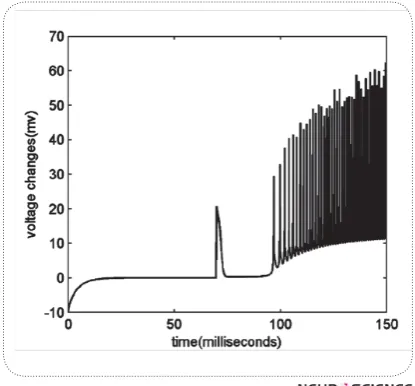
Since we ignored hf,, hs dynamics and used their steady & $-pothesis. This is the reason why we calculate INa one step before calculation of potassium currents. Next, we compare the difference between these currents in each step. If the difference increases regarding to previous steps, it means that INa has had a sudden increase and the membrane depolarization has been started. Interest-ingly, voltage changes over time, ] can show this difference since it is sum of all the membrane currents. Thus, we measure ] in each step, if it reaches a threshold, say 20; we amplify it in order to highlight the effect of depolarization before activation of potassium !<"$%# "% this assumption. In Fig.3 you can see voltage changes
versus time. An obvious change is seen at around 70th millisecond after running the model, exactly at the mo-ment we applied the depolarization.
Whenever the membrane voltage reaches to a thresh-old voltage, say 0 mili-volts, an action potential arises and we reset the voltage to -65 mili-volts.
All the simulations are performed using MATLAB 2008, and Euler method with step size equal to 0.01 is used for solving differential equations. Calculations were double checked by step size of 0.001 but the re-sults were rather the same. During this study, our main &# # #" DCN neurons.
3. Results
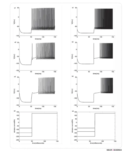
Z # " $ $ response to a hyperpolarization followed by a depolar-izing stimulus, show a good correspondence with three #" Z`"$!/#!# patterns of our minimal model is compared with those of KM-LIF model under applying similar stimuli. Fig.4 (a1-a3) exhibit KM-LIF model responses to step stimuli with hyperpolarizing amplitudes of -80, -147 and -200 pA during 50 milliseconds and a post depolarization with amplitude of 130 pA. Chopper, pauser and buildup pat-terns are obtained, respectively. In Fig.4 (b1-b3) same patterns are generated by applying step stimuli with hy-perpolarizing amplitudes of -80, -130 and -200 pA during 70 milliseconds and a post depolarization with amplitude of 130 pA to the minimal model. Applied step stimuli to the models are shown in Fig 4(a4, b4).
< ##" $ && Z` "$#" !/ # Rinzel (2010) demonstrated that the delay before spik# &spik#34;% &spik#34; -um currents and can be controlled by them. In fact these currents are inactivated at rest. If the membrane voltage decreases, e.g. a hyperpolarizing current applies, inac-tivation gating variables, hf , h s increases. Then, with some depolarizing current, IKif,, IKis activate fast and in balance with other membrane currents make a long de-lay. Since IKif,, IKis inactivations are slow, this delay lasts until inactivation variables, hf , h s&" " ! < # # # started. Stronger or longer hyperpolarizations leads to more deinactivation of IKif,, IKis.
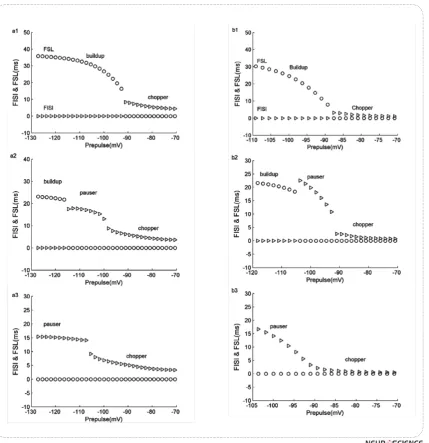
/^#!&& " "% ^>¦ X"% ^/>/ # of pre hyperpolarization. Our results match well with X¦/^$!<#& ^/>/ and FSL duration versus pre hyperpolarization. When prehyperpolarized voltage is weak, these values are low since there is no delay or latency, but stronger hyper-polarization causes these values change suddenly. Fig.5 (a1-a3) shows the dependency of FISI and FSL extract-ed from KM-LIF model to the pre hyperpolarizextract-ed volt-age while following depolarizing currents are 70, 120, 160, respectively. In Fig.5 (b1- b3), the minimal mod-el FSL and FISI dependency to preset voltage is also shown when depolarizing currents are 100, 150, 200. "" &% # & chopper pattern but when hyperpolarization is stronger, " "# # " change into pauser or build-up.
Winter 2012, Volume 3, Number 2
Basic and Clinical
Figure 5. FISI and FSL of KM-LIF model versus hyperpolarizations prior to different depolarizing currents: 70, 120,160 (a1-a3). FSL and FISI of the minimal model versus hyperpolarizations prior to different depolarizing currents: 100, 150, 200 (b1-b3). Sudden change in FISI and FSL represents pattern changing. Circles represent FSLs and triangles show FISIs.
Fig.6.a shows IKif, inactivation variable changes over the time. The more it hyperpolarizes, the more hf in-creases.
Fig.6b exhibits the effect of depolarizing current, I0 on hf changes. Each point represents the value of hf during a spike. If we look into hf decreasing path from right "/& & two adjacent spots is large at the beginning. While we approach to the left, the distance becomes smaller, i.e. hf rarely changes among two adjacent spikes,
^>¦^/>/#!hf starts from the right triangle, delay of spiking is high, thus FSL or FISI is
rath-er high, but if hf $ ##
regularly and thus FISI and FSL would be low.
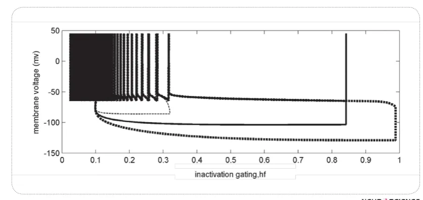
Fig7 shows three different responses of the minimal model versus hf changes. The initialization for all re-sponses is (h =0.1,v =-65) representing the rest
condi-goes on where hyperpolarization stops, and then by ap-plying a depolarization, membrane voltage increases. Depend on the value of hf #" $ occur. An example of weak pre hyperpolarization in this ##hf to about 0.3. This value is not enough to deinactivate I Kif , so applying depolarization increas-es membrane voltage and spiking starts with no delay -chopper pattern (thin dashed trend). When pre
hyper-Figure 7."%%<[%`%%V"[%%
Winter 2012, Volume 3, Number 2
Basic and Clinical
#"%$ !& we would have a long delay before ht returns to low val-ues. Then regular spiking starts - pauser or buildup pat-tern (solid black and thick dashed trends respectively)
4. Discussion
In this study, a minimal model of two variables has been presented for simulation of DCN pyramidal cells #"!<$ & inactivation properties of the transient potassium cur-rents to generate different patterns. The responses con-$& #"X¦/^$ thus of DCN neurons.
The stimuli applied to this model have a hyperpolariz-ing part followed by a depolarizhyperpolariz-ing part. Depend on the value of stimulus in each part; the model represents the following behaviors:
1) If the hyperpolarization is low and depolarization # $ & # #!< #" ""!
2) If the hyperpolarization is rather high and depo-larization is also high, a leading spike may occur and # $& # -!<#" "!
3) If the hyperpolarization is too high and & #" & long latency. This pattern is called buildup.
Appendix A:
Appendix B
The equations describing the minimal two-variable model dynamics are as follows:
Where all parameters are identical with those of KM-LIF model in appendix A.
References
Av-Ron E (1994) The role of a transient potassium current in a bursting neuron model. J Math Biol , 33, 71-78.
Goldberg, J.M. Brownell, W.E. (2003) Discharge characteristics of neurons in anteroventral and dorsal cochlear nuclei of cat. Brain Research, 64, 35-54.
Godfrey, D.A. Kiang, N.Y. & Norris, B.E.(1975) Single unit ac-tivity in the dorsal cochlear nucleus of the cat. J Comp
Neu-Kanold P.O. and Manis P.B. (1999) Transient potassium cur-rents regulate the discharge patterns of dorsal cochlear nu-cleus pyramidal cells. J Neurosci 19, 2195-2208.
Kanold, P.O. & Manis, P.B. (2000) A physiologically based model of discharge pattern regulation by transient K cur-rents in cochlear nucleus pyramidal cells J Neurophysiol, 85, 523–538.
Winter 2012, Volume 3, Number 2
Basic and Clinical
Manis, P.B. (1990) Membrane Properties and Discharge Char-acteristics of guinea Pig Dorsal Cochlear Nucleus Neurons Studied in vitro. J. Neurosci, 10 (7), 2338-2351.
#`[`<`=>%V" by two transient potassium currents: leading spike, latency, bistability. J Comput Neurosci, 31 (1), 117-136.
%%%`=>V%""%" discharges for units in the cochlear nucleus: tone-burst stim-ulation. Exp Brain Res, 1, 220–235.
Rhode, W.S. Smith P.H. & Oertel D. (1983) Physiological re-sponse properties of cells labeled intracellularly with horse-radish peroxidase in cat dorsal cochlear nucleus. J Comp Neurol , 213, 426–447.
Rhode, W.S. Smith P.H. (1986) Physiological studies of neurons in the dorsal cochlear nucleus of the cat. J Neuophysiol, 56, 287-307.
Rothman, J. & Manis, P. B. (2003) The roles potassium currents play in regulating the electrical activity of ventral cochlear nucleus neurons. Journal of Neurophysiol, 89, 3097–3113. Street, S.E. & Manis, P.B. (2007) Action Potential Timing