*Corresponding author: Elahe Elahi: [email protected], [email protected] © 2011 Progress in Biological Sciences Tel.: 0098-21-61113561; Fax: 0098-21-66492992
Preliminary data suggest possible association between
IL-32 expression level and time of MS attack
Mehrzad Roghani1, Fereidoun Mahboudi 2, Mohammad Ali Sahraian 3, Masoud Etemadifar4,
Elahe Elahi1,5,6*
1 School of Biology, College of Science, University of Tehran, Tehran, Iran 2 Department of Biotechnology, Pasteur Institute of Iran, Tehran, Iran
3MS Research Center, Sina Hospital, Tehran University of Medical Sciences, Tehran, Iran 4Isfahan University of Medical Sciences, Isfahan, Iran
5Department of Biotechnology, College of Science, University of Tehran, Tehran, Iran
6Center of Excellence in Biomathematics, School of Mathematics, Statistics and Computer Science, College of Science, University of
Te-hran, TeTe-hran, Iran
IL-32 and TNFαare important cytokines in autoimmune and inflammatory diseases. IL-32 has not been previously studied with respect to MS. Here, we report IL-32 and TNFαtranscript levels in peripheral blood mononuclear cells of MS patients and control individuals by real time PCR. A significant difference in IL-32 and TNFαmRNA levels of patients as compared to controls was not observed. However, patients with more recent attacks had highly significant increased levels of IL-32 mRNAs. As the sample size was small, this potentially relevant preliminary observation needs to be corroborated using a larger number of patients with well defined clinical features..© 2011 Progress in Biological Sciences. Vol. 1, No. 2, 44-49.
Introduction
It is generally accepted that cytokines and che-mokines are involved in inflammation and autoimmune disease processes, including those pertaining to the central nervous system(Kiyoto et al., 1998, Murphy et al., 2003, Alsaleh et al., 2010).
Although the etiology of multiple sclerosis (MS) is shrouded in unknowns, it is regarded by most as an inflammatory demyelinating autoimmune disease(Hafler and Weiner, 1989).
Pro-inflammatory cytokines, including TNFα,
TNFβ, IFNγ, IL-4, IL-6, and IL-15 are among
immune system related agents believed to have roles in the etiology of MS(Navikas and Link, 1996, KivisÄkk et al., 1998). Relevant observa-tions include presence of pro-inflammatory cy-tokines in active MS plaques, their increase in the peripheral circulation during active phases of MS, and changes in expression of certain che-mokine receptors and cheche-mokines in correlation with clinical course of MS during pregnancy (Beck et al., 1988, Martino et al., 1998, López et al., 2006). TNFα, may be particularly
important because it affects permeability at the blood-brain barrier and promotes demyelina-tion(Sharief and Thompson, 1992). IL-32 was originally identified as natural killer cell tran-script 4 in 1992, and designated a cytokine in 2005(Kim et al., 2005). It is a pro-inflammatory
cytokine that was shown to induce TNFα and to
be involved in TNFα related functions(Kim et
al., 2005). TNFα has roles in autoimmune and
inflammatory diseases such as rheumatoid arth-ritis and colits, and IL-32 can exacerbate mouse
models of these diseases via TNFα(Targan et al.,
1997, Shoda et al., 2006). IL-32 is expressed mainly in lymphoid tissues and leukocytes (Tar-gan et al., 1997, Kim et al., 2005, Shoda et al., 2006). It is now known that IL-32 production is induced in response to a wide range of stimuli
including IL-18, IL-1β, IFN-γ, and IL-12, and
that it can induce several cytokines in addition
to TNFα including MIP-2 (macrophage
induction of IL-32 by cytokines and its induc-tion of cytokines creates a positive feedback loop and that IL-32 affects the immune response mainly in this manner(Shoda et al., 2006). There is evidence that IL-32 directly or indirectly af-fects various functions including phagocytosis and killing of bacteria, prostaglandin E2 related intracellular processes, and apoptosis of T cells(Goda et al., 2006, Conti et al., 2007). This latter function is expected to be relevant in the important process of clearing of activated T cells after the peak of an immune response(Hildeman et al., 2002, Krueger et al., 2003) .
To the best of our knowledge, IL-32 has not yet been studied in MS patients. Here, we meas-ure IL-32 transcript levels in peripheral blood mononuclear cells (PBMCs) of MS patients and control individuals by real time PCR. We also
measure the levels of TNFα transcripts in the
same individuals. We report that the levels of these transcripts were not significantly different when the cohorts of patients and controls were compared at large. However, there were signifi-cant differences in the levels of both IL-32 and
TNFα transcripts between subgroups of patients.
Specifically, the levels of the transcripts differed between patients with recent attacks and patients without recent attacks. Implications of these ob-servations are discussed.
Material and methods
Patients and control subjects
This research was performed in accordance with the Helsinki Declaration. Initially 13 unrelated Iranian MS patients recruited from MS referral clinics in Esfahan and Tehran in 2010 were stu-died. The strongly limiting inclusion criterion of not having received any medication or treatment within at least six months prior to recruitment was implemented. Otherwise they were re-cruited on a consecutive basis. Subsequently, seven additional untreated patients were also recruited. None of the Iranian MS patients in whom serum NO levels were previously re-ported are included in this study(Roghani et al., 2010). The patients were diagnosed with MS according to the criteria of McDonald et al. (McDonald et al., 2001). The disability status of the patients was rated using Kurtzke’s expanded
disability status score (EDSS)(Kurtzke, 1983). Fifteen unrelated controls from these cities who self-reported not to be under any medication and to have no indications of disease were also re-cruited from the same cities.
Real time PCR
PBMCs were separated from 4cc blood of par-ticipants by Ficoll-Isopaque density centrifuga-tion (Gibco BRL, Life Technologies Ltd, UK). RNA was isolated from the cells using RNX+ according to the manufacturer’s instructions (Cinnagen Tehran, Iran). RNA yield was deter-mined spectrophotometrically and RNA quality was assessed after electrophoresis by using den-sity ratio of 28S to 18S ribosomal RNA bands. Complementary DNA (cDNA) was synthesized with the Bioneer cDNA synthesis kit (Alameda, CA, USA). Candidate control genes for
assess-ment of levels of IL-32 and TNFα mRNA levels
were ACTB, GADPH, and Β2M. Real time PCR
on the candidate control genes was done using cDNA of 5 patients and 6 control individuals on an ABI7500 machine (Foster City, CA, USA) using the QuantiFast SYBR Green PCR Kit (QIAGEN; Germantown, MD, USA). Results of the real time experiments were submitted to ge-Norm program (http://medgen.ugent.be / ~ jvde-somp/genorm/) for identification of internal con-trol gene with least variable transcript level among the samples. Subsequently, real time
PCRs for IL-32 and TNFα were performed on
the cDNAs of the 13 patients and 15 control
in-dividuals. Real time PCR was done only for
IL-32 on the cDNA of the 7 patients later recruited.
All gene specific primers for real time
experi-ments were purchased from QIAGEN (ACTB:
QT01680476; GADPH: QT01192646; Β2M:
QT00088935; IL-32: QT00199766; TNFα:
QT01079561). Expression levels of IL-32 and
TNFα between patients and controls were
com-pared by comparing their ∆CT values, and
com-parison of expression of the genes between pa-tient subgroups was done by comparing 2-
∆∆CT values(Livak and Schmittgen, 2001).
Statistical analysis
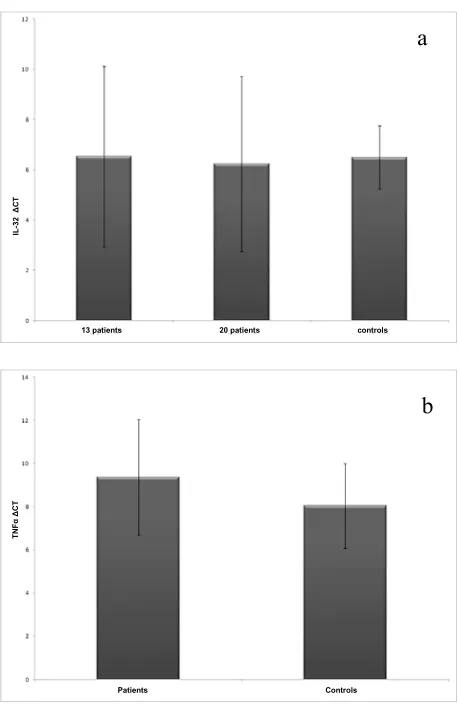
Fig. 1. Comparison of mean IL-32 (A)and TNFα (B)
mRNA levels between MS patients and controls.
The SPSS 17.0 and Minitab 15 statistical pro-gram software were used.
Results
The clinical features of the patients are pre-sented in Table 1. The fact that almost all our patients were women is a reflection of the nota-bly skewed female to male ratio among Iranian MS patients(Roghani et al., 2010). Course of disease in the vast majority of our patients was relapsing-remitting, consistent with previous reports from Iran (Roghani et al., 2010).
GeNorm identified ACTB as the gene with the
best stability value (M=0.076). IL-32 and
TNFα mRNA levels were not significantly
dif-ferent between the cohorts consisting of 13 MS patients and 15 control individuals (p=0.70 and 0.20, respectively; Fig. 1A,B.). Absence of
sig-nificant difference in IL-32 levels was still ap-parent after inclusion of data pertaining to seven patients recruited in 2010 (p=0.61; Fig. 1A.).
The raw data on IL-32 and TNFα mRNA levels
showed clustering of the values for each gene among control individuals and dispersion among the patients. This is reflected in the relatively large standard deviations associated with the
average values for patients (IL-32 ∆CT:
6.24+/-3.48; TNFα ∆CT: 9.38+/- 2.67) as compared to
control individuals (IL-32 ∆CT: 6.50+/-1.25;
TNFα ∆CT: 8.04+/- 1.97). Dispersal of TNFα
and other cytokine levels at the protein level is also evident in the data presented by other au-thors(Kiyoto et al., 1998).
We thought this may suggest heterogeneity among the MS patients and that the levels of some of cytokines may differ among patient subgroups.
We observed no correlation between levels of
IL-32 and TNFα mRNAs and EDSS scores of
the patients (IL-32: Pearson correlation =0.142,
p=0.50; TNFα: Pearson correlation =-0.426,
p=0.15). There was also no significant correla-tion between the Progression Index (EDSS
changes per year) of the patients and IL-32 and
TNFα mRNA levels (IL32: Pearson
correla-tion=0.035, p=0.890; TNF-α: Pearson
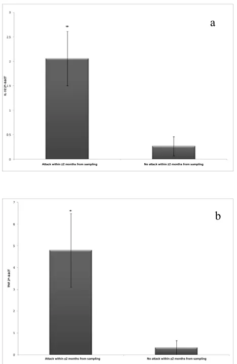
correla-tion+-0.318, p=0.340). Subsequently, we com-pared the levels of the mRNAs between relapse-remitting (RR) patients who had and who had not experienced a relapse within two months before or after time of sample collection. Among the 15 and 10 RR patients in whom,
re-spectively, IL-32 and TNFα were measured,
in-formation on time of most recent relapse on 12
and 10 were available. Based on 2-∆∆CT
val-ues, IL-32 mRNA levels were significantly
higher (p=0.008) among the six patients who had had a relapse within this time period as compared to the six who had not had a relapse
(Fig. 2A.). TNFαmRNA levels were also
signif-icantly higher (p=0.013) among the four patients who had had a relapse within this time period as compared to the six who had not had a relapse
(Fig. 2B.). Notably, the levels of IL-32 and
TNFα mRNAs in primary progressive (PP) and
secondary progressive (SP) patients were similar a
b
Patients Controls 13 patients 20 patients controls
TN
F
α
∆
CT
IL
-32
∆
Fig. 2. Comparison of IL-32 (A) and TNFα (B)
mRNA levels between relapsing remitting MS pa-tients with and without attack in period +/- two months of blood withdrawal.
to RR patients without recent attacks, and signif-icantly lower than RR patients with recent at-tacks (not shown); clearly PP and SP patients have not experienced a recent attack. The corre-lations we have were always in the same
direc-tion for IL-32 and TNFα mRNAs. Consistent
with this, we observed a close correlation
be-tween IL-32 and TNFα mRNA levels among
both our patient (Pearson correlation =0.920, p=0.00) and control (Pearson correlation =0.985, p=0.00) cohorts. Finally, there was no
signifi-cant correlation between age at onset (IL-32:
Pearson correlation =0.059, p=0.85; TNFα:
Pearson correlation =-0.176, p=0.57) or disease
Table 1. Clinical features of MS patients
TNF-α IL-32
No. of patients* 13 20
No. females 12 19
Average age at onset 29.0±6.3 yrs. 28.9±7.1 yrs.
Average age at examination 35.5±8.8 yrs. 35.3±8.9 yrs.
Duration 6.4±7.1 yrs. 6.8±6.8 yrs.
Average EDSS score 2.42±1.8 2.7±2.0
Clinical course**
RR 10 15
SP 1 1
PP 2 4
*the 20 patients include 13 originally recruited and 7 later recruited.
**RR, relapsing-remitting; SP, secondary progressive; PP, primary progressive.
duration (IL-32: Pearson correlation = -0.192,
p=0.53;TNFα: Pearson correlation =-0.351,
p=0.24) and IL-32 and TNFαmRNA levels.
Discussion
IL-32 levels at the mRNA or protein levels have been shown to be lower in controls as compared to patients affected with various au-toimmune and inflammatory diseases including rheumatoid arthritis, myeloblastic leukemia, HIV,and Crohn’s disease(Dinarello and Kim, 2006, Marcondes et al., 2008, Rasool et al., 2008, Alsaleh et al., 2010). We did not observe a
significant difference in IL-32 mRNA levels
be-tween MS patients and controls.
We expect medical treatments were not relevant to these observations as none of our patients had received recent treatment. In most previous stu-dies on other diseases, IL-32 was measured in the target tissue of the disease e.g. the synovial tissue in rheumatoid arthritis and in intestinal epithelial cells in Crohn’s disease(Dinarello and Kim, 2006, Shioya et al., 2007, Alsaleh et al., 2010). The fact that we did not have access to MS target tissue and used PBMCs as surrogate may partly explain why we were unable to detect a difference. Perhaps more important is the consideration that cytokines including IL-32 may exhibit an expression pattern that is not uni-form at all stages of disease activity. We
ob-served a more non-uniform distribution of IL-32
mRNA levels among our patients as compared a
b
to controls. Grouping our patients based on some MS relevant parameters showed that they
did not correlate with IL-32 levels. Notably,
there was no significant correlation with EDSS scores or with Progression Index. However, grouping based on proximity in time to attack did show statistically significant differences in
IL-32 expression. Patients who had recently or
were about to experience attacks had higher
le-vels of IL-32 mRNAs in their PBMCs. The
sta-tistical significance of the correlation (p=0.008) was notable.
TNFα is a multifunctional cytokine with
impor-tant roles in the immune system (Cope, 1998). It
isconsidered a driving factor in the
demyelina-tion process associated with MS(Lock et al.,
1999). IL-32 and TNFα levels have been shown
to be concordant in several studies and it has
been suggested that iinteractions between TNFα
and IL-32 are important in the functions of both
cytokines in various disease conditions (Kim et al., 2005). It was because of these considerations
that we measured TNFα mRNA levels while
al-so measuring IL-32 mRNA levels.
Consistent with observations in other autoim-mune and inflammatory diseases, we observed a strong correlation between the expressions of
these two cytokines. During the couple of months before and after attack, there were in-creases in the levels of both. These preliminary results suggest that monitoring the levels of these cytokines in easily accessible PBMCs might be informative for prediction of oncoming attacks and may suggest targets for effective an-ti-infalmmatory treatments for prevention or control of attack. Similarly, they may be tools for monitoring effectiveness of treatment proto-cols. Although our results are interesting, our sample size was small and the data need to be corroborated using a larger number of patients with well defined clinical features. Although our results are interesting, our sample size was small due to patient recruitment criterion of not having received recent medication. The preliminary findings need to be corroborated using a larger number of patients with well defined clinical features.
Acknowledgments
We thank the patients for participating in this study. We acknowledge The College of Science of The University of Tehran for funding the research. All authors declare ab-sence of conflict of interest.
References
Alsaleh G, Sparsa L, Chatelus E, Ehlinger M, Gottenberg J-E, Wachsmann D, Sibilia J (2010) Innate immunity triggers IL-32 expression by fibroblast-like synoviocytes in rheumatoid arthritis. Arthritis Res Ther 12, R135 Beck J, Rondot P, Catinot L, Falcoff E, Kirchner H, Wietzerbin J (1988) Increased production of interferon gamma and tumor necrosis factor precedes clinical ma-nifestation in multiple sclerosis: Do cytokines trigger off exacerbations? Acta Neurol Scand 78, 318-323
Conti P, Youinou P, Theoharides TC (2007) Modulation of autoimmunity by the latest interleukins (with special emphasis on IL-32). Autoimmun Rev 6, 131-137
Cope AP (1998) Regulation of autoimmunity by proin-flammatory cytokines. Curr Opin Immunol 10, 669-676 Dinarello CA, Kim S-H (2006) IL-32, a novel cytokine with a possible role in disease. Ann Rheum Dis 65, iii61-iii64
Goda C, Kanaji T, Kanaji S, Tanaka G, Arima K, Ohno S, Izuhara K (2006) Involvement of IL-32 in activation-induced cell death in T cells. Int Immunol 18, 233-240 Hafler DA, Weiner HL (1989) MS: a CNS and systemat-ic autoimmune disease. Immunol Today 10, 104-107
Hildeman DA, Zhu Y, Mitchell TC, Kappler J, Marrack P (2002) Molecular mechanisms of activated T cell death in vivo. Curr Opin Immunol 14, 354-359
Kim S-H, Han S-Y, Azam T, Yoon D-Y, Dinarello CA (2005) Interleukin-32: A Cytokine and Inducer of TNF-α. Immunity 22, 131-142
Kivisakk P, Matusevicius D, He B, Soderstrom M, Fre-drikson S, Link H (1998) IL-15 mRNA expression is up-regulated in blood and cerebrospinal fluid mononuclear cells in multiple sclerosis (MS). Clin Exp Immunol 111, 193-197
Kiyoto H, Atsushi I, Chang-Sung K (1998) Elevated se-rum levels of IFN-γ, IL-4 and TNF α /unelevated serum levels of IL-10 in patients with demyelinating diseases during the acute stage. J Neuroimmunol 87, 27-32 Krueger A, Fas SC, Baumann S, Krammer PH (2003) The role of CD95 in the regulation of peripheral T-cell apoptosis. Immunol Rev 193, 58-69
Kurtzke JF (1983) Rating neurologic impairment in mul-tiple sclerosis: An expanded disability status scale (EDSS). Neurology 33, 1444-52
and cytokine expression during pregnancy in multiple sclerosis patients. Mult Scler 12, 421-427
Livak KJ, Schmittgen TD (2001) Analysis of Relative Gene Expression Data Using Real-Time Quantitative PCR and the 2-[Delta][Delta]CT Method. Methods 25, 402-408
Lock C, Oksenberg J, Steinman L (1999) The role of TNF α and lymphotoxin in demyelinating disease. Ann Rheum Dis 58, I121-I128
Marcondes AM, Mhyre AJ, Stirewalt DL, Kim S-H, Di-narello CA, Deeg HJ (2008) Dysregulation of IL-32 in myelodysplastic syndrome and chronic myelomonocytic leukemia modulates apoptosis and impairs NK function. P Natl Acad Sci 105, 2865-2870
Martino G, Grohovaz F, Brambilla E, Codazzi F, Consig-lio A, Clementi E, Filippi M, Comi G, Grimaldi LME (1998) Proinflammatory cytokines regulate antigen-independent T-cell Activation by two separate calcium-signaling pathways in multiple sclerosis patients. Ann Neurol 43, 340-349
McDonald WI, Compston A, Edan G, Goodkin D, Har-tung HP, Lublin FD, McFarland HF, Paty DW, Polman CH, Reingold SC, et al. (2001) Recommended diagnos-tic criteria for multiple sclerosis: Guidelines from the international panel on the diagnosis of multiple sclerosis. Ann Neurol 50, 121-127
Murphy CA, Langrish CL, Chen Y, Blumenschein W, McClanahan T, Kastelein RA, Sedgwick JD, Cua DJ (2003) Divergent Pro- and Antiinflammatory Roles for IL-23 and IL-12 in Joint Autoimmune Inflammation. J Exp Med 198, 1951-1957
Navikas V, Link H (1996) Review: cytokines and the pathogenesis of multiple sclerosis. J Neurosci Res 45, 322-333
Rasool ST, Tang H, Wu J, Li W, Mukhtar MM, Zhang J, Mu Y, Xing HX, Wu J, Zhu Y (2008) Increased level of IL-32 during human immunodeficiency virus infection suppresses HIV replication. Immunol Lett 117, 161-167 Roghani M, Mahboudi F, Saharian MA, Etemadifar M, Esfahani AN, Nahrevanian H, Elahi E (2010) Concentra-tions of nitric oxide metabolites in the serum of Iranian multiple sclerosis patients. J Neurol Sci 294, 92-94 Sharief MK, Thompson EJ (1992) In vivo relationship of tumor necrosis factor-[alpha] to blood-brain barrier dam-age in patients with active multiple sclerosis. J Neu-roimmunol 38, 27-33
Shioya M, Nishida A, Yagi Y, Ogawa A, Tsujikawa T, Kim-Mitsuyama S, Takayanagi A, Shimizu N, Fujiyama Y, Andoh A (2007) Epithelial overexpression of inter-leukin-32α in inflammatory bowel disease. Clin Exp Immunol 149, 480-486
Shoda H, Fujio K, Yamaguchi Y, Okamoto A, Sawada T, Kochi Y, Yamamoto K (2006) Interactions between IL-32 and tumor necrosis factor alpha contribute to the ex-acerbation of immune-inflammatory diseases. Arthritis Res Ther 8, R166
Targan SR, Hanauer SB, van Deventer SJH, Mayer L, Present DH, Braakman T, DeWoody KL, Schaible TF, Rutgeerts PJ (1997) A Short-Term Study of Chimeric Monoclonal Antibody cA2 to Tumor Necrosis Factor- α
for Crohn's Disease. New Engl J Med 337, 1029-1036