Abstract
Objective: Modified anterior tension wiring with K-wires and cannulated lag screws with anterior tension wiring are currently the fixation of choice for patellar fractures. Failure of fixation, migration of the wires, postoperative pain and resulting revision surgery, how-ever, are not uncommon. after preliminary biome-chanical testing of a new fixed-angle plate system es-pecially designed for fixation of patella fractures the aim of this study was to evaluate the surgical and anatomical feasibility of implanting such a plate-de-vice at the human patella.
Methods: In six fresh unfixed female cadavers without history of previous fractures around the knee (average age 88.8 years) a bilateral fixed-angle plate fixation of the patella was carried out after previous placement of a transverse central osteotomy. operative time, intra-operative problems, degree of retropatellar arthritis (following outerbridge), quality of reduction and exis-tence of any intraarticular screw placement have been raised. In addition, lateral and anteroposterior radi-ographs of all specimens were made.
Results: due to the high average age of 88.8 years no patella showed an unimpaired retropatellar articular surface and all were severely osteoporotic, which made a secure fixation of the reduction forceps during surgery difficult. the operation time averaged 49 min-utes (range: 36-65). although in postoperative x-rays the fracture gap between the fragments was still visi-ble, the analysis of the retropatellar surface showed no residual articular step or dehiscence > 0.5 mm. also in a total of 24 inserted screws not one intraarticular malposition was found. no intraoperative complica-tions were noticed.
Conclusions: osteosynthesis of a medial third patella fracture with a bilateral fixed-angle plate-device is sur-gically and anatomically feasible without difficulties. Further studies have to depict whether the bilateral fixed-angle plate-osteosynthesis of the patella displays advantages over the established operative procedures.
Key words: Patella fracture; fixed-angle plate; angle-sta-ble plate; feasibility study; knee
I
ntRoductIonthe patella is the largest sesamoid bone in the body and its subcutaneous location makes it susceptible to direct injury [1]. only 1 % of all injuries to the human
skeleton are patella fractures. of those, only approxi-mately one-third requires surgical attention [2, 3]. the aim of surgical treatment in addition to the preserva-tion of most of the kneecap, is anatomical reducpreserva-tion of the articular surface, followed by stable fixation, restoration of the extensor mechanism and early mo-bilization [4-11].
currently, the use of modified anterior tension wire with or without circumferential wiring is the standard surgical treatment of patellar fractures [6, 12-14, 3, 10]. despite several technical modifications of the an-terior tension wiring, early fracture dislocation can be observed in 22-30% of all cases [15, 16]. apart from failure of fixation, migration of the wires, postopera-tive pain and resulting revision surgery are not uncom-mon, making the long-term outcome in clinical studies quite poor [4, 17, 5, 7, 9, 18]. secondary postoperative pain due to skin irritation caused by the K-wires also is a common problem in the tension-band wiring of patellar fractures [14, 4, 5, 9, 19, 18]. thus, revision surgery with K-wire removal becomes necessary in up to 65% of cases [2, 4, 5, 9].
the development of fixed-angle plates, which are based on the concept of an internal fixator , has led to an improvement in the biomechanical stability of the plate-bone interface [20, 21]. Based on this improved stability, fixed-angle plate systems differ substantially in their biomechanical properties from conventional plates making their application possible even in skele-tal segments that were previously denied of conven-tional plating. Furthermore it has been possible to de-velop even smaller and thinner fixed-angle plates. For use in transverse patella fractures a 2.7 mm bilateral, polyaxial, fixed-angle plate system (manufacturer: Königsee® allendorf, germany) has been designed. In a preliminary biomechanical testing until failure us-ing a sawbone® patella fracture model it could be demonstrated that a bilateral, fixed-angle plate os-teosynthesis results in significantly superior strength and rigidity compared to modified anterior tension wiring or cannulated lag screws with anterior tension wiring [22].
since the biomechanical examination was conduct-ed on polyurethane foam patellae, the objective of this study was to determine the technical and anatomical feasibility of reducing and fixing a horizontal patellar fracture with two 2.7 mm polyaxial locking plates which are attached to the medial and lateral rim of the patella.
Eur J Med Res (2011) 16: 41-46 © I. Holzapfel Publishers 2011
F
IxEd
-
anglE
P
latEs In
P
atElla
F
RactuREs
- a P
Ilot
c
adavER
s
tudy
M. Wild, s. thelen, P. Jungbluth, M. Betsch, d. Miersch, J. Windolf, M. Hakimi
M
EtHodsthe operations were carried out in six fresh unfixed female cadavers of an average age of 88.8 years (range 80-96 years) provided by the Institute of anatomy of the Heinrich Heine university düsseldorf. In order to ensure that none of the bones presented with patho-logical qualities, clinical and radiopatho-logical examinations were conducted and patient histories were evaluated. through a longitudinal incision of 10 cm of length above the patella, the dissection was executed down to the anterior surface of the patella so its medial and lat-eral margins were made visible. after identification of
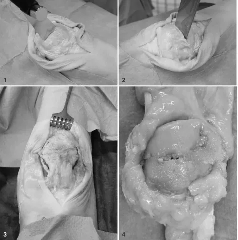
base and apex of the patella a transverse incision of the soft tissues in the middle part of the patella was conducted. the dissection was extended to the medial and lateral retinaculum, as usually in dislocated trans-verse patellar fractures it comes to a tear of the reti-nacula. then, using a surgical saw a transverse os-teotomy was placed in the middle of the patella just to the subchondral bone of the articular surface (Fig.1). the osteotomy were completed by dissection of the subchondral bone and articular cartilage with a chisel (Fig. 2). subsequently, the transverse patellar fracture was reduced under digital control of the articular sur-face which was made accessible via the cut through
Fig. 1. transverse osteotomy in the middle of the patella using a surgical saw.
Fig. 2. completing the osteotomy through the subchondral bone and articular cartilage with a chisel.
Fig. 3. Positioning of the fixed angle plates after open reduction.
Fig. 4. articular surface of a patella after open reduction and internal fixation with bilateral fixed-angle plates
1
2
the retinacula. temporary reduction was secured with a pointed reduction clamp, with one point placed at the base of the patella, the other at the apex.
then the specifically for the patella designed bilat-eral, fixed-angle plate system (Königsee Implants®, allendorf, germany) with four respectively five lock-ing holes, accordlock-ing to the given anatomy of the patel-la, was applied. the titanium plates had a thickness of 2.7mm with polyaxial angle-stable locking options for the osteosynthesis screws. Both locking plates were bent semicircularly, and placed each to the medial and lateral margin in an 80 degree angle to the anterior sur-face of the patella. these plates have an additional eyelet on each end allowing a mutual tensioning of the implants in the direction of the pull of the extensor mechanism in order to enable greater stability of the construct. Both in the proximal and distal eyelet of each plate a pointed reduction forceps was introduced and by closing of the forceps both plates were tensed longitudinally. then, the most proximal hole of both locking plates was filled each with a 3.5 mm locking screw after placing the drill hole. this was followed by inserting 3.5 mm fixed-angle screws in the most distal plate holes. When using a 5-hole plate, 3.5 mm locking screws were inserted into all holes except the one at the fracture-site, so each plate was fixed to the patella with 4 angle-stable locking screws. at the 4-hole plates
all holes were filled with 3.5 mm locking screws, as there was a greater distance between the two inner-most plate holes, thus none of the screws were located at the level of the fracture. afterwards the reduction forceps as well as the pointed forceps placed in the eyelets of the plates were removed (Fig. 3). Finally, the entire patella with adherent quadriceps and patellar tendon was excised for further analysis and the skin was closed by suture (Fig. 4).
In addition to the demographic data of the cadaver specimens, we recorded operative time, any intraoper-ative problems, the degree of retropatellar arthritis (following the outerbridge-classification [23]), the quality of reduction by measurement of a remaining gap or step at the fracture site and the existence of any intraarticular screw placement. at last, lateral and a.p. radiographs of all specimens were made.
R
Esultsdue to the high average age of the specimens of 88.8 years (range: 80-96 years), all samples showed an ad-vanced degree of retropatellar arthritis with an average scale of 3.2 (range: 3-4) according to outerbridge’s classification [23]. not in a single specimen a regular retropatellar cartilage covering was found (Fig. 4). the main problem during the performed plate fixation
a
b
proved to be the partially very soft bone structure that made it difficult to securely position the reduction for-ceps and often led to a loss of reduction when drilling, requiring to repeat the reduction. In addition, there were sometimes collisions of the drill with already in-serted screws on the opposite, since occasionally the direction of the drill hole was chosen to flat in order to avoid an intraarticular screw positioning, even though all patellae were thick enough to avoid colli-sion of the screws with the drill.
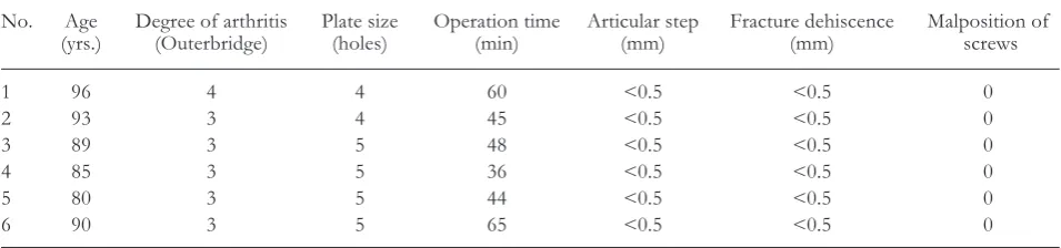
despite these problems and the relatively small fe-male patellae always at least 3 screws crossed the frac-ture gap. the operation time averaged 49 minutes (range: 36-65). although in the postoperative x-rays a fracture gap between the fragments was still visible (Fig. 5a and 5b), the analysis of the articular surface of each patella showed no residual articular step or dehis-cence > 0.5 mm. the minimal (<0.5 mm) steps or de-hiscences observed were a result of the defect in the bony substance after osteotomy of the patella. after placing a centered transverse osteotomy, in all speci-mens it was possible to attach a 2.7mm fixed-angle plate on the lateral and medial border of the patella, without interfering with the quadriceps or patellar ten-don. (Fig. 3) also, in a total of 24 inserted screws not a single intraarticular malposition was found, although during the implantation of the screws no fluoroscopic control was available. (table 1)
d
IscussIonthe main function of the patella is an increase of the potential force produced by the quadriceps muscle due to elevation of the extensor axis out of the knee joint [24, 25]. In order to function properly, a patella must be able to withstand significant axial forces. Everyday activities increase patello-femoral pressure by a signifi-cant factor of the body weight. Walking generates a patello-femoral stress amounting to about half the body weight, which increases e.g. when climbing stairs by a factor of 3.3 and when assuming a crouching po-sition by a factor of 7.6 of the body weight [26]. giv-en the fact that every internal fixation of the patella is inevitably exposed to such high tensile forces [3], a discussion over the appropriate fixation method was unavoidable from the beginning.
the technique of tension wiring was first described by Pauwels 1966 [27] and later popularized by 2 addi-tional longitudinal parallel Kirschner wires as a modi-fied ao tension band. [10, 7, 8] after numerous clini-cal and biomechaniclini-cal studies currently a modified
tension band wiring has been implemented as the most commonly used technique in almost all types of fractures. [7, 10, 14, 28, 6, 3, 2, 5, 18] another fre-quently used alternative, which however can only be applied for simple central transverse fractures with good bone quality, is the cannulated lag screw os-teosynthesis with anterior tension band wiring. the combination of lag screws with a tension band promises thereby greater stability with fewer disloca-tions and higher loads to failure than any single method for themselves. [12, 14, 6] Both procedures are technically demanding and have high complication rates. In particular, the use of tension band wires of-ten causes irritation of the soft tissues, so it has to be removed in 30-65% of cases in another surgery. [29, 4, 9, 19, 2, 15, 30, 5] Further frequently observed side ef-fects are fractures and dislocations of the wire or wire components. [31, 29, 5] catalano et al. [4] reported that in patients with tension wires metal removal was necessary because of symptomatic wires in 65% of cases. gosal et al. [17] had a reoperation rate of 38% using stainless steel wires.
despite technical modifications of tension band wiring early fracture dislocation may take up to 22-30% in observed cases [15, 16]. a failure of fixation, the migration of wires, postoperative pain and revi-sion surgery are not uncommon and some clinical studies have also shown that the outcome of the mod-ified tension band in 25-42% of cases depicts only modest results [4, 17, 5, 7, 9, 18].
Hung [5] followed up 68 patients after tension band osteosynthesis, and only 72% were subjectively satis-fied with the results, 37% had broken implants and 15% needed a revision surgery. Bostrom [2] reported in a sample of 422 patients of a 50-70% chance of long-term problems after surgical treatment of a patel-lar fracture, 37% having a degree of functional impair-ment, and 21% requiring revision surgery. In a differ-ent follow-up of 49 patellar fractures, a dislocation of the fragments over 2 mm could be detected in 22% of the fractures that were treated with a tension band wiring [15].
In a preceding study conducted by our study group, the x-rays of 50 patients who were treated surgically using a modified tension band were analysed. a joint dislocation level of less than 2 mm became apparent in 50% of patients and a dislocation or step in the ar-ticular surface of > 2 mm appeared in 30% of pa-tients [16]. although it is considered in the literature that a dislocation or step of the fracture gap of 2 mm does not affect the outcome after surgery of patellar
Table 1.Results after open reduction and bilateral fixed-angle plate fixation.
no. age degree of arthritis Plate size operation time articular step Fracture dehiscence Malposition of
(yrs.) (outerbridge) (holes) (min) (mm) (mm) screws
1 96 4 4 60 <0.5 <0.5 0
2 93 3 4 45 <0.5 <0.5 0
3 89 3 5 48 <0.5 <0.5 0
4 85 3 5 36 <0.5 <0.5 0
5 80 3 5 44 <0.5 <0.5 0
fractures [2, 18] the anatomical reduction should be the objective of any surgical treatment of articular fractures in particular in the heavily loaded patello -femoral joint [32]. Because most of the load passes over the patellofemoral joint, post-traumatic os-teoarthritis after patellar fracture is often seen [33]. sorensen [34] was able to demonstrate in a 10 to 30 year followup period more than 45 cases of patello -femoral osteoarthritis (70%) in 64 knees after patellar fracture, compared to 20 cases of patello femoral os-teoarthritis (31%) on the uninjured opposite side. al-though the realistic assessment of joint injuries is dif-ficult, many studies have shown that the best clinical results can be expected after the best possible reduc-tion of the fracture [2, 4, 33, 5, 7, 9].
the goal of treatment is to restore the articular sur-face of the patella and to achieve a strong quadriceps-apparatus in order to allow for early mobilization of the knee joint [6, 35, 4, 5, 7-10].
the development of angle-stable plate systems with their excellent biomechanical properties brought about implants with a significantly reduced size but superior stability. this enhanced stability is the result of the bet-ter anchorage of the plate construct in the bone, which is based on the principle of internal fixation [20, 21].
the objective of developing a fixed-angle plate for the patella was providing an osteosynthesis with in-creased stability and lower deformation under load, compared to the currently most frequently used meth-ods of modified tension band or cannulated lag screws with tension band and to prevent secondary loss of reduction. also, the frequent problems of wires with irritation of the soft tissues and the subsequent re-moval of the material should be avoided. due to the three-point loading of the patella with bending as well as tensile forces occurring at the fracture site and to prevent a deterioration of the skin by the implant, two plates were used, each of which being applied to the medial and lateral margin of the patella.
as part of a previously performed biomechanical testing on polyurethane foam patellae (sawbone®) it could be demonstrated that the application of two polyaxial fixed-angle plates, each of which being at-tached to the medial and lateral border of the patella, led to a stability twice as high (mean tensile strength: 2396 n) compared to the cannulated lag screws with tension band (mean tensile strength: 1015 n). com-pared to modified tension band osteosynthesis with Kirschner wires (mean tensile strength: 625 n) a four-fold greater stability could be achieved [22].
In addition to the significantly higher stability, the bilateral angle-stable patella-plate also shows signifi-cantly lower dehiscence of the fracture gap under ten-sile stress and a significantly higher modulus of elas-ticity (9.01 MPa), which surpasses the cannulated lag screws with tension band (4.98 MPa) and the modified tension band osteosynthesis with Kirschner wires (2.85 MPa) (p<0,01). thus, the fixed-angle plate is by far the most stable and most rigid construct with the lowest loss of reduction in comparison to the other two most commonly used methods of internal fixa-tion of the patella [22].
attempts have already been made to fix patellar fractures with plates. “Basket plates” have been
suc-cessfully used in clinical practice for fractures of the distal pole of the patella [36]. However, these are not angle-stable and are suitable only for distal pole frac-tures of the patella. they are also much larger and are located directly at the patellar tendon, so irritation of the soft tissues could arise here. Because of their act-ing as an internal fixator, fixed-angle plates do not lie directly on the periosteum of the bone so the soft tis-sue underneath the plate is not damaged by pressure. thereby the risk of a circulatory disorder of the patel-la, as it may be caused by cerclage-wires [37] or a di-rect injury to the quadriceps or patellar tendon through the implant can possibly be reduced.
this feasibility study carried out on 6 fresh female cadaver knees showed that despite the osteoporotic bone of the very old body donors, the reduction of the patellar fracture was easily possible in the conven-tional technology, and the bilateral fixed-angle plate osteosynthesis could be accomplished not requiring too much time. due to the thickness of the patella that exists even in these elderly patients [38] the risk of intraarticular screw placement under operational con-ditions is considered low and did not occur once in our series.
not a single patella stabilized with this technique had an articular step or dehiscence > 0.5 mm. the minimal dehiscences observed were a result of the defect in the bony substance after osteotomy of the patella. despite the relatively small female patella it was possible to place a plate on the lateral and medial border of the patella without stripping the quadriceps tendon or patellar tendon. due to the internal fixator principle the plate must not lie directly on the bone, so the soft tis-sue of the extensor mechanism can be spared. the plate system presented here is certainly not suitable for comminuted fractures of the patella, just for simple transverse patellar fractures, which make up the majori-ty of patellar fractures with 34 % [2].
Whether this change in principle, offers an advan-tage under clinical conditions compared to the estab-lished procedures - modified tension band or cannu-lated lag screw with tension band wire - must be shown in further clinical studies.
R
EFEREncEs1. Koval KJ, Kim, yH. Patella fractures. Evaluation and treatment. am J Knee surg. 1997; 10(2): 101-8.
2. Bostrom a. Fracture of the patella. a study of 422 patel-lar fractures. acta orthop scand suppl. 1972; 143: 1-80. 3. lotke Pa, Ecker, Ml. transverse fractures of the patella.
clin orthop Relat Res. 1981; (158): 180-4.
4. catalano JB, Iannacone, WM, Marczyk, s, dalsey, RM, deutsch, ls, Born, ct, delong, Wg. open fractures of the patella: long-term functional outcome. J trauma. 1995; 39(3): 439-44.
5. Hung lK, chan, KM, chow, yn, leung, Pc. Fractured patella: operative treatment using the tension band princi-ple. Injury. 1985; 16(5): 343-7.
6. Benjamin J, Bried, J, dohm, M, McMurtry, M. Biome-chanical evaluation of various forms of fixation of trans-verse patellar fractures. J orthop trauma. 1987; 1(3): 219-22.
8. Mueller ME, allgöwer, M, schneider, R, Willenegger, H. Manual of internal fixation: techiques recommended by the ao group. springer-verlag, Berlin. 1992.
9. torchia ME, lewallen, dg. open fractures of the patel-la. J orthop trauma. 1996; 10(6): 403-9.
10. Weber MJ, Janecki, cJ, Mcleod, P, nelson, cl, thomp-son, Ja. Efficacy of various forms of fixation of trans-verse fractures of the patella. J Bone Joint surg am. 1980; 62(2): 215-20.
11. yu Z, Zheng, l, Zhang, y, li, J, Ma, B. Functional and radiological evaluations of high-energy tibial plateau frac-tures treated with double-buttress plate fixation. Eur J Med Res. 2009; 14(5): 200-5.
12. Berg EE. open reduction internal fixation of displaced transverse patella fractures with figure-eight wiring through parallel cannulated compression screws. J or-thop trauma. 1997; 11(8): 573-6.
13. Burvant Jg, thomas, Ka, alexander, R, Harris, MB. Evaluation of methods of internal fixation of transverse patella fractures: a biomechanical study. J orthop trau-ma. 1994; 8(2): 147-53.
14. carpenter JE, Kasman, Ra, Patel, n, lee, Ml, goldstein, sa. Biomechanical evaluation of current patella fracture fixation techniques. J orthop trauma. 1997; 11(5): 351-6. 15. smith st, cramer, KE, Karges, dE, Watson, Jt, Moed, BR. Early complications in the operative treatment of patella fractures. J orthop trauma. 1997; 11(3): 183-7. 16. Wild M, Khayal, t, Miersch, d, Windolf, J, Hakimi, M.
[dynamic cerclage wiring of patellar fractures. complica-tions and midterm functional results]. unfallchirurg. 2008; 111(11): 892-7.
17. gosal Hs, singh, P, Field, RE. clinical experience of patellar fracture fixation using metal wire or non-ab-sorbable polyester--a study of 37 cases. Injury. 2001; 32(2): 129-35.
18. Bostman o, Kiviluoto, o, santavirta, s, nirhamo, J, Wilppula, E. Fractures of the patella treated by operation. arch orthop trauma surg. 1983; 102(2): 78-81.
19. Wu cc, tai, cl, chen, WJ. Patellar tension band wiring: a revised technique. arch orthop trauma surg. 2001; 121(1-2): 12-6.
20. Egol Ka, Kubiak, En, Fulkerson, E, Kummer, FJ, Ko-val, KJ. Biomechanics of locked plates and screws. J or-thop trauma. 2004; 18(8): 488-93.
21. Haidukewych gJ. Innovations in locking plate technolo-gy. J am acad orthop surg. 2004; 12(4): 205-12. 22. Wild M, Eichler, c, thelen, s, Jungbluth, P, Windolf, J,
Hakimi, M. Fixed-angle plate osteosynthesis of the patella - an alternative to tension wiring? clin Biomech. 2010; doI: 10.1016/j.clinbiomech.2009.12.010.
23. outerbridge RE. the etiology of chondromalacia patellae. J Bone Joint surg Br. 1961; 43-B: 752-7.
24. Perry J, antonelli, d, Ford, W. analysis of knee-joint forces during flexed-knee stance. J Bone Joint surg am. 1975; 57(7): 961-7.
25. Kaufer H. Mechanical function of the patella. J Bone Joint surg am. 1971; 53(8): 1551-60.
26. Reilly dt, Martens, M. Experimental analysis of the quadriceps muscle force and patello-femoral joint reac-tion force for various activities. acta orthop scand. 1972; 43(2): 126-37.
27. Pauwels F. [surprising success with the use of a traction binding in patellar fracture]. langenbecks arch chir. 1966; 316: 221-4.
28. Mehdi M, Husson, Jl, Polard, Jl, ouahmed, a, Poncer, R, lombard, J. [treatment results of fractures of the patella using pre-patellar tension wiring. analysis of a se-ries of 203 cases]. acta orthop Belg. 1999; 65(2): 188-96. 29. nummi J. Fracture of the patella. a clinical study of 707
patellar fractures. ann chir gynaecol Fenn suppl. 1971; 179: 1-85.
30. cedidi cc, Ingianni, g. compression therapy after com-plex soft tissue trauma, and flap coverage: optimization of scar development, swelling, function, and aesthetic result. Eur J Med Res. 2006; 11(2): 85-9.
31. chen yJ, Wu, cc, Hsu, RW, shih, cH. the intra-articu-lar migration of the broken wire: a rare complication of circumferential wiring in patellar fractures. changgeng yi xue Za Zhi. 1994; 17(3): 276-9.
32. Koppelmann J. [clinical significance of the biomechanical demand on transverse patellar fractures]. arch orthop unfallchir. 1973; 75(3): 226-38.
33. Edwards B, Johnell, o, Redlund-Johnell, I. Patellar frac-tures. a 30-year follow-up. acta orthop scand. 1989; 60(6): 712-4.
34. sorensen KH. the late Prognosis after Fracture of the Patella. acta orthop scand. 1964; 34: 198-212.
35. carpenter JE, Kasman, R, Matthews, ls. Fractures of the patella. Instr course lect. 1994; 43: 97-108.
36. Matejcic a, smiljanic, B, Bekavac-Beslin, M, ledinsky, M, Puljiz, Z. the basket plate in the osteosynthesis of com-minuted fractures of distal pole of the patella. Injury. 2006; 37(6): 525-30.
37. scapinelli R. Blood supply of the human patella. Its rela-tion to ischaemic necrosis after fracture. J Bone Joint surg Br. 1967; 49(3): 563-70.
38. gass R. the early preclinical diagnosis of osteoporosis measuring the pure trabecular bone density. Eur J Med Res. 2001; 6(5): 228-30.
Received: January 27, 2010 / Accepted: February 8, 2010
Address for correspondence:
dr med. simon thelen
department of trauma and Hand surgery Heinrich Heine university Hospital Moorenstrasse 5
40225 düsseldorf germany
tel.: +49 (2 11) 81 04400 Fax: +49 (211) 81 04902