IJPSR (2017), Vol. 8, Issue 2 (Research Article)
Received on 24 July, 2016; received in revised form, 21 September, 2016; accepted, 26 September, 2016; published 01 February, 2017
VIRTUAL SCREENING, SYNTHESIS OF NEWER HETEROCYCLES AS PPAR γ AGONISTS WITH ANTIDIABETIC ACTIVITY
R. Priyadarsini *, V. Durga and Shahana Ahmed
Department of Pharmaceutical Chemistry, College of Pharmacy, Madras Medical College, Chennai-03, Tamilnadu India.
ABSTRACT: Recent decades have experienced a sharp increase in the incidence and prevalence of diabetes mellitus. The major therapeutic approach is to diabetes is to reduce gastrointestinal glucose production and absorption through the inhibition of carbohydrate digesting enzymes such as α-amylase and α-glucosidase. The agonists of PPAR γ is of great interest to the pharmaceutical industry since they regulate the expression of genes Associated with diseases like cancer, diabetes, atherosclerosis and obesity The aim of the current study was to synthesize newer PPAR γ agonists with alleged antidiabetic properties for amylase and α-glucosidase inhibitory activities. Newer PPAR γ agonist ligands containing three parts acidic head, a linker, and a hydrophobic tail as Pharmacophore features were designed. Four different compounds containing thiazole as heterocyclic nucleus have been synthesised and characterised by analytical methods like IR, NMR, spectral data. All the synthesised molecules were Optimizied using Lipinksi’s rule of five and subjected to docking studies using ARGUS LAB 4.0. Docking studies showed the important interactions of lead molecules posses with some of the common active site residues like ARG 288, CYS 285, HIS 323, HIS 449, LEU 353, LYS 367, MET 364, MET 348, PHE 363 of different PPAR γ receptors like 3FEJ, 3V9V. The synthesised compounds were evaluated for their antidiabetic activity by α amylase and α glucosidase inhibitory methods and all were found to exhibit an effective inhibition against both the enzymes. Out of the four synthesised compounds, KPSV 4 was found to be most effective with percentage growth inhibition of 61.92% and 80..46% at 1000μg when compared with other three synthesised compounds like KPSV 1, KPSV2, KPSV3.
INTRODUCTION: Diabetes mellitus is a group of metabolic diseases in which there are high blood sugar levels over a prolonged period 1. Diabetes Mellitus is responsible for 7% of global deaths (WHO, 2015) Type 2 diabetes is caused by a decreased sensitivity of target cells to insulin accompanying serious, potentially life-threatening complications like atherosclerosis, retinopathy, neuropathy, foot problems, nephropathy 2.
QUICK RESPONSE CODE
DOI:
10.13040/IJPSR.0975-8232.8(2).631-45
Article can be accessed online on:
www.ijpsr.com
DOI link: http://dx.doi.org/10.13040/IJPSR.0975-8232.8 (2).631-45
Peroxisome Proliferator-Activated Receptors (PPARs) are a group of receptor proteins that function as transcription factors regulating the expression of genes. PPARs play essential roles in the regulation of cellular differentiation, development, and metabolism (carbohydrate, lipid, protein), and tumorigenesis of higher organisms 3. Three types of PPARs have been identified: alpha, gamma, and delta (beta):γ (gamma): Two types 1. γ1 - expressed in heart, muscle, colon, kidney,
pancreas, and spleen. 2. γ2 - expressed mainly in
adipose tissue (30 amino acids longer), macrophages, large intestine 4.
The course of the study and research has been to identify novel molecule for Anti- diabetic activity Keywords:
PPAR- Peroxisome Proliferator Activated Receptors;PDB –Protein Data Bank;T2DM-Type 2 Diabetes Mellitus; NCE-Newer Chemical Entities; IR-Infra Red spectroscopy; NMR-Nuclear Magnetic Resonance
Correspondence to Author: Dr. R. Priyadarsini
M. Pharm., Ph.D., Asst. Prof in Pharmacy, Department of Pharmaceutical Chemistry, College of Pharmacy, Madras Medical College, Chennai-03, Tamilnadu India.
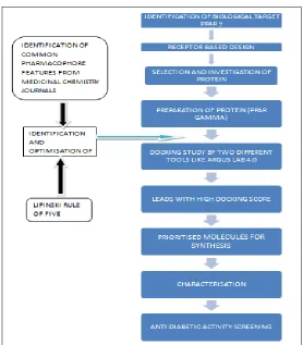
PPAR γ receptors, so following steps were performed. Identification of common Pharmacophore features responsible for inhibiting PPAR γ from the literature review which is carried out as part of the current research 5. Designing of a series of compounds that selectively modulate the activities of Peroxisome Proliferator- Activated Receptors PPAR γ for exhibiting anti diabetic activity. The current study, the binding mechanism of PPAR γ receptors and newly designed ligands were studied using molecular docking using
[image:2.612.175.441.218.389.2]docking tools like ARGUS LAB 4.0.Finally the newly designed ligands of high dock value were selected and optimised using Lipinski’ rule. Based on the synthetic feasibility, certain compounds containing thiazoleheterocycles were synthesised and purified. All the synthesised molecules were characterised by analytical methods like IR, NMR, spectral data. In vitro anti -diabetic screening was carried out for all the four synthesised compounds by α -Amaylase and α-Glucosidase inhibitory method.
FIG. 1: 3D STRUCTURE OF PPAR
MATERIALS AND METHODS:
[image:2.612.167.445.420.737.2]Target Selection:
PPAR γ As Antidiabetic Agents: Recent research suggests that PPAR γ has a therapeutic potential to treat diabetes mellitus inflammatory diseases and certain cancers. The metabolic regulatory role of PPAR δ is recently recognised and clinical trials for PPAR δ agonists are key regulators of adipocyte differentiation and used to treat type 2diabetes 6. The agonists of PPAR γ is of great interest to the pharmaceutical industry since they regulate the expression of genes associated with diseases like cancer, diabetes, atherosclerosis and obesity7.
Selection of X-ray crystal PDB: The protein selection is carried out from the RCSB PDB(Protein Data Bank). Protein data bank is resource for studying biological macromolecules. It contains information about experimentally determined structures of proteins, nucleic acids, and complex assemblies 8.
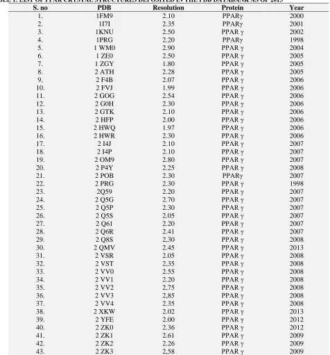
[image:3.612.63.539.236.747.2]A search in the PDB database retrieved a large number of crystal structure PPAR γ receptors. PDB codes, resolution of crystal structures are shown in tabular column below 9.
TABLE 1: LIST OF PPAR CRYSTAL STRUCTURES DEPOSITED IN THE PDB DATABANK AS OF 2013 10
S. no PDB Resolution Protein Year
1. 1FM9 2.10 PPARγ 2000
2. 1I7I 2.35 PPARγ 2001
3. 1KNU 2.50 PPAR γ 2002
4. 1PRG 2.20 PPARγ 1998
5. 1 WM0 2.90 PPAR γ 2004
6. 1 ZE0 2.50 PPAR γ 2005
7. 1 ZGY 1.80 PPAR γ 2005
8. 2 ATH 2.28 PPAR γ 2005
9. 2 F4B 2.07 PPAR γ 2006
10. 2 FVJ 1.99 PPAR γ 2006
11. 2 GOG 2.54 PPAR γ 2006
12. 2 G0H 2.30 PPAR γ 2006
13. 2 GTK 2.10 PPAR γ 2006
14. 2 HFP 2.00 PPAR γ 2006
15. 2 HWQ 1.97 PPAR γ 2006
16. 2 HWR 2.30 PPAR γ 2006
17. 2 I4J 2.10 PPAR γ 2007
18. 2 I4P 2.10 PPAR γ 2007
19. 2 OM9 2.80 PPAR γ 2007
20. 2 P4Y 2.25 PPAR γ 2008
21. 2 POB 2.30 PPARγ 2007
22. 2 PRG 2.30 PPAR γ 1998
23. 2Q59 2.20 PPAR γ 2007
24. 2 Q5G 2.70 PPAR γ 2007
25. 2 Q5P 2.30 PPAR γ 2007
26. 2 Q5S 2.05 PPAR γ 2007
27. 2 Q61 2.20 PPAR γ 2007
28. 2 Q6R 2.41 PPAR γ 2007
29. 2 Q8S 2,30 PPAR γ 2008
30. 2 QMV 2.45 PPAR γ 2013
31. 2 VSR 2.05 PPAR γ 2008
32. 2 VST 2,35 PPAR γ 2008
33. 2 VV0 2.55 PPAR γ 2008
34. 2 VV1 2.20 PPAR γ 2008
35. 2 VV2 2,75 PPAR γ 2008
36. 2 VV3 2,85 PPAR γ 2008
37. 2 VV4 2.35 PPAR γ 2008
38. 2 XKW 2.02 PPAR γ 2013
39. 2 YFE 2.00 PPAR γ 2012
40. 2 ZK0 2.36 PPAR γ 2012
41. 2 ZK1 2.61 PPAR γ 2009
42. 2 ZK2 2.26 PPAR γ 2009
44. 2 ZK4 2.57 PPAR γ 2009
45. 2 ZK5 2.45 PPAR γ 2009
46. 2 ZK6 2.41 PPAR γ 2010
47. 2 ZNO 2.01 PPAR γ 2009
48. 2 ZVT 1.90 PPAR γ 2009
49. 3 ADS 2.25 PPAR γ 2010
50. 3 ADT 2.70 PPAR γ 2010
51. 3 ADU 2.77 PPAR γ 2010
52. 3 ADV 2.27 PPAR γ 2010
53. 3 ADW 2.07 PPAR γ 2010
54. 3 ADX 1.95 PPAR γ 2010
55. 3 AN3 2.30 PPAR γ 2010
56. 3 AN4 2.30 PPAR γ 2011
57. 3 B0Q 2.10 PPAR γ 2013
58. 3 B0R 2.15 PPAR γ 2013
59. 3 B1M 1.60 PPAR γ 2011
60. 3 BC5 2.60 PPAR γ 2008
61. 3 CDP 2.80 PPAR γ 2013
62. 3 CDS 2.65 PPAR γ 2008
63. 3 CS8 2.30 PPAR γ 2008
64. 3 CWD 2.40 PPAR γ 2008
65. 3 D6D 2.40 PPAR γ 2008
66. 3 DY6 2.90 PPAR γ 2008
67. 3 ET0 2.40 PPAR γ 2009
68. 3 ET3 1.95 PPAR γ 2009
69. 3 FEJ 2.01 PPAR γ 2009
70. 3 FUR 2.30 PPAR γ 2009
71. 3 G9E 2,30 PPAR γ 2009
72. 3 HO0 2.60 PPAR γ 2009
73. 3 HOD 2.60 PPAR γ 2009
74. 3 IA6 2.30 PPAR γ 2009
75. 3 K8S 2.55 PPAR γ 2008
76. 3 KDT 2.70 PPAR γ 2010
77. 3 KMG 2.10 PPAR γ 2013
78. 3 LMP 1.90 PPAR γ 2010
79. 3 NOA 1.98 PPAR γ 2013
80. 3 OSI 2.70 PPAR γ 2011
81. 3 OSW 2.55 PPAR γ 2011
82. 3 PBA 2.30 PPAR γ 2011
83. 3 PRG 2.90 PPAR γ 1998
84. 3 QTO 2.50 PPAR γ 2013
85. 3 R5N 2.00 PPAR γ 2011
86. 3 R8A 2.41 PPAR γ 2011
87. 3 R8I 2.30 PPAR γ 2011
88. 3 S9S 2.55 PPAR γ 2011
89. 3 SZI 2.30 PPAR γ 2012
90. 3 TY0 2.00 PPAR γ 2011
91. 3 T03 2.10 PPAR γ 2012
92. 3 U9Q 1.52 PPAR γ 2012
93. 3 V9T 1.65 PPAR γ 2012
94. 3 V9V 1.60 PPAR γ 2011
95. 3 VJH 2,20 PPAR γ 2012
96. 3 VJI 2.61 PPAR γ 2012
97. 3 V9Y 2.10 PPAR γ 2012
98. 3 VN2 2.18 PPAR γ 2012
99. 4PRG 2.90 PPAR γ 1999
100. 4 A4V 2.00 PPAR γ 2013
101. 4 A4W 2.00 PPAR γ 2013
Q-Site Software: Using Q-site finder software tool, Some of the recent and efficient PDB file γ receptors with low resolution were selected and further evaluated by its Resolution value, R Free, R
value and optimised crystal ligand interaction details. Some of the selected receptors listed below from which the highlighted best PDB targets were selected for present study.
TABLE 2: SELECTION OF PDB USING Q-SITE FINDER SOFTWARE
PPAR-γ Resolution R- Value BEST PBD TARGET
3FEJ 2.01 0.206 Ligand-Good PDB
1FM9 2.10 0.239 Ligand-Good PDB
3B1M 2.60 0.221 Peptide Linked
3KMG 2.10 0.245 Ligand-Good PDB
3V9V(Best) 1.60 0.226 Ligand-Excellent PDB
TABLE 3: ACTIVE SITE OF SELECTED PPAR RECEPTORS WERE IDENTIFIED USING THE SOFTWARE Q-SITE FINDER AND TABULATED AS BELOW
Pharmacophore Identification: When reviewed the efficient journals and research articles, the target-naive screening library was already designed by using compounds from a relatively narrow and low molecular weight range (350-5000D), selected
diversity at both the putative “scaffold” core 11 The review of the literature shows that a typical PPAR γ agonist consists of an acidic head attached to an aromatic scaffold, a linker, and a hydrophobic tail.
[image:5.612.127.479.428.497.2]Common structural features of PPAR agonists: 12
FIG. 3: STRUCTURAL COMPONENTS OF PPAR γ AGONISTS 13
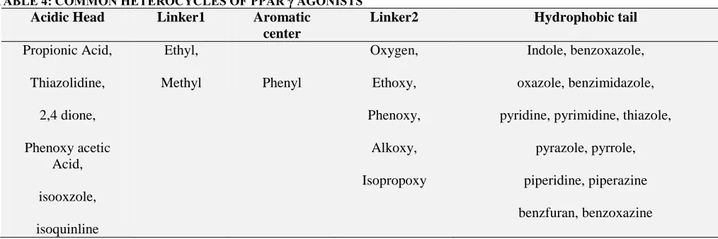
From the literature survey, the Common heterocycles used in designing PPAR γ agonists
[image:5.612.52.559.565.734.2]resembling the common features of PPARγ agonists have been listed below 14
TABLE 4: COMMON HETEROCYCLES OF PPAR γ AGONISTS
Acidic Head Linker1 Aromatic
center
Linker2 Hydrophobic tail
Propionic Acid,
Thiazolidine,
2,4 dione,
Phenoxy acetic Acid,
isooxzole,
isoquinline
Ethyl,
Methyl Phenyl
Oxygen,
Ethoxy,
Phenoxy,
Alkoxy,
Isopropoxy
Indole, benzoxazole,
oxazole, benzimidazole,
pyridine, pyrimidine, thiazole,
pyrazole, pyrrole,
piperidine, piperazine
benzfuran, benzoxazine
PPAR
Receptors PDB code Active amino acids site
Gamma 3V9V ARG280,288,CYS285,GLY286,284,GLU291,343HYS323,449,ILE341,326,262,281,LEU330, 353,KYS367,MET329,334,364,348,PHE287,368,363,SER289,342,TYR327,VAL339,446 Gamma 3FEJ ARG288,ALA282,278,333,CYS275,278,367,GLU286,GLU343,291,HIS323,328,440,449,LE
Based on the above literature facts, some new ligands have been identified, designed for further
[image:6.612.49.565.517.723.2]molecular docking using ARGUS LAB 4.0.
TABLE 5: LIST OF DESIGNED LEADS
Designed leads IUPAC name Structure
Lead 1 2-{[2-(4-methoxy phenyl)-1,3-thiazol-4yl]methyl-2-methoxy phenyl}3-(morpholin-4-yl)
butanoic acid
Lead 2 6-[2-(4-{[2-(4-chlorophenyl)-1,3-thiazol-4yl]oxy}
phenyl)ethyl]pyridine-3-carboxylic acid
Docking Study of Leads: Docking procedures aim to identify correct posses of ligands in the binding
pocket of the protein and to identify and to predict the affinity between the ligands and the protein.15
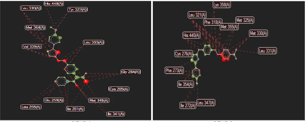
Snapshots: 3FEJ
3V9V
LEAD 1 LEAD 2 FIG. 4: INTERACTIONS OF LEADS 1 AND 2 WITH 3FEJ AND 3V9V
Stereo view of LEAD 1 and 2 in the active amino acid binding site of PPARγ receptors, suggested by molecular docking studies. The colors are as
[image:7.612.48.566.77.286.2]follows: H BONDS: Blue, Steric Interaction: Red, Interaction Overlay: Red and Green dots.
TABLE 6: DOCKINGSCORES OF LEADS USINGARGUS LAB 4.0
Protein Ligand Argus lab-binding energy (Kcal/ m)
3FEJ LEAD 1 -11.0846
LEAD 2 -11.0578
3V9V LEAD 1 -11.0686
LEAD 2 -10.3116
From the above obtained docking results and also based on the synthetic feasibility certain compounds containing thiazoleheterocycles were
[image:7.612.50.572.487.748.2]synthesised and optimised using Lipinski rule of Five. They were also further subjected to molecular docking using ARGUS LAB 4.0.
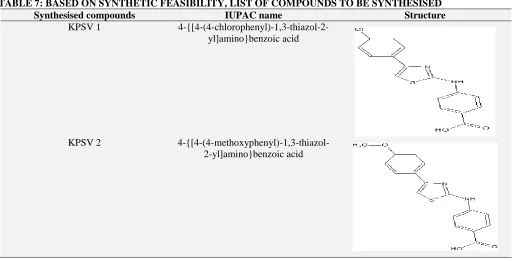
TABLE 7: BASED ON SYNTHETIC FEASIBILITY, LIST OF COMPOUNDS TO BE SYNTHESISED
Synthesised compounds IUPAC name Structure
KPSV 1
4-{[4-(4-chlorophenyl)-1,3-thiazol-2-yl]amino}benzoic acid
KPSV 2
KPSV 3 4-fluoro-2-{[4-(4-methoxy phenyl)-1,3-thiazol-2-yl]amino}benzoic acid
KPSV 4 4-fluoro-2-{[4-(4-chloro
phenyl)-1,3-thiazol-2-yl]amino}benzoic acid
N
S NH
Cl
F
O
OH
Lead Optimization Using Lipinskis Rule 16: All the synthesised analogues were checked for Lipinski rule using online version of Mol
[image:8.612.54.564.55.338.2] [image:8.612.50.563.414.510.2]inspiration software and the results are depicted in table below.
TABLE 8: OPTIMISATION OF COMPUNDS TO BE SYNTHESISED
Compound Log P Mol. Wt TPSA nOHNH NON No. of rotatable
bonds
No. of violations
LEAD 1 4.629 468.575 81.131 1 7 9 0
LEAD 2 3.997 466.881 119.61 2 9 9 0
KPSV 1 5.25 330.788 62.22 2 4 4 0
KPSV 2 4.62 326.369 71.45 2 5 5 0
KPSV3 4.90 344.360 71.45 2 5 5 0
KPSV 4 5.52 348.779 62.22 2 4 4 0
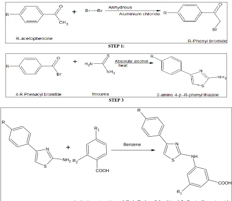
Synthesis: 17-19 Procedure:
Step 1: Synthesis of p- (chloro /methoxy) phenacyl bromide: Dissolve 0.25 mol of p- (chloro/ methoxy) acetophenone in 100ml of anhydrous diethyl ether in a 500ml flask. To this add 1gm of anhydrous aluminium chloride followed by the addition of 0.25 mol of bromine in a drop wise manner from a dropping funnel for about 30 minutes. Shake the mixture vigorously during the addition and also maintain the temperature around 20-300C. The final product commences to separate as needles after about half of the bromine has been introduced. When the addition is complete, Cool the mixture in ice water, filter the crude product and recrytallise it using rectified spirit. The purity of product was
established by single spot on T.L.C. plate. The percentage of yield was found to be 85%w/w.
Step 3: Synthesis of 4-{[4-(4-chlorophenyl)-1,3-thiazol-2-yl]amino}benzoic acid: 2- Amino-4- (p-chloro/methoxy) phenyl thiazole (1.5 gm,0.02mol) and 4 ml of dry benzene were placed in a 100 ml round bottomed flask fitted with water condenser. To this 4-chloro benzoic acid (0.02mol) was added drop wise. The reaction mixture was refluxed on
water bath at 800 c for 4 hrs. The resulting solid residue was washed with aqueous solution of sodium bicarbonate (50ml) followed by ice cold water. The resulting product obtained was recrystallised using ethanol. The purity of product was established by single spot on T.L.C. plate. The percentage of yield was found to be 70%w/w.
Scheme:
STEP 1:
STEP 3
N
S NH
COOH R
R1 N
S
NH2 R
+
COOH R1
R2
Benzene
2-Amino 4- p- R phenyl thiazole R1,R2 Benzoic acid 4-{[4-(4-R-phenyl)-1,3-thiazol-2-yl]amino}benzoic acid STEP 3
Where R= Cl / OCH3 R1= F, R2= Cl
[image:9.612.73.538.181.584.2]Physicochemical Properties of Substituted Thiazole:
TABLE 9: PHYSICOCHEMICAL PROPERTIES OF SUBSTITUTED THIAZOLE
S.no. Code R R1 R2 Molecular formula Molecular
weight
% yield
Melting
point TLC(Rf)
1. KPSV-1 Cl H Cl C16H11ClN2O2S 330.788 70.25 1600 0.8
2. KPSV-2 OCH3 H Cl C17H14N2O3S 326.369 75.16 1640 0.7
3. KPSV-3 OCH3 F Cl C17H13FN2O3S 344.360 80.32 1700 0.7
Characterisation of Synthesised Compounds: Comp. code. KPSV 1: (C16H11ClN2O2
S);4-{[4-(4-chlorophenyl) - 1, 3-thiazol-2-yl] amino}benzoic acid; Mol. wt: 330.788; C(58.09%), H(3.35%), Cl (10.72%), N(8.47%), O(9.67%), S(9.69%); IR Values: 3448.47cm-1 (O-H str); 3070.45cm-1(C=H str); 1689.52cm-1(C=O str); 1596.94cm-1(C=N str); 678.89cm-1 (C-Clstr)
Comp. code: KPSV 2: (C17H14N2O3S);
4-{[4-(4-methoxyphenyl)-1,3-thiazol-2-yl] amino} benzoic acid; Mol. wt : 326.369; C(62.56%), H(4.32%), N(8.58%), O(14.71%), S(9.83%) IR Values: 3456.18cm-1 (O-H str); 3070.45cm-1 (C=H str); 1596.94cm-1 (C=N str); 678.89cm-1 (C-Clstr)
Comp. code: KPSV 3: C17H13FN2O3
S;4-fluoro-2-{[4-(4-methoxy phenyl)-1,3-thiazol-2-yl] amino} benzoicacid:Mol.Wt:344.360;C(59.29%),H(3.81%,
N(8.13%), O(13.94%), S(9.83%), F(5.52%); IR Values:1311.50cm-1(C-C str); 2923.87cm-1 (C-H str); 1689.52cm-1(C-Ostr); 1596.94cm-1(C-N str); 678.89cm-1(C-Cl str)
Comp.code:4 KPSV;C16H10ClFN2O2
S;4-fluoro-2-{[4-(4-chloro phenyl)-1,3-thiazol-2-yl] amino} benzoic acid; Mol. wt: 348.77; C(55.10%), H(2.89%), N(8.03%), O(9.17%), S(9.19%), F(5.45%) 1311.50cm-1 (C-C str); 3085.88cm-1 (C-H str); 1689.52cm-1 (C-O str); 1596.94cm-1 (C-Nstr); 678.89cm-1; (C-Cl str); 3425.33cm-1 (O-H str);
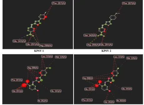
Docking Studies of Synthesised Compounds: Snap Shots: ARGUS LAB 4.0 docking results between Peroxisome Proliferator Activated Receptor (PPAR γ) along with four synthesised ligands PPAR γ:
3FEJ
KPSV 1 KPSV 2
KPSV 3 KPSV 4 FIG. 5: INTERACTIONS OF KPSV 1, 2, 3, 4 WITH 3FEJ
Stereo view of KPSV 1, 2, 3, 4, in the active amino acid binding site of PPARγ receptor 3 FEJ, suggested by molecular docking studies. The colors
3V9V
KPSV 1 KPSV 2
[image:11.612.73.535.69.407.2]KPSV 3 KPSV 4
FIG. 6: INTERACTIONS OF KPSV 1, 2, 3, 4 WITH 3V9V
Stereo view of KPSV 1, 2, 3, 4 in the active amino acid binding site of PPARγ receptors 3V9V, suggested by molecular docking studies. The
colors are as follows: H Bonds: Blue, Steric Interaction: Red, Interaction Overlay: Red and Green dots.
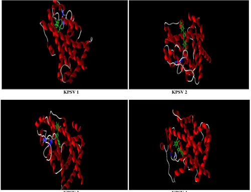
Docking View with 3FEJ
KPSV 3 KPSV 4
FIG.7: DOCKING VIEW OF KPSV 1, 2, 3, 4 WITH 3FEJ
Docking View with 3V9V
KPSV 1 KPSV 2
KPSV 3 KPSV 4
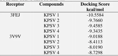
[image:12.612.56.557.309.692.2]TABLE 10: RESULTS OF MOLECULAR DOCKING STUDIES USING ARGUS LAB 4.0
Receptor Compounds Docking Score
kcal/mol
3FEJ KPSV 1 -10.5584
KPSV 2 -9.7660
KPSV 3 -9.4585
KPSV 4 -9.3435
3V9V KPSV 1 -9.0188
KPSV 2 -8.4113
KPSV 3 -8.0190
KPSV 4 -8.7298
Evaluation of Anti-Diabetic Activity:
α Amylase Inhibitory Method: 20, 21 α-amylase was dissolved in phosphate buffer saline (PBS, 0.02 mol/L, pH 6.8) at a concentration of 0.1 mg/mL. Various concentrations of sample solutions
(0.25 mL) were mixed with α-amylase solution (0.25 mL) and incubated at 37°C for 5 min. Then the reaction was initiated by adding 0.5 mL 1.0% (w/v) starch substrate solution to the incubation medium. After incubation at 37°C for 3 min, the reaction was stopped by adding 0.5mL DNS reagent (1% Dinitrosalicylic acid, 0.05% Na2SO3
and 1% NaOH solution) to the reaction mixture and boiling at 100 °C for 5 min. After cooling to room temperature, the absorbance (Abs) at 540 nm was recorded by a spectrophotometer. The inhibition percentage was calculated by the following equation:
Inhibition (%) = [(Abs1 − Abs2)/Abs1] × 100
[image:13.612.48.298.73.193.2]where, Abs1=sample and Abs2 = control.
TABLE 11: α AMYLASE INHIBITORY ACTIVITY
S.no Compound code 10 μg 50μ 100μ 500μg 1000μg
1. KPSV 1 13.88+ 0.26 18.42+0.45 27.06+0.78 36.73+0.76 51.18+0.67
2. KPSV 2 14.1+0.28 19.35+0.53 35.9+0.91 44.86+0.53 53.91+0.83
3. KPSV 3 20.51+0.17 23.45+0.34 34.04+0.83 43.12+0.28 58.11+0.78
4. KPSV 4 21.6+0.35 25.32+0.42 36.2+0.52 45.12+0.34 61.92+0.65
5. ACARBOSE 21.52+0.29 34.73+0.61 60.75+0.48 70.89+0.48 82.23+0.24
6. CONTROL 0.062 0.062 0.062 0.062 0.062
FIG. 9: PERCENTAGE GROWTH INHIBITION
GRAPH 1: α AMYLASE INHIBITORY ACTIVITY OF
SYNTHESISED SUBSTITUTED THIAZOLES AND
ACARBOSE
α-Glucosidase Inhibitory Activity: 20, 21 The α glucosidase reaction mixture contains 2.9 mM p-nitrophenyl-α D-glucopyranoside (p NPG) (Sigma-Aldrich),0.25 ml of extract(varying concentrations) in DMSO and 0.6 U/ml bakers yeast α-glucosidase in sodium phosphate buffer, pH 6.9.Control tubes contain only DMSO, enzyme and substrate, while in positive control acarbose replaced the synthesised compounds. Mixture without enzyme, synthesised compounds and acarbose served as blanks. The reaction mixture were incubated at 25 C for 5 min, after which the reaction was stopped by boiling for 2 min.
Absorbance of the resulting p-Nitrophenol (pNP) was determined at 405 nm and was considered directly to the activity of the enzyme. Glucosidase activity inhibition was determined as percentage of control as follows:
% Glucosidase inhibition (%) = [(Abs1 − Abs2)/Abs1] × 100
[image:13.612.51.496.285.716.2]TABLE 11: α - GLUCOSIDASE INHIBITORY ACTIVITY
S.no Compound code 10 μg 50μ 100μ 500μg 1000μg
1. KPSV 1 12.52+0.18 23.77+0.37 30.44+0.59 50.24+0.18 78.10+0.23
2. KPSV 2 11.65+0.61 20.98+0.82 32.61+0.63 48.55+0.22 76.78+ 0.26
3. KPSV 3 14.89+0.54 25.10+0.53 28.34+0.77 52.79+0.34 72.65+0.17
4. KPSV 4 15.47+0.32 26.56+0.34 36.56+0.80 57.32+0.57 80.46+0.46
5. ACARBOSE 20.44+0.46 31.50+0.73 40.24+0.57 61.77+0.61 85.10+0.38
6. CONTROL 0.026 0.026 0.026 0.026 0.026
FIG. 10: PERCENTAGE GROWTH INHIBITION
GRAPH 2: α GLUCOSIDASE INHIBITORY ACTIVITY OF
SYNTHESISED SUBSTITUTED THIAZOLES AND
ACARBOSE
RESULTS AND DISCUSSIONS:
PPAR (Peroxisome Proliferator Activated Receptor) agonist play a critical role in treating metabolic diseases, especially the type-2 diabetes mellitus (T2DM). In a search for more effective anti-diabetic treatment, we first aimed to work on a series of compounds that selectively modulate the activities of Peroxisome Proliferator-activated receptor γ. By reviewing the literature many scientific medical journals of existing PPAR γ agonists, it was found that a typical PPAR γ agonist consists of an acidic head attached to an aromatic scaffold, a linker, and hydrophobic tail. Based on these facts, new ligands containing thiazole heterocyclic nucleus have been designed and made ready for docking studies.
The PPAR activities of the designed NCEs were predicted by the developed models and then they were subjected to docking studies using ARGUS LAB 4.0 PPARγ (PDB code 3FEJ, 3V9V) to predict the binding affinities an interactions of the ligands at the active sites of the receptors. The hydrogen bonding interactions of the selected molecules at the active sites of PPARγ was also determined to identify which lead interacts better with the target proteins and results were tabulated. Based on the synthetic feasibility, certain compounds containing thiazoleheterocycles were synthesised and optimised using Lipinski rule of five. The synthesised compounds were characterised by IR, NMR, and Spectraldata’s.
All the four synthesised compounds revealed a significant inhibitory action on α amylase and α glucosidase enzyme when evaluated for their antidiabetic activity. The percentage inhibition at 10μg to 1000μg/ml concentrations of all the four synthesised compounds showed a concentration dependent increase in percentage inhibition in both the α- amylase and α –glucosidase inhibitory methods. Compound KPSV 4 exhibited a percentage inhibition varying from 61.92±0.65 to 21.6±0.35 for highest concentration1000μg/ml to lowest concentration 100μg/ml against α amylase enzyme and 80.46±0.46 to 15.47±0.32 for highest concentration 1000μg/ml to lowest concentration 100μg/ml against α glucosidase enzyme.
For Acarbose, 10 μg of sample exhibits 21.52 % growth of inhibition. 50μg of sample exhibits 34.73 % growth of inhibition. 100μg of sample exhibits 60.75 % growth of inhibition. 500μg of sample exhibits 70.83 % growth of inhibition. 1000μg of sample exhibits 82.23 % growth of inhibition.
[image:14.612.50.301.335.493.2]pharmacophore acidic head, a linker ,and a hydrophobic tail. Four different compounds have been synthesised and subjected to docking studies. Then the synthesised compounds were subjected to optimization using Lipinksi’s rule of five.
Docking is performed by using ARGUS LAB 4.0 docking studies showed the important interactions of lead molecules posses with some of the common active site residues like ARG 288, CYS 28, HIS 323, HIS 449, LEU 353, LYS 367, MET 364, MET 348, PHE 363 of different PPARγ receptors like 3FEJ, 3V9V.
The synthesised compounds were evaluated for their antidiabetic activity by α amylase and α glucosidase inhibitory methods and all were found to exhibit an effective inhibition against both the enzymes. Out of the four synthesised compounds, KPSV 4 was found to be most effective with percentage growth inhibition of 61.92% and 80. 46% at 1000μg when compared with other three compounds.
ACKNOWLEDGEMENT: The Authors are thankful to IIT, Chennai for providing us analytical and spectral datas and also to Dr. Suresh M. Pharm Ph.D for helping us to carry out in vitro antidiabetic studies.
CONFLICT OF INTEREST: The authors have no conflict of interest.
REFERENCES
:
1. American diabetes association diagnosis and classification of diabetes mellitus Diabetes Care. 35 suppl 1, 2012; S64-S71.
2. Shoback, edited by David G. Gardner, Dolores.et al.,
Greenspan’s basic & clinical endocrinology (9th
ed.). Newyork: McGraw-Hill Medical. pp. Chapter.17.2011. 3. Mitchel, Richard Sheppard; Kumar, Vinay; Abbas, AbulK., Robbins., et al., Basic Pathology. Philadelphia: Saunders. Unless otherwise specified, reference is: Table 20-5 in 8th edition.
4. Markt P, Petersen RK, Flindt EN, Kristiansen K, Kirchmair J, Spitzer G, et al., Discovery of Novel PPAR Ligands by a virtual Screening Approach Based on 8, 2013,51 (20): 6303-6317.
5. Zulma Garcia, Nikki Lewis, Josep Bassagnaya-Riera and David Bevan Structure based pharmacophore sampling of PPAR., The FASEB Journal, 2012, 26:760.
6. Klebe G, et al., Virtual ligand screening: strategies, perspectives and limitations, Drug Discovery Today, 2006, 11 (13-14): 580-594.
7. Billin AN, et al., PPAR-beta/delta agonists for Type 2 diabetes and dyslipidemia: an adopted orphan still looking for a home, 2008, 17 (-7110):1465.
8. Kitchen DB, Decornez H,. Furr JR, Bajorath J, et al.,
Docking and scoring in virtual screening, 2004, 3(11): 935-949.
9. Kuhn B, Hilpert H, Benz J, et al., “Structure- based design of indole propionic acids as novel PPAR alpha/gamma co-agonists” Bioorganic and Medicinal chemistry letters, 2006,16 (15): 4016-4020.
10. Haffner CD, Lenhard JM, Miller AB , et al., Structure-based design of potent retinoid X receptor alpha agonists, Journal of Medicinal Chemistry, 2004, 47/980: 2010-2029.
11. N. Mahindroo, C.-C. Wang, C.-C. Liao, et al., Indol-1-yl acetic acids as a PPAR agonists design synthesis, structural biology and molecular docking studies and journal of medicinal chemistry 2006, 49(3):1212-1216. 12. Wilson TM, Brown PJ, Sternbach DD ,Henke BR., et al., The PPARS: from orphan receptors to drug
discovery .J .Med Chem.,2000,42:527-550.
13. Shearer BG, Hoekstra WJ; et al., Recent Advances in PPAR Sceince. Curr Med Chem., 2003, 10:267-280. 14. Mahindroo N, Huang CF, Peng YH, Wang CC, Liao
CC ,Lien TW, et al., Novel Indole –based Peroxisome Proliferator –Activated Receptor Agonists: Design, SAR, Structural Biology and Biological activities .J Med Chem., 2005,48(26):8194-8208.
15. Schrodinger, L. L.c., New York, www. Schrodinger. com
16. Lipinski CA., Lambardo F., Dominy BW., et al., Feeny PJ Experimental and computational approaches to estimate and solubility and permeability in drug development studies.; Adv drug Del Rev .,1997,23;3-25.
17. Bernthsen, A., et al., Textbook of organic chemistry. London: Blackie and Son limited; 1947.327,678-681,685,700,701
18. Vogel Textbook of practical organic chemistry 1978: 6.12
19. International journal of Chemical science, volume. 7(4), 2009:2222-2938, 2584.
20. Rajagopal PL., Sreejith KR., Kiron SS., et al.,
Antidiabetic activity (in vitro) of the rhizomes of
Anaphyllum wightiis chott., International Journal of Research in Pharmacy and chemistry., 2013,3(4),790-792.
21. Mogale, M A., Lebeloo, S L., et al., αamylase and
αglucosidase inhibitory effects Sclerocarya birrea
subspecies caffra (sond) Kokwaro (Anacardiaceae) stem- bark extracts. African Journal of Biotechnology, 2011, Vol.10 (66):15033-15039.
All © 2013 are reserved by International Journal of Pharmaceutical Sciences and Research. This Journal licensed under a Creative Commons Attribution-NonCommercial-ShareAlike 3.0 Unported License.
This article can be downloaded to ANDROID OS based mobile. Scan QR Code using Code/Bar Scanner from your mobile. (Scanners are available on Google Playstore)
How to cite this article:
Priyadarsini R, Durga V and Ahmed S:Virtual screening, synthesis of newer heterocycles as PPAR γ agonists with antidiabetic activity.