R E S E A R C H
Open Access
Defective structural RNA processing in
relapsing-remitting multiple sclerosis
Charles F Spurlock III
1, John T Tossberg
1, Yan Guo
2, Subramaniam Sriram
3, Philip S Crooke III
4and Thomas M Aune
1,5,6*Abstract
Background:Surveillance of integrity of the basic elements of the cell including DNA, RNA, and proteins is a critical element of cellular physiology. Mechanisms of surveillance of DNA and protein integrity are well understood. Surveillance of structural RNAs making up the vast majority of RNA in a cell is less well understood. Here, we sought to explore integrity of processing of structural RNAs in relapsing remitting multiple sclerosis (RRMS) and other inflammatory diseases.
Results:We employed mononuclear cells obtained from subjects with RRMS and cell lines. We used quantitative-PCR and whole genome RNA sequencing to define defects in structural RNA surveillance and siRNAs to deplete target proteins. We report profound defects in surveillance of structural RNAs in RRMS exemplified by elevated levels of poly(A) + Y1-RNA, poly(A) + 18S rRNA and 28S rRNAs, elevated levels of misprocessed 18S and 28S rRNAs and levels of the U-class of small nuclear RNAs. Multiple sclerosis is also associated with genome-wide defects in mRNA splicing. Ro60 and La proteins, which exist in ribonucleoprotein particles and play different roles in quality control of structural RNAs, are also deficient in RRMS. In cell lines, silencing of the genes encoding Ro60 and La proteins gives rise to these same defects in surveillance of structural RNAs.
Conclusions:Our results establish that profound defects in structural RNA surveillance exist in RRMS and establish a causal link between Ro60 and La proteins and integrity of structural RNAs.
Background
Relapsing remitting multiple sclerosis (RRMS) affects ap-proximately 0.1% of the population. RRMS is characterized by de-myelination of neurons but disease mechanisms are incompletely understood. Involvement of innate and adaptive arms of the immune system is detected in in-flammatory demyelinating lesions implicating these in pathogenesis. Both genetic and environmental factors are also implicated in the origins of RRMS [1-8].
Studies have demonstrated an expanding complexity of non-protein-coding RNAs (ncRNA) in higher eukary-otes [9]. Non-vertebrate metazoans and microbes pos-sess a single Y RNA while humans encode up to four Y RNAs, termed Y1, Y3, Y4, and Y5 RNA [10]. Y RNAs are approximately 100 nucleotides in length and are
transcribed by RNA polymerase III. Y RNAs possess sequence-specific binding sites for the Ro60 protein [11]. All four human Y RNAs assemble into ribonucleotein (RNP) particles including Ro60, La, and other pro-teins [12]. Precise functions of Y RNAs and Ro nucleoprotein particles are incompletely understood but studies suggest they contribute to ncRNA quality control and ribosomal RNA (rRNA) processing [13,14]. To pro-duce mature rRNAs, RNA polymerase 1 transcribes a 47S precursor rRNA that is processed via endonucleo-lytic, exonucleoendonucleo-lytic, and additional modifications produ-cing mature 5.8S, 18S, and 28S rRNAs [15-17]. Small nuclear U RNAs are another form of ncRNA making up the RNA portion of the RNP complex known as the spli-ceosome required for splicing of pre-mRNAs to mature mRNAs [18,19]. In vertebrates, major small nuclear U RNAs include U1, U2, U4, U5, and U6 [20,21]. Defects or mutations in specific small nuclear U RNAs or the genes encoding proteins required for biogenesis of U-RNA RNP complexes, such as SMN1 (survival of motor
* Correspondence:[email protected] 1
Department of Medicine, Vanderbilt University School of Medicine, Nashville, TN 37232, USA
5
Department of Pathology, Microbiology and Immunology, Vanderbilt University School of Medicine, Nashville, TN 37232, USA
Full list of author information is available at the end of the article
neuron 1), give rise to specific neurodegenerative disor-ders rather than global defects [22-26].
Polyadenylation of mRNAs at the 3’end is a necessary step in their synthesis and maturation [14,19,27-32]. Ma-ture, fully processed ncRNAs lack 3’ poly(A) tails. Addition of poly(A) tails to ncRNAs represents a critical quality-control step to promote degradation of mis-folded or mis-processed ncRNAs. Specific protein com-plexes exist to add poly(A) tails to RNA substrates to promote their degradation by stimulating exonucleolytic activity of the exosome thus leading to degradation of mis-processed or mis-folded ncRNAs.
It is fundamentally unknown how normal cellular pro-cesses or responses to extracellular stimuli may invoke polyadenylation and degradation of ncRNA substrates or if human disease processes exhibit defects in polyadeny-lation of ncRNA substrates as part of their pathogenesis. Here, our results demonstrate that mononuclear cells from subjects with RRMS exhibit pervasive increases in levels of polyadenylated ncRNAs including Y1 RNA, 18S and 28S rRNA, and U1, U2, and U4 snRNAs and these defects are unique to RRMS. Defects in expression of both Ro60 and La proteins in RRMS appear to contrib-ute to increased polyadenylation of ncRNAs. Further, IFN-β1b, a common RRMS therapy [33], restores both Ro60 and La levels to normal as well as levels of polya-denylated Y1 RNA and U1 snRNA suggesting that aber-rant polyadenylation of ncRNA substrates may have pathogenic consequences.
Results
Elevated polyadenylation of ncRNA substrates in RRMS To initiate our studies, we determined levels of Y RNAs in different subject cohorts by quantitative PCR using ei-ther random hexamers or oligo-dT for synthesis reason-ing that random hexamers should allow amplification of all Y RNAs independent of whether or not they were polyadenylated but that oligo-dT would allow amplifica-tion of only those Y RNAs that were polyadenylated con-sistent with previously established methodologies [31]. We also confirmed that >95% of total cellular Y RNAs were retained during the PaxGene total RNA isolation procedure. Sanger sequencing of the PCR products con-firmed identity of all human Y RNAs. We found that levels of total Y RNAs (random hexamers) were not markedly different between CTRL and RRMS subjects (Figure 1A). In marked contrast, levels of polyadenylated Y1 RNA (oligo-dT), but not other Y RNAs, were in-creased by about 20-fold in RRMS relative to CTRL (Figure 1B). This elevation of polyadenylated Y1 RNA was not seen in systemic lupus erythematosus (SLE), rheumatoid arthritis (RA), neuromyelitis optica (NMO), or Parkinson’s disease (PD) (Additional file 1: Table S1 for a description of subjects) (Figure 1C). In a
cross-sectional analysis, we compared levels of polyadenylated Y1 RNA in blood samples obtained from subjects ex-periencing a first clinically isolated syndrome who went on to develop RRMS at a later date (CIS-MS), from sub-jects at the time of their diagnosis of RRMS but prior to onset of any therapies (RRMS-NAÏVE) and from subjects with RRMS of >1 year’s duration (RRMS) (Figure 1D). We found that levels of polyadenylated Y1 RNA were elevated in both RRMS-NAÏVE and RRMS cohorts but that levels of polyadenylated Y1 RNA were similar to CTRL levels in the CIS-MS cohort. Thus, increased polyadenylation of Y1 RNA, but not other Y RNAs, was observed in RRMS, but not other autoimmune diseases.
18S RNA 28S RNA 0
1000 2000
3000 CTRL
RRMS
18S RNA 28S RNA 0
200 400 600 800
p
o
ly
(A
)+ r
RNA
le
v
e
l
*
*
A
B
C
D
5'-90 5'-60 5'-30 18S3'+303'+603'+90 5'-90 5'-60 5'-30 28S3'+303'+603'+90
0 2 4 6 8 10
F
ol
d C
hange
RRM
S
:CT
RL
r
RNA
*
*
*
*
*
*
*
*
*
*
to
ta
l r
RNA
le
v
e
[image:3.595.60.538.89.220.2]l
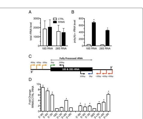
Figure 2Increased polyadenylation and mis-processing of 18S and 28S rRNAs in RRMS. (A) Total rRNA levels in blood determined after cDNA synthesis using random hexamers in CTRL (N = 24) and RRMS (N = 22). (B) As in (A) except oligo-dT was used for cDNA synthesis. (C) Schematic illustrating PCR strategy to detect misprocessed 18S and 28S rRNAs. (D) Transcript levels of misprocessed rRNAs (-90, -60, -30) in RRMS (N = 12) relative to CTRL (N = 12). Results were normalized to total 18S or 28S RNA and are presented as fold difference between CTRL and RRMS. *P<0.05.
Y1 RNAY3 RNAY4 RNAY5 RNA
0.001 0.01 0.1 1 10
pol
y
(A
)+
Y
R
N
A
**
Y1 RNAY3 RNAY4 RNAY5 RNA
0.001 0.01 0.1 1 10 100
T
o
ta
l Y
RNA
CTRL RRMS
A B C
Y1 RNA Y3 RNA Y4 RNA Y5 RNA 0.0001
0.001 0.01 0.1 1 10
p
o
ly
(A)
+
Y R
N
A
CTRL SLE RA NMO PD
*
CTRL CIS-MS
RRMS-NAIVE RRMS 0
1 2 3 4
p
o
ly
(A)
+
Y1
R
N
A
[image:3.595.61.539.287.684.2]* * D
Next, we determined levels of U1 RNA in blood (PaxGene tubes) from CTRL subjects, subjects with RRMS or with other inflammatory diseases. As above, synthesis of cDNA was performed using random hexamers and oligo-dT and transcript levels were determined by quantitative PCR. We found a modest increase in total levels of U1 RNA (random hexamers) in RRMS relative to CTRL (Figure 3A). In contrast, poly(A) + U1 tran-script levels were markedly elevated in subjects with RRMS relative to CTRL (Figure 3B). The CIS-MS co-hort did not exhibit increased poly(A) + U1 RNA levels nor did subjects with other inflammatory diseases, SLE, RA, and NMO. We also extracted RNA-seq data and confirmed that poly(A) + U1 RNA levels were elevated in RRMS and that U2 and U4 RNA levels were also ele-vated in RRMS relative to CTRL (Figure 3C). We also examined expression of total and poly(A) + U11 and U12 transcripts, components of the minor spliceosome pathway, and found elevated poly(A) + U11 and U12 transcript levels in RRMS versus CTRL and increased total U11 transcript levels. Total levels of U12 snRNA were not significantly different between RRMS and CTRL (Figure 3D). Thus, multiple ncRNA species ex-hibit increased polyadenylation in RRMS relative to CTRL or other autoimmune diseases.
Alterations in mRNA processing in RRMS
The U1, U2, U4, U5, and U6 RNAs play critical roles in determining mRNA length and isoform expression via several mechanisms including protection from prema-ture cleavage and polyadenylation of nascent pre-mRNAs [21,26,34]. Disruption of levels of individual U RNAs is sufficient to cause global alterations in alterna-tive splicing and mRNA length. It is not known how im-balances or altered polyadenylation of multiple U snRNAs may impact alternative splicing and mRNA lengths. Since these defects include mRNA shortening
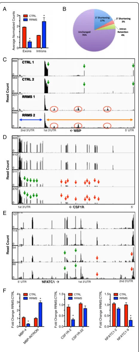
by loss of 5’ or 3’ exons, choice of alternative 5’and 3’ UTRs, choice of initial and final exons and partial reten-tion of intron sequences, we searched our RNA-seq data for differences in splicing and intron retention between CTRL subjects and subjects with RRMS. For each chromosome, we counted the number of reads per exon across the genome in CTRL and RRMS subjects. We found a genome-wide loss of exon expression in RRMS relative to CTRL (Figure 4A). Genome-wide analysis of expressed genes demonstrated that 17% of transcribed genes exhibited loss of 5’exons (5’shortening) (Figure 4B). Loss of 3’ exons (3’shortening, 3% of transcribed genes) was less frequent. Intron retention was also less frequent and only 4% of transcribed genes exhibited forms of intron retention. Visual inspection of 3,000 or 30% of expressed genes using the Integrative Genome Viewer (IGV) con-firmed these results. We defined 5’ and 3’shortening to mean that at least one exon at the 5’or 3’end of the tran-script, respectively, exhibited a >5-fold reduction in expressed counts or FPKM relative to exons at the 3’or 5’ end of the transcript, respectively, in RRMS subjects com-pared to CTRL subjects. Intron retention was similarly de-fined as an increase in read counts localized to an intron by >5-fold in RRMS compared to CTRL subjects. The ma-jority of genes transcribed in mononuclear cells from RRMS compared to CTRL did not display differences in isoform distribution or retention of intron sequences.
We analyzed the number of read counts per intron and found that subjects with RRMS exhibited higher in-tron read counts across chromosomes compared to CTRL. Examples of these alterations included MBP,
CSF1R, and NFATC1 (Figure 4C to E). We found that
the myelin basic protein gene, MBP, which encodes both classic myelin basic protein expressed primarily by mye-lin forming cells and a second family of proteins, called golli proteins, expressed by T lymphocytes, neurons, and oligodendrocytes, displayed increased intron retention in
CT RL
CIS-MSRRMS SLE RANMO 0.0
0.5 1.0 1.5
p
o
ly
(A
)+ U1
RNA **
U1 U2 U4
0 5 10 15 20 25
p
o
ly
(A
)+ U RNA
RRMS CTRL *
* *
A
B
C
CTRL RRMS 0.00 0.02 0.04 0.06 0.08 0.10
*
poly(A)+ Tota
l 0.001
0.01 0.1 1
U
1
1 E
x
pr
es
s
ion
RRMS CTRL
* *
*
Poly(A)+ Total 0.00001
0.0001 0.001 0.01 0.1 1
U
1
2 E
x
pr
es
s
ion
RRMS
CTRL
* *
D
To
ta
l U
1
R
N
[image:4.595.62.539.543.665.2]A
the mature mRNA (Figure 4C, note that transcription is from right to left) [35-37]. Individual exons are labeled with green arrows and regions of intron retention seen in RRMS are labeled with red circles. Retained intron se-quences were present in both ‘golli’ and ‘golli-MBP’ mRNAs; classic MBP gene depicted by the blue arrow, golli-MBP gene depicted by the orange arrow. Thus, U snRNA imbalance in RRMS was associated with intron retention in MBP mRNA.
We also found a marked reduction in transcript levels of the exons comprising CSF1R mRNAs. In CTRL sub-jects, each of the 22 exons was present at approximately equal abundance. However, in RRMS, there was de-creased transcript abundance of exons 1 to 11 (green ar-rows identify exons expressed at similar levels in CTRL and RRMS and red arrows show exon loss) (Figure 4D). Thus, U snRNA imbalance in RRMS was also associated with 5’ shortening. 3’ shortening was also found in RRMS (Figure 4E). All nine exons of NFATC1 mRNA and the 3’ UTR exhibited similar transcript abundance assembled into a continuous mRNA in CTRL subjects. RRMS subjects exhibited a specific loss of the eighth and ninth exons (note that the ninth exon is continuous with the 3’ UTR, red arrows). The remaining expressed exons were present at similar levels in CTRL and RRMS (green arrows). We then validated these findings in a dif-ferent cohort of CTRL and RRMS subjects using quanti-tative PCR (Figure 4F). Transcript levels of MBP exon 4, CSF1R exon 2, and NFATC1 exon 9 were significantly reduced in RRMS versus CTRL. Levels of CSF1R exon 22 and NFATC1 exon 2 were only modestly reduced in RRMS similar to our RNA-sequencing findings. We also found increased transcript levels of the MBP intron be-tween MBP exons 3 and 4 in RRMS compared to CTRL. Thus, using both quantitative PCR and RNA-sequencing, we were able to confirm specific examples of intron retention, 5’shortening, and 3’shortening in this independent sample set of RRMS subjects. These
NFATC1
5’ UTR 1st 3’UTR 2nd 3’UTR
5’
CSF1R
1st 3’UTR
MBP
CTRL 1
CTRL 2
RRMS 1
RRMS 2
5’ UTR 1st 3’UTR
2nd 3’UTR
Read Count
866
0 969
0 401
692 0
0
Read Count
605
0 810
0 511
870 0
0
Read Count
127
0 114
0 54
80 0
0
A B
C
D
CSF1R-2 CSF1R-2 2
0.0 0.5 1.0 1.5
F
o
ld
C
han
g
e R
R
M
S
:C
T
R
L
CTRL RRMS
*
MBP-4
MBP-INTRON
0 1 2 3 4
Fo
ld
C
h
a
n
g
e
RRM
S
:CT
RL CTRL
RRMS
* *
NFA TC1-2
NFATC1-9
0.0 0.5 1.0 1.5
Fo
ld
C
h
a
n
g
e R
R
M
S
:C
T
R
L
CTRL RRMS
* Exons Introns
0 1 2 3
A
v
er
a
g
e N
or
m
al
iz
ed C
ount
s
CTRL RRMS
* *
E
[image:5.595.58.291.86.674.2]F
shortened mRNA isoforms seen in RRMS are consist-ent with mRNA premature cleavage and polyadenyla-tion, a property that is produced by imbalances of U snRNAs or loss of U snRNA function. U snRNA imbal-ance observed in RRMS may contribute to intron reten-tion as well as exon loss and mRNA shortening.
Reduced expression of Ro60 and La in RRMS
Ro60 and La proteins are components of ribonucleopro-tein particles, bind discrete structural ncRNAs, and are thought to play important roles in ncRNA processing and quality control. For these reasons, we measured
TROVE2(Ro60) and SSB(La) expression levels in blood
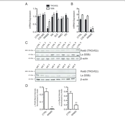
samples harvested in PaxGene tubes from the following cohorts of subjects: CTRL, CIS-MS, RRMS, RA, SLE, NMO, and PD. We found that TROVE2 and SSB tran-script levels were markedly reduced in the established RRMS cohort compared to CTRL. This difference was unique to RRMS and not observed in other autoimmune disease cohorts or in other inflammatory (NMO) or non-inflammatory (PD) neurologic conditions (Figure 5A). We replicated these findings by whole-genome RNA se-quencing (RNA-seq) and obtained equivalent results (Figure 5B). We also determined levels of protein ex-pression of Ro60 and La in PBMC by western blotting. We found that both Ro60 and La proteins were pro-foundly diminished in RRMS PBMC relative to CTRL PBMC (Figure 5C and D). Thus, bothTROVE2andSSB transcripts and Ro60 and La proteins were profoundly diminished in RRMS and these mRNA and protein ex-pression differences were not seen in several other autoimmune diseases.
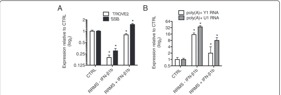
Interferon-β1b (Betaseron) and structural defects in RRMS For all analyses above, RRMS subjects were either on copaxone or no immunomodulatory therapy. Thus, we compared these subjects to RRMS on stable betaseron therapy. We found that levels ofTROVE2,SSB, poly(A) + Y1 RNA and poly(A) + U1 snRNA were close to CTRL levels in RRMS subjects on betaseron therapy compared to RRMS subjects on either copaxone or no immunomod-ulatory therapy (Figure 6A, B). We examined responses of three individuals in longitudinal studies who initiated betaseron and found that correction of levels ofTROVE2,
SSB, poly(A) + Y1 RNA and poly(A) + U1 RNA was very rapid. We hypothesize that signaling pathways either dir-ectly or indirdir-ectly activated by betaseron interfere with signaling pathways driving defects in polyadenylation of structural RNAs in and expression ofTROVE2andSSBin RRMS and that betaseron will be a useful tool to identify underlying mechanisms. Further, measurement of polya-denylated species of ncRNAs may provide a useful means to monitor responses to betaseron or other immunomod-ulatory therapies in RRMS.
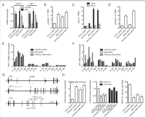
TROVE2 and SSB silencing disrupts structural RNA surveillance and alters mRNA length
To establish causal links between Ro60 and La imbal-ance and polyadenylation and processing of structural ncRNAs observed in RRMS, we designed siRNAs spe-cific for human TROVE2 and SSBand transfected them into the human THP-1 monocyte or the Jurkat T cell line. Transfection with TROVE2 siRNA caused specific reduction ofTROVE2transcripts but notSSBtranscripts (Figure 7A). Similarly, the SSB siRNA caused specific loss of SSBtranscripts but notTROVE2 transcripts. We asked if reduced TROVE2 or SSB levels resulted in an increase in the amount of poly(A) + Y1 RNA in THP-1 cells. We found that knockdown of eitherTROVE2,SSB, or the combination effectively increased levels of poly (A) + Y1 RNA (Figure 7B). Additionally, knockdown of
SSB resulted in marked accumulation of poly(A) + 18S rRNA (Figure 7C). Knockdown of TROVE2 resulted in only a modest increase in poly(A) + 18S RNA. In con-trast, levels of poly(A) + 28S rRNA were largely un-affected by knockdown of either TROVE2 or SSB but were modestly increased by the knockdown of both
TROVE2 and SSB. We also examined the impact of
TROVE2 or SSB knockdown on levels of poly(A) + U1
RNA and found that knockdown of TROVE2, but not knockdown ofSSB, caused a marked increase in levels of poly(A) + U1 RNA (Figure 7D). rRNA misprocessing was also analyzed as described above. In both THP-1 cells and Jurkat cells, transfection of TROVE2 and/or SBB siRNAs increased levels of misprocessed 18S rRNA and, to a lesser extent, misprocessed 28S rRNA (Figure 7E, F). Since the combination of TROVE2and SSB knockdown increased the amount of poly(A) + U1 RNA and alter-ations in levels of U1 RNA result in isoform switching, we determined ifTROVE2andSSBRNA knockdown was suf-ficient to alter mRNA lengths in a cell [21]. We trans-fected TROVE2 and SSB siRNAs into THP-1 cells and employed a PCR strategy to test for different lengths of
MBP, CSF1R, and NFATC1 transcripts (Figure 7G).
Similar to what we observedin vivo, we found that re-duced levels of either Ro60 or La by siRNA silencing were sufficient to increase expression levels of the
processing, and proper mRNA splicing; key compo-nents of RNA function in the cell.
Discussion
We have found that extensive polyadenylation of struc-tural RNAs, including Y1 RNA, U RNAs, and rRNAs is a feature of mononuclear cells from subjects with RRMS but not from subjects with other autoimmune or neuro-degenerative conditions. Increased polyadenylation of rRNAs is associated with mis-processing of both 18S and 28S rRNAs. Alterations in polyadenylation and total
U RNAs, essential components of the spliceosome, are associated with extensive 5’ shortening, 3’ shortening, and intron retention of mRNAs. Ro60 and La, two pro-tein components of ribonucleopropro-tein complexes in the cell, are under-expressed in RRMS and siRNA-mediated
‘knockdown’ of Ro60 and/or La recapitulates these ef-fects in cell lines resulting in increased polyadenylation of structural RNAs, misprocessing of 18S and 28S rRNAs, and isoform switching of mRNAs. Finally, IFN-β1b or betaseron, a common therapy for RRMS, but not other autoimmune diseases, restores the balance of CTRL
CIS-MSRRMS
RA SLENMO PD
0.0 0.5 1.0 1.5
m
R
N
A
E
x
pr
es
s
ion
*
*
SSB TROVE2
CTRL RRMS
0.0 0.1 0.2 0.3
-R
o60 B
and I
n
tens
it
y
Nor
m
aliz
ed t
o
-Act
in
**
CTRL RRMS
0.0 0.2 0.4 0.6 0.8 1.0
-La B
a
nd I
n
te
ns
it
y
Nor
m
aliz
ed t
o
-Act
in
** CTRL
CIS-MS RRMS
0 2 4 6 8
m
R
N
A
E
x
pr
es
s
ion
*
*
*
Ro60 (TROVE2)
La (SSB)
-actin MW: 60 kDa
47 kDa
42 kDa
Ro60 (TROVE2)
La (SSB)
-actin 42 kDa
MW: 60 kDa
47 kDa
A
B
C
[image:7.595.60.538.88.532.2]D
polyadenylated structural RNAs to normal and reverses the loss of Ro60 and La that is seen in RRMS.
A general model is that misfolded or misprocessed RNA substrates are polyadenylated in ribonucleoprotein particles, termed theTrf4p/Air2p/Mtr4p polyadenylation complex or TRAMP complex in yeast [29]. Trf4p and Trf5p are poly(A) polymerases. Mammalian orthologues include PAPD5 and PAPD7. This complex or similar complexes in higher eukaryotes catalyzes oligoadenyla-tion or polyadenylaoligoadenyla-tion of RNA substrates, which, in turn, stimulates the exosome to degrade these poly(A) + substrates. Thus, in RRMS mononuclear cells, alter-ations in levels of misfolded or misprocessed structural RNAs or increased activity of the TRAMP complex or its equivalent may lead to increased polyadenylation of structural RNA substrates. Alternatively, decreased func-tion of the exosome may lead to increased levels of poly (A) + structural RNA substrates. In fact, the eubacterium orthologue of Ro60 functions with Y RNA and the exo-some to degrade misprocessed or misfolded structural RNAs [38]. Thus, defects in expression of Ro60 in RRMS may lead to defective function of the exosome and contribute to increased levels of polyadenylated structural RNAs seen in RRMS. Future studies will be required to delineate among these possibilities.
Ribosomal RNA processing proceeds via both endonu-cleolytic cleavages and exonuendonu-cleolytic processing to yield mature 18S and 28S rRNAs. Levels of misprocessed rRNAs in cells are extremely low if effective RNA sur-veillance mechanisms outlined above are fully functional. Various manipulations, such as inhibition of RNA poly-merase 1 by low concentrations of actinomycin D [30], produce aberrant pre-rRNA transcripts that are polyade-nylated. Thus, abortive synthesis and processing of 18S
and 28S rRNAs as well as other structural RNAs may also contribute to increased levels of polyadenylated structural RNAs in RRMS.
The U RNAs are another class of structural RNAs. These snRNAs constitute the RNA portion of the spli-ceosome required to remove introns from pre-mRNAs to produce mature mRNAs for translation. Polyadeny-lated forms of U1, U2, and U4 RNAs, as well as U11 and U12 components of the minor spliceosome pathway, are markedly increased in RRMS. There is also genome-wide disruption of splicing and processing of mRNAs resulting in 5’ and 3’ mRNA shortening and intron re-tention. An example of intron retention is the MBP gene. MBP has long been considered a candidate auto-antigen for RRMS. In animal models, injection of foreign MBP gives rise to cross-reactivity to native MBP and in-flammation, autoimmunity and clinical symptoms simi-lar to human RRMS [39]. In a simisimi-lar vein, intron retention in the MBP mRNA could produce MBP pro-teins with altered C-termini viewed by the immune sys-tem as foreign. Resulting immune responses to these altered MBP proteins could lead to cross-reactivity with native MBP and immune attack against myelin in the central nervous system thus contributing to develop-ment of RRMS.
Ro60 and La proteins are components of ribonucleo-protein particles. Both ribonucleo-proteins bind certain structural RNAs. For example, Ro60 binds Y RNAs while La binds tRNAs, certain rRNAs, and microRNAs [13]. Both pro-teins are thought to play important roles in surveillance and quality control of structural RNAs. We found that both Ro and La proteins exhibit substantially reduced expression in RRMS but not other autoimmune diseases or other neurodegenerative diseases. In cell lines, we
CTRL
RRMS - IFN-1b
RRMS + IFN-1b
0.125 0.25 0.5 1 2
E
x
pr
es
s
ion r
el
at
iv
e t
o C
T
R
L
(l
o
g2
)
TROVE2
SSB
* *
* *
CTRL
RRMS - IFN-1b
RRMS + IFN-1b
0.5 1 2 4 8 16 32 64
E
x
p
re
s
s
io
n
r
e
la
tiv
e
t
o
CT
RL
(l
o
g2
)
poly(A)+ Y1 RNA poly(A)+ U1 RNA
* *
* *
[image:8.595.57.541.89.252.2]A
B
found that siRNA-mediated depletion of either Ro60 or La or both recapitulated many of the defects in struc-tural RNA surveillance seen in RRMS mononuclear cells. Thus, depletion of Ro60 and La proteins in RRMS probably contributes to loss of structural RNA quality control in RRMS. Exact mechanisms are not completely clear. Ribonucleoprotein particles are heterogeneous complex structures that contain multiple proteins and RNA species with different functions. Loss of Ro60 and/ or La may disrupt these ribonucleoprotein particles lead-ing to loss of function, some of which are intimately
involved in structural RNA quality control. Loss of Ro60 or La may also produce other defects in polyadenylation and/or degradation of structural RNAs. Further studies will be required to better understand mechanistic rela-tionships between Ro60 and/or La deficiency and polya-denylation and processing of structural RNAs.
The relationship between deficiencies of Ro60 and La proteins in RRMS, defective quality control of structural RNAs in RRMS and disease pathogenesis is not immedi-ately apparent. It is noteworthy that deletion of the Ro60 orthologue in mice results in selective development of a 15kb
’ 3 ’
5 NFATC1
NFATC1 Short NFATC1 Long
CTRL siRNA TROVE2 siRNA
SSB siRNACTRL siRNA TROVE2 siRNA SSB siRNA 0.0 0.5 1.0 1.5 m RNA t ra n s c rip ts TROVE2 SSB * * * * THP-1 JKT
CTRL siRN A TROVE2 siRNA SSB s iRNA TROVE2+SSB siRNA 0 2 4 6 8 10 pol y A + r R N A 28S 5' 18S 5' * * * CTRL siRN A TROVE2 siRNA SSB siRNA TROVE2+SSB siRNA 0 2 4 6 8 10 p o ly (A )+ U1 RNA * * B
A C D
CTRL s iRNA TROVE2 siRNA SSB siRNA TROVE2+SSB siRNA 0 1 2 3 4 5 p o ly (A) + Y1 R N A * * * E CTRL siRNA TROVE2 siRN A SSB siRNA TROVE2+SSB siRNA 0 2 4 6 F ol d I n c reas e ( tr ans c ri p t) * MBP-INTRON * * F H
5'-90 5'-60 5'-30 18S3'-30 3'-60 3'-9 0
5'-30 5'-60 5'-90 28S3'-30 3'-6 0 3'-9 0 0 5 10 15 T H P -1 r R N A F o ld I nc reas e TROVE2 siRNA SSB siRNA TROVE2+SSB siRNA
5'-90 5'-60 5'-30 18S3'-30 3'-6 0
3'-90 5'-90 5'-6 0
[image:9.595.57.539.88.474.2]5'-30 28S3'-30 3'-60 3'-90 0 2 4 6 J K T r R N A Fo ld I n c re a s e TROVE2 siRNA SSB siRNA TROVE2+SSB siRNA G CTRL siRNA TROVE2 siRN A SSB siRNA TROVE2+SSB siRNA 0 5 10 15 20 NF A T C1 S hor t/ Lon g R a ti o * * * CTRL siRNA TROVE2 siRN A SSB siRNA TROVE2+SSB siRNA CTRL siRNA TROVE2 siRNA SSB siRNA TROVE2+SSB siRNA 0.0 0.5 1.0 1.5 F ol d I n c reas e ( tr ans c ri p t) * * * CSF1R-22 CSF1R-2 ’ 3 ’ 5 CSF1R 15kb 15kb ’ 3 ’ 5 MBP INTRON
lupus-like autoimmunity syndrome including increased photosensitivity, membranoproliferative glomeruloneph-ritis and production of autoantibodies suggesting that one role of Ro60 may be to prevent certain forms of autoimmunity [40]. Loss of Ro60 orthologues in various species also results in aberrant responses to UV irradi-ation, a form of cellular stress, defective ribosome bio-genesis, including rRNA biobio-genesis, results in activation of stress responses mediated via the p53 pathway [41]. Numerous studies have implicated aberrant stress re-sponses in the genesis of autoimmunity. This interpret-ation is consistent with our results that show that Ro60 and La deficiency are associated with human RRMS. It is also noteworthy that therapy IFN-β1b (betaseron), a common treatment for RRMS, restores levels of Ro60 and La proteins and polyadenylated structural RNA sub-strates to near normal. As such, monitoring levels of Ro60 and La proteins and polyadenylated structural RNA substrates in response to therapy may be useful predictors of disease activity or progression. It may also be informative to determine how levels of Ro60 and La proteins and polyadenylated structural RNA substrates respond to newer therapies for RRMS, such as Tysabri™, that are designed to keep mononuclear cells out of the central nervous system [42]. Studies such as these may define whether stimuli driving loss of Ro60 and La and accumulation of polyadenylated structural RNAs are de-rived from the central nervous system or the periphery.
It may seem unexpected that defects in expression of Ro60 and La proteins and defective structural RNA pro-cessing are seen in RRMS but not other autoimmune diseases. In some respects this is reminiscent of the find-ings that selective defects in RNA metabolism that are thought to be causative for specific neurodegenerative diseases [43-45]. In large part, these neurodegenerative diseases result from death of specific classes of neurons but are often caused by mutations in ubiquitously expressed genes but produce a very specific neurologic deficit. For example, spinal muscular atrophy is caused by deletions or mutations in the gene that encodes sur-vival of motor neuron 1 (SMN1) and the SMN1 protein is ubiquitously expressed and plays a critical role in as-sembly of the spliceosome and mRNA processing. Simi-larly, defects in spliceosome integrity are also associated with the motor neuron disease amyotrophic lateral sclerosis. Other examples exist where defects in these universal cellular processes give rise to unique neurode-generative disorders. It is also relevant to note that we have not examined defects in mRNA processing in other inflammatory or autoimmune disorders and these studies are in progress. It cannot be ruled out that in-tegrity of mRNA processing is a common defect in multiple autoimmune diseases that may arise from dif-ferent molecular pathways.
Conclusion
Origins of defects in expression of Ro60 and La proteins, structural RNA processing, and mRNA processing in RRMS may arise from inheritance or from environmen-tal events or responses to environmenenvironmen-tal events. Our cross-sectional analyses demonstrate that these defects are not seen in subjects at the time of their initial CIS event who go on to develop RRMS but are seen in sub-jects at their initial diagnosis of RRMS. The period of time between an initial CIS event and diagnosis of RRMS is quite variable but can be up to five years (1 to 4). For these reasons, we do not believe that defects described here arise via inheritance but rather propose that environ-mental events or responses to environenviron-mental changes are causative. We cannot rule out the possibility that these de-fects arise via somatic mutation and this will be the sub-ject of future investigations. Further, if other cells, especially cells within affected tissues in the CNS, exhibit these defects is unknown and will be the subject of future investigations.
Finally, biologic pathways that control surveillance and quality control of structural RNAs are largely conserved throughout evolution. Our results show that acquired defects in these pathways are associated with human dis-ease and may pave the way for a deeper understanding of how these pathways may be involved in normal bio-logic processes such as responses to extracellular and intracellular stimuli, responses to various forms of stress, and contributions to human disease.
Materials and methods Study populations
was obtained. All subjects provided written informed consent. Additional patient characteristics are summa-rized in Additional file 1: Table S1.
siRNA knockdown and cell culture
Cells were cultured in RPMI 1640 medium supple-mented with fetal bovine serum (FBS) at 10% (Jurkat) or 20% (THP-1), 1% penicillin/streptomycin, and 1% L-glutamine at 37°C in a humidified atmosphere of 5% CO2. Jurkat T cells were obtained from the American
Type Culture Collection (ATCC, Manassas, VA). The THP-1 monocytic cell line was from Dr. Jacek Hawiger (Vanderbilt University Medical Center). Single, inventor-ied silencer select siRNAs (Ambion) against each mRNA were transfected into Jurkat T cells using the Amaxa Cell Line Nucleofector Kit V (Lonza) or into THP-1 cells by Lipofectamine RNAiMAX (Invitrogen).
RNA isolation, cDNA synthesis, and real-time PCR
Peripheral whole blood was drawn into PreAnalytiX PaxGene tubes (VWR, West Chester, PA, USA). Cell cultures were treated with Tri-Reagent (Molecular Re-search Center). RNA was isolated following the supplied protocol and purified with the RNEasy MinElute Cleanup kit (Qiagen) and quantified using a Nano Drop 1000 spectrophotometer [46,47]. Complementary DNA (cDNA) was reverse transcribed from total RNA using the SuperScript III First-Strand Synthesis Kit (Life Tech-nologies) using oligo-dT or random hexamer primers and purified using the Qiagen QiaQuick PCR purifica-tion kit. Real-time qPCR (Bio-Rad iCycleriQ Real Time PCR System) was performed in duplicate using SYBR green in 15 μL reaction volumes. Primers used in this study are described in Additional file 3: Table S2.
Western blotting
Western blotting was performed as described previously [48,49]. Briefly, peripheral blood mononuclear cells were isolated from whole blood using BD Vacutainer Cell Preparation Tubes. Whole cell lystates were resolved by SDS polyacrylamide gel electrophoresis and transferred to polyvinylidene fluoride (PVDF) membranes overnight at 4°C. Membranes were washed and blocked using Odyssey Blocking Buffer (Li-COR Biosciences, Lincoln, NE, USA) for 1 h at room temperature. Membranes were rinsed and incubated with primary antibodies over-night at 4°C. Antibodies used in this study: monoclonal mouse anti-SSB (ab75927; Abcam), polyclonal rabbit anti-TROVE2 (NBP1-86998; Novus Biologicals), and mouse monocloncal anti-actin (sc-8432; Santa Cruz Bio-technology). Membranes were then washed and incubated with fluorescently labled IRDye 700/800 antibodies diluted in blocking buffer in the dark. Blots were washed and re-suspended in TBS prior to scanning and band
quantification using the Li-COR Odyssey Infrared Imaging System (Li-COR Biosciences, Lincoln, NE, USA).
RNA-Immunoprecipitation (RIP)
RIP analysis was performed as described previously [50,51]. Briefly, Jurkat T cells were harvested, nuclei isolated, lysed, and chromatin sheared, followed by in-cubation with monoclonal mouse anti-SSB (ab 75927; Abcam) or polyclonal rabbit anti-TROVE2 (NBP1-86998; Novus) overnight at 4°C. Protein A/G beads were added to the lysate and incubated at 4°C for an additional 3 h. Beads were pelleted, supernatants har-vested, and beads washed and suspended in Tri-Reagent. RNA bound to the immunoprecipitate was purified following the manufacturer’s supplied protocol (Molecular Research Center).
RNA-seq sample preparation and data analysis
We extracted RNA from healthy controls (N = 8) and established relapsing-remitting multiple sclerosis pa-tients (N = 6) using PaxGene tubes according to the manufacturer’s protocol. Library preparation was then performed using the Illumina Tru-Seq RNA kit using oligo-dT primers. RNA-sequencing was conducted in the Vanderbilt Technologies for Advanced Genomics (VANTAGE) core. One hundred bp paired-end reads were generated with an Illumina HiSeq 2500. A quality control step was initially performed on the raw data to identify potential outliers before any advanced analysis using tools such as Fastx Toolkit and FastQC [52]. The RNA data were aligned with TopHat and gene expres-sion levels were quantified using Cufflinks [53,54]. FPKM (fragments per kilobase per million reads) based approaches (Cuffdiff ) were used to detect differentially expressed genes [55]. False discovery rate (FDR <0.05) was used for multiple test correction.
MatLab (version R2013a) was used to construct a matrix of counts from the data. In particular, MatLab functions included in theBioinformatics Toolbox (for ex-ample, getCounts, estimateBaseParams, computePVal, and so on) were employed to extract counts on basepair (bp) intervals specified by library GTF annotation files or by customized un-annotated locations along each chromosome in annotated exons and introns. These raw counts were normalized using the medians of the sets formed by the ratios of the raw counts and the non-zero geometric mean of the raw counts on each interval. These normalized counts were then used to perform the statistical analysis comparing the control and multiple sclerosis groups. In particular, the P values and adjusted
sampling. Next, the log2 of the ratio of the mean
nor-malized multiple sclerosis counts to the mean normalize control counts was computed for bp intervals that had non-zero geometric means. Lastly, bp intervals where the adjustedP value was less than a prescribed number (for example, 0.01) and the absolute value of the log2
ra-tio was larger than a second prescribed number (for ex-ample, 4) were then selected for further analysis. Output from MatLab calculations were inputted into
Mathema-tica (version 9.0.1) whose GeonomeData database was used to further explore coding and non-coding regions along the chromosomes. We considered loss of one or more exons at the 5’ end of an individual mRNA tran-script to be an example of 5’mRNA shortening and loss of one or more exons at the 3’ end of an individual mRNA to be an example of 3’mRNA shortening. Intron retention was defined as gain of specific RNA transcripts within introns.
Statistical analysis
Data are expressed as the mean ± SD of three or more in-dependent experiments. Significance was determined by Student’st-test using GraphPad Prism Software (La Jolla, CA, USA).Pvalues <0.05 were considered significant.
Data availability
The RNA-sequencing data used in this study are access-ible through NCBI’s Gene Expression Omnibus using accession code GSE66573.
Additional files
Additional file 1: Table S1.Summary of patient characteristics.
Additional file 2: Figure S1.RNA-immunoprecipitation analysis of target mRNA binding to Ro60 and La proteins. Ro60 or La proteins were immunoprecipitated with specific antibodies. Levels of each mRNA (X-axis) were determined by quantitative PCR using exon-specfic primers described in Figure 4F. Result are expressed as fraction of the total of each mRNA unbound or recovered in the Ro60 or La immunoprecipitates (± S.D).
Additional file 3: Table S2.Primers used in quantitative PCR reactions.
Abbreviations
CIS:Clinically isolated syndrome; ncRNA: Non-coding RNA; NMO: Neuromyelitis optica; PD: Parkinson’s disease; RA: Rheumatoid arthritis; RRMS: Relapsing remitting multiple sclerosis; SLE: Systemic lupus erythematosus.
Competing interests
The authors declare that they have no competing interests.
Authors’contributions
CFS and JTT performed all experiments. YG and PSC performed whole genome RNA-sequencing analysis. CFS and TMA conceived the study, designed experiments, analyzed the data, and wrote the paper. SS provided critical patient samples and provided valuable input to the studies. All authors contributed to drafting the manuscript and read and approved the final manuscript.
Acknowledgements
This work was supported by grants from the National Institutes of Health (AI053984, AI044924, and ULITR000445), the National Science Foundation Research Fellowship Program (DGE0909667), and the National Multiple Sclerosis Society (RG4576A2/1).
Author details
1
Department of Medicine, Vanderbilt University School of Medicine, Nashville, TN 37232, USA.2Department of Cancer Biology, Vanderbilt
University School of Medicine, Nashville, TN 37232, USA.3Department of Neurology, Vanderbilt University School of Medicine, Nashville, TN 37232, USA.4Department of Mathematics, Vanderbilt University, Nashville, TN 37232, USA.5Department of Pathology, Microbiology and Immunology, Vanderbilt
University School of Medicine, Nashville, TN 37232, USA.6Medical Center North T3113, Vanderbilt University Medical Center, 1161 21st Avenue South, Nashville, TN, USA.
Received: 21 August 2014 Accepted: 11 March 2015
References
1. Compston A, Coles A. Multiple sclerosis. Lancet. 2002;359:1221–31. 2. Compston A, Coles A. Multiple sclerosis. Lancet. 2008;372:1502–17. 3. Degenhardt A, Ramagopalan SV, Scalfari A, Ebers GC. Clinical prognostic
factors in multiple sclerosis: a natural history review. Nat Rev Neurol. 2009;5:672–82.
4. Gourraud PA, McElroy JP, Caillier SJ, Johnson BA, Santaniello A, Hauser SL, et al. Aggregation of multiple sclerosis genetic risk variants in multiple and single case families. Ann Neurol. 2011;69:65–74.
5. Noseworthy JH, Lucchinetti C, Rodriguez M, Weinshenker BG. Multiple sclerosis. N Engl J Med. 2000;343:938–52.
6. Sawcer S, Hellenthal G, Pirinen M, Spencer CC, Patsopoulos NA, Moutsianas L, et al. Genetic risk and a primary role for cell-mediated immune mechanisms in multiple sclerosis. Nature. 2011;476:214–9.
7. Ascherio A, Munger KL. Environmental risk factors for multiple sclerosis. Part II: Noninfectious factors. Ann Neurol. 2007;61:504–13.
8. Ascherio A, Munger KL. Environmental risk factors for multiple sclerosis. Part I: the role of infection. Ann Neurol. 2007;61:288–99.
9. Taft RJ, Pheasant M, Mattick JS. The relationship between non-protein-coding DNA and eukaryotic complexity. Bioessays. 2007;29:288–99. 10. Perreault J, Perreault JP, Boire G. Ro-associated Y RNAs in metazoans: evolution
and diversification. Mol Biol Evol. 2007;24:1678–89.
11. Green CD, Long KS, Shi H, Wolin SL. Binding of the 60-kDa Ro autoantigen to Y RNAs: evidence for recognition in the major groove of a conserved helix. RNA. 1998;4:750–65.
12. Langley AR, Chambers H, Christov CP, Krude T. Ribonucleoprotein particles containing non-coding Y RNAs, Ro60, La and nucleolin are not required for Y RNA function in DNA replication. PLoS One. 2010;5, e13673.
13. Wolin SL, Sim S, Chen X. Nuclear noncoding RNA surveillance: is the end in sight? Trends Genet. 2012;28:306–13.
14. Sim S, Wolin SL. Emerging roles for the Ro 60-kDa autoantigen in noncod-ing RNA metabolism. Wiley Interdiscip Rev RNA. 2011;2:686–99. 15. Eichler DC, Craig N. Processing of eukaryotic ribosomal RNA. Prog Nucleic
Acid Res Mol Biol. 1994;49:197–239.
16. Preti M, O’Donohue MF, Montel-Lehry N, Bortolin-Cavaille ML, Choesmel V, Gleizes PE. Gradual processing of the ITS1 from the nucleolus to the cytoplasm during synthesis of the human 18S rRNA. Nucleic Acids Res. 2013;41:4709–23.
17. Sloan KE, Mattijssen S, Lebaron S, Tollervey D, Pruijn GJ, Watkins NJ. Both endonucleolytic and exonucleolytic cleavage mediate ITS1 removal during human ribosomal RNA processing. J Cell Biol. 2013;200:577–88.
18. Nilsen TW. The spliceosome: the most complex macromolecular machine in the cell? Bioessays. 2003;25:1147–9.
19. Wahl MC, Will CL, Luhrmann R. The spliceosome: design principles of a dynamic RNP machine. Cell. 2009;136:701–18.
20. Kaida D, Berg MG, Younis I, Kasim M, Singh LN, Wan L, et al. U1 snRNP protects pre-mRNAs from premature cleavage and polyadenylation. Nature. 2010;468:664–8.
22. Clermont O, Burlet P, Lefebvre S, Burglen L, Munnich A, Melki J. SMN gene deletions in adult-onset spinal muscular atrophy. Lancet. 1995;346:1712–3. 23. Lefebvre S, Burglen L, Reboullet S, Clermont O, Burlet P, Viollet L, et al.
Identification and characterization of a spinal muscular atrophy-determining gene. Cell. 1995;80:155–65.
24. Mordes D, Luo X, Kar A, Kuo D, Xu L, Fushimi K, et al. Pre-mRNA splicing and retinitis pigmentosa. Mol Vis. 2006;12:1259–71.
25. Zhang Z, Lotti F, Dittmar K, Younis I, Wan L, Kasim M, et al. SMN deficiency causes tissue-specific perturbations in the repertoire of snRNAs and wide-spread defects in splicing. Cell. 2008;133:585–600.
26. Jia Y, Mu JC, Ackerman SL. Mutation of a U2 snRNA gene causes global disruption of alternative splicing and neurodegeneration. Cell. 2012;148:296–308.
27. LaCava J, Houseley J, Saveanu C, Petfalski E, Thompson E, Jacquier A, et al. RNA degradation by the exosome is promoted by a nuclear
polyadenylation complex. Cell. 2005;121:713–24.
28. Nakamura R, Takeuchi R, Takata K, Shimanouchi K, Abe Y, Kanai Y, et al. TRF4 is involved in polyadenylation of snRNAs in Drosophila melanogaster. Mol Cell Biol. 2008;28:6620–31.
29. Houseley J, Tollervey D. The many pathways of RNA degradation. Cell. 2009;136:763–76.
30. Shcherbik N, Wang M, Lapik YR, Srivastava L, Pestov DG. Polyadenylation and degradation of incomplete RNA polymerase I transcripts in mammalian cells. EMBO Rep. 2010;11:106–11.
31. Slomovic S, Fremder E, Staals RH, Pruijn GJ, Schuster G. Addition of poly(A) and poly(A)-rich tails during RNA degradation in the cytoplasm of human cells. Proc Natl Acad Sci U S A. 2010;107:7407–12.
32. Fasken MB, Leung SW, Banerjee A, Kodani MO, Chavez R, Bowman EA, et al. Air1 zinc knuckles 4 and 5 and a conserved IWRXY motif are critical for the function and integrity of the Trf4/5-Air1/2-Mtr4 polyadenylation (TRAMP) RNA quality control complex. J Biol Chem. 2011;286:37429–45.
33. Paty DW, Li DK. Interferon beta-1b is effective in relapsing-remitting multiple sclerosis. II. MRI analysis results of a multicenter, randomized, double-blind, placebo-controlled trial. UBC MS/MRI Study Group and the IFNB Multiple Sclerosis Study Group. Neurology. 1993;43:662–7.
34. O’Reilly D, Dienstbier M, Cowley SA, Vazquez P, Drozdz M, Taylor S, et al. Differentially expressed, variant U1 snRNAs regulate gene expression in human cells. Genome Res. 2013;23:281–91.
35. Tosic M, Rakic S, Matthieu J, Zecevic N. Identification of Golli and myelin basic proteins in human brain during early development. Glia. 2002;37:219–28. 36. Filipovic R, Rakic S, Zecevic N. Expression of Golli proteins in adult human
brain and multiple sclerosis lesions. J Neuroimmunol. 2002;127:1–12. 37. Feng JM, Fernandes AO, Campagnoni CW, Hu YH, Campagnoni AT. The
golli-myelin basic protein negatively regulates signal transduction in T lymphocytes. J Neuroimmunol. 2004;152:57–66.
38. Chen X, Taylor DW, Fowler CC, Galan JE, Wang HW, Wolin SL. An RNA degradation machine sculpted by Ro autoantigen and noncoding RNA. Cell. 2013;153:166–77.
39. Sriram S, Steiner I. Experimental allergic encephalomyelitis: a misleading model of multiple sclerosis. Ann Neurol. 2005;58:939–45.
40. Xue D, Shi H, Smith JD, Chen X, Noe DA, Cedervall T, et al. A lupus-like syndrome develops in mice lacking the Ro 60-kDa protein, a major lupus autoantigen. Proc Natl Acad Sci U S A. 2003;100:7503–8.
41. Chen X, Smith JD, Shi H, Yang DD, Flavell RA, Wolin SL. The Ro autoantigen binds misfolded U2 small nuclear RNAs and assists mammalian cell survival after UV irradiation. Curr Biol. 2003;13:2206–11.
42. Kivisakk P, Healy BC, Viglietta V, Quintana FJ, Hootstein MA, Weiner HL, et al. Natalizumab treatment is associated with peripheral sequestration of proinflammatory T cells. Neurology. 2009;72:1922–30.
43. Tsuiji H, Iguchi Y, Furuya A, Kataoka A, Hatsuta H, Atsuta N, et al. Spliceosome integrity is defective in the motor neuron diseases ALS and SMA. EMBO Mol Med. 2013;5:221–34.
44. Cooper TA, Wan LL, Dreyfuss G. Rna and Disease. Cell. 2009;136:777–93. 45. Coovert DD, Le T, McAndrew PE, Strasswimmer J, Crawford TO, Mendell JR,
et al. The survival motor neuron protein (SMN) in spinal muscular atrophy (SMA). Am J Hum Genet. 1997;61:A329.
46. Spurlock 3rd CF, Tossberg JT, Fuchs HA, Olsen NJ, Aune TM. Methotrexate increases expression of cell cycle checkpoint genes via JNK activation. Arthritis Rheum. 2012;64:1780–9.
47. Spurlock 3rd CF, Gass 4th HM, Bryant CJ, Wells BC, Olsen NJ, Aune TM. Methotrexate-mediated inhibition of nuclear factor kappaB activation by
distinct pathways in T cells and fibroblast-like synoviocytes. Rheumatology (Oxford). 2015;54:178–87.
48. Spurlock 3rd CF, Aune ZT, Tossberg JT, Collins PL, Aune JP, Huston 3rd JW, et al. Increased sensitivity to apoptosis induced by methotrexate is mediated by JNK. Arthritis Rheum. 2011;63:2606–16.
49. Spurlock 3rd CF, Tossberg JT, Matlock BK, Olsen NJ, Aune TM. Methotrexate inhibits NF-kappaB activity via long intergenic (noncoding) RNA-p21 induction. Arthritis Rheum. 2014;66:2947–57.
50. Rinn JL, Kertesz M, Wang JK, Squazzo SL, Xu X, Brugmann SA, et al. Functional demarcation of active and silent chromatin domains in human HOX loci by noncoding RNAs. Cell. 2007;129:1311–23.
51. Khalil AM, Guttman M, Huarte M, Garber M, Raj A, Rivea Morales D, et al. Many human large intergenic noncoding RNAs associate with chromatin-modifying complexes and affect gene expression. Proc Natl Acad Sci U S A. 2009;106:11667–72.
52. Patel RK, Jain M. NGS QC Toolkit: a toolkit for quality control of next generation sequencing data. PLoS One. 2012;7, e30619.
53. Trapnell C, Pachter L, Salzberg SL. TopHat: discovering splice junctions with RNA-Seq. Bioinformatics. 2009;25:1105–11.
54. Trapnell C, Williams BA, Pertea G, Mortazavi A, Kwan G, van Baren MJ, et al. Transcript assembly and quantification by RNA-Seq reveals unannotated transcripts and isoform switching during cell differentiation. Nat Biotechnol. 2010;28:511–5.
55. Bernstein BE, Birney E, Dunham I, Green ED, Gunter C, Snyder M. An integrated encyclopedia of DNA elements in the human genome. Nature. 2012;489:57–74.
Submit your next manuscript to BioMed Central and take full advantage of:
• Convenient online submission
• Thorough peer review
• No space constraints or color figure charges
• Immediate publication on acceptance
• Inclusion in PubMed, CAS, Scopus and Google Scholar
• Research which is freely available for redistribution