Interaction between the Wilms tumour factor-1
element in the promoter of Amh and a downstream
enhancer is required for a strong expression of
the gene in pre-pubertal sertoli cells
David W. Dresser
The Ashworth Laboratory, The University of Edinburgh, Edinburgh, UK Email: [email protected]
Received 12 March 2013; revised 15 April 2013; accepted 10 June 2013
Copyright © 2013 David W. Dresser. This is an open access article distributed under the Creative Commons Attribution License, which permits unrestricted use, distribution, and reproduction in any medium, provided the original work is properly cited.
ABSTRACT
Amh (anti-Müllerian hormone) is a single copy gene
which is expressed strongly in Sertoli cells in the
foe-tal testis and participates in the onset of sexual
dif-ferentiation. Its promoter driving the expression of a
reporter gene (d2EGFP) has been used to analyse the
role of certain defined putative elements and a down-
stream enhancer element in gene expression. These
experiments were carried out
in vitro
using a line of
pre-pubertal mouse Sertoli cells, transienly trans-
fected with circular DNA constructs with variously
mutated promoter elements. A downstream enhancer
element, situated immediately 3’ of the polyadenyla-
tion (PA) signal for Amh, has been inserted in an
equivalent position in the d2EGFP construct. When
the Amh promoter is unmodified, the downstream
enhancer (DE) is positively associated with a large
increase in EGFP expression. This is at least partly
the consequence of an increased rate of expression by
individual cells. Experiments using variously trun-
cated Amh promoters indicate that an upstream re-
gion (
−
214 to
−
336) may play a minor role in facili-
tating enhancement. However mutation of the Wilms
tumour factor-1 element, situated between the tata
box and the start of translation, results in an almost
complete suppression of enhancement.
Keywords:
Mouse Cell Lines;
In Vitro
; Amh Promoter;
SMAT; Pre-Pubertal Sertoli; Downstream Enhancer;
Wilms Tumour Factor Element
1. INTRODUCTION
Amh, a member of the TGFbeta (BMP) family of
trans-forming growth factor genes, plays a key role in early
sexual differentiation in male mammals [1] and possibly
a more subtle and protracted role in females [2].
Under-standing the way in which gene expression is controlled
in pre-Sertoli cells in the foetal testis, may help in
decy-phering the differentiation cascade triggered by the
ini-tial expression of Sry [3]. In the experiments to be
de-scribed here the Amh promoter has been
used
to drive
expression of a reporter gene (d2EGFP)
in vitro
, thus
allowing a simple and quick way to investigate the role
of constituent elements of the Amh promoter [4]. This
approach largely confirms results obtained
in vivo
with
an Amh promoter driving the expression of AMH [5-9].
It therefore seems likely that the
in vitro
system affords a
convenient and economical approach to understanding
other aspects of the control of gene expression by the
Amh promoter.
Previously it was suggested that 1 - 3 kb of DNA im-
mediately downstream of the Amhpolyadenylation (PA)
signal, might play a role as a modulator of expression
[10]. Further analysis of this region now shows that the
3’ part of this sequence is in the open reading frame
(ORF) of yet another gene in the cluster around Amh.
This gene is transcribed in an anti-sense direction and
codes for JP45 (Jsrp1—junctional sarcoplasmic reticu-
lum protein [11]. JP45 runs from 15260 to 11973 in
mouse genomic sequence X83733—the Amh enhancer
sequence (DE) runs from 11106 to 11195.
pofectAmine and TM4 cells with 1200 ng DNA and 3 µl
Lipofect-Amine per well.
Details of the flow cytometric analysis of d2EGFP
reporter gene expression and of the maintenance of the
cell lines, have been described previously [4,10]. An
index of EGFP (green) fluorescence was measured using
aflowcytometer monitoring red (Iexas red) and green
(fluorescein) channnels. This enables autofluorescence
to be defined accurately to allow a window for cells
ex-pressing EGFP specific fluorescence to be measured
accurately. The index of expression is the product of the
number of cells in the green window expressed as
per-cent of total live cells (%) and the geometric mean
brightness (Gm) of these cells (I = % × Gm).
Free DE and control DNA was prepared by synthesiz-
ing complimentary 90 nt oligo-nucleotides which were
annealed by mixing them in equal molar proportions,
heating them to 98˚ and allowing the mixture to cool
very slowly overnight in a Dewar flask.
3. RESULTS
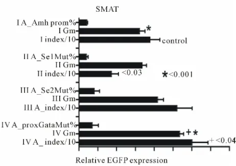
Previous results [4] hinted that the increased response
index seen when an Amh promoter with a mutated prox-
Gata element was used to drive expression of the re-
porter gene (d2EGFP), was explicable in terms of an
increase in Gm. This implies that the mean expression
per cell is increased.
Figure 1
illustrates this point but
also indicates that the same conclusion cannot be used to
explain fully a decreased index with a mutated Se1 element.
Figure 2
illustrates the effectiveness of a downstream
enhancer (DE) on responses driven by three different
truncations of the Amh promoter in pre-pubertal Sertoli
cells. Truncation X (see
Figure 3
) has virtually no effect
[image:2.595.309.540.84.247.2] [image:2.595.312.537.407.586.2]while truncations Y and Z lead to a highly significant
reduction in the level of EGFP expression. The presence
of DE immediately 3’ of the Amh PA signal, leads to a
very large increase in the response driven by the intact
(control) Amh promoter. The efficacy of DE on the
re-sponses driven by all three of the truncations is very
much less. This result hints that something in the
Figure 1. Data which confirms that an Amh promoter with a mutated Se1 element shows a significantly lower response Index than the control group and an increased EGFP expres-sion Index when the proxGata element was mutated. It is rele-vant to note that since the Index is the product of % of cells in the green window and their geometric mean brightness (Gm), it is interesting to note that d2EGFP gene expresses a version of EGFP with an intracellular half-life of 2 hours, consequently the Gm (brightness) value is a good approximation to a meas-urement of rate of synthesis. The increased index seen with the mutated proxGata element can be accounted for by a signifi-cant increase in Gm and hence an increase rate of synthesis per cell. The lower index value seen with the mutated Se1 element cannot be completely interpreted in the same manner.
Figure 3. Nucleotide sequence (5’ to 3’) of a mouse Amh pro-moter. SF3a2-PA is the polyadenylation signal of an upstream gene coding for a spliceosome component [13]. Potential pro-moter elements are identified on the basis of sequence similar-ity with human, rat and other mammalian Amh promoter se-quences: the order of elements is conserved. These potential elements are highlighted in black and identified by superscript titles, with mutated sequences indicated as subscripts. The grey highlighted sequence is an inverted repeat. Where possible the superscript titles are defined by their affinity for known tran-scription factors. Superscripts tX and tY indicate the start of two truncated Amh promoters terminating at the start of trans-lation; tZ- and -tZ indicate the range of a third truncation. The start of translation (0) is position 8647 in GenBank genomic nucleotide sequence X83733. DE is a downstream enhancer starting at the polyadenylation signal for Amh. This 3’ UTR element was inserted in the d2EGFP vector at a MluI site as indicated in this figure. The MluI site replaces an AflII site which was in the vector as supplied by Invitrogen.
Amh promoter upstream of the distSF1 element (
Figure 3
;
−
215 to
−
336) may play a small role in the action of DE.
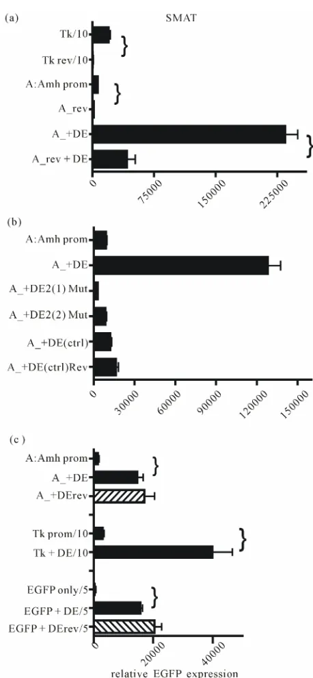
Both Amh and the control minimal thymidine kinase
(Tk) promoter are ineffective when in reverse orientation.
The presence of DE relatively increases responsiveness
(
Figure 4(a)
), and it also is shown in part B that muta-
tion of DE2 or replacement of the entire 89 nt of the en-
hancer, completely reduces the effectiveness of the en-
hancer. This suggests that DE is nucleotide sequence spe-
cific and is not merely an inert spacer which could act by
changing the flexibility of the DNA. Part 4C shows that
the orientation of DE is not important and that DE can
enhance expression driven by other promoters.
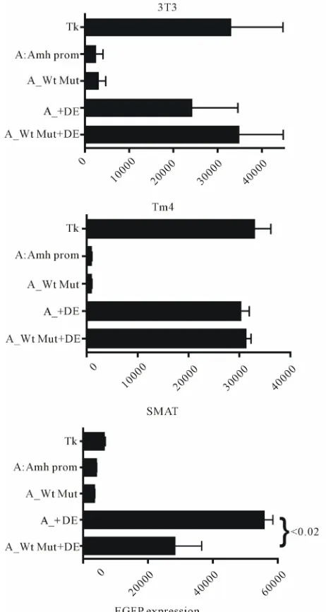
Figure 5
extends the results illustrated in
Figure 4(a)
and shows that the pattern of response in post-pubertal
(TM4) Sertoli cells resembles that seen in 3T3
(fibro-blast) cells and is different to that of pre-pubertal (SMAT)
Sertoli cells. In the TM4 and 3T3 cells an Amh promoter
in reverse orientation seems to do slightly better than the
promoter in the forward orientation.
[image:3.595.309.538.79.574.2]In
Figure 6
a comparison is made between EGFP ex-
[image:3.595.57.283.85.273.2]Figure 5. This figure illustrates the effectiveness of various combinations of Amh promoter and downstream enhancer (DE). EGFP expression was measured in three lines of mouse cells. The pattern of response in 3T3 (fibroblast) and TM4 (post-pubertal Sertoli) cells are similar and are markedly dif- ferent to that seen in SMAT (pre-pubertal Sertoli) cells. As in Figure 4 “DE only” is the response elicited by a pd2EGFP vector with a DE inserted 3’ but lacking any 5’ promoter. The MCS of this vector may have a weak non-specific promoter- like effect which can be reduced by truncation of the MCS.
Figure 6. EGFP expression in TM4 (post-pubertal) and SMAT (pre-pubertal) Sertoli cells. Expression driven by a range of promoters with different elements mutated with and without an additional downstream enhancer element (DF). The presence of DE makes no difference in TM4 cells but in SMAT cells shows a small enhancement with the SF1 mutated elements. In contrast there is a significant enhancement of expression in the group with a mutated Sox element.
elements. However as shown in
Figure 7(b)
, with the
[image:4.595.58.287.364.649.2]exception of element distSF1, double mutation with the
addition of a mutated Wt results in a reduction of both
the negative and positive effects of single mutated
ele-ments recorded in part A. The exception seems to be that
distSF1 + Wt mutation leads to an augmention of
sup-pression.
In
Figure 8
combination of truncation X of the Amh
promoter with mutated Wt and the presence of DE was
tested in TM4 (post-pubertal) Sertoli cells and in SMAT
(pre-pubertal) Sertoli cells. There is little measurable
effect in TM4 cells but a significant reduction in the
en-hancer effect of DE in SMAT cells when Wt is mutated.
This implies that an intact Wt element may be necessary
for enhancement.
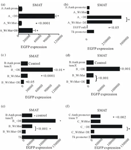
The experiment illustrated in
Figure 9
extends the
results shown in
Figure 8
to show additionally that a
mutated Wt is ineffective in permitting enhancement by
DE in SMAT cells. Further confirmation of this
observa-tion is provided in
Figures 10(a)-(f)
, where an intact
Amhpromoter, and promoters with truncation X and
truncation Y were used.
Addition of a relatively large amount of free
dou-ble-stranded oligo-nucleotide DE DNA to the DNA-Li
[image:5.595.307.538.86.519.2]Figure 8. EGFP expression driven by a truncated Amh pro-moter in TM4 and SMAT Sertoli cells. The effect of adding the downstream enhancer (DE) in the presence of a mutaed or non-mutated Wilms tumour factor-1 (Wt) element. There is a strong suggestion from these results that in pre-pubertal Sertoli cells, DE requires the presence of an intact Wt element to be an effective enhancer.
Figure 9. A comparison of in vitro EGFP expression responses
in three lines of mouse cells. It can be seen that mutation of the Wilms tumour factor (Wt) element has on its own no effect in any of the cells tested, confirming previous observations. How- ever in SMAT cells but not in either 3T3 or TM4 cells, it can be seen that Wt mutation shows a significant but not total, sup- pression of enhancement by the downstream element (DE). This result confirms the data in Figure 8 but using a “full length” Amh promoter.
pofectAmine mixture at the time of transfection, results
in a significant specific lowering of expression. The
re-sults of these experiments are made available as a
sup-plementary data file.
4. DISCUSSION
[image:5.595.56.284.374.656.2]Figure 10. Six experiments were designed to illustrate the dependence of the downstream (3’) enhancer on the upstream (5’) Wilm’s tumour factor. These experiments used a line of mouse pre-pubertal Sertoli cells (SMAT) with differently mo- dified Amh promoters. This supplements the data in Figures 8 and 9. A mutated Wt significantly reduces enhancement by DE in all cases.
an enhancer (DE) situated immediately downstream of
the PA signal for Amh. Furthermore it is shown that for
full functionality this enhancer depends on the presence
of an intact Wilms Tumour factor-1 element (Wt). Wt is
situated in the promoter immediately upstream of the
Amh gene and lies between the tata box and the start of
translation. Interrogation of the PubMed data base shows
that sequences with a close similarity to DE only occur
immediately downstream of Amh/AMH genes. As shown
in
Figure 4
replacement of the DE sequence with a ran-
dom DNA sequence of the same length, or mutation of
the central portion of DE (DE2), led to a total ablation of
enhancer activity. This suggests that enhancer function is
both position and nucleotide sequence specific, although
change of the orientation of DE makes no difference
(
Figure 4(c)
).
As shown in
Figure 2
analysis of enhancer (DE) ac-
tivity in constructs with truncated Amh promoters indi-
cates that a stretch of DNA at the 5’ end of the promoter
sequence, as illustrated in
Figure 3
(
−336 to
−215), plays
a small but significant role in enhancement. The as-
sumption that position
−336 is the effective 5’ end of the
mouse Amh promoter is based on the observation that
immediately upstream is the polyadenylation (PA) signal
for SF3a2 which codes for a spliceosome component
point in development. Furthermore enhancement is also
dependent on the presence of an intact Wilms tumour
factor element (Wt). A question therefore arises as to the
molecular mechanism of enhancement. The results sum-
mariesed in
Figure 4(b)
support the view that enhance-
ment depends on the DE2 sequence, which might be a
specific anchor point which is instrumental in the folding
back of the entire DE close to the promoter at the Wt
element. Such a folding across 4 - 5 kb of ORF may be
mediated by a duplex transcription-like bridging factor
[14], with specificity for both DE2 and Wt. There is no
hint in the DNA itself that sequence complementarity
plays a role in such an interaction. The relatively small
specific inhibition of DE activity by free DE DNA, in-
troduced at the time of transfection (see Supplementary
Data) (
Figures S1
and
S2
), is compatible with this view
but does not rule out other possible mechanisms [15-19].
For example once DE is juxtaposed close to Wt, part of
DE (DE1; DE3) might take on the role of supplementary
promoter elements. It is not clear how this could affect
the efficiency of use or recycling of mRNA. The results
summarised in
Figures 4(a)
and
(c)
show that enhance-
ment is sensitive to the orientation of the promoter but
not to that of DE. This is compatible with the view that
the promoter is an ordered assembly template and DE is
an anchor point for a bridge.
[image:6.595.57.287.82.344.2]assem-bly of the constituents of the transcription mechanism.
The Wt element lies between the tata box and the start of
translation—this region may include different cap sites
leading to alternative transcripts which may play a part
in control of gene expression. There may be many other
potential examples which have a role in the control of
gene expression during sex development [23,24].
5. ACKNOWLEDGEMENTS
I thank Dr. N. di Clemente for providing the SMAT (prepubertalSertoli) cells; the University of Edinburgh for an Emeritus Fellowship; Profes-sor Rick Maizels and his colleagues for the hospitality of the lab; and Daniel Guerrier for a critical reading of a draft manuscript.
REFERENCES
[1] Josso, N., Picard, J.Y., Rey, R. and di Clemente, N. (2006) Testicular anti-Mullerian hormone: History, ge- netics, regulation and clinical applications. Pediatric En-
docrinology Reviews, 3, 347-358.
[2] Durlinger, A.L., Visser, J.A. and Themmen, A.P. (2002) Regulation of ovarian function: The role of anti-Muller- ian hormone. Reproduction, 124, 601-609.
doi:10.1530/rep.0.1240601
[3] Koopman, P., Gubbay, J., Vivian, N., Goodfellow, P. and Lovell-Badge, R. (1991) Male development of chromo- somally female mice transgenic for Sry. Nature, 351,
117-121. doi:10.1038/351117a0
[4] Dresser, D.W. (2012) Mutated elements of a complex promoter (Amh) can help to demonstrate the role of cer- tain elements in controlling differential gene expression.
American Journal of Molecular Biology, 2, 351-358.
doi:10.4236/ajmb.2012.24036
[5] de Santa Barbara, P., Moniot, B., Poulat, F. and Berta, P. (2000) Expression and subcellular localization of SF-1, SOX9, WT1, and AMH proteins during early human tes- ticular development. Developmental Dynamics, 217,
293-298.
doi:10.1002/(SICI)1097-0177(200003)217:3<293::AID-DVDY7>3.0.CO;2-P
[6] Oreal, E., Mazaud, S., Picard, J.Y., Magre, S. and Carre- Eusebe, D. (2002) Different patterns of anti-Mullerian hormone expression, as related to DMRT1, SF-1, WT1, GATA-4, Wnt-4, and Lhx9 expression, in the chick dif- ferentiating gonads. Developmental Dynamics, 225, 221-
232. doi:10.1002/dvdy.10153
[7] Arango, N.A., Lovell-Badge, R. and Behringer, R.R. (1999) Targeted mutagenesis of the endogenous mouse Mis gene promoter: In vivo definition of genetic
path-ways of vertebrate sexual development. Cell, 99, pp.
409-419. doi:10.1016/S0092-8674(00)81527-5
[8] Münsterberg, A. and Lovell-Badge, R. (1991) Expression of the mouse anti-mullerian hormone gene suggests a role in both male and female sexual differentiation. De-
velopment, 113, 613-624.
[9] Schepers, G., Wilson, M., Wilhelm, D. and Koopman, P. (2003) SOX8 is expressed during testis differentiation in
mice and synergizes with SF1 to activate the Amh pro- moter in vitro. Journal of Biological Chemistry, 278, 28101-28108. doi:10.1074/jbc.M304067200
[10] Dresser, D.W. and Guerrier, D. (2005) Candidate sertoli cell specific promoter element for a TGFbeta family member (Amh) and a 3’ UTR enhancer/repressor for the same gene. Gene, 363, 159-165.
doi:10.1016/j.gene.2005.08.004
[11] Anderson, A.A., Treves, S., Biral, D., Betto, R., Sandonà, D., Ronjat, M. and Zorzato, F. (2003) The novel skeletal muscle sarcoplasmic reticulum JP-45 protein. Molecular cloning, tissue distribution, developmental expression, and interaction with alpha 1.1 subunit of the voltage- gated calcium channel. Journal of Biological Chemistry,
278, 39987-39989. doi:10.1074/jbc.M305016200 [12] Belville, C., Jamin, S.P., Picard, J.Y., Josso, N. and di
Clemente, N. (2005) Role of type I receptors for anti- Mullerian hormone in the SMAT-1 Sertoli cell line.
On-cogene, 24, 4984-4992.
[13] Dresser, D.W., Hacker, A., Lovell-Badge, R. and Guer- rier, D. (1995) The genes for a spliceosome protein (Sap 62) and the anti-Mullerian Hormone are contiguous.
Human Molecular Genetics, 4, 613-618.
doi:10.1093/hmg/4.9.1613
[14] Anderson, A.M., Weasner, B.M., Weasner, B.P. and Kumar, B.P. (2012) Dual transcriptional activities of SIX define their roles in normal and ectopic eye development.
Development, 139, 991-1000. doi:10.1242/dev.077255
[15] Miyamota, Y., Taniguchi, H., Hamel, F., Silversides, D.W. and Viger, R. (2008) A GATA4/WT1 cooperation regulates transcription of genes required for mammalian sex derermination and differentiation. BMC Molecular
Biology, 9, 44-62. doi:10.1186/1471-2199-9-44
[16] Viger, R.S., Taniguchi, H., Robert, N.M. and Tremblay, J.J. (2004) Role of the GATA family of transcription factors in andrology. Journal of Andrology, 25, 441-452.
[17] Klattig, J., Sierig, R., Kruspe, D., Besenbeck, B. and Englert, C. (2007) Wilms’ tumor protein Wt1 is an acti-vator of the anti-Mullerian hormone receptor gene Amhr2.
Molecular and Cellular Biology, 27, 4355-4364.
doi:10.1128/MCB.01780-06
[18] Guittot, S.M., Tetu, A., Legault, E., Pilon, N., Silversides, D.W. and Viger, R.S. (2007) The proximal Gata4 pro- moter directs reporter gene expression to sertoli cells during mouse gonadal development. Biology of Repro-
duction, 76, 85-95. doi:10.1095/biolreprod.106.055137
[19] Sekido, R. and Lovell-Badge, R. (2008) Sex determina- tion involves synergistic action of SRY and SF1 on a specific Sox9 enhancer. Nature, 453, 930-934.
doi:10.1038/nature06944
[20] Merritt, C., Rasojoson, D., Ko, D. and Seydoux, G. (2008) 3’ UTR are the primary regulators of gene expression in
the C. elegansgermline. Current Biology, 14, 1476-1482.
doi:10.1016/j.cub.2008.08.013
[21] Derrigo, M., Cestelli, A., Savettieri, G. and diLiegro, I. (2000) RNA-protein interactrions in the control of stabil- ity and localization of messenger RNA. International
Journal of Molecular Medicine, 5, 111-123.
Amh promoter, suggest that nucleotide sequence
speci-ficity is key in the interaction. Therefore experiments
were carried out to check the effect of free DE DNA on
the “enhancer effect”.
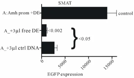
Figure S2. Experiments which confirm that free DE DNA interferes with the interaction of DE with the Wilms tumour factor element in the promoter of Amh used to drive the ex-pression of the reporter gene (EGFP). Two independently pre-pared control Amh promoter constructs, (a) and (b), are in-cluded. The addition of 2 µl of 50 pMol/µl DNA to the trans-fection mixture resulted in a significant reduction in EGFP expression. A similar experiment using a truncated Amh pro-moter (truncation X; AtX) showed broadly similar results. Figure S1. Free 89 nt DE DNA and a control DNA (ctrl) were
[image:8.595.58.287.322.456.2]