Copyright © 2002, American Society for Microbiology. All Rights Reserved.
Multicenter Evaluation of a New Automated Fourth-Generation Human
Immunodeficiency Virus Screening Assay with a Sensitive
Antigen Detection Module and High Specificity
Bernard Weber,
1,2* Lutz Gürtler,
3Rigmor Thorstensson,
4Ulrike Michl,
5Annelies Mühlbacher,
5Philippe Bürgisser,
6Roberto Villaescusa,
7Adolfo Eiras,
7Christian Gabriel,
8Herbert Stekel,
9Srivilai Tanprasert,
10Sinenaart Oota,
10Maria-Jose Silvestre,
11Cristina Marques,
11Maria Ladeira,
11Holger Rabenau,
2Annemarie Berger,
2Urban Schmitt,
12and Walter Melchior
12Laboratoires Réunis Kutter-Lieners-Hastert, Junglinster, Luxembourg
1; Institut für Med. Virologie, Universitätskliniken Frankfurt,
Frankfurt,
2Friedrich-Loeffler-Institute für Med. Microbiology, Ernst-Moritz-Arndt University Greifswald, Greifswald,
3and
R&D Infectious Diseases, Roche Diagnostics GmbH Laboratory Systems, Penzberg,
12Germany; Swedish Institute for
Infectious Disease Control, Solna, Sweden
4; Blutzentrale der Landeskrankenanstalten Salzburg, Salzburg,
5and
Red Cross Transfusion Service Upper Austria
8and Allgemeines Krankenhaus Linz, Zentrallabor,
9Linz,
Austria; Division d’Immunologie et d’Allergie, Département de Médecine Interne, Centre Hospitalier
Universitaire Vaudois, Lausanne, Switzerland
6; Centro de Transfusión de Galicia, Santiago de
Compostela, Spain
7; National Blood Centre, Thai Red Cross Society, Bangkok, Thailand
10;
and Laboratorio Microbiologia, Hospital de Curry Cabral, Lisbon, Portugal
11Received 24 October 2001/Returned for modification 5 January 2002/Accepted 13 February 2002
Fourth-generation assays for the simultaneous detection of human immunodeficiency virus (HIV) antigen
and antibody that were available on the international market until now have antigen detection modules with
relatively poor sensitivity and produce a higher rate of false-positive results than third-generation enzyme
immunoassays (EIAs). The new Cobas Core HIV Combi EIA with an improved sensitivity for HIV p24 antigen
was compared to alternative fourth- and third-generation assays, the p24 antigen test, and HIV type 1 (HIV-1)
RNA reverse transcriptase PCR (RT-PCR). A total of 94 seroconversion panels (
n
ⴝ
709 sera), samples from
the acute phase of infection after seroconversion (
n
ⴝ
32), anti-HIV-1-positive specimens (
n
ⴝ
730) from
patients in different stages of the disease, 462 subtyped samples from different geographical locations,
anti-HIV-2-positive sera (
n
ⴝ
302), dilutions of cell culture supernatants (
n
ⴝ
62) from cells infected with different
HIV-1 subtypes, selected performance panels from Boston Biomedica Inc., 7,579 unselected samples from
blood donors, 303 unselected daily routine samples, 997 specimens from hospitalized patients, and potentially
interfering samples (
n
ⴝ
1,222) were tested with Cobas Core HIV Combi EIA. The new assay showed a
sensitivity comparable to that of the Abbott HIV-1 AG Monoclonal A for early detection of HIV infection in
seroconversion panels. The mean time delay of Cobas Core HIV Combi EIA (last negative sample plus 1 day)
in comparison to that for HIV-1 RT-PCR for 87 panels tested with both methods was 2.75 days. The diagnostic
window was reduced with Cobas Core HIV Combi EIA by between 3.6 and 5.7 days from that for
third-generation assays. The specificities of Cobas Core HIV Combi EIA in blood donors were 99.84 and 99.85%
(after repeated testing). Overall, 30 repeatedly reactive false-positive results out of 10,031 HIV-negative
samples were obtained with Cobas Core HIV Combi EIA. Our results show that a fourth-generation assay with
improved specificity such as Cobas Core HIV Combi EIA is suitable for blood donor screening because of its
low number of false positives and because it detects HIV p24 antigen with a sensitivity comparable to that of
single-antigen assays.
Since the first enzyme immunoassays (EIA) for blood donor
screening and laboratory diagnosis of human
immunodefi-ciency virus (HIV) infection were licensed over 15 years ago,
the quality of these tests has been continuously improved by
the use of recombinant antigens and synthetic peptides
(sec-ond test generation) and the sandwich EIA technology (third
test generation) (10, 36). There is however a residual risk for
false-negative results. The potential causes include the
diag-nostic window in the preseroconversion phase, genetic
variabil-ity, atypical seroconversions, a delayed or absent immune
re-sponse in the very early or advanced stages of infection,
respectively, and laboratory reporting errors (6).
The highest risk (
⬎
90%) of a false-negative result is
ob-served in the preseroconversion phase during primary HIV
infection (diagnostic window) (6). The residual risk of an HIV
infection by a seronegative blood donor during acute HIV
infection is estimated to be 1/493,000 to 1/1,866,000 per
trans-fused unit in healthy, unpaid donors in the United States and
Germany (2). In emergency department patients and in
high-risk groups, it ranges between 0.14 and 0.17% (9, 18).
Early detection of HIV infection is important for reasons of
infection security, prevention, and individual prognosis. An
antiretroviral combination therapy during primary HIV
infec-tion reduces the likelihood of a rapid progression to the AIDS
stage. Moreover, the frequency of opportunistic infections,
* Corresponding author. Mailing address: Laboratoires Réunis
Kut-ter-Lieners-Hastert Centre, Langwies L-6131, Junglinster,
Luxem-bourg. Phone: (352) 78 02 90 1. Fax: (352) 78 88 94. E-mail: web@labo
.lu.
1938
on May 15, 2020 by guest
http://jcm.asm.org/
skin and mucous membrane diseases, and respiratory
infec-tions is reduced (4).
Nucleic acid amplification technology (NAT) and HIV
an-tigen (Ag) detection make it possible to reduce the residual
risk of HIV transmission by blood and blood products and to
improve the early detection of primary HIV infection in
high-risk groups. With NAT testing, the diagnostic window (about
21 days) is reduced by 11 days and the residual risk is reduced
by over 50% (2). In the primary HIV infection, a localized viral
replication (eclipse) takes place first and lasts for
approxi-mately 10 days. In exceptional cases, it can last for many
months. Experiments conducted with the animal model
indi-cate that the HIV-infected subject is not infectious during this
phase of the incubation period. In the subsequent viremic
phase, HIV RNA is the first and only detectable virus-specific
marker for 1 to 5 days. In theory, all potentially infectious viral
carriers are excluded by using the NAT technique, because no
infectivity is observed during primary infection in the animal
model before appearance of HIV type 1 (HIV-1) RNA (22).
The advantages of NAT are offset by its technical limitations.
The automated processing of large numbers of samples,
espe-cially nucleic acid extraction, is not possible at this time or is
possible only with limitations. HIV-1 RNA amplification is
currently performed on pooled samples from donors in
Euro-pean blood banks as a cost-effective alternative to
single-sam-ple testing (25). Depending on the pool size and detection
threshold of NAT, the gain in sensitivity may be relatively low
in comparison to HIV p24 Ag testing (17). However, NAT
testing of pools is more cost-effective than Ag determination in
individual donations (24).
Fourth-generation HIV screening assays that make
com-bined HIV Ag and antibody detection possible in one test
batch have been licensed in Europe since 1997 (12, 19, 20, 23,
26, 30–35). These combined assays offer the advantage of early
detection of HIV infection via p24 Ag detection at a technical
burden and financial cost more or less equal to those for HIV
antibody testing using third-generation EIAs. The diagnostic
window is reduced by 4 to 5 days compared with that for
antibody detection alone (third-generation assays).
Approximately one-third of the solid phase is coated with
monoclonal antibodies for HIV p24 Ag detection. The
remain-ing bindremain-ing capacity is available for antibody detection
(sand-wich or indirect EIA). Generally, fourth-generation assays
ex-hibit a relatively high limit for detection of the HIV Ag module
(
⬎
30 pg of p24 Ag/ml). For this reason, they cannot replace
single-Ag determination with sensitive commercially available
EIAs (detection limit: 3 to 5 pg/ml) (34). The risk of
interfer-ence is potentially higher with the HIV combined assays than
with single-Ag or -antibody EIAs, since both serological
mark-ers are determined in one test batch. The specificity for blood
donor screening (99.6 to 99.8%) is lower than that of
third-generation HIV EIAs (12, 19). The higher rate of false-positive
results (0.2%) of HIV combined assays has a particularly
neg-ative effect in cases of high sample throughput and low
prev-alence as in blood donor screening mainly because a separate
Ag detection must be carried out in addition to the antibody
confirmatory assay (Western blotting) in order to rule out an
HIV infection (12).
MATERIALS AND METHODS
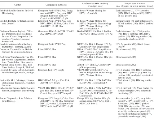
Recently, new fourth-generation assays with an improved sensitivity of the Ag detection module and optimized specificity have been established. In a multi-center study (Table 1 shows a list of the participants), the new automated Cobas Core HIV Combi EIA was compared to different fourth- and third-generation HIV screening assays, p24 Ag tests, and HIV-1 reverse transcriptase PCR (RT-PCR).
Cobas Core HIV Combi EIA.Cobas Core HIV Combi EIA is a double-Ag immunoassay for the detection of total antibodies to HIV-1 (including group O) and HIV-2, combined with a sandwich assay for the detection of HIV-1 p24 Ag. HIV-specific antibodies and the HIV-1 p24 Ag are detected within one deter-mination.
Samples, digoxigenin-conjugated anti-p24 antibodies, and digoxigenin-conju-gated RT were incubated with polystyrene beads coated with recombinant Ags derived from the Pol and Env regions of HIV-1 and HIV-2 as well as monoclonal antibodies against HIV-1 p24 Ag.
For Ag detection, HIV-1 p24 Ag reacted simultaneously with the anti-p24 antibodies on the bead and with the digoxigenin-labeled anti-p24 antibodies to form a sandwich complex.
For antibody detection, antibodies directed to RT formed a complex with immobilized Ag on the bead and digoxigenin-labeled Ag. Antibodies directed to HIV-1 Env Ag (including group O) and HIV-2 bound to the immobilized Ags. After beads were washed, the Env-specific antibodies were detected by incuba-tion with recombinant Ags from the Env regions of HIV-1 and HIV-2 conjugated to horseradish peroxidase and a peroxidase-labeled antidigoxigenin monoclonal antibody.
After a second washing step, the bead was incubated with Cobas Core sub-strate. The intensity of the resulting color was roughly proportional to the amount of anti-HIV antibodies and HIV-1 p24 Ag detected in the specimen.
The cutoff was calculated from the following formula: 0.12⫻positive control
⫹negative control. A result was considered negative if the index value (signal
sample/cutoff value) was⬍0.9. A sample is positive if its index value isⱖ1.0. The
grey zone ranges between index valuesⱖ0.9 and⬍1.0.
Alternative HIV screening assays.Cobas Core HIV Combi EIA was compared to different third- and fourth-generation assays established as routine methods in the different centers participating at the study. Included were HIV-1/HIV-2
3rd-gen. Plus EIA, IMx HIV-1/HIV-2 III Plus, AxSYM HIV-1/2 gO, Prism
HIV-O Plus (Abbott, North Chicago, Ill.), VIDAS HIV DUO (Biomérieux, Marcy-l’Etoile, France), Genscreen HIV-1/2 (Bio-Rad, Marnes la Coquette, France), Enzygnost HIV1/2 Plus, Enzygnost HIV Integral, Enzygnost Anti-HIV (Dade Behring, Marburg, Germany), Enzymun-Test Anti-HIV Combi, and
Co-bas Core Anti-HIV 1⫹2⫹O EIA (Roche Diagnostics, Penzberg, Germany). All
the tests were performed and interpreted in accordance with the manufacturers’ recommendations.
HIV Ag detection assays.For p24 Ag detection, different commercially avail-able assays, i.e., HIV-1 AG Monoclonal A (Abbott), VIDAS HIV p24 II assay (Biomérieux), Coulter HIV p24 Ag assay, Elecsys HIV Ag (Roche Diagnostics), were used. All the HIV Ag tests were performed and interpreted in accordance with the manufacturers’ recommendations.
HIV antibody confirmatory assays.Repeatedly reactive samples were sub-jected to Western blot or immunoblot testing. The assays used included com-mercially available reagents, i.e., NEW LAV Blot I, NEW LAV Blot II (Bio-Rad), Diagnostic Biotechnology Western blot HIV 1, version 2.2 (Genelabs, Geneva, Switzerland), HIV Blot 2.2 (Abbott Diagnostics, Delkenheim, Germa-ny), INNO-LIA HIV Confirmation (Innogenetics, Ghent, Belgium), RIBA HIV-1/-2 SIA (Chiron, Emeryville, Calif.), and in house Western blots: SBL-6669 (HIV-2; Swedish Institute for Infectious Disease, Solna, Sweden) and MVP-899/87 (HIV-1), MVP-5106/91 (HIV-O), and MVP 11971/87 (HIV-2) (Fried-rich-Löffler-Institut für Med. Mikrobiologie, Greifswald, Germany).
Positive results were defined in accordance with World Health Organization (WHO) and Centers for Disease Control and Prevention (CDC) criteria (NEW LAV Blot I and II), Food and Drug Administration and WHO criteria (Diag-nostic Biotechnology Western blot), and the Deutsches Institut für Normierung (DIN 58969-41 [1992]) definition (Friedrich-Loefler-Institute für Med. Microbi-ology).
Nucleic acid amplification.HIV-1 RNA was amplified by using different assays including Amplicor Monitor HIV 1, version 1.5, Ampliscreen HIV 1, version 1.5 (Roche Diagnostics, Branchburg, N.J.), and in-house HIV-1, HIV-2, and HIV-O PCR (Friedrich-Loefler-Institute für Med. Microbiology).
Specimens.The following specimens were tested to evaluate the sensitivity of Cobas Core HIV Combi EIA in comparison to different third- and fourth-generation assays and p24 Ag detection (Table 1). (i) Ninety-four HIV-1
on May 15, 2020 by guest
http://jcm.asm.org/
seroconversion panels were provided by Boston Biomedica Inc. (BBI; West Bridgewater, Mass.), BioClinical Partners (Franklin, Mass.), North American Biologicals Inc. (NABI; Boca Raton, Fla.), and the Swedish Institute for Infec-tious Disease Control. For most of the seroconversion panels, HIV-1 RNA detection was performed by using quantitative PCR (Amplicor HIV-1 Monitor; Roche Diagnostics; see Table 2). The detection limits of the PCR protocol employed for panels BCP 9010 to 9034 and ANT 6240 to 6248 were 50 and 400 copies of HIV-1 RNA/ml of plasma, respectively. All seroconversion samples were tested by Western blotting. HIV Ag detection (HIV-1 AG Monoclonal A) was performed for all the seroconversion panels with the exception of SIIDC35.
(ii) Single serum samples (n⫽32) from the acute phase of infection after
seroconversion were obtained. (iii) Anti-HIV-1-positive specimens (n⫽610)
from patients in different stages of the disease were obtained. (iv) For the assessment of the effect of genetic variability on HIV-1 detection, 373 subtyped samples from different geographical locations (Cameroon, Germany, Luxem-bourg, Belgium, Portugal, Switzerland, South Africa, Thailand, and Zimbabwe), including group M (subtypes A to J and putative subtype S) and group O sera were selected. The total number of group M non-B sera was 324. Samples from patients infected with recombinant group M virus and simian immunodeficiency virus (SIV)-positive monkey sera were analyzed. Subtype determination was by competitive EIA using HIV-1 subtype A- to E-specific gp120 V3 peptides in accordance with the protocol of Kasper et al. (P. Kasper, A. N. Smith, G. Duraisamy, B. Ofenloch, and E. Faatz, Abstr. Deutscher AIDS-Kongreß, abstr. V089, p. 32, 1996) or by using a V3 loop-based research enzyme-linked immu-nosorbent assay from Dade-Behring (14, 27) and PCR-amplified sequencing of the C2V3 region after isolation of HIV from peripheral blood mononuclear cells
from citrated blood. Alignment of the amino acids to the known consensus sequences was done by using the Los Alamos database (15). Also included were the Worldwide HIV-1 performance panel WWRB301 (BBI) consisting of 47 HIV-1 subtyped and HIV-2-positive samples from different geographic locations (Argentina, Cameroon, Canada, United States, China, Egypt, Ghana, India, South Africa, Thailand, Uganda, and Zimbabwe) and panel AfrRB1 (BBI) including 42 HIV-1 group M (A, B, and C) and 5 HIV-2-positive samples from
Africa. (v) Anti-HIV-2-positive samples (n⫽302) were from different
geo-graphic regions in West Africa and Portugal and included three SIV type 2 (SIV-2)-positive samples from monkeys. (vi) HIV-1 Ag- and HIV-1
antibody-positive samples were from patients at different stages of disease (n⫽120). (vii)
Dilutions of cell culture supernatants (n⫽57) from cells infected with different
HIV-1 subtypes, including group M subtypes A to H, group O, HIV-2, and unknown subtypes were tested in order to investigate the influence of the genetic variability of HIV on Ag detection. Virus isolates had been genotyped by se-quencing PCR-amplified fragments of the C2V3 genome region (16). All the supernatants were diluted in anti-HIV-negative serum. (viii) Selected perfor-mance panels from BBI included low-titer panels PRB104, -105, -106, and -107 and anti-HIV-1 mixed-titer panels PRB202 and PRB203. Criteria for inclusion in mixed-titer performance panels were a positive Western blot using CDC criteria or an indeterminate blot when HIV Ag is present. Inclusion criteria for low-titer panels were sample absorbance-to-cutoff ratios of less than 4.0 on the basis of at least two Food and Drug Administration-licensed tests and a positive Western blot by Association of State and Territorial Public Health Laboratory Directors and CDC criteria or an indeterminate Western blot when HIV p24 Ag is present. For the evaluation of specificity, the following unselected and selected
speci-TABLE 1. Participants involved in the study, methods used, and samples tested
Center Comparison method(s) Confirmation HIV antibodyor antigen assay (no. of panels or serum samples tested)Sample type or source
Friedrich-Loeffler-Institut für Med.
Microbiology Enzygnost Anti-HIV1/2 Plus, Enzyg-nost anti-HIV 1, Enzygnost HIV
Integral, Enzymun-Test HIV Combi, AxSYM HIV-1/2 gO
In-house Western blotting for
HIV-1, -2, and -O Early infection (2), HIV-1 positive(112), HIV-1 subtyped (89), HIV-2
or SIV positive (10) Swedish Institute for Infectious
Dis-ease Control Enzygnost Anti-HIV1/2 Plus, IMxHIV-1/HIV-2 III Plus, Cobas Core
Anti-HIV 1⫹2⫹O EIA
In-house Western blotting for HIV-2, Diagnostic Biotechnology HIV 1 Western blotting, HIV AG-1 Monoclonal A
Seroconversion (7), early infection (7), HIV-1 positive (100), HIV-2 positive (100)
Division d’Immunologie et d’Aller-gie, Département de Médecine Interne, Centre Hospitalier Uni-versitaire Vaudois, Lausanne, Switzerland
Genscreen HIV1/2, AxSYM HIV-1/2
gO, VIDAS HIV DUO BioRad NEW LAV Blot 1, BioRadNEW LAV Blot 2, Elecsys HIV Ag Early infection (13), HIV-1 positive(71), HIV-1 subtyped (15), HIV-2
positive (16), HIV Ag positive (30), potentially interfering (140) Landeskrankenanstalten Salzburg
Blutzentrale, Salzburg, Austria Enzygnost Anti-HIV1/2 Plus NEW LAV Blot 1, Elecsys HIV Ag,Coulter HIV p24 antigen assay HIV Ag positive (20), Blood donors(3,550)
Centro de Transfusión de Galicia,
Santiago de Compostela, Spain Prism HIV-O Plus RIBA HIV-1/-2 SIA,
aAmpliScreen
HIV-1, version 1.5, Ortho HIV-1/2 antibody capture ELISA
Blood donors (1,522)
Red Cross Transfusion Service Up-per Austria, Allgemeines Kranken-haus, Zentrallabor, Linz, Austria
Prism HIV-O Plus NEW LAV Blot 1, Coulter HIV p24
antigen assay Blood donors (1,025)
National Blood Centre, Thai Red
Cross Society, Bangkok, Thailand Prism HIV-O Plus Abbott HIV Blot 2.2, Coulter HIVp24 antigen assay Blood donors (1,482)
Hospital de Curry Cabral,
Laborato-rio Microbiologia, Lisbon, Portugal Enzymun-Test HIV Combi VIDAS HIV p24 II, NEW LAVBlot II, Diagnostics Biotechnology
HIV-1 Western blotting
HIV-1 infected (87), HIV-1 subtyped (50), HIV-2 positive (20), HIV Ag positive (14), unselected hospitalized patients (420)
Institut für Med. Virologie, Univer-sitätskliniken Frankfurt, Frankfurt, Germany
HIV-1/HIV-2 3rd gen. Plus EIA,
AxSYM HIV-1/2 gO NEW LAV Blot I, NEW LAV BlotII, HIV AG-1 Monoclonal A HIV-1 infected (69), HIV Ag positive(27)
Laboratories Réunis,
Kutter-Lieners-Hastert, Junglinster, Luxembourg VIDAS HIV DUO, HIV-1/HIV-2 3
rd
gen. Plus EIA, Enzymun-Test HIV Combi, Enzygnost HIV Integral
NEW LAV Blot I, NEW LAV Blot II, HIV AG-1 Monoclonal A, VI-DAS HIV p24 II, INNO-LIA HIV Confirmation
HIV-1 subtyped (17), Virus lysates (5), Routine samples (303), potentially interfering (37)
Roche Diagnostics, R & D
Infec-tious Diseases Enzygnost HIV Integral, Cobas CoreAnti-HIV 1⫹2⫹Q EIA, Genscreen
HIV 1/2, version 2, Enzymun-Test HIV Combi, AxSYM HIV-1/2 gO
Elecsys HIV Ag, Coulter HIV p24 antigen assay, NEW LAV Blot I, NEW LAV Blot II
Seroconversion (n⫽87), early
infec-tion (10), 1 positive (181), HIV-1 subtyped (HIV-197), HIV-2 positive (159), HIV Ag positive (29), virus lysates (57), dilutions of HIV positive sera (10), performance panels (8), selected hospitalized patients (577), potentially interfering (1,048)
aSIA, strip immunoblot assay.
on May 15, 2020 by guest
http://jcm.asm.org/
[image:3.587.44.541.80.465.2]mens were comparatively tested with Cobas Core HIV Combi EIA and alterna-tive assays (see Table 3): (i) 7,579 unselected samples from blood donors from different blood transfusion centers, (ii) 303 unselected daily routine samples, (iii) 420 unselected specimens from hospitalized patients, (iv) 577 selected samples from hospitalized patients tested negative for acute or chronic hepatitis B, (v) a
high number of potentially interfering samples (n⫽1,222) including rheumatoid
factor-, anti-hepatitis C virus-, and human T-cell leukemia virus-positive serum samples, sera from patients suffering from acute viral, bacterial, and fungal infections, liver cirrhosis, or autoimmune diseases, dialysis patients, and pregnant women.
Data evaluation and statistical analysis.Determinations were carried out in single measurements. Initially reactive (IR) specimens and discrepant specimens were repeated in single measurements or double determinations if enough sam-ple material was available. Repeatedly reactive (RR) and discrepant samsam-ples were subjected to antibody and/or Ag confirmation with Western blotting and/or a single-Ag assay.
The performance of Cobas Core HIV Combi EIA was compared with that of alternative screening assays and HIV Ag and HIV-1 RNA detection for the seroconversion panels. The mean number of days by which the diagnostic win-dow period was reduced with Cobas Core HIV Combi EIA in comparison to results for alternative third- and fourth-generation assays was calculated. The statistical significance of the reduction for each test was determined by using the Wilcoxon test for matched pairs (5).
The time delay between blood sampling points in commercially available seroconversion panels used for the present study is on average relatively short (2 to 7 days) but may last up to 37 days, for example, for panel BBI W. The calculation model for time delays between assays established by the Paul Ehrlich Institute (11) was used. This method considers that seroconversion is theoreti-cally possible the following day after the last negative follow-up sample. The total number and the average number of days of time delay for the 94 panels were compared with those for the most sensitive assay.
For the calculation of sensitivity and specificity, samples were considered HIV-1 positive if any of the following tests were positive: Western blotting (interpreted according to CDC criteria [7]), HIV-1 p24 Ag assay, and HIV-1 RNA assay. Patients were considered HIV negative if all the screening assays were negative or, for the EIA reactive samples, if the Western blotting result was negative or indeterminate and the HIV-1 p24 Ag assay was negative.
RESULTS
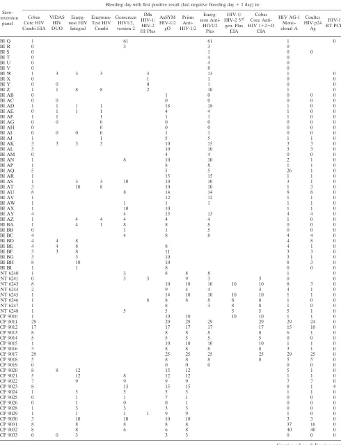
HIV Ag was detected with Cobas Core HIV Combi EIA one
bleeding earlier in 4 of 93 seroconversion panels tested in
parallel with HIV AG-1 Monoclonal A (Table 2). HIV Ag was
not detected with HIV AG-1 Monoclonal A until the end of
follow-up in four cases of primary infection (panels BBI AQ,
BCP 9014, 9031, and 9032). Conversely, HIV-1 AG
Monoclo-nal A detected acute HIV infection one or more bleedings
earlier than Cobas Core HIV Combi EIA in seven
serocon-version panels. The Coulter HIV p24 Ag assay detected HIV-1
Ag one or two bleedings earlier in 26 of 64 seroconversion
panels tested in parallel with Cobas Core HIV Combi EIA (see
Table 2). Cobas Core HIV Combi detected HIV-1 primary
infection one or more bleedings earlier in four
seroconver-sions.
Cobas Core HIV Combi EIA was more sensitive than the
alternative fourth-generation assays for early detection of HIV
infection. HIV Ag was detected earlier in 2 of 16, 6 of 15, and
21 of 29 seroconversion panels than with VIDAS HIV DUO,
Enzymun-Test HIV Combi, and Enzygnost HIV Integral,
re-spectively (Table 2). In panel BCP 9029, Enzygnost HIV
Inte-gral was transiently positive on day 16 and remained negative
until the end of follow-up. Since this sample was HIV-1
RT-PCR, HIV Ag, and Western blotting negative, the result
should be considered false positive. Primary HIV infection was
detected one or more bleedings earlier than with AxSYM
HIV-1/2 gO, Prism HIV-Oplus, Enzygnost Anti-HIV1/2 Plus,
and Genscreen HIV1/2, version 2, in 49 of 62, 25 of 36, 34 of
40, and 20 of 31 seroconversion panels, respectively.
Con-versely, seroconversion was detected one bleeding earlier with
one or more third-generation assays in panels BCP 9017 and
9032 and NABI SVO-0251-1. A second diagnostic window with
Cobas Core HIV Combi EIA was observed in three
serocon-version panels. A borderline result (index value, 0.92) was
observed on day 45 in BCP 9010. Cobas Core HIV Combi EIA
became negative in panel SIIDC13 on day 50 when
antigen-emia was no longer detectable; this sample reacted weakly in
the IMx HIV-1/HIV-2 III Plus and was tested negative with
Enzygnost Anti-HIV1/2 Plus. Cobas Core HIV Combi EIA
was borderline reactive (index values on duplicate testing: 0.87
and 0.92) in a follow-up sample obtained 4 days later that was
positive in the third-generation assays. A borderline result was
observed in the second follow-up sample of SIIDC35 with
Cobas Core HIV Combi. In panel BCP 9012, the HIV-1 RNA
copy number was transiently under the detection limit of
HIV-1 RT-PCR (day 2). The viral load was below the
detec-tion limit in panel BCP 9014 in the last four follow-up samples
(days 12 to 31).
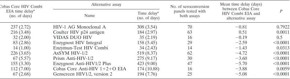
The mean time delay of Cobas Core HIV Combi EIA (last
negative sample plus 1 day) in comparison to HIV-1 RT PCR
for 87 panels tested with both methods was 2.75 days. The
difference in sensitivity between Cobas Core HIV Combi EIA
and HIV-1 RT-PCR was statistically significant (
P
⬍
0.0001).
Table 3 gives an overview of the mean time delays obtained for
Cobas Core HIV Combi EIA and alternative assays in
com-parison to PCR for seroconversion panels. Cobas Core HIV
Combi EIA showed a significantly higher sensitivity for the
detection of primary HIV infection than alternative
fourth-generation (with the exception of VIDAS HIV DUO) and
third-generation assays (
P
⬍
0.05) (Table 3).
All the 32 single-serum samples obtained during
serocon-version were tested positive with Cobas Core HIV Combi EIA.
A variable number of samples gave false-negative results with
third-generation EIAs (Table 4).
All 368 HIV-1 group M subtypes and group O and five
samples positive for SIV-1 or -3 were found positive with
Cobas Core HIV Combi EIA and the alternative assays. Only
one sample (putative HIV-1 subtype S) was negative with
En-zygnost Anti-HIV1/2 Plus and positive with Cobas Core HIV
Combi EIA.
All the 302 sera from HIV-2-infected individuals, 3
SIV-2-positive sera from monkeys, and subtyped samples from BBI
panel WWRB301 (M) (
n
⫽
47) and AfrRB1 (
n
⫽
42) were
tested positive with Cobas Core HIV Combi EIA and the
alternative assays (Table 4). One HIV-2-positive serum sample
from Guinea-Bissau gave an index value (1.16) close to the
cutoff with Cobas Core HIV Combi EIA and Enzygnost
Anti-HIV1/2 Plus.
All the cell culture supernatants (
n
⫽
62) infected with
different HIV-1 subtypes, including group M subtypes A to H,
group O, HIV-2, and unknown subtypes were detected with a
twofold- to more-than-fivefold-higher sensitivity with Cobas
Core HIV Combi EIA than with Enzymun-Test HIV Combi
(data not shown). Of 57 dilutions of virus lysates, including 1
HIV-1 subtype B, 2 group O, and 8 untyped HIV-1 lysates,
tested in parallel with the Coulter HIV p24 Ag assay, HIV Ag
was detected in higher dilutions in 11 lysates with Coulter HIV
on May 15, 2020 by guest
http://jcm.asm.org/
TABLE 2. Time delay in days for the detection of HIV primary infection in comparison with the most sensitive assay for each panel
Sero-conversion
panel
Bleeding day with first positive result (last negative bleeding day⫹1 day) in:
Cobas Core HIV Combi EIA
VIDAS HIV DUO
Enzyg-nost HIV
Integral
Enzymun-Test HIV Combi
Genscreen HIV1/2, version 2
IMx HIV-1/
HIV-2 III Plus
AxSYM HIV-1/2 gO
Prism Anti-HIV-1/2
Enzyg-nost
Anti-HIV1/2 Plus
HIV-1/ HIV-2 3rd
gen. Plus EIA
Cobas Core Anti-HIV 1⫹2⫹O
EIA
HIV AG-1 Mono-clonal A
Coulter HIV p24
Ag
HIV-1 RT-PCR
BBI Q 1 61 61 1 0
BBI R 0 3 3 0
BBI S 0 1 0 0
BBI T 0 4 0
BBI U 0 4 0
BBI V 0 8 0
BBI W 1 3 3 3 3 13 1 0
BBI X 0 1 1 0 0
BBI Y 0 0 0 1 0 0
BBI Z 1 1 8 8 2 10 1 0
BBI AB 0 1 0 0 0 0
BBI AC 0 0 0 0 0 0 0
BBI AD 1 1 1 1 18 18 1 0 0
BBI AE 0 1 1 1 4 4 1 0 0
BBI AF 1 1 1 1 1 1 0 0
BBI AG 0 0 0 0 0 0 0 0
BBI AH 0 0 0 0 0 0 0
BBI AI 0 0 0 0 1 1 0 0 0
BBI AJ 1 1 5 5 1 1 0
BBI AK 3 3 3 3 10 15 3 3 0
BBI AL 3 10 10 3 3 0
BBI AM 0 4 4 0 0 0
BBI AN 1 8 10 10 2 1 0
BBI AP 1 8 8 1 1 0
BBI AQ 5 5 5 26 1 0
BBI AR 1 15 15 1 1 0
BBI AS 1 3 3 10 10 10 3 1 0
BBI AT 3 10 8 10 10 1 3 0
BBI AU 8 8 14 14 8 8 0
BBI AV 1 12 12 1 1 0
BBI AW 1 1 1 1 1 1 0
BBI AX 1 10 10 1 1 0
BBI AY 4 4 13 13 4 4 0
BBI AZ 1 4 4 4 4 4 1 0 0
BBI BA 1 4 1 8 8 8 0 0 0
BBI BB 0 1 1 5 0 0 0
BBI BC 4 4 8 8 4 4 0
BBI BD 4 4 8 4 8 0
BBI BE 4 4 8 8 4 1 0
BBI BF 3 3 8 11 3 3 0
BBI BG 3 3 10 3 1 0
BBI BH 8 10 10 8 3 0
BBI BI 1 1 8 0 0 0
ANT 6240 1 3 8 8 8 1 0
ANT 6241 0 3 3 9 3 3 0 0
ANT 6243 8 10 10 10 10 10 8 3 0
ANT 6244 2 9 4 4 4 4 1 0
ANT 6245 1 14 10 10 10 10 1 1 0
ANT 6246 1 8 8 8 8 8 8 1 0 0
ANT 6247 1 8 3 8 8 1 0 0
ANT 6248 1 5 5 5 5 5 1 0
BCP 9010 1 10 10 10 10 1 1 0
BCP 9011 29 29 29 29 29 29 24 0
BCP 9012 17 17 17 17 17 15 10 0
BCP 9013 8 8 8 8 8 6 1 0
BCP 9014 5 5 5 5 5 0 0 0
BCP 9015 1 10 10 10 10 1 1 0
BCP 9016 3 8 8 8 8 3 1 0
BCP 9017 29 25 25 25 25 29 25 0
BCP 9018 5 8 8 8 8 5 5 0
BCP 9019 0 0 0 0 0 0 0
BCP 9020 8 8 12 15 12 5 1 0
BCP 9021 5 12 8 12 12 1 1 0
BCP 9022 7 9 9 9 9 7 7 0
BCP 9023 8 13 15 15 8 1 4
BCP 9024 1 5 5 5 5 1 1 0
BCP 9025 0 1 1 7 1 0 0 0
BCP 9026 0 1 0 0 1 0 0 0
BCP 9028 1 3 3 3 3 0 0 0
BCP 9029 1 1 1 1 9 9 1 0 0
BCP 9030 3 10 10 10 10 3 3 0
BCP 9031 8 8 8 8 8 37 16 0
BCP 9032 8 8 6 6 8 40 40 0
BCP 9033 0 0 3 3 3 0 0 0
Continued on following page
on May 15, 2020 by guest
http://jcm.asm.org/
p24 Ag assay than with Cobas Core HIV Combi EIA.
Con-versely, Cobas Core HIV Combi EIA showed a higher
sensi-tivity for one HIV-1 group O lysate, two HIV-2 lysates, and
one untyped HIV lysate than the Coulter HIV p24 Ag assay.
Low index values were observed for two plasma samples
(PRB107-06 [HIV p24 Ag positive, Western blot negative] and
PRB 104-15 [HIV p24 Ag low positive, Western blot negative])
from BBI low-titer and mixed-titer performance panels.
PRB107-06 was highly reactive in alternative third-generation
assays. No results of third-generation assays were available for
PRB104-15. One false-negative result was obtained with the
Cobas Core Anti-HIV 1
⫹
2
⫹
O EIA in an HIV-1 p24-positive,
Western blot-negative sample (PRB107-09).
From 7,579 unselected samples from blood donors, 16
(0.21%) were IR, and 14 (0.19%) were RR, in the Cobas Core
HIV Combi EIA. Three samples were confirmed positive by
Western blotting. The remaining 11 samples were negative in
the Western blot and HIV-1 Ag test (Table 4). The
Amplis-creen test was performed for 7 of the 11 RR samples from
blood donors; all 7 were HIV-1 RNA negative. Cobas Core
HIV Combi EIA allowed a high discrimination between
neg-ative and positive samples, since samples from only 3 of 7,579
blood donors gave an initially borderline reaction (index
val-ues: 0.9 to 1.0). The specificity of Cobas Core HIV Combi EIA
for blood donors was high: 99.84% for IR samples and 99.85%
for RR samples. The specificity of Enzygnost Anti-HIV-1/2
Plus was 99.83% (IR and RR samples). Prism Anti-HIV 1/2
showed 99.80% specificity for IR samples and 99.83%
speci-ficity for RR samples.
One hemolytic sample of 303 unselected routine sera was
RR in the Cobas Core HIV Combi EIA but negative in the
alternative assays, Western blotting and HIV p24 Ag EIA
(Table 4). In a group of 420 unselected hospitalized patients
with an extremely high prevalence of HIV, 50 HIV-positive
confirmed samples were found with Cobas Core HIV Combi
EIA and Enzymun-Test HIV Combi (Table 4). Three RR
samples were Western blot indeterminate and HIV Ag
nega-tive. No follow-up sera were available, and samples were
ex-cluded from specificity analysis. One sample was initially
neg-ative in the Cobas Core HIV Combi EIA but positive in the
Enzymun-Test HIV Combi. The sample was positive on
peated testing. Western blotting was indeterminate (gp160
re-active), and the sample was HIV Ag negative. This result was
excluded from specificity analysis since no follow-up sample
was available.
Fourteen false-positive results were obtained in the
collec-TABLE 3. Time delay in comparison to HIV-1 RT-PCR of Cobas Core HIV Combi EIA and alternative
HIV screening assays in seroconversion panels
Cobas Core HIV Combi
EIA time delaya
(no. of days)
Alternative assay No. of seroconversion
panels tested with both assays
Mean time delay (days) between Cobas Core HIV Combi EIA and alternative assay
P
Name (no. of days)Time delaya
237 (2.72)
HIV-1 AG Monoclonal A
308 (3.54)
70
⫺0.81
0.7922
216 (3.48)
Coulter HIV p24 antigen
184 (2.97)
63
0.51
0.0011
32 (2.00)
VIDAS DUO HIV
35 (2.19)
16
⫺0.19
0.5
83 (2.86)
Enzygnost HIV Integral
158 (5.45)
29
⫺2.59
⬍0.0001
14 (1.00)
Enzymun-Test HIV Combi
34 (2.43)
14
⫺1.43
0.0313
226 (3.65)
AxSYM HIV-1/2
519 (8.37)
62
⫺4.72
⬍0.0001
67 (5.57)
Prism Anti-HIV-1/2
275 (9.17)
30
⫺3.60
⬍0.0001
155 (3.30)
Enzygnost Anti-HIV1/2 Plus
423 (9.00)
47
⫺5.70
⬍0.0001
112 (7.00)
Cobas Core Anti-HIV 1⫹2⫹O EIA
174 (10.88)
16
⫺3.88
0.0059
67 (2.68)
Genscreen HIV1/2, version 2
194 (7.76)
25
⫺5.08
⬍0.0001
aTime delay is the last negative sample plus 1 day in comparison to result of HIV-1 RT-PCR. Values are totals. Values in parentheses are means. The calculation
[image:6.587.46.541.89.253.2]model for time delays between assays established by the Paul Ehrlich Institute (11) was used.
TABLE 2—
Continued
Sero-conversion
panel
Bleeding day with first positive result (last negative bleeding day⫹1 day) in:
Cobas Core HIV Combi EIA
VIDAS HIV DUO
Enzyg-nost HIV
Integral
Enzymun-Test HIV Combi
Genscreen HIV1/2, version 2
IMx HIV-1/
HIV-2 III Plus
AxSYM HIV-1/2 gO
Prism Anti-HIV-1/2
Enzyg-nost
Anti-HIV1/2 Plus
HIV-1/ HIV-2 3rd
gen. Plus EIA
Cobas Core Anti-HIV 1⫹2⫹O
EIA
HIV AG-1 Mono-clonal A
Coulter HIV p24
Ag
HIV-1 RT-PCR
BCP 9034 6 6 10 13 10 10 6 1 0
NABI SVO-0211-1 0 4 4 0
NABI SVO-0241-1 0 4 4 0
NABI SVO-0251-1 1 0 1 4
NABI SVO-0261-1 0 0 1 0
NABI SVO-0271-1 0 0 0 1 1 0
NABI SVO-0281-1 1 1 1 0
SIIDC1 0 0 0 0 0
SIIDC5 0 0 0 0 0
SIIDC13 0 0 34 0
SIIDC18 0 0 0 0 38
SIIDC22 0 0 0 0 0
SIIDC24 0 1 1 1 0
SIIDC35 0 4
on May 15, 2020 by guest
http://jcm.asm.org/
[image:6.587.45.542.575.711.2]tion of 1,222 potentially interfering serum samples (Table 4).
Most of the false positives (
n
⫽
5) were among the group of
hepatitis A virus (HAV)-immunoglobulin M (IgM)-positive
sera. Two sera each from pregnant women and human T-cell
leukemia virus-infected patients and one sample each from
dialysis patients, patients with autoimmune diseases or chronic
hepatitis B virus infection,
Toxoplasma gondii
IgG-positive
in-dividuals, and one person vaccinated against influenza virus
were also false positive.
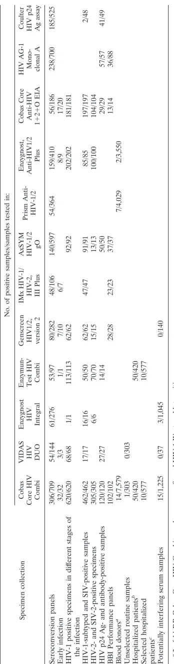
Cobas Core HIV Combi EIA showed a sensitivity of 100%
for HIV-1- and HIV-2-positive samples (Table 5). Overall, 30
RR false-positive results out of 10,031 HIV-negative samples,
including a high number of potentially cross-reactive
speci-mens, were obtained with Cobas Core HIV Combi EIA (Table
4). It is to be expected that about 1 of 334 specimens analyzed
will be incorrectly identified as false positive. The specificity of
Cobas Core HIV Combi EIA was 99.73% (Table 5). In most
cases, the total number of samples measured with each one of
the alternative assays was too low to get a statistically reliable
result for specificity (Table 4).
DISCUSSION
The results of our study demonstrate that Cobas Core HIV
Combi EIA permits an earlier diagnosis of HIV infection than
third-generation EIAs. The time delay in comparison to HIV-1
RNA amplification is about 2.7 days (Table 3). By using
fourth-generation assays with a more sensitive HIV Ag detection
module (detection limit,
ⱕ
10 pg of p24 Ag/ml), such as Cobas
Core HIV Combi EIA, the diagnostic window is reduced by
about 1.5 to 2.5 days in comparison to that for less-sensitive
combined p24 Ag and antibody assays (detection limit,
ⱖ
30 pg
of p24 Ag/ml). As shown by the results obtained with Coulter
HIV p24 Ag assay, the diagnostic window would be reduced by
a further 0.5 days with a lower detection limit of the Ag module
(Table 3). Of note, the new VIDAS Duo Ultra, with a
detec-tion limit of 3 pg of p24 Ag/ml (20), not investigated here,
shows a sensitivity which is equal to that of one of the most
sensitive HIV p24 Ag assays (detection limit, 2.2 pg of p24
Ag/ml) available on the market (32).
The diagnostic window would be reduced by 2.7 days in
comparison to that for Cobas Core HIV Combi EIA by
am-plification of HIV-1 RNA by RT-PCR from plasma or serum
TABLE
4.
Specimens
tested
and
results
obtained
with
comparative
assays
for
the
evaluation
of
sensitivity
and
speci
ficity
of
Cobas
Core
HIV
Combi
Specimen collection No. of positive samples/samples tested in: Cobas Core HIV CombiVIDAS HIV DUO Enzygnost HIV Integral Enzymun- Test
HIV
Combi
Genscreen HIV1/2, version
2
IMx
H
IV-1/
HIV-2, IIIPlus AxSYM HIV-1/2 gO
[image:7.587.72.246.68.729.2]Prism Anti-HIV-1/2 Enzygnost, Anti-HIV1/2 Plus Cobas Core Anti-HIV 1 ⫹ 2 ⫹ O EIA HIV A G-1 Mono- clonal A Coulter HIV p24 Ag assay Seroconversion panels 306/709 54/144 61/276 53/97 80/282 48/106 140/597 54/364 159/410 56/186 238/700 185/525 Early infection 32/32 3/3 1/1 7/10 6/7 8/9 17/20 HIV-1 positive specimens in dif ferent stages of the infection 620/620 68/68 1/1 113/113 62/62 92/92 202/202 181/181 HIV-1-subtyped and SIV-positive samples 462/462 17/17 16/16 50/50 62/62 47/47 91/91 85/85 197/197 2/48 HIV-2-and SIV-2-positive specimens 305/305 6/6 70/70 15/15 13/13 100/100 104/104 HIV p24 Ag-and antibody-positive samples 120/120 27/27 14/14 50/50 29/29 57/57 41/49 BBI Performance panels 102/102 28/28 23/23 37/37 13/14 36/88 Blood donors a 14/7,579 7/4,029 2/3,550 Unselected routine samples 1/303 0/303 Hospitalized patients b 50/420 50/420 Selected hospitalized 10/577 10/577 patients c Potentially interfering serum samples 15/1,225 0/37 3/1,045 0/140 a3 of 14 RR Cobas Core HIV Combi samples were con firmed HIV-1 Western blot positive. b50 samples were con firmed positive with HIV-1 Western blotting. c10 samples were con firmed positive with HIV-1 Western blotting.
TABLE 5. Sensitivities and specificities of assays
Assay Sensitivityseroconversionsawithout SpecificityaRR
Cobas Core HIV Combi 100 (99.82–100) 99.73 (99.61–99.82)
VIDAS HIV DUO 100 (99.43–100) 100 (99.12–100)
Enzygnost HIV Integral 100 (87.79–100) 99.71 (99.16–99.94)
Enzymun-Test HIV Combi 100 (98.8–100) 100 (99.68–100)
Genscreen HIV1/2, version 2 98.31 (95.13–99.65) 100 (97.88–100)
IMx HIV-1/HIV-2 III Plus 98.70 (92.98–99.97) NDb
AxSYM HIV-1/2 gO 100 (98.50–100) ND
Prism Anti-HIV1/2 ND 99.88 (99.64–99.93)
Enzygnost Anti-HIV1/2 plus 99.75 (98.60–100) 99.94 (99.80–99.99)
Cobas Core Anti-HIV 1⫹2⫹O
EIA 99.27 (98.13–99.8) ND
HIV AG-1 Monoclonal A ND ND
Coulder HIV p24 Ag assay ND ND
aValues are percentages. Values in parentheses are 95% confidence intervals.
bND, not determined.
on May 15, 2020 by guest
http://jcm.asm.org/
[image:7.587.300.542.83.224.2](single-unit testing). Cost-effectiveness analysis of expanded
HIV testing protocols for donated blood has shown that RNA
PCR testing would prevent annually eight more cases of
trans-fusion-associated HIV infection than combined p24 and
anti-body detection at a net additional cost of $96 million per year
in the United States (2). Current commercially available PCR
protocols are not adapted to large-scale screening of individual
blood donations. For these reasons, fourth-generation assays
represent a valuable alternative in terms of cost-effectiveness
calculations for the improvement of the sensitivity of blood
donor screening.
The prevalence of HIV-1 group M non-B subtypes (subtypes
A and C to J) and HIV-2 infections is increasing relatively
quickly in Europe. HIV-1 group O infections remain a rarity.
The epidemiological significance of HIV-1 group N, described
only recently, is not yet known (28). The genetic variability
represents a challenge in particular for the early detection of
HIV infection. In the seroconversion phase, false-negative
re-sults and delayed detection of antibody response are observed
in infections with HIV-1 non-B subtypes and HIV-2 (1, 8). In
recent years, the sensitivity for detection of HIV-1 non-B
sub-types has been optimized. However, obtaining seroconversion
samples with HIV-1 non-B subtypes in order to permit an
extensive evaluation of the sensitivities of newly developed
tests is difficult. In the present evaluation, one seroconversion
from a patient with HIV-1 subtype E infection (SIIDC24) was
tested with Cobas Core HIV Combi EIA and HIV-1 AG
Monoclonal A. Both assays were highly reactive in the first
blood sample obtained 7 days after onset of symptoms.
Ters-mette et al. (29) reported the failure of a monoclonal antibody
to detect p24 Ag from certain strains of HIV. HIV-1 subtype
O, which is highly divergent from other HIV-1 subtypes known
so far (13), may not be detected by assays using monoclonal
antibodies for the capture of p24 Ag. Our results from dilution
series of cell supernatants infected with different HIV-1
sub-types, including HIV-1 group O isolates and HIV-2, show that
no commercially available assay (HIV p24 Ag EIA or
fourth-generation EIA) is capable of detecting every HIV-1 virus
lysate with an optimal sensitivity. Although a limited number
of lysates were tested, Cobas Core HIV Combi EIA was likely
to have a higher sensitivity for HIV-2 Ag detection than the
Coulter HIV p24 Ag assay. Previous studies reported a
vari-able sensitivity of fourth-generation assays for HIV-1 group M
non-B subtypes and HIV-2 (12, 20, 33).
There is a potential risk for false-negative results due to the
absence of p24 Ag in the Ag mixture of Cobas Core HIV
Combi. In general, anti-p24 antibodies are, together with
an-tienvelope antibodies, the first to appear during
seroconver-sion (21). HIV RT has been included in analogy to
Enzymun-Test HIV Combi in the antigenic mixture of Cobas Core HIV
Combi EIA in order to avoid false-negative results in the early
seroconversion phase after disappearance of HIV antigenemia
since p24 Ag would have interfered with anti-p24 for HIV Ag
capture. As shown by the results of our study, in only 3 of 94
seroconversions were transient borderline-negative results
measured with Cobas Core HIV Combi EIA in the early phase
of seroconversion; this effect was also observed with
Enzymun-Test HIV Combi in a former study (12) and could therefore
possibly result from the absence of anti-p24 detection. The
second reason for including RT as the Ag in the Cobas Core
HIV Combi EIA is that, with gp41 alone, the sensitivity of the
assay would have been impaired not only during
seroconver-sion but also at all stages of the disease. The RT is highly
conserved among HIV-1 group M and O isolates and HIV-2
and induces a cross-reactive, predominantly IgG response (W.
Melchior, unpublished data).
A second diagnostic window may be observed not only in
serological assays, since, in two cases of primary infection, the
viral load decreased to under the detection limit of 50 copies of
HIV-1 RNA/ml of plasma for HIV-1 RT-PCR after initially
positive results. In panel BCP 9014 HIV-1 RNA was not
de-tectable from day 12 (fourth follow-up sample) until the end of
follow-up (day 31, seventh follow-up sample). With NAT,
false-negative results are observed in infected subjects with a
low viral load independent of HIV-1 subtype and stage of
disease (3).
A second potential risk for impaired sensitivity is that a more
limited area of the solid phase can be used for antibody
de-tection since about one-third of the binding sites are occupied
by anti-p24 antibody for HIV Ag detection. A time delay
be-tween third-generation assays and Cobas Core HIV Combi
EIA was observed in three seroconversions with weak, delayed,
or absent antigenemia. Gürtler et al. reported a delayed
de-tection of primary infection with Enzymun-Test Combi in an
HIV p24 Ag-negative seroconversion panel (12).
Anti-HAV-IgM antibody-positive samples possibly can lead
to elevated signals with Cobas Core HIV Combi EIA, since 5
out of 125 anti-HAV-IgM-positive samples were tested as false
positives. This interference cannot be explained by
cross-reac-tive epitopes of HIV RT and HAV polymerase since these
enzymes are not related, but rather most probably by immune
complex formation.
The risk of interference is potentially higher with the HIV
combined assays than with single-Ag or -antibody EIAs, since
both serological markers are determined in one test batch. The
specificity for blood donor screening with licensed
fourth-gen-eration assays (99.6 to 99.8%) was lower than that of
third-generation HIV EIAs (12, 18). The higher rate of false-positive
results (0.2%) of HIV combined assays has a particularly
neg-ative effect in blood donor screening, where a high sample
throughput and low prevalence are to be expected, because a
higher number of donations must be withdrawn and a separate
Ag detection in addition to the antibody confirmatory assay
(Western blotting) must be carried out in order to rule out an
HIV infection. The specificities of the new Cobas Core HIV
Combi EIA for 7,579 blood donors (99.84%) and for more
than 10,000 samples (99.73%), including potentially interfering
sera, were very high. Cobas Core HIV Combi EIA showed a
better performance than Enzymun-Test HIV Combi, which
achieved a specificity of 99.60% in 7,659 negative samples (12).
The specificity of Cobas Core HIV Combi EIA was equivalent
to that of third-generation assays, such as Enzygnost
Anti-HIV1/2 Plus and Prism HIV-O Plus, which are routinely used
for blood donor screening. Our results show that
fourth-gen-eration assays with improved specificity are suitable for blood
donor screening in terms of specificity. Furthermore they
de-tect HIV p24 Ag with a sensitivity comparable to that of
sin-gle-Ag assays. Cobas Core HIV Combi EIA may represent an
alternative to separate Ag determination in the diagnostic
on May 15, 2020 by guest
http://jcm.asm.org/
oratory and blood donor screening and possibly to NAT in
(mini)pools.
ACKNOWLEDGMENTS
We are very grateful to Roche Diagnostic for providing Cobas Core
HIV Combi reagents and analyzers.
Roche Diagnostic provided financial support for purchasing
alter-native and confirmatory assays.
REFERENCES
1.Apetrei, C., I. Loussert-Ajaka, D. Descamps, F. Damond, S. Saragosti, F. Brun-Vezinet, and F. Simon.1996. Lack of screening test sensitivity during
HIV-1 non-subtype B seroconversions. AIDS10:57–60.
2.Aubuchon, J. P., J. D. Birkmeyer, and M. P. Busch.1997. Cost-effectiveness of expanded human immunodeficiency virus-testing protocols for donated
blood. Transfusion37:45–51.
3.Barlow, K. L., J. H. C. Tosswill, J. V. Parry, and J. P. Clewley.1997. Performance of the Amplicor human immunodeficiency virus type 1 PCR and analysis of specimens with false-negative results. J. Clin. Microbiol.
35:2846–2853.
4.Berrey, M. M., T. Schlaker, A. C. Collier, T. Shea, S. J. Brodie, D. Mayers, R. Coombs, J. Krieger, T. W. Chun, A. Fauci, S. G. Self, and L. Corey.2001. Treatment of primary human immunodeficiency virus type 1 infection with potent antiretroviral therapy reduces frequency of rapid progression to
AIDS. J. Infect. Dis.183:1466–1475.
5.Bünung, H., and G. Trenkler. 1978. Nicht-parametrierbare Methoden. Springer Verlag, Berlin, Germany.
6.Busch, M. P., S. H. Kleinman, B. Jackson, S. L. Stramer, J. Hewlett, and S. Preston.Committee report. Nucleic acid amplification testing of blood do-nors for transfusion-transmitted infectious diseases: report of the Interorga-nizational Task Force on Nucleic Acid Amplification Testing of Blood
Do-nors. Transfusion40:143–159.
7.Centers for Disease Control.1989. Interpretation and use of the Western blot assay for serodiagnosis of human immunodeficiency virus type 1
infec-tions. Morbid. Mortal. Wkly. Rep.38(Suppl. S-7):1–7.
8.Christiansen, C. B., T. E. Jessen, C. Nielsen, and P. Staun-Olsen.1996. False negative anti-HIV-1/HIV-2 ELISAs in acute HIV-2 infection. Vox Sanguis
70:144–147.
9.Clark, S. J., G. D. Kelen, D. R. Henrard, E. S. Daar, S. Craig, G. M. Shaw, and T. C. Quinn.1994. Unsuspected primary human immunodeficiency virus type 1 infection in seronegative emergency department patients. J. Infect.
Dis.170:194–197.
10.Constantine, N. T.1993. Serologic tests for the retroviruses: approaching a
decade of evolution. AIDS7:1–13.
11.Couroucé, A. M., and Groupe de Travail Rétrovirus de la S.F.T.S.1999. Tests de dépistage combiné des anticorps anti-VIH et de l’antigène p24. Gaz.
Transfus. Sanguine155:4–18.
12.Gürtler, L., A. Mühlbacher, U. Michl, H. Hofmann, G. G. Paggi, V. Bossi, R. Thorstensson, R. G. Villaescusa, A. Eiras, J. H. Hernandez, W. Melchior, F. Donié, and B. Weber.1998. Reduction of the diagnostic window with a new combined p24 antigen and human immunodeficiency virus antibody
screen-ing assay. J. Virol. Methods75:27–38.
13.Gürtler, L., P. H. Hauser, J. Eberle, A. von Brunn, S. Knapp, L. Zekeng, J. Tsague, and L. Kaptue.1994. A new subtype of human immunodeficiency
virus type 1 (MVP-5180) from Cameroon. J. Virol.68:1581–1585.
14.Hoelscher, M., S. Hanker, F. Barin, R. Cheingsong-Popov, U. Dietrich, B. Jordan-Harder, D. Olaleye, E. Nägele, A. Markuzzi, D. Mwakagile, F. Minja, J. Weber, L. Gürtler, and F. Sonnenburg.1998. HIV type 1 V3 serotyping of Tanzanian samples: probable reasons for mismatching with genetic
subtyp-ing. AIDS Res. Hum. Retrovir.14:139–149.
15.Korber, B. T., S. Osmanov, J. Esparza, and G. Myers.1994. The World Health Organization Global Programme on AIDS proposal for standardiza-tion of HIV sequence and nomenclature: W. H. O. network for HIV
isola-tion and characterizaisola-tion. AIDS Res. Hum. Retrovir.10:1355–1358.
16.Korber, B. T. M., E. E. Allen, A. D. Farmer, and G. L. Myers.1995.
Heter-ogeneity of HIV-1 and HIV-2. AIDS9:S5–S18.
17.Le Corfec, E., F. Le Pont, H. C. Tuckwell, C. Rouzioux, and D. Costagliola. 1999. Direct HIV testing in blood donations: variation of the yield with
detection threshold and pool size. Transfusion39:1141–1144.
18.Ly, T. D., C. Edlinger, and A. Vabret. 2000. Contribution of combined
detection assays of p24 antigen and anti-human immunodeficiency virus (HIV) antibodies in diagnosis of primary HIV infection by routine testing.
J. Clin. Microbiol.38:2459–2461.
19.Ly, T. D., S. Laperche, and A. M. Couroucé.2001. Early detection of human immunodeficiency virus infection using third- and fourth-generation
screen-ing assays. Eur. J. Clin. Microbiol. Infect. Dis.20:104–110.
20.Ly, T. D., L. Martin, M. Daghfal, A. Sandridge, D. West, R. Bristow, L. Chalouas, X. Qiu, S. C. Lou, J. C. Hunt, G. Schochetman, and S. G. Devare. 2001. Seven human immunodeficiency virus (HIV) antigen-antibody combi-nation assays: evaluation of HIV seroconversion sensitivity and subtype
detection. J. Clin. Microbiol.39:3122–3128.
21.McRae, B., J. A. M. Lange, M. S. Ascher, F. De Wolf, H. W. Sheppard, J. Goudsmit, and J. P. Allain.1991. Immune response to HIV p24 core protein during early phases of human immunodeficiency virus infection. AIDS Res.
Hum. Retrovir.7:637–643.
22.Murthy, K. K., D. R. Henrard, J. W. Eichberg, K. E. Cobb, M. P. Busch, J. P. Allain, and H. J. Alter.1999. Redefining the HIV-infectious window period in the chimpanzee model: evidence to suggest that viral nucleic acid testing
can prevent blood-borne transmission. Transfusion39:688–693.
23.Portincasa, P., R. Grillo, P. Pauri, M. G. Colao, P. P. Valcavi, D. Speziale, G. Mazzarelli, E. De Majo, P. E. Varaldo, G. Fadda, C. Chezzi, and G. Dettori.2000. Multicenter evaluation of the new HIV DUO assay for
simul-taneous detection of HIV antibodies and p24 antigen. New Microbiol.23:
357–365.
24.Quinn, T. C., R. Brookmeyer, R. Kline, M. Shepherd, R. Paranjape, S. Mechendale, D. A. Gadkari, and R. Bollinger.2000. Feasibility of pooling sera for HIV-1 viral RNA to diagnose acute primary HIV-1 infection and
estimate HIV incidence. AIDS14:2751–2757.
25.Roth, W. K., H. Weber, and E. Seifried.1999. Feasibility and efficacy of routine PCR screening of blood donations for hepatitis C virus, hepatitis B
virus, and HIV-1 in a blood bank setting. Lancet353:359–363.
26.Saville, R. D., N. T. Constantine, F. R. Cleghorn, N. Jack, C. Bartholomew, J. Edwards, P. Gomez, and W. A. Blattner.2001. Fourth-generation enzyme linked immunosorbent assay for the simultaneous detection of human
im-munodeficiency virus antigen and antibody. J. Clin. Microbiol.39:2518–2524.
27.Sherefa, K., A. Sönnerborg, J. Steinbergs, and M. Sällberg.1994. Rapid grouping of HIV-1 infection in subtypes A to E by V3 peptide serotyping and
its relation to sequence analysis. Biochem. Biophys. Res. Commun.205:
1658–1664.
28.Simon, F., P. Mauclere, P. Roques, I. Loussert-Ajaka, M. C. Muller-Trutwin, S. Saragosti, M. C. Georges-Courbot, F. Barré-Sinoussi, and F.
Brun-Vézi-net.1998. Identification of a new human immunodeficiency virus type 1
distinct from group M and Group O. Nat. Med.4:1032–1037.
29.Tersmette, M., I. Winkel, M. Groenick, R. A. Gruters, R. P. Spence, E. Saman, G. van der Groen, F. Miedema, and J. G. Huisman.1989. Detection and subtyping of HIV-1 isolates with a panel of characterized monoclonal
antibodies to HIV p24 gag. Virology171:149–155.
30.van Binsbergen, J., A. Siebelink, A. Jacobs, W. Keur, F. Bruynis, M. van de Graaf, J. van der Heijden, D. Kambel, and J. Toonen.1999. Improved performance in seroconversion with a 4th generation HIV antigen/antibody
assay. J. Virol. Methods82:77–84.
31.van Binsbergen, J., W. Keur, A. Siebelink, F. M. van de Graaf, A. Jacobs, D. de Rijk, L. Nijholt, J. Toonen, and L. G. Gürtler.1998. Strongly enhanced sensitivity of a direct anti-HIV-1/-2 assay in seroconversion by incorporation of HIV p24 Ag detection: a new generation vironostika HIV Uni-Form II.
J. Virol. Methods76:59–71.
32.Weber, B., A. Berger, H. Rabenau, and H. W. Doerr.2002. Evaluation of a new combined antigen and antibody human immunodeficiency virus
screen-ing assay, VIDAS HIV DUO Ultra. J. Clin. Microbiol.40:1420–1426.
33.Weber, B., E. M. B. Fall, A. Berger, and H. W. Doerr.1998. Reduction of diagnostic window by new fourth-generation human immunodeficiency virus
screening assays. J. Clin. Microbiol.36:2235–2239.
34.Weber, B., A. Mühlbacher, U. Michl, G. Paggi, V. Bossi, C. Sargento, R. Camacho, E. M. B. Fall, A. Berger, U. Schmitt, and W. Melchior.1999. Multicenter evaluation of a new rapid automated HIV antigen detection
assay. J. Virol. Methods78:61–70.
35.Yerly, S., F. Simon, and L. Perrin.1999. Early diagnosis of primary HIV infections: using a combined screening test (p24 antigen and anti-HIV
an-tibodies). Schweiz. Med. Wochenschr.27:319–322.
36.Zaaijer, H. L., P. V. Exel-Oehlers, T. Kraaijeveld, E. Altena, and P. N. Lelie. 1992. Early detection of antibodies to HIV-1 by third-generation assays.
Lancet340:770–772.