DOI 10.3349/ymj.2007.48.5.748
Purpose: Wegener’s granulomatosis (WG) is a rare multisystem inflammatory disease, which infrequently involves the subglottic area and trachea. Treatment usually involves the use of immunosuppressive agents with corticosteroids. Some patients, however, continue to have symptoms of airway obstruction after clinical remission following the standard therapeutic regimen. Objective: To investigate laser treatment for subglottic stenosis in five patients suffering from WG. Materials and Methods:We endoscopically treated 5 patients with subglottic stenosis due to WG and airway obstruction by Nd:YAG and CO2lasers. One of the patients had preoperative tracheostomy and after treatment was decannulated and could not breathe without dyspnea. Another patient required stenting of the subglottic area.Results:All five patients were able to breathe without dyspnea after the treatment. Three patients were treated with an Nd:YAG laser but needed repeated laser treatment every four to six months, whenever they complained of dyspnea. The other two patients were treated with a CO2 laser; one of these patients had preoperative tracheostomy and was treated twice by CO2 laser and decannulated, with no further difficulty in breathing. The follow-up period was 1-5 years.Conclusions:Nd:YAG and CO2 lasers are recommended in the treatment of subglottic stenosis (SS) due to WG, particularly when the stenosis is in continuity or close proximity to the vocal cords.
Key Words:Subglottic, stenosis, Wegener’s granulomatosis, laser, endoscopy
INTRODUCTION
Wegener’s granulomatosis (WG) is a rare
multi-system inflammatory disease of unknown origin, characterized by necrotizing granulomatous in-flammation and necrotizing vasculitis of the upper and lower respiratory tract, glomerulonephritis and a variable degree of small vessel vasculitis. Nasal and oral ulceration, epistaxis, sinusitis, or otitis is found in more than 90% of the cases.1 Ulcers in the larynx and trachea were seen in about a quarter of the cases of WG.2,3 Subglottic stenosis (SS) is considered to be less common.3,4 This rare form of the disease (SS due to WG) often responds to medical treatment of cyclophospha-mide with or without corticosteroids,5,6 but when patients continue to complain of respiratory dif-ficulty due to SS, surgical treatment is necessary.7,8 There are a wide variety of possible surgical interventions including dilatation, stents, or major open operations, such as laryngofissure with or without skin or mucosal grafts and segmental resection of the stenotic area.6,9,10
Research has concentrated on the use of a CO2 laser for the treatment of SS.2,11-13
In this study we report on a series of five patients with subglottic stenosis related to WG, who were treated by endoscopic laser surgery: three with an Nd : YAG laser and two with a CO2 laser. We also review the literature concerning tracheal reconstruction options for cases of SS due to WG.
MATERIALS AND METHODS
Patients
We treated 5 patients (4 female, 1 male) with SS
Endoscopic Laser Surgery for Subglottic Stenosis in
Wegener’s Granulomatosis
Jacob Shvero,1 David Shitrit,2 Rumelia Koren,3 Dekel Shalomi,2 and Mordechai Reuven Kramer2
1Department of Otorhinolaryngology Head & Neck Surgery,2Pulmonary Institute and 3Department of Pathology, Rabin Medical
Center, Petah Tikva, and Sackler Faculty of Medicine, Tel Aviv University, Tel Aviv, Israel.
Received February 13, 2006 Accepted January 15, 2007
Reprint address: requests to Dr. Jacob Shvero, Department of Otorhinolaryngology Head and Neck Surgery, Rabin Medical Center, Petah Tikva, Israel. Tel: 03-9376451, Fax: 03-5322890, E-mail: [email protected]
due to WG by laser. The patients' ages ranged between 21 and 83 years, with a mean age of 35.8 years. All patients were positive for complement antineutrophil cytoplasm antibody (c-ANCA) according to blood tests.
The patients were evaluated by CT scan, chest x-ray, and pulmonary function studies. Preopera-tive fiber optic laryngoscopy and bronchoscopy provided valuable information regarding vocal cord mobility, the extent of the stenosis, and the distance from the vocal cords.
The stenoses were firm, with a granulomatous rim, and extended below the vocal cord 0.5 to 1.5 cm, with an average thickness of 1.7 cm. Com-puter tomography showed circumferential steno-sis causing narrowing of the lumen in the subglottic area in three patients. One patient had a preoperative tracheostomy (Table 1).
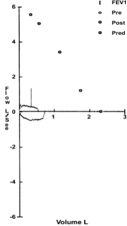
All patients were treated by the immuno-suppressive agent, cyclophosphamide, with corti-costeroids, but they continued to suffer from dyspnea on exertion and stridor. Pulmonary function testing revealed a flattening of the flow-volume loop with inspiration, consistent with an extra-thoracic airway obstruction (Fig. 1).
Surgical procedure
Direct microlaryngoscopy was performed under general anesthesia in two patients (one required an endotracheal tube and one was anesthetized via a prior tracheostomy). A Storz laser laryngo-scope was used to expose the glottic and the sub-glottic areas. A Zeiss microscope with a 400-mm
objective lens coupled to a CO2 laser was used in both cases. The laser setting was 10 W in a con-tinuous mode. Moistened neurosurgical pledgets were placed in the subglottic space to protect the trachea and endotracheal tube during excision of the stenotic area in order to reduce the lateral thermal damage of the CO2 laser.
Three patients underwent bronchoscopy under local anesthesia and an Nd:YAG laser was used with Olympus fiber-optic bronchoscopy.
[image:2.595.363.489.297.521.2]The laser energies ranged from 2500 to 6000 Joules, applied in a 50 W continuous mode. In one patient, we placed a 12/30 mm wall stent. The
Table 1. Clinical Data of Five Patients with Subglottic Stenosis
No. (yrs)Age (M/F)Sex Nose/Sinus Tracheostomy Stenosis
Preop. Postop. Thickness/cm Diameter/mm Distance/cm*
1 29 F Saddle nose Chronic
sinusitis Yes No 1 Total 1
2 20 M Saddle nose Septal
perforation No No 2 5, circular 1.5
3 25 F Saddle nose No No 3 4, circular 1
4 83 F - No No 1 6, anterior 0.5
5 25 F - No No 1.5 6, anterior 0.5
[image:2.595.62.522.583.727.2]*Distance of the stenosis from the vocal cord (cm).
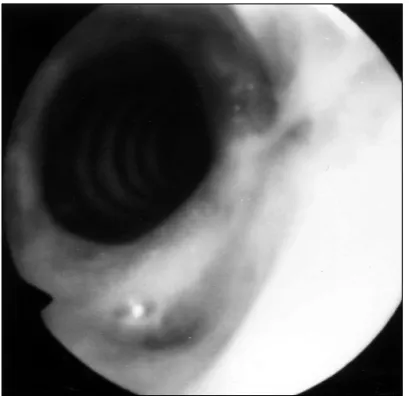
preoperative SS is shown in Fig. 2. Both the CO2 and Nd: YAG laser procedures took no longer than 20 minutes.
RESULTS
We treated two patients by CO2 laser because the SS was in close proximity to the vocal cords (within 0.5 - 1 cm). One patient, who had gone a preoperative tracheostomy, also under-went two interventions with dilatation and the other patient underwent four additional inter-ventions. These patients were in the hospital for only 24 hours after CO2 laser treatment.
The other three patients were treated by an Nd : YAG laser and underwent 5 - 18 interventions (the SS was between 1 - 1.5 cm below the vocal
cords), and these patients were discharged one hour after the procedure. None of the five patients needed emergency tracheostomy. The follow-up period for all of our patients ranged from 1 to 5 years (Table 2).
The resolution of dyspnea on exertion or decannulation was the criteria for success.
[image:3.595.69.275.107.301.2]Four patients had no dyspnea during the fol-low-up period. The fifth patient needed repeated intervention after two and three months because of episodes of dyspnea on exertion, and this patient ultimately required an SS stent. The SS in this patient had an opening diameter of 4 mm. The stenotic segment was short and had soft
Table 2. Laser Treatment and Follow-up
No. CO2 laser Nd : YAG laser Interventions (n) Outcome Follow-up/months
1 Yes + dilatation - 2 Decannulated 12
2 - Yes 10 Favorable 60
3 - Yes 18 Favorable 6
4 - Yes 5 Favorable 24
5 Yes - 4* Favorable 6
[image:3.595.362.494.111.351.2]*stent.
Fig. 2. Tracheoscopic image of preoperative subglottic
stenosis.
[image:3.595.65.536.618.726.2]tissue. Another patient who had preoperative tracheostomy was decannulated after two inter-ventions by CO2 laser.
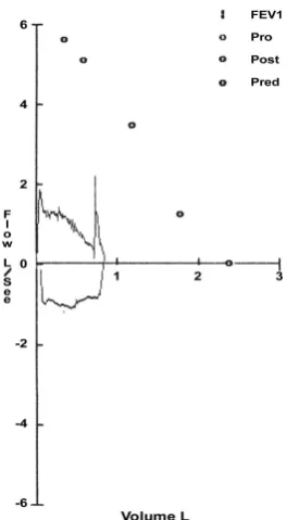
There was no voice disturbance after treatment in our patients. Postoperative pulmonary function testing showed an improvement (Fig. 3). Fig. 4 shows the postoperative results after laser treat-ment.
Histological examination
All specimens were fixed in 10% buffered formalin, embedded in paraffin, and stained with haematoxylin and eosin. The biopsies demons-trated focal acute necrosis surrounded by a zone of fibroblastic proliferation with giant cells and leukocytic infiltrate. The vasculitis affected small arteries and veins. These granulomatous inflam-matory changes were consistent with the dia-gnosis of WG.
DISCUSSION
SS occurs in nearly 16 to 23% of patients with WG.1,4,14 Significant SS is defined as the presence of symptomatic airway compromise, usually con-sisting of dyspnea, voice changes, or cough. For unknown reasons, SS is more common in young
patients. The clinical course of SS generally runs independently of systemic WG activity.14 Patients with WG are at great risk for severe airway obstruction, which can rapidly run a fatal course if not treated.15In the absence of other features of acute WG, isolated SS is optimally managed with intratracheal laser treatment and does not require systemic therapy; this is the best approach in cases, such as our patients, where SS is in close proximity to or in continuity with the vocal cords. A CO2 laser emits an invisible beam of light at 10.6 m, which is selectively absorbed by water.μ
The CO2 laser has distinct advantages over con-ventional steel knives. It seals blood vessels less than 0.5 mm in diameter, resulting in superior haemostasis, and it can be used in ablation mode for vaporizing tissue and for incisions. Therefore, this method is frequently used for micro-laryngeal surgery. However, a major disadvantage of the CO2 laser is that fiber-optic transmission is not possible. The ultra spot micromanipulator deli-very system allows a spot size of 200 m, at aμ
400-mm focal distance.
The Nd:YAG laser, which is an excellent photo coagulator and can be transmitted through fiber-optic cables, emits a beam of light at 1.06 m. Theμ
[image:4.595.71.276.107.305.2]ND:YAG laser utilizes a 220-V energizer and needs an exterior cooling device, is less precise, having a 20 to 40% scatter, and its beam of light penetrates deeply and is absorbed by water. The Nd : YAG laser can also cause edema and scarring. Because of its excellent ability to evaporate and coagulate, it is used as surgical intervention for obstructive lesions in the tracheobronchial area.6 In 1984, Dedo et al.2 described a new technique for the treatment of posterior glottic, subglottic, and tracheal stenosis with a CO2 laser and microtrapdoor mucosal flap. Nine of the ten patients in their study were given an adequate airway and good voice. In 1979, Strong et al.13 presented a study involving 18 patients with glottic or subglottic partial or total stenosis. Using a laser, they successfully treated 4/7 and 8/11 SS cases. Six of the latter also had temporary placement of Silastic roll stents. Lebovics et al.12 described 8 out of 25 cases of CO2 laser Treatment, and they reported that subglottic scarring recurred rapidly and was more extensive after each laser procedure. All 8 of their patients
ultimately required other therapies to adequately manage the stenosis.
In 2003, Gluth et al.14described their experience in repairing SS in WG and recommended mini-mizing the airway manipulation during episodes of systemic disease activity. Once the disease process is under control, consideration can then be given to either an endoscopic or an open approach based on the extent of stenosis. Either laryngotracheal reconstruction or CO2 laser resection and dilation can be safely used to produce a favorable outcome.
Alternative approaches that minimize the frequency of episodes of recurrent disease, such as trimethoprim-sulfamethoxazole therapy, are currently under investigation.14,16
SS at the level of the cricoids cartilage and/or upper tracheal rings present a management dilemma12 as the stenosis is in close proximity to the vocal cords. In these cases, laser treatment should be used to preserve the quality of the voice as any other open surgery might damage the vocal cords. In patients, especially young patients, with SS in close proximity or in continuity with the vocal cords, if the patient complains of dyspnea, repeated laser intervention is preferred over permanent tracheostomy or laryngotracheal resection.
Our patients were diagnosed with WG by a positive c-ANCA blood test and by histological examination. For treatment, we used CO2laser for two patients with SS close to the vocal cords (0.5-1 cm) since the Nd : YAG laser can penetrate deeply, causing edema and scarring that would affect the quality of the voice. The Nd : YAG laser was used in the other three patients with SS located far from the vocal cords (1 - 1.5 cm). The Nd:Yag laser is preferred over the CO2 laser when the SS is near the upper trachea and because it can be done under local anesthesia and allows for multiple interventions.
As a result of our study, we recommend laser treatment for treatment of SS for the following reasons: it requires a short surgical intervention (no longer than 20 minutes) and short hospi-talization; there is no bleeding and no scarification of the recurrent nerve; laser treatment can be done without tracheostomy; it does not produce cos-metic defects in the neck skin; it is suitable for
high risk patients; it can be performed if the SS is in close proximity or in continuity with the vocal cords; and it produces favorable results. In cases where endoscopic treatment by laser has failed, we suggest performing laryngotracheal resection with reconstruction.
REFERENCES
1. O'Devaney K, Ferlito A, Hunter BC, Devaney SL, Rinaldo A. Wegener's granulomatosis of the head and neck. Ann Otol Rhinol Laryngol 1998;107:439-45. 2. Dedo HH, Sooy CD. Endoscopic laser repair of
posterior glottic, subglottic and tracheal stenosis by division or micro-trapdoor flap. Laryngoscope 1984;94: 445-50.
3. Walton EW. Giant-cell granuloma of the respiratory tract (Wegener's granulomatosis). Br Med J 1958;34:265-70.
4. DeRemee RA, McDonald TJ, Harrison EG, Coles DT. Wegener's granulomatosis. Anatomic correlates, a pro-posed classification. Mayo Clin Proc 1976;51:777-81. 5. Arauz JC, Fonseca R. Wegener's granulomatosis
appe-aring initially in the trachea. Ann Otol Rhinol Laryngol 1982;91:593-6.
6. McDonald TJ, Neel HB 3rd, DeRemee RA. Wegener's granulomatosis of the subglottis and the upper portion of the trachea. Ann Otol Rhinol Laryngol 1982;91:588-92.
7. Alaani A, Hogg RP, Drake Lee AB. Wegener's granu-lomatosis and subglottic stenosis: management of the airway. J Laryngol Otol 2004;118:786-90.
8. Nishiike S, Kato T, Nagai M, Konishi M, Sakata Y. Management and follow-up of localized Wegener's gra-nulomatosis: a review of five cases. Acta Otolaryngol 2004;124:1103-8.
9. Eliachar I, Chan J, Akst L. New approaches to the management of subglottic stenosis in Wegener's granu-lomatosis. Cleve Clin J Med 2002;69 Suppl 2:SII149-51. 10. Herridge MS, Pearson FG, Downey GP. Subglottic stenosis complicating Wegener's granulomatosis: sur-gical repair as a viable treatment option. J Thorac Cardiovasc Surg 1996;111:961-6.
11. Crockett DM, Reynolds BN. Laryngeal laser surgery. Otolaryngol Clin North Am 1990;23:49-66.
12. Lebovics RS, Hoffman GS, Leavitt RY, Kerr GS, Travis WD, Kammerer W, et al. The management of subglottic stenosis in patients with Wegener's granulomatosis. Laryngoscope 1992;102:1341-5.
15. Utzig MJ, Warzelhan J, Wertzel H, Berwanger I, Hasse J. Role of thoracic surgery and interventional bron-choscopy in Wegener's granulomatosis. Ann Thorac Surg 2002;74:1948-52.
16. Stegeman CA, Tervaert JW, de Jong PE, Kallenberg CG.