University of Warwick institutional repository:
http://go.warwick.ac.uk/wrap
A Thesis Submitted for the Degree of PhD at the University of Warwick
http://go.warwick.ac.uk/wrap/55725
This thesis is made available online and is protected by original copyright.
Please scroll down to view the document itself.
STUDIES ON NEURAMINMASES
by
JohnP. Kabayo, B. Sc., M. Sc.
A thesis submitted in fulfilment of the
requirements for the degree of Ph. D. of the
University of Warwick
CONTENTS
nage
Preface
Acknowle, dgements it
Summary
List of Tables IV
List of Figures v
Chapter One GENERAL INTRODUCTION
1.1 Discovery of neuram in idases 1
1.2 Occurrence and distribution of neuraminidases 6
1.2.1 Neuraminidaseln viruses 6
1.2.2 Neuraminldase in protozoa and bacteria 8
1.2.3 Distribution of neuraminidases in vertebrates
9
1.3 Importance of neuraminidase 10
1.3.1 Some effects of neuraminidase on cell surface
properties 10
1.3.2 Some effects of neuraminidase on glycoprotelns 13
1.3.3 Neuraminidase in metabolismand physiology 14
1.3.4 Neuraminidase and disease 16
1.3.5 Direct applications of neuraminidase 22
1.3.6 Interpretations of the experimental use of
neuram in ida se 23
1.4 Aim of the project 25
Chapter Two PROPERTIES OFNEURAMINIDASES
2.1 Introduction 27
2.2 Isolation purification and characterisation of a
,
neuraminidase from S. griseus 32
2.2.1 Growth of S. gr iseus 32
2.2.2 Assay ofneuraminidase in the culture fluid 34
2.2.3 Purification of neuraminidase from the culture filtrate 37
2.2.4
Enzymepurity
41
2.2.5 Characterisation of S. griseus neuraminidase 41
2.2.6 Catalytic properties of S. griseus neuraminidase 51
page
Chapter Three MECHANISM OF NEURAMINIDASE ACTION
3.1
Introduction
64
3.2 Chemical modification of amino acid residues in
proteins 68
3.3 Active site studies on neuraminidase 70
3.4 Chemical modification reactions employed 72
3.4.1 Modification of lysine residues 72
3.4.2 Modification ofhistidine residues 73
3.4.3 Modification of arginine residues 73
3.4.4 Modification of carboxylgroups 75
3.4.5 Modification of tryptophan residues 76
3.5 Chemical modification of neuraminidases:
experimental procedure 77
3.5.1 Preparation of neuraminidases 77
3.5.2 Enzyme assays 77
3.5.3 Lysine modification 78
3.5.4 Histidinemodification 78
3.5.5 Arginine modification 80
3.5.6 Carboxylmodification 88
3.5.7 Modification of tryptophan in S. griseus and
Cl. perfringens neuraminidases 91
3.6
Discussion
93
3.7 Conclusions 101
Appendix I Practical application ofneuraminidase 102
Appendix II Attempt to design a radio immunoassay for
slalic acid 107
A. 2.1 Introduction 107
A. 2.2 Experimental procedure 110
Appendix III Design of a longitudinal gel slicer 120
Appendix IV Sources of materials 123
REFERENCES 125
(i)
PREFACE
The work described in this thesis was carried out in the Department
of Chemistry and Molecular Sciences, University of Warwick, Coventry,
England, during the period between October, 1975 and September, 1978.
It is the original work of the author, except where specific acknowledgement
is made or implied. This thesis has been submitted at the University of
(ii )
ACKNOWLEDGEMENTS
I am grateful to all those people who have, in their various ways,
made this thesis possible. More specifically I owe a special debt of
gratitude toMakerere University, the Humanitarian Trust, the
Commonwelath Inter-University Exchange Fund and the Africa
Educat Iona 1 Trust for financial support. It is my duty and pleasure
to express my gratitude to the chairmen and staff of the departments of Molecular Sciences and Biological Sciences, University of Warwick, for the permission to use their research facilities throughout this work.
The largest single debt of all lowe to Dr. D. W. Hutchinson, who has
succeeded in creating around himself a unique climate for research,
characterised alike by friendliness, by scientific enthusiasm and by
helpful supervision and encouragement. I am deeply indebted to
Drs. N. J. Dimmock and J. M. Morser for their guiding suggestions and
comments. A special vote of thanks goes to my colleagues in the
laboratory who have not only provided pleasant company but who have
also influenced my thoughts on my work.
My sincere thanks are due to my wife and son, who have both been
deprived of my company through many solitary evenings, in appreciation
of their unlimited patience and forebearance to enable me to complete
this work.
Finally I am grateful to Jacynth McKeand for her help in typing this
(iii)
SUMMARY
in this thesis, the work undertaken in an attempt to gain insight into the catalytic function ofneuraminidase is described.
The history, properties and importance of neuram in idases are
reviewed in Chapter One. Chapter Two contains an account of the
production of neuraminidasefrom Streptomyces rig seus by induction,
of its preparation and purification to homogeneity, of its character-
isation as a glycoprotein of 32,000 molecular weight and of its
structural and catalytic properties. In Chapter Three, chemical
modification methods were employed to seek information regarding
the nature of the amino acid residues essential in the activity of S. griseus, Cl. perfringens and influenza virus neuraminidases.
In all the three enzymes the results obtained suggested that
argin[ne, tryptophan and carboxylic groups were crucial for the
enzyme activity. Based on these findings, a mechanism for neura-
minidase action was proposed.
The extensively purified neuraminidase from S. griseus was used, in conjunction withan isoelectric focussing technique, to investigate
the sialylation differences in human Interferons. The results to
these experiments, reported in Appendix I, suggest that fibroblast
(and not leucocyte or lymphoblastoid) interferon contains neuramini-
dase -releasable s ialic acid residues . The experiments reported in Appendix Il, in which rabbits were immunised with colominic acid or
fetuin, were conducted to raise antibodies specific to sialic acid in
(iv)
LIST OF TABLES
page
1.1 Occurrence of neu ram inidases in viruses 8
2.1 Natural substrates for neuraminidase 29
2.2 Subunit relationship of some neuraminidases 31
2.3 Neuram inidase levels in the culture fluid
37
2.4 Sialic acid content of fetuin-sepharose 38
2.5 Purification of neuraminidase from S. griseus 39
2.6 Molecular weight determination of neuraminidase by
electrophoresis 45
2.7 Amino acid content of S. griseus neuraminidase 47
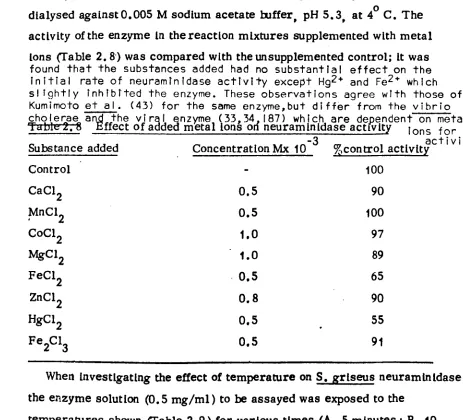
2.8 Effect of added metal ions on neuraminidase activity 54
2.9 Effect of temperature on S. griseus neuraminidase 54
3.1 Incorporation of [ 14C ]phenylglyoxal in S. griseus
neuramin idase 87
3.2 Analysis of 3H-glyc[neethyl ester hydrochloride 90
3.3 Incorporation of 3H-glyc[ne in S. griseus
neuram in Ida se 91
(v)
LIST OF FIGURES
Figure 2age
1.1 Hirst's Interpretation of the erythrocyte
heamagglutinin phenomena 2
1.2 Structures of some sialic acids 4
1.3 Hydrolysis of an a-ketoside of sialic acid by
neuramin idase 5
1.4 Diagrammatic representation of a segment of an
influenza virus 7
1.5 Inhibitors of viral neuram[nidase 20
2.1 Action of neuraminidase on the possible glycosidic
linkages 28
2.2 Phase contrast photomicrograph of S. griseus 33
2.3 Absorption spectra of chromgens in TBA assays 35
2.4
Production of Nase during the growth of S. griseus
36
2.5 Gelfiltration and affinity chromatography of
S. griseus neuraminidase 40
2.6 Electrophoretic detection of proteolytic contaminants
In neuraminidase preparations 42
2.7 Polyacrylamide gel electrophoresis of purified
S. griseus neuraminidase 43
2.8 Molecular weight determination of S. griseus
neurandnidase by gel filtration 44
2.9 Molecular weight determination of S. griseus
neuraminidase by SDS -PAGE 45
2.10 Determination of disulphide bonds In S. griseus
neuraminidase 48
2.11 Determinationof Sulphydrylgroups in S. griseus
neuram in idase 50
2.12 S. griseus neuraminidase staining with dansyl-
hydrazine 52
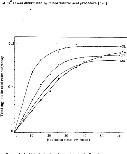
2.13 Hydrolysis of various substrates by S. griseus
neuraminidase 53
2.14 Effect of pH on reaction rates of substrate hydrolysis
(vi)
Figure pie
2.15 Inhibition of S. griseus neuraminidase by NANA 56
2.16 Dixon plot for the inhibition of S. griseus
neuraminidase by NANA 57
2.17 Determination of K and V of S. griseus
neuraminidase m max 58
3.1
Derivatives of N-acetylneuraminic acid with lipo-
philic sidechains of cyclic acetals 65
3.2 Colominlc acid showing the esterification of the
carboxyl by C7 or C9 hydroxylgroups 67
3.3 Reaction of TNBS with amino groups in proteins 73
3.4 Modification of histidine with diethyl pyrocarbonate 74
3.5 Reaction of arginyl sidechains in proteins with 2,4-
pentanedione and phenylglyoxal 74/75
3.6 Regeneration of 2 4-pentanedione-modifiedlysines
,
by reaction with hydroxylamine 75
3.7 Reaction of a water soluble carbodiimide with
protein carboxyl groups 76
3.8 Effect of lysinemodification on the activity of
neuraminidases 79
3.9 Modification of histidine residues in neuraminidase 81
3.10 Effect of modifying arginyl residues with 2 4-pentane-
,
d ione 82
3.11 Protection of S. griseus and viral neuraminidase by
NANA againstarginine modification 83
3.12
Reaction of phenylglyoxalwith S. griseus
neuram in ida se 84
3.13 Reaction of phenylglyoxal' with Cl. perfringens
neuram in ida se 85
3.14 Modification of neuraminidase with EDC 88
3.15 Effect of protection by NANA and sialyllactose on
EDC modification 89
3.16 The effect of NBS on S. griseus and Cl. perfringens
neuraminidase activity 92
3.17A Structure of the cyclic oxocarbonium Ion of N-acetyl-
neuram inis acid 99
3.17B Structure of 2-deoxy-2,3-dehydro-N-acetylneura-
(vii )
page
3.18 Possible mechanisIn of substrate binding by
neuramiliidase 100
3.19 Possible hydrolytic mechanism of neuraminidase 101
A. 1 Isoelectric focussing of human interferons 103
A. 2 Isoelectric focussing of neuramin idase -treated
fetu in 104
A. 3 Relationship between the sialic acid content of fetuin
and its migration in isoelectric focussing 105
B. 1 Summary of the principles of radiolmmunoassay 107
B. 2 Structure of the possible carbohydrate immuno-
determinant moiety in fetuin 110
B. 3
Double Immunodiffus ion of rabbit antifetuin anti-
serum 111
B. 4 14C -NANA binding capacity of antiserum to
colominic acid and fetuin 113
B. 5 Diagram of a microequilibrium dialysis apparatus 114
B. 6 Determination of completion of equilibrium 115
B. 7 14C -NANA binding by FAb and CAb determined by
equilibrlumdialysls 116
B. 8 Competition of 14C-NANA and unlabelled NANA for
CAb 117
B. 9 Competition of 14C -NANA and other sugars for CAb 118
1.
1. GENERAL INTRODUCTION
1.1 Discovery of Neuraminidase
As early as 1902 Kraus and Ludvig [1] described bacterial
haemagglutinlns which were destroyed by heating at 580 C; the ability to haemagglutlnate red blood cells was later observed as a property of
many protozoa and bacteria. Thomsen [2) and Friedenrich [31 found
that contrary to the high specificity of native human erythrocytes in their reaction with iso-antibodies (isoagglutination ), the red blood cells
exposed to different bacteria or to their culture fluids are agglutinated by all human sera (panagglutination). They wondered whether this
phenomenon derived in a bacterially-caused alteration of the red cell membranes.
About ten years later, Hirst [ 41 and McClelland and Hare [ 51 reported
that chicken erythrocytes were haemagglutinated by influenza virus. Hirst
[ 6,7] later showed thatwhen influenza viruses area Rowed to interact with
red blood cells at temperatures near 00C. the viruses remain attached
to the cells but are spontaneously eluted from the cells at 370 C. Hirst
observed that the red cells from which the virus had spontaneously eluted
would neither adsorb, nor become reagglutinated by, freshly added virus.
The eluted virus, however, would still reagglutinate fresh cells. Something
had changed the surface of the red cells, and Hirst proposed that an enzyme
present on the virus particle must have been functional in destroying
certain agglutinating receptors on the cells.
Hirst [7] suggested the analogy of the agglutination reaction with the
interaction of an enzyme E (on the virus particle) and its substrate S
(receptors present at the red cell surface),, resulting in the complex ES
2.
'S SS
SSE --aE SS
US
SS ESS
near 0ý CEEw
SS at 3
101
S
E
SSS4.0
SSE,,,
(_ tl.
Jýý_
ESS
ESSS
E+S cý ES 0E+ products
virus fresh haemagglutination intact altered
cells virus cells
Fig. I. I Hirst's Interpretation of the erythrocyte haemagglutin in phenomena
Hirst's postulate contradicted a view previously held that viruses, unlike bacteria or higher organisms, were devoid of enzymes. However, following
the discovery that other viruses, e. g. mumps, Newcastle disease, also had
agglutinating activity [ 8], It was found that cells from which one strain of
myxovirus had eluted were still agglutinable by certain other strains,
implying that the amount or destructibility of the enzyme-susceptible
receptors required for binding and haerrngglutination differed from strain
to strain.
In the course of the studies on the virus -cell interactions Burnet and his
colleagues [ 81 furnished strong evidence in support of Hirst's view when
they found that pretreatment of erythrocytes or host cells with a culture
filtrate from certain bacteria, e . g. Vibrio cholerae, abolished their
properties to agglutinate or adsorb Influenza virus. This observation was
correlated with the earlier reports of Friedenrich [ 31 and Thomsen [ 21
[image:13.551.95.536.49.358.2]3,
/
that the carbohydrate moiety of erythrocyte surfaces played a role
in haemagglutination reactions and that this process could be inhibited
by a number of glycoproteins [ 9,10,111. They found that the carbo-
hydrate receptor sites on the erythrocyte surfaces and on the glyco-
proteins which inhibited haemagglutination could be removed or
inactivated by treatment with an enzyme present in the culture
filtrate of Vibrio cholerae [ 12] or other bacteria [ 13], and since this
enzyme destroyed the receptor sites for influenza viruses on the
surface of erythrocytes it was termed 'receptor-destroying enzyme' [ 12].
Following the observation that influenza viral infections were focussed on the respiratory tract, the lining of which contains mucinous
substances [ 14], it became necessary to conduct biochemical studies on the interaction between influenza viruses and mucins [ 9,15,16,17 ]. Thus, by associating a particular substrate with an enzymic function,
these studies further substantiated the enzymic nature of erythrocyte
agglutination by virus. They also showed that the phenomena observed on treatment of cells with either bacterial filtrates or viral particles were due to one and the same enzyme found in both viruses and bacteria.
More information regarding the chemical nature of the substrate and
the enzymic destruction of haemagglutination receptors came from
further experiments in which (a) the product of the interaction between
the influenza virus enzyme and ovomucin was characterised as an
oligosaccharide containing one or more N-asyl hexosam ine residues
[9,18], (b) the viral enzyme and the receptor destroying enzyme
from bacteria were classified as glycosidases [ 19,24], (c) the
product of the interaction of the receptor destroying enzyme with
mucoproteins was isolated in crystalline form and identified as
N-acetyl neuraminic acid, a sialic acid [20], and (d) the enzyme was
recognised as an a-O glycosidase [ 21,23].
According to the nomenclature suggested by Blix et al. [24], sialic
acid is the group name for acylated derivatives of a nine-carbon sugar,
4.
Neuraminic acid never occurs in nature unsubstituted; usually it Is found in its N-acetylated form, or as the N-glycolyl derivative or
as various disubstituted derivatives (ems. N, O-diacetyl). N-adetyl neuraminic acid and probably all other stalic acids occur in the
pyranose formand have the
IC
conformation.
H2
H
CH2OH
neuram in is acid
(I)
H2
H OH
CH3COFIN
iH CHZO
HOCH CH2OH N-acetyl neuraminic acid (11) OH
HOCH2 H
OH H CH3CO `H HOCH t HfOH CH2OH N-glycolyl neuram inic
acid
(III)
COO
N-acetylnauraminic acid in pyranose form
(V ) OOH C=O 2 HýOCCH3 CH3COHNCH HOCH HCOH CH2OH Ný O-diacetyl neuram in is
acid (IV )
F
S.
Heimer and Meyer [25] suggested that the n=e'sialidase' be
employed generally for the responsible enzyme whenever the product of the enzymatic action could be identified as slalic acid; since the destruction of receptor sites on mucolds capable of inhibiting
influenza virus haemagglutination can be mediated by enzymes other than sialldase, the general term 'receptor destroying enzyme' was retained to describe those cases where destruction of biological
activity alone was determined. A year later, Gottschalk [ 26 ] suggested the use of an alternative term 'neuraminidase' which has gained fairly wide acceptance. The enzyme was defined as the specific o'-glycosidase which catalyses the hydrolytic cleavage of the a-ketosidic linkage joining the potential keto group of a terminal N-acetylated neuraminic
acid to an adjacent sugar in a disaccharide, oligosaccharide or poly- saccharlde [ 250 26 ]. Figurel3 shows the action of neuraminidase on
the glycosides of N-acyl neuraminic acids.
OH H
HOCH 2ý
H\ CH3CONH--, d
ý`ý ý+
HOCH2
DR H-
neuramt i as CH3CONH
COO
H
+ROH
F iZ. 1.3 The hydrolysis of an a-ketos ide of s ialic (N-acetyl neurarr_in is )
acid by neuraminidase
R= monosaccharides, oligosacchar ides, glycoproteins, glycolipids,
aliphatic or aromatic alcohols.
Today the enzyme is listed by the International Union of Biochemistry
under the systematic name 'Mucopolysaccharicje (or glycoprotein )
N-acetyl neu raminylhydrolase EC 3.2.1.18'according to Enzyme
Nomenclature, 1965, [281, while retaining 'neuraminidase' as the
G.
by Helmer and Meyer [ 251 is preferred by many investigators who
feel that this name is in better agreement with the presently proposed
nomenclature of slalic acids [24] and that 'sialidase' is the more
correct term because neuraminic acid (implied In the alternative term
'neuraminidase') is neither the product of the enzymatic action nor
the
stable enough to exist per se in nature. Nevertheless/term'neuraminidase'
appears to have a more universal use as is clearly attested by the titles
and texts of many papers and reports (including this thesis ).
1.2 Occurrence and Distribution of Neuraminidases
Neuraminidases comprise a group of rather diverse origin. They
have been found in viruses, bacteria, protozoa and vertebrates.
Neither neuraminidase nor sialic acid has so far been detected in
plants.
1.2.1 Neuraminidase in viruses
As mentioned above, neuraminidase was the first enzyme to be found
in a virus [6] and indeed was for a long time , after its discovery the
only virus specific enzyme known to be an integral part of myxoviruses ;
it is only In the last few years that other enzymes, e. g. RNA- and DNA-
polymerases, endo- and exo-nucleases and phosphohydrolases have
also been associated with the virus particle.
Occurrence of tieuraminidase in viruses is restricted to the ortho-
myxovirus and paramyxovirus groups and not in other classes of virus.
Indeed, myxoviruses were originally defined as those viruses 'having
special affinity for mucins' [29], reflecting the presence of neuraminidase
on their surfaces. From electron microscopic examinations, it has been
suggested that viral neuraminidases are located on the surface of the virus
where they form a significant feature of the surface morphology of the
virus [30]. For parainfluenza viruses , the haemagglutinin and neura-
minidase activities appear to reside on a single glycoprotein, although in
the case of the Influenza viruses the activities reside in different glyco-
7.
Membrane protein
Nucleoprotein
Ribonucleic acid
Polymerase
Lipid
Haemagglutin in
Neuram in idase
Fi
. 1.4 Diagrammatic representation of a segment of an influenza
virus 30
Since the synthesis of this enzyme is directed by the viral genome,
neuraminidase from different viral strains have different antigenic, kinetic and physicochemical properties; also there is a several-fold difference in the amount of neuraminidase incorporated into a virus
[321. Depending on the virus strain and conditions of detection and
isolation of the enzyme, neuraminidase generally represents 5 to 10%
of the total viral proteins [ 30 ).
Table 1 summarises the occurrence of neuraminidases in myxo-
viruses, essentially as listed in recent reviews on neuraminidases
[image:18.540.39.456.74.369.2]S.
Table 1. i Occurrence of neuraminidase in viruses
Group Type of virus
Human influenza, type A
Orthomyxovtruses Avian, porcine, equine influenza
Human influenza, type B
Newcastle disease
Paramyxov[ruses Mumps
Parainfluenza, types 1,2 and 3
An earlier report that Influenza C virus contains neuraminidase
[35] has been revised, and it is now thought that influenza C has a
receptor destroying enzyme which does not possess the enzymatic
properties of neuraminidase but has an analogous effect on Its own
receptor, enzyme and haemagglutlnating activity [ 36 ]. Also measles,
distemper and rinderpest viruses classified as paramyxoviruses have
been found to Contain no neuraminidase. There has been no report in
the literature that neuraminidase occurs in parainfluenza type 4 virus.
It is interesting that viruses such as Influenza which contain neura-
minidase lack siallc acid In their surfaceglycoproteins, presumably
as a result of the viral neuraminidase activity ( 371 while viruses such
as Sindbis or rabies in which no neuraminidase has been detected contain
stalic acid residues at their surfaces.
1.2.2 Neuraminidase in protozoa and bacteria
The history of bacterial neuraminidases dates from the time of Burnet's
concept of the existence of a receptor destroying enzyme [8,12] which was
first detected in Clostridium perfringens [ 13 ] and later in Vibrio
cholerae [ 12]. The discovery of neuraminidases in bacteria and protozoa
then entered a logarithmic phase and today more than 60 neuraminidases
from different bacteria and protozoa have been described. Müller [39]
has reviewed the occurrence of neuraminidases as reported in bacteria
9.
Bacterial neuraminidases are either cell-bound or are excreted into culture filtrates. For most bacteria the production of neuraminidase can be greatly enhanced by induction witn free or bound s is lic acid. [ 38,40] . Recently one thousand microorganisms were screened for the production of neura- minidase s colmminic acid as. the sole our of carbon in the culture media[ 01. at genic microorganisms -ucn as Lorynebacteria,
Bacteriodaceae, Clostridia, Vibrios, Streptococci, Pasteurellae,
Kiebsiella aerogeaes, Erysipelothrix insidiosa, Mycoplasma
gallisepticum and the protozoa Trichomonas foetus tend to produce more
neuraminidase than non-pathogens. Indeed, some authors have
correlated the pathogenicity of these microorganisms with neuraminidase
activity [39]. A few non -pathogenic bacteria, e. g. Lactobacillus
bifidus [421 and Streptomyces griseus [431 have been found to produce
neuraminidase. Microorganisms which possess sialic acid have weak
or no neuraminidaseand acylneuraminate-pyruvate lyase (NANA lyase,
NANA aldolase EC. 4.1.33 ) activities. Bacteria which are well
established neuraminidase producers also possess NANA-lyase but
their cells do not contain sialic acid. It is possible that strong
neuraminidase-producers have evolved their ability to produce the
enzyme as Ceir adaptation to the inhabitation of sialic acid-containing
substances
, e. g. the respiratory and intestinal tracts [ 44 ].
1.2.3 Distribution of neuraminidases in vertebrates
Neuram Inidase activity has been detected in different organs of birds and mammals [ 33,34,45 ]. The enzyme activity is high in those
organs, ems. . liver, brain, kidney and mammary glands which are associated with a high metabolism of sialic acid and its derivatives [461. Whereas vertebrate species all contain substrates for neura -
minidase, viral and bacterial species which produce neuraminidase appear to contain no substrates for it. Most of the neuraminidase activity within calf brain has been found in grey matter where it is
mainly associated with the synaptosome membrane [47]. In animal
cells neuraminidase is bound to lysosomes, but has also been found associated with other organelleseg. microsomes and mitochondria (48'j. In a number of tissues neuraminida eactivity has been shown to vary
10.
1.3 Importance of Neuram in idases
Sialic acids occur widely if not universally as terminal carbo-
hydrate residues in glycoproteins, glycolipids and polysaccharides
and also form a quantitatively important structural part of the cell
surface. The participation of sialic acids in the functions of these
substances has achieved widespread recognition through extensive
studies in which the selective removal of sialic acid residues with
neuraminidase has been associated with altered properties of the
normal sialylated structures. The biological functions in which
the Involvement of sialic acid has been suggested include: cell-cell
interactions, cellular transformations, blood clot formation,
alteration of the half-life of circulating sialocompounds, neuro-
transmission, transport, Interaction of hormones with their target cells,
immunological properties and reproduction. In addition to the metabolic
significance ofneuraminidase in these physiological phenomena,
neuraminidase function has been implicated in pathogenesis and its
therapeutic efficacy has been assessed. It has also been employed as
a tool in various structural Investigations.
1.3.1 Some effects of neuraminidase on cell surface properties
Sialic acids may have direct or indirect biological roles as has
recently been emphasised in a number of reviews on the chem Istry
and biology of cell surfaces [ 49,50,51,52,53 ]. The indirect roles of
sialic acid attributed to the masking of cell surface antigens and other
receptors can be ascertained by the enhanced antigenicity or other
interattianmcreated bythe removal of sialic acid residues from the cell
surface [54,55].
The direct roles of sialic acid result either from the chemical
properties of the carboxylic group or from the rigidity of the membrane
due to the chemical properties imparted by the charged groups. In
these respects stalic acid may be involved;
(a) in nonspecific repulsion of cells or macromolecules by virtue of Its
11.
(b) as a receptor for:
(i) various lectins [58,59]
(ii) virus particles[ 60,61,621
(iii) mycoplasmas [631
(iv) antibodies to cell surface antigens [ 64,651
(v) hormones [66,67]
(vi) circulating glycoproteins (except transferrin) [68]
(vii) tetanus toxin [69],
(c) In cell-cell interactions;
(I) attachment of cells to Inactive supports (adhes ion) [ 701
(ii) attachment of cells to each other (aggregation) [ 71,72,73 ] (iii) attachment of cells to antibodies, other proteins, charged
polymers or blood components [ 74,75 ], (d) In maintaining cell surface rigidity [ 56,76
(e) In physiological events:
(i) stimulation of phagocytos is [57,771
(it) transport of Ions, amino acids, proteins [ 78,79,80]
(tit) synaptic transmission and brain excitability [ 81,82]
(iv) life -time of cells and s talo compounds [83,841 (v) reproduction [ 85,86,871
(f) In normal and malignant or transformed cells [ 88,89,90]
Although the exact role of cell surface sialic acid is not yet clear in
the cellular functions mentioned above, it is interesting to note that its
removal from the cell surface with neuraminidase is associated with a
variety of altered properties of these cells(Table 2).
It is also interesting that the total cell surface sialic acid changes
throughout the cell cycle : sialic acid increases before cell division and
decreases after the onset of mitosis [>9,100,1011. Further, old
erythrocytes have less surface sialic acid than young cells and it is now
established that sialic acid-deficient cells are recognised and removed
from circulation.
The possibility that cellular neuraminidase activity is involved in
these events, perhaps as a controlling mechanism for growth and
12.
Table 2 Some effects of neuram inidase on cell surface properties
1. Reduces electrophoretic mobility (and negative charge) of a variety
of cells [ 49,52,531.
2. Changes homing properties of lymphocytes [ 91) .
3. Increases deformability of cultured cells [ 561 .
4. Increases rosette -form Ing ability [ 92 ].
5. Decreases pinocytic behaviour of the cell surface [ 52,53].
6. Increases susceptibility to phagocytosis of cells [57].
7. Alters transport of K+ ions across cell membrane [78].
8. Decreases accumulation of n' -aminoisobutyric acid in cells [79].
9. Alters viral-induced hameagglutination [52].
10. Reduces adrenal respon se to ACTA [ 66
, 67 ].
If. Increases adrenal response to cholera enterotoxin [92].
12. Shortens erythrocyte lifetime [ 83,84 ].
13. Reduces penetration of ova by sperms [ 93 ].
14. Suppresses pregnancy [94].
15. Increases Immunogenicity of various cells [52,53,95].
16. Increases cytolysis of cells by the immune components [96]. 17. Increases platelet aggregation [ 75 1.
18. Detaches cells from glass substrate [ 70 ].
19. Delays mouse skin graft rejection and skin hypersensitivity [97].
20. Regresses mammary carcinoma [52,531.
21. Inhibits release of proteins [ 80 ].
22. Abolishes synaptic transmission in synaptic structure [ 81 ].
23. Generates cell-mediated cytotoxicity [52,53].
24. Reduces response of smooth muscle tissue to serotonin [52,531.
25. Destroys virus receptors [71.
26. Stimulates celldivision in cells at confluence [98].
27. Reduces precipitation of cells with protamine sulphate or
hexadimethr ine bromide [74].
13.
1.3.2 Some effects of neuraminidase on glycoproteins
Treatment of various glycoproteins and other sialic acid-containing
substances with neuraminidasehas been shown to destroy or modify
their biological activities and change such physicochemical properties
as electrophoretic mobility, heat- and pH-stability, solubility, and
microheterogeneity.
1.3.2.1 Biological activity of glycoproteins
The effects of experimental removal of sialic acid from biologically
important sialic acid-containing substances with microbial neuramini-
dases have been reviewed in detail by several authors [ 49,521.102,103,
104].
1.3.2.2 Stability and kinetic properties of glycoproteins
Results from several investigations have led to the thought that sialic
acid may influence the stability and kinetic properties of glycoproteins.
with neuraminidase
Treatment of human Tamm and Horsfall urinary glycoprotein / resulted
in an increase In the number of low pK tyrosine groups and in an increase
in the number of titratable arginine groups [ 105 ], possibly suggesting
that the carboxyl groups of sia lic acid and the hydroxyl groups of
tyrosine or arginine are directly hydrogen -bonded together. Some
glycoproteins show considerable resistance to enzymatic proteolysis
unless their slalic acid is first removed, e. g. with neuraminidase
[ 106,107] and, as mentioned earlier, such removal of terminal sialic
acid has been 'shown to result in rapid elimination of the glycoprotein
from plasma and uptake by the liver cells [ 84].
The stability of y-glutamyltranspeptidase in acid was increased by
neuraminidase treatment [ 1081 and treatment of acid phosphatase with
neuraminidase increased its optimum pH, substrate affinity and
sensitivity to inhibition [ 109]. Enzymatic removal of sialic acid from
choline esterase gives rise to a mixture of kinetically distinguishable
forms of enzymes, one of which was specific for acetylcholine only
14.
responsible for the low isoelectric point, high electrophoretic
mobility and chromatographic, electrophoretic and immunologic
heterogeneity. Often the enzymatic removal of s ialic acid will
reduce or remove the heterogeneity, and in the case of isoenzymes
the presence of subunits can be distinghuished from the occurrence
of different charges in different forms of the enzyme [ 111 ].
1.3.3 Neuraminldase in metabolism and physiology
The important roles of sialic acid, most of which have been
suggested above, must inevitably apply to neuraminidase as well.
In this section, therefore, the significance of neuraminidase upon the
chemical and physiological state of its endogenous substrates and upon
the functional aspects of other biological activities , ems, cell-cell
Interactions, growth and evelopment of malignancy, reproduction,
hormonal, enzymatic and Immunological activity, and neurotrans -
mission will be discussed only briefly. The turnover of glycoproteins
and their physiological functions have been discussed[ 112,113]. The
In vivo functions of neuraminidase as well as the effects of activators
and inhibitors on Its activity must assume a central role in the
metabolism of these substances.
Various effects of neuraminidase upon different hormones have been
described; for instance Lt'inhibits' insulin secretion [ 114] has Insulin-
like stimulating action [ 115] and inactivates some of the hormones
involved in reproduction of chorionic gonadotropin (HCG) and follicle-
stimulating hormone (FSH) [ 1161. Removalof sialic acid groups from
cell surfaces by neuraminidase suppresses collagen synthesis [ 1171,
inhibits the release of proteins from cells and alters the K+ and Na+
flux [ 1181. It Is Interesting that the (K+' Na+)-activated adenosine
triphosphatase in plasma membranes contains sialic acid [ 1191.
Evidence has accumulated favouring the role of neuram inidase in
regulating the catabolism of a wide rangeof sialic acid-containing
substances, In cells, glycoproteins and polysaccharides. Exposure of
terminal non-reducing galactosyl residues by removal of sialic acid
15.
molecule from circulation [ 84]. This may be their normal catabolic
pathway. It is Interesting that substances such as lysozyme and
albumin which are not normally cleared from circulation by rat liver
are cleared if covalently linked to desialysed fetuin [ 1201.
Neuraminidase may be involved in the control of Immune processes,
acting to unmask antigenic determinants on cells or other substances
by removing stalic acid residues and increasing immunogenicity [95].
Neuraminidasehas also been shown to Increase the non-specific
fixation of immunoglobulins to erytLrocytes [ 1211 and, together with its
enhancement of cytolysis of treated cells by the immune components [961,
this may be another mechanism for the destruction of defective
molecules. As well as influencing the immunological apparatus, the
principle of masking bysialic acid may well be at work in cell-cell
and cell-glycoprotein interactions, in which events neuraminidase may
be Instrumental in their regulation.
Essentially all lysosomal hydrolytic enzymes of rat liver and
kidney are glycoproteins and some contain sialic acid which is
responsible for their solubility, acidity and high electronegative charge ;
the acidic, more soluble forms of these enzymes can be converted to
the basic forms by treatment with neuraminiJase [ 1221, but the
biological significance of such enzyme multiple forms is still unclear.
However, since these forms differ in solubility, pH stability, pH optimum
and charge, they would be expected to possess different hydrolytic
activities under physiological conditions. The inhibition of alkaline
phosphatase [ 123] by sialic acid is perhaps another point of metabolic
regulation effected by neuraminidase. The induction of vertebrate
cellular neuraminidase by its substrates is not documented, but it is
well known in bacterial neuraminidases [41,42]. However, when
investigating the appearance of neuraminidase activity in the course
of vertebrate development, Carubelli [ 1241 obtained results indicating
that vertebrate neuraminidase, in analogy to bacterial neuraminidases
1h.
observed elevated neurarinidase activity in HeLa cells grown in the presence of hydrocortisone. Although this rise in activity is obviously not due to specific enzyme induction by substrates or related substances, the finding indicated that neuraminidase production in vertebrate tissues, like that in bacterial systems may be under regulatory control.
Several investigators have reported that neuraminidase content in brain
tissues is often related to the amount of its endogenous substrates [ 126] and that Its activity on'brain gangliosides may be influenced by ionic strength [ 127]. It has also been observed that Intoxication in rats leads to high levels
of lysosomal neuraminidase and decreased Golgi neuraminidase [ 128], while
hypercapnia and the associated respiratory acidosis is believed to be
responsible for the activation of brain glycosidases Including neuraminidases
[ 129]. The metabolic diseases discussed below also implicate neuraminidase
in metabolic functions.
1.3.4 Neuraminidase and disease
1.3.4.1 Metabolic diseases
In a few cases the activity or lack of activity of neuramin idase has been
thought to directly or indirectly Influence the establishment of disease
conditions in which the amounts of neuraminidase susceptible substrates are
accumulated or are deficient in abnormal proportions. In other instances,
however, the abnormal levels of the enzyme or its substrates have not been
shown to be parallel.
Deficiency of various lysosomal hydrolases, including neuraminidase, have
been associated with mucolipidoses, congenital disorders deriving in abnormal
storage of glycoproteins and/or glycolipids [ 129]. Recently another form of
mucolipidosis in fibroblasts was shown to be associated with increased sialic
acid and deficiencyof neuraminidase, but no other lysosomal hydrolase [ 130 ].
Reviewing the various forms of sphingolipidoses caused by the lack of specific
hydrolytic enzymes, Brady [98] has suggested that in Tay-Sachs disease and
in generalised gangliosidosis, neuraminidase may be present only in low
amounts or may be absent. However, the more recent finding of similar levels
of neuraminidase in brain tissues from normals and cases of Tay-Sachs disease
[ 160] is not in keeping with such a view.
A lowcontent of platelet sialic acid was observed in two cases of
congenital thrombocytopenia with giant platelets [ 1311, and can be
17.
neuramin idase into mice, rabbits and rats [ 132 1. Further, persistent
polyagglutinatibility involving all the blood cells of one patient has been
shown to be related to apartial deficiency of sialic acid residues at the
surface of the erythrocytes [ 133]. In these studies, however, the low
levels of sialic acid were not correlded with the neuraminidase content
nor were the high levels of sialic acid content of blood from patients
with coxarthrosis [ 134]; It is possible that the altered levels of siallc acid result from changes In the activity of the enzymes, including
neuraminidase, which regulate its metabolism.
Excretion of sialic acid In urine was found to be predominantly in
the bound form in kwashiokor while normal children excreted this
compound largely in the free form [ 1351. The abnormally viscous
secretions In pancreas, lungs and Intestinal tract of patients with
cystic fibrosis are thought to arise from faulty glycoprotein metabolism.
The salivary mucins of patients with this disease were found to be more
resistant toexoglycosidases e. g. neuraminidase, of bacterial and
glandular origin. A raised level of uromucoid (a substance related to
the Taman and Horsfall glycoprotein in urine) was reported in patients
with calculous disease [ 1361 but neither has the relationship between
uromucoid and the process of calcification nor the correlation between
neuraminidase activity and the high levels of this glycoprotein been
reported. However, by virtue of their sialic acid content, glycoproteins
are capable of binding calcium [ 1371 and per"haps i-iitiate the
formulation of a calculus.
1.3.4.2 The role of neuraininidase in pathogenic microorganisms
All living organisms need to adapt themselves in the most
advantageous fashion on their environment, and this is apparent in the
successful adjustment of pathogenic viruses, protozoa and bacteria to
their selected hosts. The ability to induce enzymes which enhance their
adaptation potential contributes to this success. Neuraminidase is still
probably the best example of such enzymes. Correlation between neura-
i8.
diff icult, but it is of interest to note that neuram in idases from pathogens
show higher affinity for their substrates and are produced in higher
amounts than neuraminidases from non-pathogens [ 138].
The substrates of neuram inidase are widely distributed in animal
tissues, secretions and excretions. Invading pathogens will encounter in their new environment in the host adequate numbers of inducers and
substrates; the glycoproteins and cell membrane glycoconjugates, the protective mucus liniref respiratory systems, oral cavities, gasto-
intestinal tracts, urinary genital, lachrymal and nervous systems = all
will possess chemicalgroupings which will Induce the synthesis of neuraminidase whose effect and that of other enzymes present is to remove stalic acid and subsequently other sugars which can serve as a ready source of energy for the parasites. Neuram Inidase serves to modify the physicochemical characteristics of the protective mucus meant to hinder close approach of other cells. and by Its adhesiveness to entrap invading microorganisms. Gottschalk [ 1391 described the patho- mechanism of influenza in the respiratory tract which, in molecular
terms proceeds by decreasing the viscosity of mucin. Neuraminidase may thus be a powerful weapon of these parasitic organisms to be used when they are threatened with solitary confinement in a coating of
adhesive and protective host mucin. This way the host is exposed to more infection. Further, the mechanism of the host immune response Is made less effective because:
(i) Ig A, which is the major antibody response
, occurs only after the
early highly virulentphase of the disease, and is itself an inducer and
substrate of neuraminidase. Although the desialytion of IgA by neura-
minidase affects neither the affinity nor the antibody specificity [ 140
Its efficiency as a defence mechanism will depend upon the relative
rates of proliferation of the pathogen.
(ii) The other defence mechanism is perhaps the cell-mediated immune
response involving the B and T type lymphocytes , both of which possess
terminal sialic acids. When these sialic acid residues are removed by
19.
mitogenic stimuli is enhanced and the presence of the pathogens will activate macrophages, leading to the release of hydrolytic lysosomal
enzymes, of which lysozyme will damage lysozyme-sensitive micro-
organisms and neuraminidase will destory the host's protective mucus lining and cell membranes, thus contributing to an inflammatory
response [ 142 ].
In some cases a direct link has been established between the ability
of these organisms to synthesise aeuraminidase and their pathogenicity.
The pathology of microbial neuraminidases has been reviewed;
numerous sources of the enzyme were considered and diseases
associated with neuraminidases were tabulated [39.1431. However,
some authors have not favoured such direct role for neuraminidase
activity in pathogenic microorganisms. Thus White and Mellanby [ 144]
were able to separate neuraminidase activity from the oedema-
producing, haemorrhage-producing and lecithinase activities, and
other workers [ 145] found no correlation between animal virulence
of D. pneumoniae and its ability to produce neuraminidase. It Is
Interesting, however, that Fedoseeva et al. [ 1461 observed a direct
relationship between the elevated levels of free sialic acid and the
bronchopulmonary process in children with chronic pneumonia and
asthma. Burton [ 147] reported that the neuraminidase of Clostridium
perfringens was not toxic to mice whereas, in other reports, neuramini-
dase was thought to be the causative agent in the toxicity of the
component produced by Clostridium perfringens in food poisoning [ 148].
Also the factor in the culture filtrates of Vibrio cholerae which
Inhibited the sodium pump of frog skin was shown to be different from
the Vibrio cholerae neuraminidase [ 1491.
1.3.4.3 The role of neuraminidase in viral infections
to the case of viral infections there have been a number of
conflicting lines of evidence in support of different roles of viral
neuraminidase in the replication of viruses. The original suggestion
of Hirst [7] that neuraminidase assists in the penetration of the virus
20.
has been doubted following observations that heat-inactivation [ 1501
or proteolytic removal [ 1511 or inhibition [ 1521 of viral neuramini-
dase does not affect the infectivity of the virus, suggesting that
neuraminidase is not essential in the early stages of influenza
virus replication. In other reports, however, the inhibition of
viral neuramiaidase with 2-deoxy-2,3-dehydro-N-acetylneuraminic
acid [ 153] or with 2-deoxy-2,3-dehydro-N-trifluoro-acetyl neura- minic acid (FIg . 1.5) [ 154] inhibited the replication of influenza viruses. It is possible that the attempts to exclude neuraminidase activity from the virus particle may not have been completely effective so that some residual neuraminidase activity remained functional. Other neuraminidase inhibitors such as phenylglyoxal or
phenylglyoxamlc acid have not been shown to inhibit viral replication.
cr
COCHO
Phenylgl xal
NHCOCOOH
Phenyloxamic acid
00
IH
11-11
HCOH
CF3CONHC
HOICH
HCOH
HCOH CH2OH
2-deoxy-2,3-dehydro-N-trifluoro-
acetyl-neuraminic acid
Fß, 1_5 Inhibitors of viral neuraminidase [ 1541
The role of viral neuraminidase in binding myxoviruses to host
surfaces was suggested in severa 1 reports, e. g. [ 155,156 ]
following experiments in which the removal of sialic acids from
21.
of viruses to cells. Later the function of sialic acid in such attachment was further confirmed using artificial lipoprotein
membranes which bound myxoviruses but not rhinoviruses or
herpes viruses [ 157]. Other observations, however, did not favour the idea that an enzyme-substrate interaction was responsible for the bindingof virusesto cells. For instance, it had been noted
earlier that aeuraminidase-specific antibodies neither affected the
agglutination of red cells by myxoviruses [ 158] nor the binding of
myxoviruses to erythrocytes [ 159] ; moreover, the neuraminidase
liberated and purified from the virus failed to adsorb to red cells
[1591. However, these studies are not necessarily conclusive,
especially in view of the existence of variations in both the
specificity and viral content of neuraminidases in even antigenically
identical virus recombinants [ 321.
The role of neuraminidase in the release of progeny virus from
the cell surface has also been suggested [ 1611 mainly from
observations on the effects of antibodies to viral neuramin idase on
viral replication; but Kendal and Madeley [ 1621 and Becht et al. [ 1631
have concluded that the observed effects of antibodies to neuramini- dase were not due to their Interference with neuraminidase enzymic
activity but rather arose from binding virlons to each other and
to infected cells. The viralenzyme is therefore not responsible for
the release of myxoviruses from the cell surface. In addition, the
time of maximal neuraminiclase production in an infected cell is
long before the release of the virus [ 1641.
Nevertheless, there is mounting evidence suggesting correlation
between immunity to viral infection and the presence of antineuram in i-
dase activity, Antibodies against purified viral neuraminidase have
protected mice [ 165], cattle [ 166] and man [ 167] against influenza.
Also the inhation of neuramin idase to destroy the receptors for the
22.
Cairns [ 169 ] obtained a moderate degree of protection against
intracerebral infection of a neurotrop ic variant of i of l uenzasý f ra i nssg/ innoculation of large amounts of neuram in idase. Interferon, a
cellular antiviral substance, was shown to suppress the production
of neuraminidase in thegrowth of influenza virus in chick embryo
cell cultures [ 170 ].
One so far unquestioned role of neuraminidase in the replication
of viruses is the part it plays in viral assembly by preventing
aggregation. It is known that viruses which contain neuraminidase
possess no sialic acid residues, presumably due to the function of
viral neuraminidase [371. If sialic acid residues were incorporated
in virions contalningneuraminidase, theywould serve as receptors
for the viral haemagglutinin and result in aggregation and consequent
low yield of infectious virus. Thus neuraminidase protects the virus
from its own agglutin in.
1.3.5 Direct applications of neuraminidases
Since theirdiscovery, neuraminidases have been used a great deal in research to study their effects on the removal of sialic acid
residues from cell surfaces and various other biologically important
slalic acid-containing compounds, e. g. enzymes, hormones, glyco-
lipids, etc., many of which were discussed above. Most of these studies have been conducted with microbial neuraminidases since the
viral enzyme is often unavailable in sufficient amounts. The
alternative chemical methods of removing sialic acid from its natural
partners, e. g. acid hydrolysis, methanolysis, often are not specific
and can be incompatible with most properties of the substances under
study.
In addition to their value in assessing the content
. the biological function and the nature of the linkage of sialic acid in glycoproteins etc., neuram in idases have been used to remove s is lic acids from
glycoproteins and thus correct for the amide nitrogen arising out
23.
The differences In the properties of neuraminidases from
different sources have been proposed as a basis for the
classification of viruses [ 172] and vibrios [ 138.
Some viruses have been purified by virtue of their neuramini- dase content. Thus influenza viruses can be concentrated and
purified by adsorbing on to red blood cells at 4 C, raising the
temperature to 370 C after the cells have been washed free of
impurities, and recovering the purified virus by centrifugation [173].
Similarly theneuraminidase inhibitor, 2-nitrophenyloxamic acid,
was used to purify neuraminidase-containing viruses [ 1741 and recently immobilised neuraminidase was used to purify
Interferon [ 1751. Neuraminidase has also been used as a means of assaying the biological activity of interferon [ 1761.
The potential of neuraminidase in the therapy of viral infections
has been assessed by a number of Investigators. Protection
against viral Infections by antibodies to neuraminidase [ 165-1671 or by treatment with neuraminidase Itself [ 168,169] was mentioned
above, and although these methods of Inhibiting viral replication have not so far gained clinical use, further studies may make It
possible. Treatment ofnormal or malignant cells with neuraminidase Increases their immunogen icity and imparts an increased antitumour
activity [ 52,53,95,177]. The Induction of an immune response towards tumour-specific antigens during natural antibody -initiated clearance of neuraminidase-treated cells is thought to be the mechanism underlying the therapeutic effect observed following innoculationof tumour-bearing rodents with neu ram in ida se -treated
cells [ 177], but whether this method of immunotherapy can be applied to the clinical management of human cancer is yet to be established.
1.3.6 Interpretations of the experimental use of neuraminidases
There have been many conflicting reports relating to the
interpretation of the results obtained, especially following the
Z4,
cells with neuraminidase is not considered to be too harmful since, in the case of red cells, the properties of osmotic fragidity
autohaemolysis at 370 C, K+ retention and pyruvate kinase
activity of untreated cells are maintained [ 521. However, it has been shown that neuraminidase can enter [ 178] or bind to [ 1791
cells, both of which phenomena could lead to various effects, (e. g. masking of adjacent functional groups, breakdown of metabolites, etc. ), and thus cause aberrant behaviour not related to the specific group or function under study. Recently Parker et al. have described the use of immobilised neuraminidase which promises to alleviate such technical problems [ 1881. The interpretation of the effects of neuraminidase on cells etc. and their function is further
complicated by the considerations that different cells contain different types of glycosidic linkages in various proportions which
are cleaved to different extents by neuraminidases from different sources [ 33,340 53 ]. This will be discussed in the next chapter. Although the particular biological function may be related to a
specific glycol idic linkage, the removal of s is lic acid may be non -specific and some of the sialic acid residues may be resistant
or poorly hydrolysed either by virtue of the a -ketosidic linkage or
because of steric hindrance. Often the substances on which the effect
of neuraminidase is studied may have been originally obtained from
tissues also known to contain neuraminidases, and the cases in which
experimental addition of neuraminidase were seen to have no effect
may well be due to the previous function of tissue neuramin idase
in vivo, after death or during the preparation of the substance under
investigation.
Lastly, the definitive information obtained by using neuraminidase
to modify cell membranes, glycoproteins, etc. must depend on having
a rigorously purified enzyme or otherwise establishing that the effect
is due to the enzyme under investigation. Observations by Kraemer
[ 180] that commercial neuraminidase contained cytotoxic, haemolytic
and phospholipase activities demonstrated the necessity to further
25.
sialic acid. Another serious contaminant of the commercial enzyme is proteolytic activity [ 1811. Considering that most of the neura-
minidase preparations employed in these studies were never
evaluated for contaminants and were used without purification
beyond commercial quality, the results obtained are necessarily
difficult to interpret. For instance, loss of homing properties
similar to that observed on treatment of lymphocytes with neura-
minidase has been observed after treating the cells with trypsin
[182], and cell surface trypsin -mediated proteolysis has been shown
to modify cell growth [ 183], experimental pulmonary metastasis
[ 184 ], cell agglutinability [ 1851 and aggregation /adhesion [ 4,86 ].
1.4 Aim of the Project
Clarification of these conflicting results and the definition of the
exact role of neuraminidase will inevitably follow the complete
characterisation of the enzyme.
Neuraminidase has for the most part, since its discovery, been
studied by virologists interested in the virus or by cell-biologists Interested In assessing the function of sialic acids through their
removal from cell surfaces or other sialic acid-containing cellular metabolites, and by chemists interested In it as a tool In structural Investigations. Bacterial neuraminidases have been, and are
presently, regarded by some as model substance of the viral
enzymes studied mainly because of lack of sufficient viral material. Soy although it is over thirty years since the first neuraminidase was
studied as an enzymic entity [ 12], the detailed chemistry of the structure and function of these enzymes is still obscure. However, a great deal of work has been done on the enzymic action of neuramini-
dase on differerLt natural and synthetic substrates as well as its
Inhibition by various chemically defined inhibitors. The kinetic and
physical properties described for neuraminidases from different
26.
Having introduced the biological importance of neuraminidase (although for the most part yet uncertain)
, it is difficult to exaggerate the necessity for accumulation of enough knowledge about the enzyme's chemical properties in attempts to define more exactly its function In molecular terms. The greatest emphasis in tt'e studies described below will therefore be on chemical properties of different neuraminidases with a view to understanding the way in which their functional properties are the phenomena that their structure must explain.