Intronic Control Elements
Thesis by
Stephanie June Culler
In Partial Fulfillment of the Requirements for the
Degree of Doctor of Philosophy
California Institute of Technology
Pasadena, California
2009
© 2009
Acknowledgements
The work described in this thesis represents a true „trial by fire‟ and there are many people that deserve recognition. First and foremost, I would like to thank my advisor Prof. Christina Smolke for accepting me to be in her first group of graduate students. Christina has taught me the fundamentals of molecular biology and how to be critical at every step of a project. I feel very fortunate to have been afforded the opportunity to help start this lab from scratch, witness the evolution of our initial projects and to set an example for new students joining the lab. More importantly, I thank her for always being there to answer questions and to offer guidance. I thank her for giving me the independence to develop all of my projects and for her constant support. Christina has truly been a wonderful mentor and has set a very high bar of how to write, communicate and manage every aspect of science.
I have had the fortunate experience of collaborating with a truly outstanding bioinformaticist, Rodger Voelker. He has played a significant role in all of the bioinformatic analyses performed for the ISRE project and has provided much guidance. My bioinformatic knowledge has been significantly increased because of his wisdom. I would also like to thank Prof. Gene Yeo as well, for assistance in answering bioinformatic related questions and for providing a critical review of the ISRE manuscript.
Being in the first group of graduate students in Prof. Smolke‟s lab has been a very
I would like to thank my friends for always supporting me and allowing me to share my personal triumphs and failures with them. I would like to thank Dr. Seema Patel, Dr. Sophia Chen and Delfa Mejia, for always being there for me and for their friendship. I don‟t know if I would have been able to finish without their support! Thanks to Dr. Ash Swaminathan as well. To Jenny and Dr. Tristan (my third roomie), I have enjoyed going through this experience with both of you and will miss our romantic comedy nights. Jenny your work ethic provides daily inspiration for me and I thank for you being a wonderful roommate. I would also like to thank the PCC orchestra for their support of my scientific endeavors, for the opportunity to share my music with the community and for having the opportunity to work with professional musicians from the LA Phil!
Abstract
Alternative splicing is a process by which multiple protein isoforms are generated from a single coding region by altering the ways in which exons are joined together. This pathway is used by cellular systems to both increase proteomic diversity from a limited number of genes and to precisely control gene expression. Bioinformatics and comparative genomics approaches have provided significant sequence and functional insight into the regulatory sequences that occur within exonic regions of a transcript. In vitro and in vivo strategies have also been developed to screen for exonic splicing enhancers and silencers (ESEs and ESSs) from small, randomized libraries. Much less is known about intronic splicing enhancers and silencers (ISEs and ISSs), although recent bioinformatics approaches are beginning to shed some light on these regulatory sequences. A thorough understanding of both exonic and intronic regulators is necessary to enable the programming of alternative splicing patterns, which will provide a powerful tool for interrogating and manipulating cellular function.
dominant effect on ISRE function. Overall, this screening strategy provides a general method for generating regulatory sequences of alternative splicing events, which provide powerful tools for gene expression control.
We next extended from our studies on cellular screening strategies for generating splicing regulatory elements, to build novel platforms that support the construction of protein-responsive alternative splicing control elements. Protein-binding RNA aptamers were inserted into key intronic locations of an alternatively spliced transcript to enable the detection of intracellular protein concentrations and to translate detection events to the regulation of alternative splicing patterns and thus gene expression. We demonstrate that these RNA elements can serve as autonomous control devices by linking endogenous nuclear protein levels to gene expression events and external stimuli to complex cellular phenotypes. These synthetic alternative splicing regulators can be implemented combinatorially to regulate alternative splicing patterns in response to multiple inputs. In addition, we applied these synthetic regulators to the rewiring of endogenous signal transduction pathways and building of novel regulatory networks for user-defined phenotypes. Our work provides an early example of a novel class of RNA-based “intelligent” therapeutics by directing increased signaling through pathways associated
Table of Contents
Acknowledgements iv
Abstract vii
Table of Contents ix
List of Figures xiii
List of Tables xv
Chapter I. Introduction
11.1. Post-transcriptional processing plays a significant role in
regulating gene expression 1
1.2. Pre-mRNA splicing 1
1.3. Basic principles of alternative splicing 3
1.4. Elements of a “splicing code” 5
1.5. Nonsense-mediated decay is a surveillance pathway in eukaryotes 8
1.6. Alternative splicing and human disease 10
1.7. Trans-acting technologies that alter pre-mRNA splicing 11
1.8. Cis-acting regulators of alternative splicing 12
1.9. Engineering cis-acting intronic regulators of alternative splicing 13
References 15
Chapter II. Functional selection of intronic splicing elements provides
insight into their regulatory mechanism
23Abstract 23
2.1. Introduction 24
2.2. Results 26
2.2.2.Recovered ISRE sequence composition correlates with sorted
sections 29
2.2.3.GCCS clustering of recovered ISREs identifies motifs similar to
known splicing factor binding sites 31
2.2.4. Enriched ISRE n-mers resemble known splicing regulatory elements 35
2.2.5. Genome-wide analysis demonstrates that enriched ISREs associate
with spliced exons 36
2.2.6. Recovered ISRE sequences enable tuning of alternative splicing 39
2.2.7. ISRE sequences function in a different cell type 41
2.2.8. Analysis of recovered ISRE sequences in a different transcript
supports context dependent function 42
2.2.9. Analysis of enriched hexamers confirms independent and combinatorial
function 45
2.2.10. Splicing factor depletion influences ISRE regulated splicing in vivo 49
2.2.11. Splicing factor depletion alters splicing of endogenous genes containing
ISREs 52
2.2.12. Models of ISRE mediated regulation of alternative splicing 55
2.3. Discussion 57
2.4. Materials and Methods 59
2.4.1. Base SPLICE constructs 59
2.4.2. Cell culture, transfections, stable cell lines and FACS 61
2.4.3. qRT-PCR analysis 63
2.4.4. siRNA mediated silencing of trans-acting splicing factors 63
2.4.6. Discovery of sequence motifs enriched in ISRE sequences 64
2.4.7. Overlap of ISRE sequences with known splicing regulatory elements 65
2.4.8. Statistical Analysis 66
2.4.9. ISRE library and ISS controls construction 66
2.4.10. Quantitative RT-PCR analysis 67
2.4.11. Discovery of sequence motifs enriched in ISRE sequences 67
2.4.12. Hierarchical clustering 68
2.4.13. RNA structural analysis 68
Acknowledgements 69
References 69
Supplementary Information 75
Supplementary Figures 76
Supplementary Tables 94
Chapter III. Engineering complex phenotypes by reprogramming
alternative splicing
119Abstract 120
3.1. Introduction 121
3.2. Results 121
3.3. Discussion 137
3.4. Materials and Methods 137
3.4.1. Base RNA device constructs 137
3.4.2. Cell culture, transfections, stable cell lines and flow cytometry 141
3.4.4. qRT-PCR analysis 144
3.4.5. Statistical analysis 144
Acknowledgements 145
References 145
Supplementary Information 148
Supplementary Tables 149
Chapter IV. Conclusions and future prospects
1594.1. Applications of SPLICE and further experimental characterizations 159
LIST OF FIGURES
Figure 1.1. Mechanism of splicing 3
Figure 1.2. Major forms of alternative splicing 5
Figure 1.3. Elements of a „splicing code‟ 6
Figure 1.4. NMD and the position of the exon-exon junction 10
Figure 2.1. A Screening PLatform for Intronic Control Elements (SPLICE)
provides a generalizable in vivo screening strategy for ISREs 28
Figure 2.2. Hierarchical clustering of recovered ISREs indicates sequence
composition correlates with sorted sections 30
Figure 2.3. Enriched motifs and GCCS clusters derived from recovered ISRE
sequences map to known and unknown splicing factors 34
Figure 2.4. Enriched n-mers overlap with both experimentally and computationally
derived SREs and associate with constitutive and alternative splicing 38
Figure 2.5. Functional analysis of recovered ISRE sequences 44
Figure 2.6. Enriched ISRE hexamers demonstrate silencer activity 48
Figure 2.7. The effects of in vivo depletion of splicing factors on ISRE regulated
splicing patterns of synthetic and endogenous genes 53
Figure 2.8. Three models for ISRE regulation 56
Figure S2.1. Fluorescence expression/analysis of SPLICE control constructs and
library sequence bias 76
Figure S2.2. FACS analysis and ISRE library sorting scheme 76
Figure S2.3. SPLICE-generated ISREs contain elements similar to sequences
within SMN1 78
Figure S2.5. ISRE hierarchical clusters and sequence alignment 83
Figure S2.6. The activity of additional recovered ISRE sequences is validated by
stable cell line assays 84
Figure S2.7. Additional qRT-PCR isoform analysis of recovered ISREs and control
constructs 86
Figure S2.8. Assessment of splicing regulatory activity through stable and transient
transfection assays 89
Figure S2.9. Predicted secondary structure for ISREs in the SMN1 and BRCA1
mini-genes 90
Figure S2.10. The effects of in vivo depletion of splicing factors on ISRE regulated
splicing patterns 92
Figure 3.1. RNA device framework and analysis 123
Figure 3.2. RNA device component modularity and reprogramming of endogenous
signaling pathways 128
Figure 3.3. Multi-input processing platform and analysis 131
LIST OF TABLES
Table S2.1. Identified ISRE regulatory sequences 94
Table S2.2. Significantly enriched ISRE n-mers 97
Table S2.3. GCCS clusters derived from the ISRE enriched n-mers 101
Table S2.4. Summary of the enriched ISRE n-mers and GCCS clustering
performance 105
Table S2.5. Detailed comparison of GCCS clusters consensus motifs to known
trans-acting factor binding sites. 106
Table S2.6. Overlap of enriched hexamers with extended recovered ISRE
sequences 108
Table S2.7. Primer and oligonucleotide sequences 113
Table S2.8. Plasmid constructs used in this work 114
Table S2.9. Primer sequences for SMN1 transcript isoform analysis through
qRT-PCR 115
Table S2.10. Primer sequences for endogenous transcript isoform analysis
through qRT-PCR 116
Table S2.11. siRNA duplex sequences 118
Table S3.1. Primer sequences 149
Table S3.2. Plasmid constructs used in this work 151
Table S3.3. Aptamer cassette sequences used in the construction of the RNA
Devices 153
Chapter I. Introduction
1.1. Post-transcriptional processing plays a significant role in regulating gene
expression
Proper control of gene expression is essential for normal cellular function, development and reproduction. Eukaryotic gene expression is regulated at both the transcriptional and post-transcriptional level. Post-transcriptional regulation arises through cellular control of RNA processing, splicing, export, localization, and turn-over1, and allows cells greater control over gene expression patterns2. mRNA splicing, export, and turn-over are tightly coupled processes. Alternative splicing creates proteomic diversity and controls protein isoform levels by regulating the patterns in which exons are assembled. Cells can also control gene expression by varying mRNA turnover rates for specific transcripts through nonsense-mediated decay (NMD), a cellular surveillance system in which mRNAs containing premature termination codons are selectively degraded. Recent studies indicate that alternative splicing and NMD are often coupled such that gene expression can be controlled in both a spatial and temporal manner3.
1.2. Pre-mRNA splicing
separated by significantly longer introns (hundreds to thousands of bp)4. Splicing requires exon recognition followed by accurate cleavage at exon-intron boundaries which are determined by the nearly invariant GU and AG intronic dinucleotides at the 5‟ and 3‟ exon-intron junctions, the polypyrimidine tract (Y)n and the A residue (adenosine) that
serves as the branch point5,6. Trans-acting factors are recruited to assemble across splice sites, forming a catalytically active complex known as the spliceosome which is responsible for the excision of introns and recombination of exons7. The spliceosome is composed of five small nuclear ribonucleoprotein particles (snRNPs) and over 100 protein factors8,9 and utilizes RNA–RNA, RNA–protein, and protein–protein interactions to correctly excise introns and to splice exons10.
The components of the spliceosome coordinate the stepwise associations, dissociations and conformational changes of the pre-mRNA, snRNPs, and protein complexes necessary for splicing and transcript release to occur9. As seen in Figure 1.1b, intron removal takes place in two SN2-type transesterification reactions. In the first step,
the 5‟ splice site is attacked by the 2‟-hydroxyl of the branch site adenosine, generating the exon 1 intermediate and a branched intron „lariat‟ attached to exon 2. In the second step, the 3‟ splice site is attacked by the 3‟-hydroxyl of the free exon 1 intermediate. The
Figure 1.1. Mechanism of splicing. (a) Consensus mammalian 5‟ splice site (5‟ ss), branch point (A), polypyrimidine tract (Yn), and 3‟ splice site (3‟ ss) sequences are
shown. Splicing takes place in two transesterification steps. The first step results in two reaction intermediates: the detached 5‟ exon and an intron 3‟– exon fragment in a lariat
structure. The second step ligates the two reactions and releases the intron lariat. (b) Two-step transesterification pathway of pre-mRNA splicing. First, the phosphodiester bond at the 5‟ ss is attacked by the 2‟-hydroxyl of an adenosine at the branch point, which generates a free 5‟ exon and an intron lariat-3‟ exon. Subsequently, the 3‟-hydroxyl of the 5‟ exon attacks the phosphodiester bond at the 3‟ ss, leading to exon ligation and an
intron lariat-3‟ exon.
1.3. Basic principles of alternative splicing
Alternative splicing is a process by which multiple protein isoforms are generated from a single coding region by altering the ways in which the exons are joined together,
Exon 1 Exon 2
Exon 1 +
Exon 2
(Y)nAG
(Y)nAG + Exon 1 Exon 2
intron
U G A
U G A
5’ss 3’ss
Exon 1 P P Exon 2
A
first step
Exon 1 OH Exon 2
A
Second step
Exon 1 Exon 2
A OH lariat intron
P
P
P
P
[image:18.612.115.487.81.276.2]or defined, during the splicing process12. Alternative splicing allows for the enrichment of the transcriptome and proteome of higher organisms without the need for genome expansion9. The potential of alternative splicing to generate more protein isoforms from a single gene than the number of genes in an entire organism helps to explain the discrepancy between the low number of human protein-coding genes (~26,000) and the number of human proteins, which is estimated to be more than 90,00013,14. Recent studies using high-throughput deep sequencing suggest that the extent of alternative splicing is significantly greater in humans than was previously estimated with approximately 92– 94% of human multi-exon genes being alternatively spliced15,16. While these studies have provided catalogues of splicing events, further characterization is needed to determine if these spliced isoforms have a particular function17,18. A current challenge in the field of splicing is the development of high-throughput cell-based assays to evaluate the function of these spliced variants17.
play a role in the splicing of most transcripts. These elements include specific cis-acting elements in exons and introns, which aid in splice site recognition and act as enhancers or repressors of splicing. Within the spliceosome, splicing is carried out by the interplay of these cis-acting sequences and trans-acting factors that modulate them, leading to a “splicing code”24
.
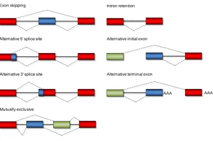
Exon skipping
Alternative 5’ splice site
Alternative 3’ splice site
Intron retention
Alternative initial exon
Mutually exclusive
Alternative terminal exon
AAA AAA
Figure 1.2. Major forms of alternative splicing. These modes are responsible for the
[image:20.612.115.535.233.512.2]1.4. Elements of a “splicing code”
The auxiliary elements that make up the splicing code include exonic and intronic splicing enhancers (ESEs and ISEs, respectively), which aid in exon recognition, and exonic and intronic splicing silencers (ESSs and ISSs, respectively), which suppress exon inclusion (Figure 1.3)11,19. These splicing regulatory elements (SREs) generally function by recruiting trans-acting splicing factors that activate or suppress splice site recognition or spliceosome assembly19. Mutating, removing, and/or shifting the location of these sequences will affect the overall splicing pattern of a transcript25. In contrast to the canonical splice sites whose sequence and position are well characterized, the complex code formed by these auxiliary SREs is only partially understood. In addition, the regulatory proteins that interact with these specific sequences to either stimulate or repress exon recognition have been only partially elucidated. Therefore the generation of a complete “splicing code” will require the elucidation of all types of SREs and the
corresponding trans-acting factors that regulate them.
Exon
U1 U2snRNP
U2AF65 35
BPS PY tract
ESE
SRp
5’ss 3’ss
Exon
hnRNP
ESS ISS ISE
ISS ISE
Figure 1.3. Elements of a “splicing code”. The diagram illustrates regulated splicing.
adjacent splice sites. Splicing is regulated by cis-elements (ESEs, ESSs, ISSs, and ISEs) and the trans-acting factors that bind them (SR proteins, hnRNP, and unknown factors).
The most well characterized family of regulatory proteins that bind both intronic and exonic cis-elements are the serine/arginine-rich (SR) and SR-like proteins. The human SR family contains ten identified members26 that are thought to mediate interactions between splicing factors bound to the 5‟ and 3‟ splice sites. All SR proteins
have a modular structure and contain either one or two copies of an RNA-recognition motif (RRM) and a C-terminal end highly enriched in arginine and serine dipeptides (RS domain)27. The RRMs serve to mediate sequence-specific binding to the RNA, which determines substrate specificity, and the RS domains are involved in protein-protein interactions that are essential for the recruitment of the splicing machinery5. Most exonic splicing enhancers (ESEs) are purine rich and different ESEs are recognized by various subsets of SR proteins8. Additionally, SR protein functions have been extended to mRNA export28, mRNA stability29, protein translation30, and nuclear export31.
binding to the transcript35. These factors can often block essential interactions between spliceosome components to inhibit splicing36,37.
Overexpression of SR proteins and hnRNP proteins has been shown to affect the splicing patterns of alternatively spliced pre-mRNAs in vivo38. Most hnRNP and SR proteins shuttle continuously between the nucleus and cytoplasm and consequently their subcellular distribution can shift in response to stress signals9. Recent studies have also demonstrated that the relative amounts of SR and hnRNP A/B proteins are important in regulating patterns of alternative splicing in a tissue-specific and developmentally-regulated manner39 . The expression of these proteins is unique to each cell type and thus the regulation of expression and activity of these proteins is critical for normal alternative splicing and cellular function. Moreover, a diverse set of diseases are associated with changes in expression of trans-acting splicing factors40. Disease-related changes in splicing factors are potentially useful biomarkers for disease diagnosis and classification. It has been suggested that the modulation of the relative stoichiometries of splicing factors can be used to regulate the alternative splicing of disease-relevant mRNAs9. As such, high-throughput small molecule screens are currently being developed to select for agents that modulate splicing factor ratios towards targeted therapies41.
1.5. Nonsense-mediated decay is a surveillance pathway in eukaryotes
The NMD pathway targets aberrant transcripts and removes potentially harmful truncated versions of proteins43. A recent investigation of the human genome sequence and databases of expressed sequence tags (ESTs) identified a coupling between NMD and alternative splicing3. NMD substrates from aberrant splicing processes include transcripts with retained introns, skipped exons and extended 5‟ or 3‟ UTRs44,45
. NMD specifically targets transcripts containing premature termination codons (PTCs) that can be introduced into transcripts as a result of DNA rearrangements, frameshifts, nonsense mutations or errors during transcription and splicing45,46. In higher eukaryotes, PTCs are recognized when they occur 50–55 nucleotides (nt) upstream of an exon-exon junction (Figure 1.4)47. The current model of the NMD mechanism is that during pre-mRNA processing the spliceosome deposits exon junction complexes (EJCs) at sites of intron removal. During the first round of translation the ribosome displaces the EJCs in its path and then dissociates from the mRNA at the stop codon45. However, if a PTC is present the ribosome will stop and will fail to remove any EJCs downstream from the PTC. Interactions between EJC proteins and several release factors48 trigger mRNA decay through a deadenylation-dependent pathway49.
disease phenotypes. Therefore, novel therapeutic approaches targeting NMD may be able to modify diseases with similar genetic phenotypes6. Therapeutic strategies targeting the coupling of NMD with alternative splicing may be also used to fine tune levels of specific trans-factors including splicing factors and other RNA binding proteins24.
3’ most exon-exon junction
Ter
Cap (A)n
PTC
50-55 nt
NMD No NMD
Figure 1.4. NMD and the position of the exon junction. Only the 3‟-most
exon-exon junction within a generic mammalian mRNA is shown. A PTC that is located in the region indicated in green, which is followed by an exon-exon junction more than 50–55 nt downstream, elicits NMD, whereas a PTC that is located in the region indicated in blue fails to elicit NMD. The normal termination codon (Ter) usually resides in the 3‟-most
exon.
1.6. Alternative splicing and human disease
are found to affect splicing43. Mutations affect splicing by disrupting canonical splice sites and auxiliary elements, by creating cryptic splice sites or by altering RNA secondary structure52. Mutations that alter trans-acting factors may result in global splicing defects which can have very specific phenotypic outputs53. Therefore, understanding the potential effects of single-nucleotide mutations that alter pre-mRNA splicing will enable researchers to develop new therapeutics and treatments that target certain genetic diseases and a variety of cancers46,54. Moreover, further elucidation of the cis-acting elements that regulate alternative splicing is needed to determine the extent of which single nucleotide polymorphisms (SNPs) modulate splicing through these elements.
1.7. Trans- and cis-acting technologies that alter pre-mRNA splicing
acid (PNA) hybrid containing RS dipeptide repeats coupled with an antisense oligonucleotide targeting a mutated ESE in the SMN2 gene to correct splice site selection
56
. ESSENCE was also demonstrated on the BRCA1 gene and was successful at suppressing the effects of a mutation on exon skipping in vitro.
SMN2 pre-mRNA transcripts have also been targeted by another AO approach which uses a tailed bi-functional antisense oligonucleotide57. In this approach, the antisense portion of the oligonucleotide targets the molecule to exon 7 in SMN2 and the tailed portion contains an ESE element such that splicing factors will be recruited to influence splice site selection. The latter technology was tested successfully in vitro and
in vivo57,58. RNAi approaches may also be used to eliminate aberrantly spliced mRNAs by targeting specific isoforms43. However, the applications of these and other antisense technologies are limited because of off-target effects, toxicity, efficiency, and issues with delivery.
studies have employed cell-based selection strategies, these screens were employed on a modest scale and have only examined the splicing patterns of a limited set of genes. The potential of small molecule based strategies targeting alternative splicing can be determined with the development of more robust, quantitative and specific cell-based splicing assays9.
In addition to trans-acting technologies, cis-acting RNA-based regulatory systems have been developed towards the regulation of pre-mRNA splicing in yeast and mammalian cells62. Regulation of splicing in these systems is exerted by small molecule responsive aptamers which were inserted into the intronic regions of spliced transcripts. The aptamer sensing components used in these platforms were shown to not be modular and consequently these systems have not been extended to sense other biomolecules. The adaptation of these platforms towards therapeutic applications have also been hampered by the lack of existing aptamers with suitable pharmacological properties, such as limited cell toxicity62. The extension of these designs towards applications in health and medicine may be realized with the identification of new aptamers and with the modification of these platforms to be responsive to protein biomarkers.
1.8. Cis-acting regulators of alternative splicing
sequence data68–70. Identified ESSs are able to control the selection of alternative 5‟ and 3‟ ss when placed between competing sites71
. Results from these in vivo studies demonstrate that not all RNA sequences that have been selected against SR proteins are splicing enhancers, underlining the importance of functional screens. Some of the selected exonic splicing regulators displayed significant similarities to naturally occurring regulators, whereas others were novel. From these and other results, it has been suggested that most exons are likely to have multiple regulators (ESEs and ESSs) that act as weak splicing signals which have an additive effect on splicing26,64. ESEs and ESSs also play critical roles in directing splicing to consensus splice sites rather than decoy sites72-74. However, few exonic regulators may be strong enough on their own to regulate splicing individually65.
In contrast, fewer ISEs and ISSs have been characterized and little is known regarding the auxiliary factors by which they are bound. These elements are generally short, variable in sequence, individually weak and present in multiple copies38. Several common intronic regulator motifs have been observed, including the GGG triplet75,76, purine-rich motifs10 or polypyrimidine tracts present in the 3‟ intronic regions10; however, most newly found auxiliary elements tend to be quite degenerate77. ISSs and ISEs have been identified near alternatively spliced exons and their mechanistic actions appear to be antagonistic78. ISSs may inhibit exon inclusion by recruiting splicing repressors, which directly antagonize splicing factor binding, or by recruiting repressors to multiple binding sites resulting in a „zone of silencing‟19
ISSs have been shown to inhibit the inclusion of pseudoexons into mature mRNAs79. Several ISEs have been characterized80, however the nuclear factors that regulate alternative splicing through these sequences have not been elucidated81. Further adding to these complexities, are observations that some intronic splicing regulatory elements (ISREs) act as inhibitors upstream of a specific splice site and as enhancers downstream of that splice site81. These results support a role for intronic elements in regulating splicing patterns in a combinatorial manner 8. Despite the widespread importance of ISREs, a systematic experimental characterization or iterative functional screen has yet to be applied towards developing a functional definition of these elements.
1.9. Engineering cis-acting intronic regulators of alternative splicing
Cis-acting regulators of alternative splicing play key roles in regulating the form and function of protein isoforms produced from a given gene in response to various signals received by the cell. Therefore, the ability to program alternative splicing patterns will provide a powerful tool to interrogate and manipulate cellular function. Despite the critical role of alternative splicing in creating phenotypic complexity and regulating gene expression, the sequence composition and function of ISREs have not been well elucidated. As a result of this lack of knowledge around how alternative splicing events are encoded at a genetic level, researchers very rarely incorporate control of alternative splicing events in synthetic genetic networks.
high-throughput in vivo screen for ISRE function. This high-throughput approach combined a systematic screening strategy with extensive genome-wide bioinformatic analyses and experimental characterization. The implementation of this strategy yielded insight into the sequence composition of ISREs, the splicing regulatory networks (SRNs) associated with these sequences and the mechanisms in which they achieve regulation. Chapter III describes an extension of this platform to support the construction of protein-responsive alternative splicing regulatory elements based on the integration of protein-binding RNA aptamers into key intronic locations of a target alternatively spliced transcript. This protein-responsive platform was adapted to detect disease biomarkers, reprogram natural signaling pathways, and control biologically-relevant processes, such as apoptosis, in response to increased signaling through pathways associated with disease. The molecular platforms described in Chapters 2 and 3 represent powerful tools to regulate alternative splicing events and thus gene expression. In addition, the ability to reprogram biological function in response to endogenous protein levels has broad applications in health and medicine, where such molecular tools can provide the basis for the design of targeted “intelligent” therapeutics. Chapter 4 provides a perspective on the general applications of
such genetically encoded technologies and future work needed to further characterize these synthetic regulatory systems.
References
1. Maquat, L.E. & Carmichael, G.G. Quality control of mRNA function. Cell 104,
2. Maniatis, T. & Reed, R. An extensive network of coupling among gene expression machines. Nature416, 499–506 (2002).
3. Lewis, B.P., Green, R.E. & Brenner, S.E. Evidence for the widespread coupling of alternative splicing and nonsense-mediated mRNA decay in humans. Proc Natl Acad Sci U S A100, 189–192 (2003).
4. Wang, Z. & Burge, C.B. Splicing regulation: from a parts list of regulatory elements to an integrated splicing code. Rna14, 802–813 (2008).
5. Cartegni, L., Chew, S.L. & Krainer, A.R. Listening to silence and understanding nonsense: exonic mutations that affect splicing. Nat Rev Genet3, 285–298 (2002). 6. Nissim-Rafinia, M. & Kerem, B. Splicing regulation as a potential genetic
modifier. Trends Genet18, 123–127 (2002).
7. Goldstrohm, A.C., Greenleaf, A.L. & Garcia-Blanco, M.A. Co-transcriptional splicing of pre-messenger RNAs: considerations for the mechanism of alternative splicing. Gene277, 31–47 (2001).
8. Smith, C.W. & Valcarcel, J. Alternative pre-mRNA splicing: the logic of combinatorial control. Trends Biochem Sci25, 381–388 (2000).
9. Cooper, T.A., Wan, L. & Dreyfuss, G. RNA and disease. Cell 136, 777–793 (2009).
10. Hastings, M.L. & Krainer, A.R. Pre-mRNA splicing in the new millennium. Curr Opin Cell Biol13, 302–309 (2001).
11. Black, D.L. Mechanisms of alternative pre-messenger RNA splicing. Annu Rev Biochem72, 291–336 (2003).
12. Maniatis, T. & Tasic, B. Alternative pre-mRNA splicing and proteome expansion in metazoans. Nature418, 236–243 (2002).
13. Modrek, B., Resch, A., Grasso, C. & Lee, C. Genome-wide detection of alternative splicing in expressed sequences of human genes. Nucleic Acids Res29,
2850–2859 (2001).
14. Woodley, L. & Valcarcel, J. Regulation of alternative pre-mRNA splicing. Brief Funct Genomic Proteomic1, 266–277 (2002).
16. Wang, E.T. et al. Alternative isoform regulation in human tissue transcriptomes.
Nature456, 470–476 (2008).
17. Ledford, H. Human genes are multitaskers. Nature456, 9 (2008). 18. Multitudes of messages. Nat Genet40, 1385 (2008).
19. Matlin, A.J., Clark, F. & Smith, C.W. Understanding alternative splicing: towards a cellular code. Nat Rev Mol Cell Biol6, 386–398 (2005).
20. Lim, L.P. & Burge, C.B. A computational analysis of sequence features involved in recognition of short introns. Proc Natl Acad Sci U S A 98, 11193–11198 (2001).
21. Cote, J., Dupuis, S. & Wu, J.Y. Polypyrimidine track-binding protein binding downstream of caspase-2 alternative exon 9 represses its inclusion. J Biol Chem 276, 8535–8543 (2001).
22. Zhang, X.H., Heller, K.A., Hefter, I., Leslie, C.S. & Chasin, L.A. Sequence information for the splicing of human pre-mRNA identified by support vector machine classification. Genome Res13, 2637–2650 (2003).
23. Sun, H. & Chasin, L.A. Multiple splicing defects in an intronic false exon. Mol Cell Biol20, 6414–6425 (2000).
24. Blencowe, B.J. Alternative splicing: new insights from global analyses. Cell 126,
37–47 (2006).
25. Graveley, B.R., Hertel, K.J. & Maniatis, T. A systematic analysis of the factors that determine the strength of pre-mRNA splicing enhancers. Embo J 17, 6747– 6756 (1998).
26. Graveley, B.R. Sorting out the complexity of SR protein functions. Rna6, 1197– 1211 (2000).
27. Chandler, S.D., Mayeda, A., Yeakley, J.M., Krainer, A.R. & Fu, X.D. RNA splicing specificity determined by the coordinated action of RNA recognition motifs in SR proteins. Proc Natl Acad Sci U S A94, 3596–3601 (1997).
28. Maatta, A.M. et al. Non-small cell lung cancer as a target disease for herpes simplex type 1 thymidine kinase-ganciclovir gene therapy. Int J Oncol 24, 943– 949 (2004).
30. Sanford, J.R., Gray, N.K., Beckmann, K. & Caceres, J.F. A novel role for shuttling SR proteins in mRNA translation. Genes Dev18, 755–768 (2004). 31. Caceres, J.F., Misteli, T., Screaton, G.R., Spector, D.L. & Krainer, A.R. Role of
the modular domains of SR proteins in subnuclear localization and alternative splicing specificity. J Cell Biol138, 225–238 (1997).
32. Zhu, J., Mayeda, A. & Krainer, A.R. Exon identity established through differential antagonism between exonic splicing silencer-bound hnRNP A1 and enhancer-bound SR proteins. Mol Cell8, 1351–1361 (2001).
33. Valcarcel, J. & Gebauer, F. Post-transcriptional regulation: the dawn of PTB.
Curr Biol7, R705–708 (1997).
34. Wagner, E.J. & Garcia-Blanco, M.A. Polypyrimidine tract binding protein antagonizes exon definition. Mol Cell Biol21, 3281–3288 (2001).
35. Caceres, J.F. & Kornblihtt, A.R. Alternative splicing: multiple control mechanisms and involvement in human disease. Trends Genet 18, 186–193 (2002).
36. Izquierdo, J.M. et al. Regulation of Fas alternative splicing by antagonistic effects of TIA-1 and PTB on exon definition. Mol Cell19, 475–484 (2005).
37. Sharma, S., Falick, A.M. & Black, D.L. Polypyrimidine Tract Binding Protein Blocks the 5' Splice Site-Dependent Assembly of U2AF and the Prespliceosomal E Complex. Mol Cell19, 485–496 (2005).
38. Ladd, A.N. & Cooper, T.A. Finding signals that regulate alternative splicing in the post-genomic era. Genome Biol3, reviews0008 (2002).
39. Hanamura, A., Caceres, J.F., Mayeda, A., Franza, B.R., Jr. & Krainer, A.R. Regulated tissue-specific expression of antagonistic pre-mRNA splicing factors.
Rna4, 430–444 (1998).
40. Ule, J. Ribonucleoprotein complexes in neurologic diseases. Current opinion in neurobiology18, 516–523 (2008).
41. Stoilov, P., Lin, C.H., Damoiseaux, R., Nikolic, J. & Black, D.L. A high-throughput screening strategy identifies cardiotonic steroids as alternative splicing modulators. Proc Natl Acad Sci U S A105, 11218–11223 (2008).
42. Green, R.E. et al. Widespread predicted nonsense-mediated mRNA decay of alternatively-spliced transcripts of human normal and disease genes.
43. Wang, G.S. & Cooper, T.A. Splicing in disease: disruption of the splicing code and the decoding machinery. Nat Rev Genet8, 749–761 (2007).
44. Hilleren, P. & Parker, R. mRNA surveillance in eukaryotes: kinetic proofreading of proper translation termination as assessed by mRNP domain organization? Rna 5, 711–719 (1999).
45. Chang, Y.F., Imam, J.S. & Wilkinson, M.F. The nonsense-mediated decay RNA surveillance pathway. Annu Rev Biochem76, 51–74 (2007).
46. Cartegni, L. & Krainer, A.R. Correction of disease-associated exon skipping by synthetic exon-specific activators. Nat Struct Biol10, 120–125 (2003).
47. Maquat, L.E. Nonsense-mediated mRNA decay: splicing, translation and mRNP dynamics. Nat Rev Mol Cell Biol5, 89–99 (2004).
48. Czaplinski, K. et al. The surveillance complex interacts with the translation release factors to enhance termination and degrade aberrant mRNAs. Genes Dev 12, 1665–1677 (1998).
49. Lejeune, F., Li, X. & Maquat, L.E. Nonsense-mediated mRNA decay in mammalian cells involves decapping, deadenylating, and exonucleolytic activities. Mol Cell12, 675–687 (2003).
50. Culbertson, M.R. RNA surveillance. Unforeseen consequences for gene expression, inherited genetic disorders and cancer. Trends Genet 15, 74–80 (1999).
51. Holbrook, J.A., Neu-Yilik, G., Hentze, M.W. & Kulozik, A.E. Nonsense-mediated decay approaches the clinic. Nat Genet36, 801–808 (2004).
52. Pagani, F. & Baralle, F.E. Genomic variants in exons and introns: identifying the splicing spoilers. Nat Rev Genet5, 389–396 (2004).
53. Garcia-Blanco, M.A., Baraniak, A.P. & Lasda, E.L. Alternative splicing in disease and therapy. Nat Biotechnol22, 535–546 (2004).
54. Venables, J.P. et al. Identification of alternative splicing markers for breast cancer. Cancer Res68, 9525–9531 (2008).
55. Singh, R.N. Evolving concepts on human SMN pre-mRNA splicing. RNA biology 4, 7–10 (2007).
56. Khoo, B., Akker, S.A. & Chew, S.L. Putting some spine into alternative splicing.
57. Skordis, L.A., Dunckley, M.G., Yue, B., Eperon, I.C. & Muntoni, F. Bifunctional antisense oligonucleotides provide a trans-acting splicing enhancer that stimulates SMN2 gene expression in patient fibroblasts. Proc Natl Acad Sci U S A 100,
4114–4119 (2003).
58. Baughan, T.D., Dickson, A., Osman, E.Y. & Lorson, C.L. Delivery of bifunctional RNAs that target an intronic repressor and increase SMN levels in an animal model of spinal muscular atrophy. Hum Mol Genet18, 1600–1611 (2009). 59. Soret, J. et al. Selective modification of alternative splicing by indole derivatives
that target serine-arginine-rich protein splicing factors. Proc Natl Acad Sci U S A 102, 8764–8769 (2005).
60. Fukuhara, T. et al. Utilization of host SR protein kinases and RNA-splicing machinery during viral replication. Proc Natl Acad Sci U S A 103, 11329–11333 (2006).
61. Muraki, M. et al. Manipulation of alternative splicing by a newly developed inhibitor of Clks. J Biol Chem279, 24246–24254 (2004).
62. Suess, B. & Weigand, J.E. Engineered riboswitches: overview, problems and trends. RNA Biol5, 24–29 (2008).
63. Coulter, L.R., Landree, M.A. & Cooper, T.A. Identification of a new class of exonic splicing enhancers by in vivo selection. Mol Cell Biol 17, 2143–2150 (1997).
64. Liu, H.X., Zhang, M. & Krainer, A.R. Identification of functional exonic splicing enhancer motifs recognized by individual SR proteins. Genes Dev12, 1998–2012 (1998).
65. Schaal, T.D. & Maniatis, T. Multiple distinct splicing enhancers in the protein-coding sequences of a constitutively spliced pre-mRNA. Mol Cell Biol 19, 261– 273 (1999).
66. Tian, H. & Kole, R. Strong RNA splicing enhancers identified by a modified method of cycled selection interact with SR protein. J Biol Chem 276, 33833– 33839 (2001).
67. Wang, Z. et al. Systematic identification and analysis of exonic splicing silencers.
Cell119, 831–845 (2004).
68. Fairbrother, W.G., Yeh, R.F., Sharp, P.A. & Burge, C.B. Predictive identification of exonic splicing enhancers in human genes. Science297, 1007–1013 (2002). 69. Fairbrother, W.G. et al. RESCUE-ESE identifies candidate exonic splicing
70. Zhang, X.H. & Chasin, L.A. Computational definition of sequence motifs governing constitutive exon splicing. Genes Dev18, 1241–1250 (2004).
71. Wang, Z., Xiao, X., Van Nostrand, E. & Burge, C.B. General and specific functions of exonic splicing silencers in splicing control. Mol Cell 23, 61–70 (2006).
72. Pozzoli, U. & Sironi, M. Silencers regulate both constitutive and alternative splicing events in mammals. Cell Mol Life Sci62, 1579–1604 (2005).
73. Zhang, X.H., Kangsamaksin, T., Chao, M.S., Banerjee, J.K. & Chasin, L.A. Exon inclusion is dependent on predictable exonic splicing enhancers. Mol Cell Biol25,
7323–7332 (2005).
74. Zhang, X.H., Leslie, C.S. & Chasin, L.A. Computational searches for splicing signals. Methods37, 292–305 (2005).
75. McCullough, A.J. & Berget, S.M. An intronic splicing enhancer binds U1 snRNPs to enhance splicing and select 5' splice sites. Mol Cell Biol 20, 9225– 9235 (2000).
76. Brudno, M. et al. Computational analysis of candidate intron regulatory elements for tissue-specific alternative pre-mRNA splicing. Nucleic Acids Res 29, 2338– 2348 (2001).
77. Blencowe, B.J. Exonic splicing enhancers: mechanism of action, diversity and role in human genetic diseases. Trends Biochem Sci25, 106–110 (2000).
78. Charlet, B.N., Logan, P., Singh, G. & Cooper, T.A. Dynamic antagonism between ETR-3 and PTB regulates cell type-specific alternative splicing. Mol Cell9, 649– 658 (2002).
79. Fairbrother, W.G. & Chasin, L.A. Human genomic sequences that inhibit splicing. Mol Cell Biol20, 6816–6825 (2000).
80. Yeo, G., Hoon, S., Venkatesh, B. & Burge, C.B. Variation in sequence and organization of splicing regulatory elements in vertebrate genes. Proc Natl Acad Sci U S A101, 15700–15705 (2004).
Chapter II. Functional selection of intronic splicing elements provides insight into their regulatory mechanism
Abstract
2.1. Introduction
Post-transcriptional gene regulatory mechanisms play central roles in programming the complexity of biological systems. One such process is alternative splicing, a dynamic mechanism that produces multiple protein isoforms from a single gene by altering the ways in which exons are joined from a single pre-mRNA1. Splicing patterns are regulated by the interplay between auxiliary cis-acting elements that include exonic and intronic splicing enhancers (ESEs and ISEs, respectively) and exonic and intronic splicing silencers (ESSs and ISSs, respectively) and the trans-acting factors that modulate them, leading to a ‘splicing code’2
. The lack of high-throughput in vivo
Several properties of ISREs have complicated their functional characterization. ISSs and ISEs have been identified near alternatively spliced exons; however, their actions appear to be antagonistic12 suggesting that they behave in a combinatorial manner13. In addition, the activities of some sequences are context dependent10,14. ISSs may inhibit exon inclusion by recruiting splicing repressors that directly antagonize splicing factor binding or by recruiting repressors to multiple binding sites resulting in a ‘zone of silencing’15
. While several ISEs have been characterized16, the trans-acting factors that bind these sequences remain unknown17.
2.2. Results
2.2.1.SPLICE: a Screening PLatform for Intronic Control Elements
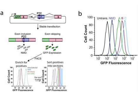
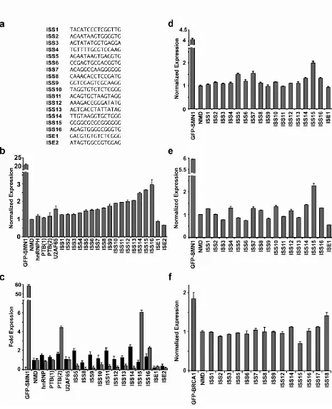
SPLICE is a high-throughput in vivo screen for ISRE function based on a reporter construct encoding the green fluorescent protein (GFP) fused 5’ of a three-exon,
two-intron mini-gene. The alternatively-spliced middle exon harbors a premature termination codon (PTC) that triggers mRNA degradation through the NMD pathway18. Auxiliary elements that regulate alternative splicing are normally positioned in proximity to splice sites16,19,20 and have been shown to vary in length between 10- to 30-nt19. We implemented SPLICE with the SMN1 mini-gene containing a random 15-nucleotide (nt) library positioned 45-nt upstream of the 3’ ss in the first intron (Figure 2.1a). Therefore, cells with a high level of exon 7 inclusion display lower GFP fluorescence than cells in which this exon is excluded. By coupling NMD to splicing efficiency, ISREs with a range of activities can be selected using fluorescence activated cell sorting (FACS).
A library of synthetic DNA oligonucleotides containing a random 15-nt region (~1x10⁹ sequences) was ligated into the NMD control construct and transformed into
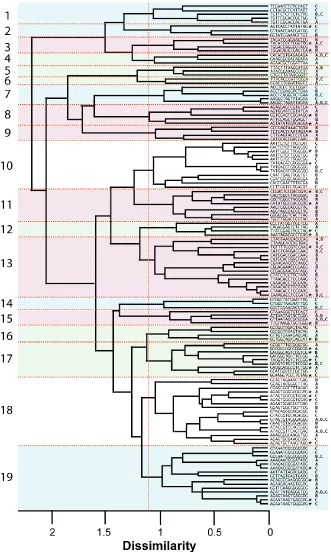
2.2.2.Recovered ISRE sequence composition correlates with sorted sections
We identified 125 unique sequences with enhanced fluorescence from 480 sequenced isolates (Table S2.1). Three of the recovered ISRE sequences exhibit significant (12 of 15-nt) similarity to portions of the SMN1 mini-gene, suggesting that these sequences may be involved in the cooperative assembly of repressor elements on the SMN1 transcript (Figure S2.3). The sequences have a higher level of G (35.8%) and reduced levels of T (22%), C (18.6%), and A (23.6%) (Figure S2.4a). The dinucleotide CC is overrepresented in the ISRE dataset, while others, such as AC, AG, CA, GT, TA, TC, and TG, are only slightly enriched (Figure S2.4b).
40% sequence representation from one sorted section are indicated (A, green; B, red; C, blue). Sorted sections for each sequence are denoted. Starred sequences were subjected to additional studies to examine regulatory activity.
2.2.3. GCCS clustering of recovered ISREs identifies motifs similar to known splicing
factor binding sites
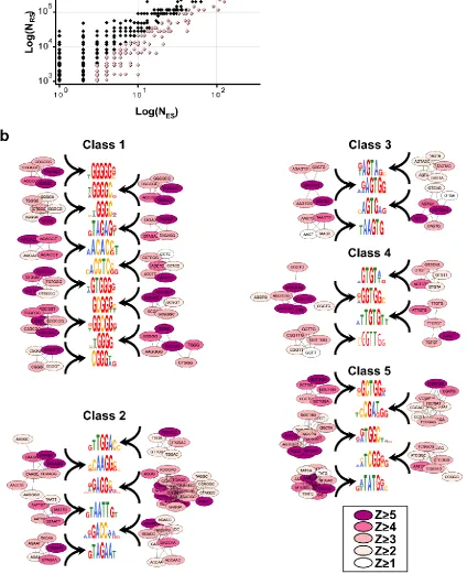
The sequence alignment results indicate that multiple motifs of varying lengths occur within the 15-nt dataset (Figure S2.5). For analyzing datasets of this nature, Graph Clustering by Common Substrings (GCCS)21 is better suited than hierarchical clustering. We analyzed a 19-nt region including the 15-mer ISRE sequence and 2-nt of the flanking regions for sequence enrichment. Since RNA binding proteins typically recognize short sequence motifs, we restricted our analysis to n-mers ranging from 4–6-nt. We determined the enrichment of n-mers in a sample of 125 sequences using a confidence interval for the binomial distribution based on probabilities expected for 19-nt oligonucleotides containing 15-nt of uniformly random bases flanked by the 2 constant bases present in the experimental system. In the ISRE dataset, 241 n-mers consisting of 39 4-mers, 93 5-mers, and 109 6-mers were significantly enriched (α1-tailed = 0.1; Figure
2.3a and Table S2.2). The GCCS analysis grouped 80.1% of the statistically enriched 4-6-nt n-mers into 30 consensus motif clusters (Figure 2.3b and Tables S2.3 and S2.4, Methods).
particular, class 1 motifs resemble binding sites for several known repressors of splicing: hnRNP A1 (TAGGG)22, hnRNP F/H (GGGGG)23, the polypyrimidine tract binding protein PTB (hnRNP I, CT-rich)24 and hnRNP L (CA-rich)25. The significant similarity between binding sites for the hnRNP family of proteins and the enriched motifs supports the possible functional role of selected ISREs.
Several identified motifs resemble known binding sites for the SR protein family (class 2) whose members act as general splicing factors1. Enriched ISREs within class 2 resemble binding sites for SF2/ASF (GAAGAA)26, SRp40 (ACAAG)27, SRp30c (CTGGATT)14, SC35 (AGGAGAT)28, 9G8 (GACC)28, and Tra2β (GAA)n29. While the
examples of SR proteins involvement in splicing repression are limited, the enrichment of motifs similar to binding sites for members of this family suggests that their role in intronic regulation may be more widespread than previously thought.
Several of the enriched motifs identified in our dataset resemble the major 5’ splice site (ss) consensus sequence GT[A/G]AGT (class 3)30. All four motifs in class 3 contain an AGT core element, and the enriched motif TAAGTG is almost identical to the canonical 5’ ss sequence and the hnRNP G binding motif AAGT31. The occurrence of 5’ ss motifs within intronic regulatory elements has been noted32 and computational analyses have identified conserved elements that are similar to the consensus 5’ ss within
mammalian intronic regions21,33. In addition, the enrichment of 5’ ss motifs was previously observed in an in vivo screen for ESSs9. Taken together, these results add support to the role of cryptic 5’ ss in regulating alternative splicing.
proteins regulates alternative splicing patterns by binding sequences that contain CTG repeats and exhibit a higher affinity for GT repeats34. The motifs GTGT and TGTG resemble binding sites to a well-characterized member of this family, CUG-BP1, which has been shown to bind TGT-containing sequences34. The GTGT motif may also serve as a binding site for hnRNP M35.
frequency of all 4–6-nt n-mers in the enriched sample set (NES) vs. a corresponding
random sample set (NRS) (black). A similar scatter-plot based on n-mers determined to be
significantly enriched in the recovered ISREs is overlaid (pink). (b) Consensus motif groupings according to resemblance to binding sites for trans-acting splicing factors. Motif classes include enriched ISREs that are similar to the binding sites of the hnRNP, SR and CELF families of proteins and the 5’ ss (classes 1–4, respectively). Class 5
consists of previously unidentified elements and may represent novel regulatory sequences. The graph clusters representing the enriched n-mers used to construct each consensus motif are shown. Vertices are colored according to the enrichment Z-scores.
2.2.4. Enriched ISRE n-mers resemble known splicing regulatory elements
and weak PY elements are G-rich elements, similar to hnRNP A/B and hnRNP F/H binding sites and the canonical 5’ ss. The results suggest that the selected elements may
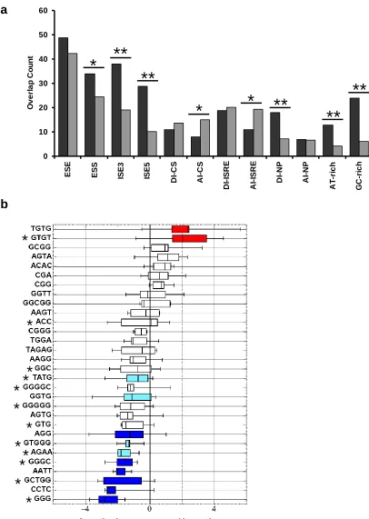
function as general splicing silencers and intronic modulators of splicing (as both silencers and enhancers) depending on their context across various cell types and are likely regulated by general splicing factors. The comparison between enriched pentamers and conserved acceptor intronic elements (AI) for CIS and ISRE datasets demonstrate some overlap (P < 0.05). However, the observed overlap is far less than expected, suggesting that SPLICE selected against these elements.
2.2.5. Genome-wide analysis demonstrates that enriched ISREs associate with spliced
exons
The biological relevance of selected motifs was examined by assessing the association of enriched motifs with naturally occurring alternative and constitutive splicing events. The occurrence of enriched motifs in the region 80-nt upstream of the AI regions flanking skipped exons was determined using a database of alternatively spliced junctions throughout the human genome21. A portion of SPLICE-generated ISREs significantly associate with alternative splicing (2 of 30; Pt-test < 0.01; class 4 only) and
constitutive splicing (10 of 30; Pt-test < 0.05; all classes except 3) (Figure 2.4b). The entire
population of consensus n-mers significantly associates with constitutive splicing (Pt-test =
0 10 20 30 40 50 60
ESE ESS ISE
3 ISE 5 D I-C S A I-C S D I-I SR E A I-I SR E D I-N P A I-N P A T -r ic h G C -r ic h O v e rl a p C o u n t
a
*
*
*
**
**
**
**
**
b
*
*
*
*
*
*
*
*
*
*
*
*
Figure 2.4. Enriched n-mers overlap with both experimentally and computationally
[image:53.612.116.522.80.651.2]enriched n-mers from recovered ISRE sequences with known classes of SREs. Observed (black) and expected overlap (gray) between datasets is shown. P-values derived from the chi-squared test of association are as follows: * P < 0.05 and ** P << 0.0001. (b) Box-plots revealing the distribution of TA-scores for GCCS derived ISREs. The GCCS consensus motifs that are significantly associated with alternative splicing are shown in red (Pt-test < 0.01) and those that are significantly associated with constitutive splicing are
shown in shades of blue (dark blue, Pt-test < 0.01; light blue, Pt-test < 0.05). In total, 9
consensus motifs are biased toward alternative splicing and 21 consensus motifs display a bias towards constitutive splicing. Elements exhibiting no significant association with either category are not shaded. Starred motifs are present in hexamers subjected to RNAi silencing studies to examine regulated splicing.
2.2.6. Recovered ISRE sequences enable tuning of alternative splicing
(P << 0.001) and 2 exhibit enhancer activity relative to the NMD control (P < 0.05) (Figure 2.5b), and over half exhibit silencing activities equal to or greater than the U2AF65 element. Similar trends were seen upon examination of an additional 12 recovered sequences (Figure S2.6b). In addition, we arranged the sequences into groups representing low (ISS1-5), medium (ISS6-10), and high (ISS11-16) silencing activities and determined the section from which each sequence was recovered (Figure 2.2). The activities of the majority of tested sequences correlated with sectioned populations, where a subset of enriched n-mers GGGGC, GGGC, and GGG correlated significantly with their sorted section (P << 0.01) and those sequences that did not correlate were shown to cluster with the appropriate group by sequence. These results support that functional regulatory activity is related to sequence.
All constructs except for the GFP-SMN1 control are expected to exhibit low levels of the exon 7 included isoform, as this isoform should be rapidly degraded through NMD. The GFP-SMN1 control exhibits a high level of exon 7 inclusion (99.7%), ~60-fold more than the NMD control. Exon inclusion levels for the ISS controls do not differ from the NMD control (P > 0.35), with the exception of PTB(2), which had a higher level of exon inclusion (P < 0.005). Exon inclusion levels for 8 of the 10 recovered ISS sequences (ISS5, ISS8-13) and the ISE sequences range from 2 to 20-fold less than the NMD control (P < 0.05), whereas ISS15 and ISS16 exhibited increased levels of the exon included isoform relative to the NMD control (P < 0.05, Figure S2.7e). The elevated levels of exon inclusion observed from several ISSs is not a result of cryptic splice sites as determined by analyzing the sizes of the RT-PCR amplification products (data not shown). Overall, the majority of sequences that display increased fluorescence have decreased levels of exon 7 inclusion compared to the NMD control, supporting their silencer function.
2.2.7. ISRE sequences function in a different cell type
display increased expression levels and population distributions relative to stable cell line assays (Figure S2.8). As such, the relative expression of the GFP-SMN1 control is only ~4.1-fold that of the NMD control (Figure 2.5d). Despite the decreased sensitivity of the transient transfection assay, the qualitative activity of 15 of the recovered ISREs was maintained and 11 sequences exhibited significantly increased expression (P < 0.05).
We next investigated whether the recovered sequences function in HeLa cells. The GFP-SMN1 control displays a approximate six fold higher level of expression than the NMD construct in HeLa cells in the transient transfection assay (Figure 2.5e). The ISRE sequences display a range of expression levels, but all are significantly different than the NMD control (P < 0.05). The majority of examined sequences (12 of 16) maintain the same trend in activity in HeLa cells as was observed in HEK-293 cells and ANOVA analysis of the activities in both cell lines shows a strong correlation (P < 0.0005). In contrast, four of the tested sequences (ISS3, 5, 6, and 13) exhibit enhancer activity relative to the NMD control in HeLa cells, which may be due to differences in levels of trans-acting factors between the cell lines. The results support that most sequences recovered from SPLICE retain function in a cell line different from which they were selected and may represent global splicing regulators.
2.2.8. Analysis of recovered ISRE sequences in a different transcript supports context
dependent function
context dependent function by examining their activity in a second NMD-based reporter, based on the BRCA1 gene consisting of exons 17, 18, and 1945, via transient transfection in HEK-293 cells. Selected ISRE sequences were inserted 50-nt upstream from the 3’ ss of exon 18. Analysis of the reporter constructs by flow cytometry reveals a approximate two fold difference between the positive and negative controls (P < 0.05, Figure 2.5f). Only 3 of the tested sequences (ISS14, 17 and 18) exhibit significant silencer activity (P
FLP-In stable cell lines generated for recovered ISRE sequences and control constructs. For all reported activities, the mean GFP levels from two independent experiments were determined and normalized to the NMD control. Normalized expression and average error are reported. ISRE sequences are labeled according to function. (c) qRT-PCR analysis of the ISS control sequences and 12 selected sequences with primer sets specific for exon 7 included (black bars) and excluded (gray bars) products. Expression levels of duplicate PCR samples were normalized to the levels of HPRT. Fold expression data is reported as the mean expression for each sample divided by the mean NMD expression value + the average error. (d) Flow cytometry analysis of recovered ISRE sequences and control constructs transiently transfected in HEK-293 cells. (e) Flow cytometry analysis of recovered ISRE sequences and control constructs transiently transfected in HeLa cells.
(f) Flow cytometry analysis of recovered ISRE sequences and control constructs in the BRCA1 mini-gene transiently transfected in HEK-293 cells.
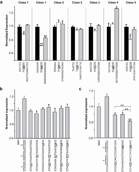
2.2.9. Analysis of enriched hexamers confirms independent and combinatorial function
We examined the silencer activities of representative hexamers from consensus motifs within the GCCS clusters through transient assays in HEK-293 cells to confirm the activity of individual motifs. Hexamers resembling the PTB, hnRNP H, SF2/ASF, Sam and the CELF protein binding sites, the 5’ ss and an unknown motif were examined
classes 1, 2, and 5). However, only one of the hexamers (class 4) displays an increase in silencer activity when present in duplicate. The results indicate that while individual hexamers exhibit silencing activity and likely represent core ISREs, they do not necessarily behave in an additive manner likely due to context and spacing requirements. For example, the duplicate hnRNP H hexamer does not exhibit increased silencing, whereas ISS15, which differs from the duplicate hexamer by 3 cytosine residues, exhibits strong silencer activity. The additional residues may provide spacing between the G-rich hexamers important for functional activity.
0.05), indicating that the regulatory function of ISS5 is likely not due to combinatorial recognition of motifs.
Representative hexamers from each class of GCCS clusters and corresponding mutant and duplicate sequences were characterized in transient transfection assays in HEK-293 cells. For all reported data, silencing activity was assessed by flow cytometry analysis, where the mean GFP levels from two independent experiments were normalized to the wild-type hexamer construct. Normalized expression and average error are reported. P
-values derived from the Student’s t-test are as follows: * P < 0.05 and ** P < 0.01. (b)
Mutational analysis of an ISS sequence supports the silencing activity of individual hexamer regions. The combined and individual activity of hexamer regions within the context of an ISS sequence was examined by introducing 2 point mutations into 3 regions, in combination and separately into all 3 hexamer regions of ISS5. (c) Mutational analysis of an ISS sequence supports the silencing activity of combined hexamer regions. The combined and individual activity of hexamer regions within the context of an ISS sequence was examined by introducing 2 point mutations separately and in combination into 2 hexamer regions of ISS8.
2.2.10. Splicing factor depletion influences ISRE regulated splicing in vivo
and displayed minimal effects on the other splicing factors examined (Figure S2.10a). Hexamers from classes 1 (ACCTCC, GGGGGG), 2 (GTAGAA), 4 (GCTGGG) and 5 (ATATGG), which harbor potential binding sites for the selected trans-acting factors, and a random insert control were subjected to the RNAi-based screen using a mini-gene lacking a PTC to avoid any siRNA-mediated effects on the NMD pathway.
We analyzed the splicing patterns of the hexamer and control constructs through qRT-PCR analysis. In the presence of the mock siRNA, four of the hexamers exhibit silencing activity, a higher ratio of exon exclusion to inclusion, relative to the GFP-SMN1 control (Figure 2.7b and Figure S2.10b). In contrast, one hexamer (GGGGGG) exhibits enhancer activity in the presence of the mock siRNA. Splicing of constructs containing the GCCS hexamers were significantly affected by the depletion of at least one, and in some cases multiple, trans-acting factors (Fig