ISSN Print: 2164-5531
DOI: 10.4236/ojanes.2017.710034 Oct. 31, 2017 341 Open Journal of Anesthesiology
A Comparative Study of Uses of Ephedrine by
Different Doses on Prevention of Hemodynamic
Changes Accompanied with Induction of
General Anesthesia through Propofol and
Fentanyl without Adverse Effects
Ayman Mohamady Eldemrdash, Mohammed Ahmed Mohammed Al-Azhary
Department of Anesthesiology, Faculty of Medicine, Aswan University, Aswan, Egypt
Abstract
Background: Propofol and fentanyl combination are common with general anesthesia. However, hypotension and bradycardia are common during in-duction of anesthetic. This study aimed to compare the response of different doses of ephedrine for attenuation of the hemodynamic changes after anes-thetic induction without adverse effects. Materials and Methods: This was a randomized, double-blinded, case-controlled clinical trial. One hundred and twenty adult patients were allocated into one of the four groups: receiving IV saline, ephedrine 0.05 mg/kg, 0.1 mg/kg, or 0.2 mg/kg respectively. Induction of anesthesia was done with propofol 3 mg/kg and fentanyl 1 mg/kg. Altera-tions in systolic and diastolic blood pressures (SBP, DBP), mean arterial pres-sure (MAP), and heart rate (HR) were calculated every 1 min after induction, and 2, 3, 4 and 5 min. Then, intubation was made. Results: Baseline hemody-namic variables were comparable between groups. Patients received 0.1 mg/kg, and 0.2 mg/kg had less drop in blood pressure both systolic and dias-tolic, MAP, and HR with no significant rise in side effects. The numbers of patients with hypotension were significantly lower in the group receiving ephedrine 0.2 mg/kg compared to other groups (P-value 0.05). Use of IV ephedrine at a dose of 0.1 mg/kg was shown to be useful for reduction of he-modynamic changes but did not eliminate the risk of blood pressure drop. Ephedrine 0.2 mg/kg was better without causing any adverse effects. We can conclude that ephedrine 0.1 mg/kg was suitable for minimizing or decreasing changes in hemodynamic at propofol-fentanyl induction but ephedrine 0.2 mg/kg was better without causing more adverse effects.
How to cite this paper: Eldemrdash, A.M. and Al-Azhary, M.A.M. (2017) A Compar-ative Study of Uses of Ephedrine by Dif-ferent Doses on Prevention of Hemody-namic Changes Accompanied with Induc-tion of General Anesthesia through Propo-fol and Fentanyl without Adverse Effects. Open Journal of Anesthesiology, 7, 341-350. https://doi.org/10.4236/ojanes.2017.710034
Received: September 16, 2017 Accepted: October 28, 2017 Published: October 31, 2017
Copyright © 2017 by authors and Scientific Research Publishing Inc. This work is licensed under the Creative Commons Attribution International License (CC BY 4.0).
DOI: 10.4236/ojanes.2017.710034 342 Open Journal of Anesthesiology
Keywords
Propofol, Fentanyl, Hypotension, Ephedrine
1. Introduction
Propofol (2.6 diisopropylphenol) is a rapidly acting ideal IV anesthetic drug widely used for induction of general anesthesia [1]. Fentanyl is widespread, commonly used short-acting analgesic agent, frequently used with propofol. The induction of general anesthesia with propofol has, however, been accompanied with a significant drop in the systolic arterial pressure [2]. The mechanism of hypotension caused by propofol is still not well understood. One of the explana-tions of the hypotensive effects of propofol was the reduction in systemic vascu-lar resistance due to the mixed venous and arterial vasodilatation [3]. Other possible mechanisms may include depression of myocardial contractility and impaired baroreflex [4] [5]. The cardiovascular effects of propofol are obviously increased when combined with fentanyl. Many approaches have been tried to limit this hypotension with unsettled evidence. Fluid preloading with colloid and crystalloid have been administered in various studies to prevent this hypoten-sion, other drugs also used as atropine, glycopyrrolate, ketamine, dopamine, dobutamine but with variable results [1]-[6].
Ephedrine is a non-catecholamine sympathomimetic alkaloid with potent al-pha and beta agonist and acts by both direct as well as indirect mechanism. Its cardiovascular effects include the increase in blood pressure, heart rate, contrac-tility, and cardiac output [7]. Ephedrine has been used to limit the hypotensive effects of induction of anesthesia with propofol and fentanyl [5] [6]. Ephedrine is a drug used to maintain blood pressure; mainly by increasing the cardiac out-put and increasing the heart rate as is it not a potent arterial vasoconstrictor. This may explain why high doses of intravenous ephedrine are accompanied with significant side effects such as reactive hypertension, which is usually con-sidered as systolic BP > 140 mmHg [8].
The purpose of this study was undertaken to compare the response of differ-ent doses of ephedrine to determine the most optimal dose of ephedrine for re-duction of the hemodynamic changes following anesthetic inre-duction with pro-pofol and fentanyl without causing significant side effects.
2. Patients and Methods
DOI: 10.4236/ojanes.2017.710034 343 Open Journal of Anesthesiology
estimated difficult airway, those with morbid obesity (BMI > 35) and pregnant females were excluded from the study.
Patients were assigned using sealed envelope technique into four groups, no drug or (normal saline) control group (group-A), 0.05 ml/kg of ephedrine (group-B), 0.1 mg/kg of ephedrine (group-C) or 0.2 mg/kg of ephedrine (group-D). The patients received no premedication.
In the anesthetic room, wide bore intravenous access was established. In the operating room, routine monitoring HR, ECG, SPO2, and NIBP were estab-lished. Baseline cardiovascular parameters, i.e., heart rate, blood pressure (sys-tolic, diastolic and mean) and oxygen saturation were recorded. Noninvasive blood pressure was measured. Patients received normal saline, ephedrine 0.05, 0.1, 0.2 mg/kg just 1 min before induction diluted in 10 cc normal saline by another person. Anesthesia was induced with fentanyl 1 - 2 μg/kg followed by propofol 2 - 3 mg/kg injected over 30 sec. Patients were given atracurium besy-late 0.5 mg/kg as a muscle relaxant. We calcubesy-lated the hemodynamic variable (arterial blood pressure, heart rate and oxygen saturation) every minute, starting 1 min after induction 2, 3, 4, 5 min (till 5 min after injection of propofol). In this period, bag and mask ventilation were used to maintain SPO2 > 95%, and no endotracheal intubation was done.
After the study period, patients were intubated, and anesthesia was continued as required. Hypotension (SBP < 20% of baseline) was treated with rapid infu-sions of ringers lactate 15 - 20 ml/min.
The statistical analysis of qualitative data was done by using Chi-square test. The quantitative data were analyzed by using one-way ANOVA test. A P-value less than 0.05 was considered statistically significant. Quantitative data were presented as mean (±SD) while qualitative data were presented as numbers and percentages. Statistical analysis was performed using SPSS version 16.
3. Results
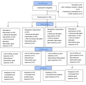
Between May 2017 and September 2017, patients scheduled for surgeries under general anesthesia at Aswan University hospitals were invited to participate in our trial. Six patients were excluded from this study, 2 patients have not meeting inclusion criteria, 1 patient declined to participate and 3 patients for other rea-sons refused to participate, and 120 patients were randomized after informed written consent. In total, 30 patients were randomized to each group. All pa-tients received the intended treatment, completed the study protocol, and were included in the analysis (Figure 1). 120 patients were recruited to the study.
Patients in each group were comparable about age, body weight, and baseline hemodynamic variables; with nonsignificant differences (Table 1).
DOI: 10.4236/ojanes.2017.710034 344 Open Journal of Anesthesiology
Figure 1. Consort flow diagram.
Table 1. Demographic data and baseline hemodynamic parameters.
Group-A
N = 30 Group-B N = 30 Group-C N = 30 Group-D N = 30 P-value Age (year) 38.47 ± 10.84 39.77 ± 9.61 39.76 ± 10.25 39.77 ± 11.25 0.80 ASA status:
I
II 27 (90%) 3 (10%) 26 (86.7%) 4 (13.3%) 28 (93.3%) 2 (6.7) 27 (90%) 3 (10%) 0.86 Sex:
Female
Male 13 (43.3%) 17 (56.7%) 14 (46.7%) 16 (53.3%) 12 (40%) 18 (60%) 14 (46.7%) 16 (53.3%) 0.95 Weight (kg) 62.18 ± 8.12 64.08 ± 6.67 62.80 ± 7.87 62.80 ± 7.87 0.13 SBP (mm. Hg) 127.36 ± 5.13 125.08 ± 8.51 124.30 ± 8.58 125.30 ± 8.58 0.15 DBP (mm. Hg) 78.68 ± 5.98 76.98 ± 7.06 77.46 ± 7.84 78.46 ± 7.74 0.13 MAP (mm. Hg) 94.18 ± 3.74 93.14 ± 7.26 93.86 ± 7.95 92.86 ± 7.85 0.11 HR (b./min) 88.06 ± 9.69 89.26 ± 12.30 87.70 ± 12.42 85.70 ± 12.42 0.33
Data are presented as mean ± Sd. *P-value < 0.05.
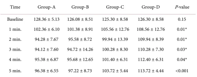
mmHg. The decrease in SBP was highest in group-A and the lowest in group-D as illustrated in Table 2 and Figure 2.
[image:4.595.210.539.396.577.2]DOI: 10.4236/ojanes.2017.710034 345 Open Journal of Anesthesiology
Figure 2. Comparison of SBP during the study period.
Table 2. Comparison of SBP during the study period.
Time Group-A Group-B Group-C Group-D P-value
Baseline 128.36 ± 5.13 126.08 ± 8.51 125.30 ± 8.58 126.30 ± 8.58 0.15 1 min. 102.36 ± 6.10 101.38 ± 8.91 105.56 ± 12.76 108.56 ± 12.76 0.01* 2 min. 94.28 ± 7.67 95.58 ± 8.72 99.94 ± 13.39 109.94 ± 8.39 0.01* 3 min. 94.12 ± 7.60 94.72 ± 14.26 100.28 ± 8.30 110.28 ± 7.30 0.03* 4 min. 95.38 ± 6.87 95.68 ± 12.65 101.40 ± 6.31 112.40 ± 6.31 0.04*
5 min. 96.38 ± 6.55 97.22 ± 8.73 103.72 ± 5.44 113.72 ± 4.44 <0.001
Data are presented as mean ± Sd. *P-value < 0.05.
pressure reduced to 60.66 ± 6.32 mmHg. The decrease in diastolic blood pres-sure was highest in group-B and the lowest in group-D as illustrated in Table 3
and Figure 3.
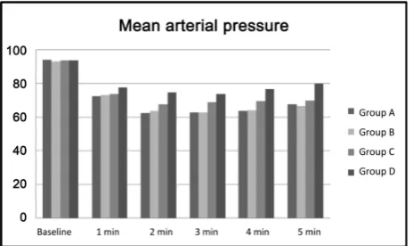
MAP decreased in all the four groups after the induction of anesthesia. The decrease was similar in group-A and group-B and no significant differences in the MAP between group-A and group-B. The decrease in the MAP in group-A and group-B was similar and more than group-C and group-D. In group-A, MAP decreased to 67.52 ± 4.92 (drop 29% from the baseline) at 5 min, in group-B MAP decreased to 66.78 ± 7.16 mmHg (drop 30% from the baseline) and in group-C mean blood pressure decreased to 69.78 ± 4.75 mmHg (drop 25% from the baseline) and in group-D mean blood pressure decreased to 72.78 ± 5.75 mmHg (drop 16% from the baseline). The decrease in mean blood pres-sure was highest in group-A, and the lowest in group-D. The decrease in the MAP in group-C and group-D was significantly less when compared to group-A and group-B as illustrated in Table 4 and Figure 4.
[image:5.595.209.541.278.408.2]DOI: 10.4236/ojanes.2017.710034 346 Open Journal of Anesthesiology
Table 3. Comparison of DBP during the study period.
Time Group-A Group-B Group-C Group-D P-value
Baseline 78.69 ± 5.88 76.98 ± 7.16 76.46 ± 7.95 78.46 ± 7.93 0.13 1 min. 57.22 ± 7.97 56.72 ± 7.03 58.14 ± 9.82 62.14 ± 9.82 0.02* 2 min. 48.64 ± 10.40 49.38 ± 6.54 54.58 ± 8.33 63.58 ± 9.33 0.02* 3 min. 47.68 ± 6.07 48.10 ± 8.52 53.34 ± 4.30 66.34 ± 5.30 0.04* 4 min. 47.94 ± 4.38 48.60 ± 10.69 54.76 ± 5.40 64.76 ± 6.40 <0.001* 5 min. 53.30 ± 4.37 52.48 ± 8.05 57.66 ± 5.32 60.66 ± 6.32 0.08
[image:6.595.255.494.234.371.2]Data are presented as mean ± Sd. *P-value < 0.05.
Figure 3. Comparison of DBP during the study period.
Table 4. Comparison of the MAP during the study period.
Time Group-A Group-B Group-C Group-D P-value
Baseline 94.18 ± 3.64 93.14 ± 7.16 93.86 ± 7.85 93.86 ± 7.85 0.11 1 min. 72.36 ± 6.04 72.98 ± 6.45 73.8 ± 10.23 77.80 ± 11.23 0.01* 2 min. 62.56 ± 7.92 63.88 ± 7.32 67.74 ± 9.60 74.74 ± 10.60 0.02* 3 min. 62.86 ± 4.18 62.70 ± 9.03 68.92 ± 4.23 73.92 ± 6.23 0.04* 4 min. 63.64 ± 4.43 64.24 ± 10.89 69.62 ± 4.11 76.62 ± 4.11 0.02* 5 min. 67.52 ± 4.92 66.78 ± 7.16 69.78 ± 4.75 79.78 ± 5.75 0.01*
Data are presented as mean ± Sd. *P-value < 0.05.
[image:6.595.208.541.421.539.2] [image:6.595.259.491.569.707.2]DOI: 10.4236/ojanes.2017.710034 347 Open Journal of Anesthesiology
decreased to 78.88 ± 11.71 (drop 11% from the baseline) and in group-C H.R increased to 88.46 ± 8.67 (increase 2% from the baseline) and in group-D H.R increased to 90.66 ± 7.57 (increase 5% from the baseline). The decrease in H.R was in group-A 20% and group-B 11% and increase in group-C 2% and group-D 5% was insignificantly in all groups as illustrated in Table 5 and Figure 5.
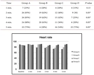
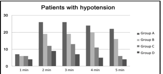
[image:7.595.209.539.269.388.2]The incidence of hypotension in the four groups during the study period was also compared. The number of patients developing hypotension at 1 min was not significant when compared to the four groups (P > 0.05). The incidence of hypotension was significant at 2 min, 3 min, 4 min and 5 min (P < 0.05). The in-cidence of hypotension was highest in group-A followed by group-B and group-C and group-D as illustrated in Table 6 and Figure 6.
Table 5. Comparison of HR during the study period.
Time Group-A Group-B Group-C Group-D P-value
Baseline 89.16 ± 9.59 87.26 ± 13.32 86.70 ± 11.40 85.70 ± 12.40 0.33 1 min. 90.46 ± 12.58 89.72 ± 16.98 87.16 ± 9.91 88.26 ± 9.91 0.48 2 min. 78.38 ± 11.94 79.58 ± 13.68 89.74 ± 7.29 90.74 ± 6.29 <0.001* 3 min. 76.98 ± 13.92 79.88 ± 13.25 85.06 ± 7.36 87.26 ± 8.36 <0.001* 4 min. 73.92 ± 12.29 77.26 ± 11.42 86.48 ± 7.55 88.58 ± 7.65 <0.001* 5 min. 72.84 ± 11.59 78.88 ± 11.71 88.46 ± 8.67 90.66 ± 7.57 <0.001*
[image:7.595.208.540.443.707.2]Data are presented as mean ± Sd. *P-value < 0.05.
Table 6. The number of patients developing hypotension and time of onset of
hypoten-sion.
Time Group-A Group-B Group-C Group-D P-value
1 min. 7 (23%) 6 (20%) 6 (20%) 4 (15%) 0.13
2 min. 26 (85%) 19 (62%) 12 (40%) 9 (30) 0.05*
3 min. 26 (85%) 19 (62%) 13 (42%) 7 (25%) 0.05*
4 min. 24 (80%) 20 (65%) 11 (36%) 6 (20%) 0.05*
5 min. 22 (75%) 6 (20%) 16 (54%) 22 (75%) 0.05*
DOI: 10.4236/ojanes.2017.710034 348 Open Journal of Anesthesiology
Figure 6. The number of patients developing hypotension and time of
onset on hypotension.
4. Discussion
Hypotension after induction with propofol is well known [9]. The cause of this hypotension has been found to be a depression of myocardial contractility and a reduced systemic vascular resistance [10]. Fentanyl was used for adjuvant induc-tion of anesthesia with propofol. Fentanyl in small doses has minimal cardi-ovascular effects [11]. However, when used with propofol for induction of anes-thesia, it may heighten the bradycardia and hypotensive effects of propofol [12].
This study confirms that induction of anesthesia with propofol combined with fentanyl in ASA-I and II patients is usually accompanied with significant sys-temic arterial hypotension. Preinduction IV injection of ephedrine in a dose of 0.1 mg/kg significantly minimized, but did not eliminate the decrease of BP, but the dose of 0.2 mg/kg was much better, where the drop in SBP from base line af-ter 5 minute was 25% and 22% in group-A and group-B but 16% in group-C and only 11% drop in group-D. Also significant decrease in systolic blood pressure from the baseline was observed in all the groups after propofol administration in our study dropped 21%, 20%, 16%, 10% from the base line respectively A-, B-, C-, D-groups after 1 min. Gamlin et al.[13] found that 15 or 20 mg of ephedrine premixed with 20 ml of 1% propofol maintained blood pressure at preinduction values, whereas ephedrine 10 mg was insufficient. The difference in observations could be connected with a higher dose of ephedrine (15, 20 and 25 mg) in other studies than in ours (0.2 mg/kg, with a mean dose of 10 mg).
In this study, we observed that prophylactic IV dose of ephedrine was effective in limiting the hypotension during propofol induction in doses 0.1 mg/kg and 0.2 mg/kg. But ephedrine did not eliminate the reduction in BP associated with induction of anesthesia with propofol and fentanyl. Our results are comparable to those of Michelsen et al.[14]. They found that prophylactic IV ephedrine 0.2 mg/kg significantly weakened, but did not eliminate the reduction in blood pressure during propofol and fentanyl induction. Similarly, El-Beheiry et al.[15]
sympa-DOI: 10.4236/ojanes.2017.710034 349 Open Journal of Anesthesiology
thoadrenal-stimulating. Though pre-induction by ephedrine attenuated the hy-potensive effects of propofol, some patients still worldly-wised in a reduction in BP to <80% of baseline. The cause for this may be that ephedrine mainly keeps the arterial blood pressure by increasing the cardiac output [16], whereas pro-pofol, under conditions similar to those in the present study, causes arterial hy-potension by reducing peripheral vascular resistance [2] [17]. Gopalakrishnan and colleagues [18] have reported ephedrine to be ineffective in preventing hy-potension after induction of anesthesia with propofol and rocuronium during rapid tracheal intubation. However, Gamlin et al. [19] had reported full effec-tiveness in obtunding hypotensive effects of propofol when ephedrine was mixed with propofol. But marked tachycardia was observed in the majority of patients in their study. In our study, we observed a decrease in heart rate in control group and increased in the ephedrine group, but it was less than 20% of the baseline and statistically insignificant. Gamlin et al. [20] reported marked ta-chycardia associated with the use of ephedrine in combination with propofol in the majority of patients. The difference in observations could be correlated with higher doses of ephedrine (20 and 25 mg) in other studies than in ours (0.2 mg/kg). Dhungana et al.[18] also reported insignificant increases in heart rate in patients receiving ephedrine. In conclusion, we found that the prophylactic intravenous injection of ephedrine 0.1 mg/kg significantly attenuated, but did not abolish the reduction in systolic blood pressure associated with induction of anesthesia with propofol and fentanyl, but 0.2 mg/kg was much better without causing any adverse effects. We recommended that ephedrine reduced the inci-dence of hypotension in a significant number of our ASA I and II grade patients, and their safety and efficacy needed to be used during routine clinical practice and in high-risk groups and critically ill patients, especially ephedrine 0.2 mg/kg.
References
[1] Smith, I., White, P.F., Nathanson, M., et al. (1994) Propofol: An Update on Its Clin-ical Use. Anesthesiology, 81, 1005-1043.
https://doi.org/10.1097/00000542-199410000-00028
[2] Fairfield, J.E., Dritsas, A. and Beale, R.J. (1991) Hemodynamic Effects of Propofol: Induction with 2.5 mg/kg−1. British Journal of Anesthesia,67, 618-620.
https://doi.org/10.1093/bja/67.5.618
[3] Muzi, M., Berens, R.A., Kampine, J.P. and Ebert, T.J. (1992) Venodilation Contri-butes to Propofol-Mediated Hypotension in Humans. Anesthesia and Analgesia, 74, 877-883. https://doi.org/10.1213/00000539-199206000-00017
[4] Robinson, B.J., Ebert, T.J., O’Brien, T.J., Colinco, M.D. and Muzi, M. (1997) Me-chanisms Whereby Propofol Mediates Peripheral Vasodilation in Humans: Sympa-thoinhibition or Direct Vascular Relaxation? Anesthesiology, 86, 64-72.
https://doi.org/10.1097/00000542-199701000-00010
[5] Cullen, P.M., Turtle, M., Prys-Roberts, C., Way, W.L. and Dye, J. (1987) Effect of Propofol Anesthesia on Baroreflex Activity in Humans. Anesthesia and Analgesia, 66, 1115-1120.
DOI: 10.4236/ojanes.2017.710034 350 Open Journal of Anesthesiology Comparison of Ephedrine and Ketamine in Prevention of Injection Pain and Hypo-tension due to Propofol Induction. European Journal of Anesthesiology, 22, 44-48.
https://doi.org/10.1097/00003643-200501000-00010
[7] Ralston, D.H., Shnider, S.M. and DeLorimier, A.A. (1974) Effects of Equipotent Ephedrine, Metaraminol, Mephentermine, and Methoxamine on Uterine Blood Flow in the Pregnant Ewe. Anesthesiology, 40, 354-370.
https://doi.org/10.1097/00000542-197404000-00009
[8] Vercauteren, M.P., Coppejans, H.C., Hoffmann, V.H., Mertens, E. and Adriaensen, H.A. (2000) Prevention of Hypotension by a Single 5-Mg Dose of Ephedrine during Small-Dose Spinal Anesthesia in Prehydrated Cesarean Delivery Patients. A nesthe-sia and Analgenesthe-sia, 90, 324-327.
[9] Skues, M.A., Richards, M.J., Jarvis, A.P. and Prys-Roberts, C. (1989) Preinduction Atropine or Glycopyrrolate and Hemodynamic Changes Associated with Induction and Maintenance of Anesthesia with Propofol and Alfentanil. Anesthesia and Analgesia, 69, 386-390. https://doi.org/10.1213/00000539-198909000-00020
[10] Kasaba, T., Yamaga, M., Iwasaki, T., Yoshimura, Y. and Takasaki, M. (2000) Ephe-drine, Dopamine, or Dobutamine to Treat Hypotension with Propofol during Epi-dural Anesthesia. Canadian Journal of Anesthesia, 47, 237-241.
https://doi.org/10.1007/BF03018919
[11] Chiu, C.L., Tew, G.P., and Wang, C.Y. (2001) The Effect of Prophylactic Metara-minol on Systemic Hypotension Caused by Induction of Anesthesia with Propofol in Patients over 55 Years Old. Anaesthesia, 56, 893-897.
https://doi.org/10.1046/j.1365-2044.2001.02059-4.x
[12] Turner, R.J., Gatt, S.P., Kam, P.C.A., Ramzan, I. and Daley, M. (1998) Administra-tion of a Crystalloid Fluid Preload Does Not Prevent the Decrease in Arterial Blood Pressure after Induction of Anesthesia with Propofol and Fentanyl. British Journal of Anesthesia, 80, 737-741. https://doi.org/10.1093/bja/80.6.737
[13] Gamlin, F., Vucevic, M., Winslow, L. and Berridge, J. (1996) The Hemodynamic Effects of Propofol in Combination with Ephedrine. Anaesthesia, 51, 488-491.
https://doi.org/10.1111/j.1365-2044.1996.tb07799.x
[14] Michelsen, I., Helbo-Hansen, H.S., Kohler, F., Lorenzen, A.G., Rydlund, E. and Bentzon, M.W. (1998) Prophylactic Ephedrine Attenuates the Hemodynamic Re-sponse to Propofol in Elderly Female Patients. Anesthesia and Analgesia, 86, 477-481. https://doi.org/10.1213/00000539-199803000-00004
[15] El-Beheiry, H., Kim, J., Milne, B. and Seegobin, R. (1995) Prophylaxis against the Systemic Hypotension Induced by Propofol during Rapid Sequence Intubation.
Canadian Journal of Anesthesia, 42, 875-878. https://doi.org/10.1007/BF03011034
[16] Critchley, L.A.H., Stuart, J.C., Conway, F. and Short, T.G. (1995) Hypotension dur-ing Subarachnoid Anesthesia: Hemodynamic Effects of Ephedrine. British Journal of Anesthesia, 74, 373-378. https://doi.org/10.1093/bja/74.4.373
[17] Claeys, M.A., Gepts, E. and Camu, F. (1988) Haemodynamic Changes during Anesthesia Induced and Maintained with Propofol. British Journal of Anesthesia, 60, 3-9. https://doi.org/10.1093/bja/60.1.3
[18] Dhungana, Y., Bhattarai, B.K., Bhadani, U.K., Biswas, B.K. and Tripathi, M. (2008) Prevention of Hypotension during Propofol Induction: A Comparison of Preload-ing with 3.5% Polymers of Degraded Gelatin (Haemaccel) and Intravenous Ephe-drine. Nepal Medical College Journal, 10, 16-19.
DOI: 10.4236/ojanes.2017.710034 351 Open Journal of Anesthesiology
Anesthesiology, 17, 959-968.