Diffusion-Tensor MR Imaging of the Brain
in Human Immunodeficiency
Virus–Positive Patients
Majda M. Thurnher, Mauricio Castillo, Alfred Stadler, Armin Rieger, Brigitte Schmid, and Pia C. Sundgren
BACKGROUND AND PURPOSE:There is early evidence that diffusion-tensor imaging (DTI) is useful in demonstrating subtle white matter alterations in different diseases of brain. We hypothesize that DTI in several brain regions in human immunodeficiency virus–positive (HIVⴙ) patients is useful in the early detection of HIV-related brain injury.
METHODS:MR imaging and DTI were performed in 60 HIVⴙ patients and in 30 controls. Fractional anisotropy (FA) and apparent diffusion coefficient (ADC; mm/s2) maps were gen-erated and coregistered on T2-weighted images. Regions of interest were placed in the splenium and genu of the callosum, the frontal white matter, and the hippocampus. HIVⴙpatients were divided into those whose CD4 count were <250 cells/mm3or >250 cells/mm3. According to plasma viral loads, patients were divided into those whose viral loads were <50 copies/mL, 50 –100,000 copies/mL, or >100,000 copies/mL.
RESULTS: Statistically significant decrease of FA was found in the genu of the corpus callosum in HIVⴙpatients compared with controls. FA was reduced in the frontal white matter and hippocampi in HIVⴙ patients compared with controls. Differences, however, were not statistically significant. No statistically significant differences were found between the HIVⴙ groups for FA of the splenium or between these groups and the controls. ADC values were significantly increased in the genu of HIVⴙ patients when compared with controls and were also increased in other locations, but without statistical significance.
CONCLUSION:As used in this study, DTI was not helpful in identifying patients with early HIV infection.
Central nervous system (CNS) involvement is an early feature of infection with the human immunodefi-ciency virus (HIV). The precise mechanism of the HIV-related changes in the brain is incompletely un-derstood. CSF analysis demonstrates that HIV enters the CNS soon after exposure, even before antibodies are detectable in blood (1, 2). The neuropathologic features of AIDS dementia (HAD) include multigiant
cell encephalitis (MGCE) and HIV leukoencephalop-athy (3). Although MR imaging is a sensitive tech-nique for depicting the effects of HIV in the brain, it cannot show early pathologic involvement. MR imag-ing features of brain HIV infection include bilateral symmetric increased T2 signal intensity in the white matter and cerebral atrophy (4). MR spectroscopy may show acutely a mild elevation of choline (prob-ably due to an inflammatory response) and low N -acetyl-asparatate due to neuronal dysfunction (5). Unfortunately, even with multiple volume techniques, the assessment of the overall involvement of the brain is difficult due to restrictions of voxel size. Whole-brain NAA measurements may potentially be useful in this regard, but this technique is not widely avail-able. Measurement of magnetization transfer may also help in detecting subclinical disease, but this technique has not been widely incorporated into clin-ical imaging (6).
Data from recent studies suggest that diffusion-tensor imaging (DTI) may be sensitive in detecting early CNS involvement of HIV (7–9). Studies dem-Received May 28, 2004; accepted after revision April 18, 2005.
From the Department of Radiology, Neuroradiology Section (M.M.T., A.S.), and the Department of Dermatology, HIV Section (A.R.), University Hospital Vienna, Vienna, Austria; the Depart-ment of Radiology, Neuroradiology (M.C.), University of North Carolina, Chapel Hill, NC; Department of Internal Medicine, HIV Section, Hospital Otto-Wagner-Spital (B.S.), Vienna, Austria; and the Department of Radiology, Neuroradiology (P.C.S.), University of Michigan, Ann Arbor, MI.
Presented in part at the 17th Symposium Neuroradiologicum, Paris, August 18 –24, 2002.
Address correspondence to Majda M. Thurnher, MD, Depart-ment of Radiology, Neuroradiology Section, University Hospital Vienna, Waehringer Guertel 18-20, 1090-Vienna, Austria.
©American Society of Neuroradiology
onstrate abnormal fractional anisotropy (FA) in fron-tal white matter and internal capsules of HIV-positive (⫹) patients (8). In another study, the largest de-creases in FA were found in the corpus callosum and the largest elevations in mean diffusivity were seen in the subcortical white matter in patients who had ad-vanced disease, high viral loads, and low CD4 counts (7). Also, pathologic studies have shown that hip-pocampal injury is common in HIV encephalitis and may play a role in the development of HAD (10 –13). The purpose of this prospective study was to deter-mine whether DTI obtained in several brain regions in HIV⫹ patients with and without dementia and compared with healthy volunteers might be helpful in the detection of early HIV infection of the brain.
Methods
Subjects
During a 16-month period (2000 –2002), a total of 60 HIV⫹ patients (48 men and 12 women; 21– 64 years of age; mean age, 42 years) and 30 healthy age-matched controls (22 men and 8 women; 13– 64 years of age; mean age, 40 years) were studied. Inclusion criteria were HIV seropositive on the basis of self-report and confirmed by enzyme-link immunosorbant assay and Western blot, either meeting the Centers for Disease Control and Prevention criteria for AIDS or demonstrating cognitive impairment. Patients with current or past opportu-nistic CNS infection, schizophrenia, severe affective disorder, or a history of chronic neurologic disorder (such as multiple sclerosis) or uncontrolled epilepsy were excluded. The patients were not receiving steroids or other medication that could have altered the measurements.
MR Examinations
All MR studies were performed on a clinical 1.5T scanner. The following sequences were obtained in controls and pa-tients: 2-mm-thick coronal T2-weighted images (TR/TE/flip angle, 4000 ms/90 ms/90°), 4-mm-thick axial fluid-attenuated inversion recovery images (TR/TE/IR, 11000 ms/80 ms/2800 ms), and 1.25-mm-thick axial T1-weighted images (TR/TE/flip angle, 20 ms/1.76 ms/35°). A single-shot, multisection, spin-echo, echo-planar pulse sequence (TR/TE/flip angle, 1935 ms/89 ms/90°) was used for DTI. We performed diffusion-weighted imaging in 7 directions (X, Y, Z, X⫹Y, X⫹Z, Y⫹ Z, X ⫹Y ⫹Z) with the maximumb value of 1000 s/mm2, field-of-view of 230 mm, 128 ⫻128 matrix reconstructed to 256⫻256 (acquisition time, 34 seconds). We calculated FA, which is defined as
FA§
冑
1⫺ Dsurf 23Darg2 ⫺2Dsurf2
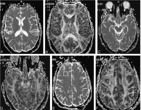
FA and apparent diffusion coefficient (ADC) maps were gen-erated from DTI datasets and coregistered onto T2-weighted images. Round regions of interest of uniform size (100 mm2) were placed in the splenium and genu of the corpus callosum. Square regions of interest of uniform size (200 mm2) were placed in the left frontal white matter, and an oval region of interest was placed (⬃250 mm2) in the region of left hippocam-pus (Fig 1). The differences in shape of the region of interest were chosen to provide the best fit in the structure analyzed.
Two experienced reviewers (M.M.T. and A.S.) who were blinded to the HIV status of the patients performed the mea-surements. Inter- and intraobserver variability were not measured.
Clinical Examinations
Symptomatic HIV⫹involvement was confirmed by a neu-rologist and a neuropsychologist at baseline. The following data were obtained: clinical history (demographic data); neurologic signs (including presence of HAD), and symptoms. CD4⫹ T-lymphocyte counts (cells/mm3) and HIV-1 RNA level (viral load, copies/mL) in plasma were obtained.
HIV⫹patients were divided into those with CD4 counts ⬍250 cells/mm3or⬎250 cells/mm3. According to plasma viral loads, patients were divided on the basis of clinical literature into those with viral loads⬍50 copies/mL, 50 –100,000 copies/ mL, or ⬎100,000 copies/mL. Viral load ⬍50 is considered under the detectable level, the second group represents pa-tients with mild increase of the viral load, and the last group patients with high level of viral load.
Statistical Analyses
Statistical analysis was performed by using commercially available software (SPSS version 12; SPSS Inc., Chicago, IL). Descriptive statistical analysis included the calculation of means and SDs of the obtained data. Normality was tested by using the Kolmogorov-Smirnov test. We compared the mean FA and ADC values obtained in each of the 4 locations per individual (total of 240 measurements in HIV⫹) with patients grouped in different categories depending on their viral loads and CD4 cell counts. All FA and ADC measurements obtained in HIV⫹patients were then compared with those of the con-trols. Statistical analysis was performed by using thettest. The level of statistical significance was established atP⬍.05.
Results
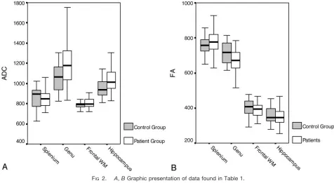
The results of the ADC and FA measurements in all 4 locations in HIV⫹ patients and controls are summarized in Table 1 and the related Fig 2A,-B.
FA was statistically significantly reduced in the genu (mean 673 in HIV⫹ patients vs mean 717 in controls). FA was reduced in the frontal white matter (mean 389 in HIV⫹ patients vs mean 404 in con-trols), and hippocampi (mean 358 in HIV⫹ patients vs mean 366 in controls) in HIV⫹patients compared with controls. FA was increased in splenium of the corpus callosum in HIV⫹ patients compared with controls. The differences were not statistically signif-icant for splenium, white matter, or hippocampi.
ADC values were significantly higher in the genu of HIV⫹ patients (mean, 1191) when compared with controls (mean, 1054). ADC values were higher in the other 3 locations, but not statistically significant.
Table 2 summarizes FA and ADC values measured in 4 locations of the brain in 3 groups of patients according to their viral load level in plasma.
No correlation was found between viral load level in plasma and FA values; FA was reduced in genu, white matter, and hippocampi in all viral load groups compared with the controls, but it did not reach the level of statistical significance. Furthermore, no sta-tistically significant differences were found in FA val-ues between the 3 groups of patients. FA valval-ues were higher in splenium in all viral load groups compared with controls, without statistical difference.
compared with controls, with statistical significance only when controls were compared with the HIV⫹ group with the lowest viral load (P ⫽ .028). No dif-ferences were found between HIV⫹ groups. The highest ADC values in splenium compared with con-trols were found in the lowest viral load group (P⫽ .023).
No statistically significant correlation was found between FA and ADC values and CD4 counts (Table 3).
Discussion
Research on the pathogenesis of HIV infection of the CNS has reached a pivotal stage. HIV commonly invades the brain soon after the peripheral infection. The virus has been found in the brain as early as 15 days after accidental intravenous inoculation (1). It is known that the neuron dysfunction or death that underlies the clinical symptoms of HIV/CNS disease does not result from direct infection of neurons. Thus, the mechanism of HIV-related brain injury remains poorly understood. It is believed that the predominant pathogenesis involves a combined influ-ence of both HIV infection and activation of immune-competent cells and their subsequent release of toxins leading to neuron and astrocyte dysfunction (2). Neu-ropathologic hallmarks of HIV brain infection are MGCE and progressive diffuse leukoencephalopathy (3). The chief cellular targets of HIV infection within
FIG 1. ADC (A) and FA (B) images in axial plane with region of interest placed in the genu and splenium of the corpus callosum.
[image:3.585.53.536.58.438.2]CandD,Region of interest placed in the frontal white matter on ADC and FA. EandF,Region of interest placed in the hippocampus.
TABLE 1: FA and ADC values in all four locations in HIV-positive patients and controls
Brain Region
HIV⫹Patients (n⫽60)
Controls (n⫽30)
Significance (P⬍.05)
FA Splenium 774⫾76 752⫾56 NS
Genu 673⫾75 717⫾68 .037
Frontal WM 389⫾45 404⫾52 NS
Hippocampus 358⫾63 366⫾65 NS
ADC Splenium 867⫾111 859⫾114 NS
Genu 1191⫾216 1054⫾146 .019
Frontal WM 809⫾58 792⫾44 NS
Hippocampus 1043⫾147 968⫾129 NS
[image:3.585.54.282.506.619.2]the CNS are the microglia/macrophages (14). After the early entry of HIV into the brain, the key question remains: what happens to the virus in the CNS? Most studies support the concept of autonomous infection, which suggests that the CNS is an independent
res-ervoir for HIV (2). The CNS infection is persistent and self-sustaining and does not depend on subse-quent spread of the virus from systemic circulation. Clinically, HIV brain infection manifests as neuropsy-chologic impairment that ranges from mild cognitive motor disorder to HAD (2, 15).
Although MR imaging is the most sensitive imaging technique for depicting the effects of HIV in the brain, it is nevertheless not sensitive enough to show early pathologic involvement. The characteristic MR imaging features of HIV infection in the brain include bilateral symmetric white matter disease (hyperinten-sities on T2-weighted MR images), as well as cerebral atrophy (4).
In the present study, we used DTI in an attempt to detect abnormalities in the brain of HIV⫹ patients. DTI detects diffusion-driven displacement of mole-cules during random motion through axonal fibers, which reflects the tissue structure and geometry found at a microscopic level. ADC values, MD and FA are specific DTI indices describing water diffusion in tissue (16 –18). FA is a scalar measure of tissue
[image:4.585.54.530.57.318.2]FIG 2. A,BGraphic presentation of data found in Table 1.
TABLE 2: FA and ADC measurements in all four locations in HIV-positive patients and controls according to the viral load in plasma
Controls (n⫽16)
HIV⫹Patients: Viral Load (Copies/mL)
⬍50
(n⫽12) PValue
50–100 000
(n⫽19) PValue
⬎100 000
(n⫽22) PValue
Splenium FA 753⫾56 770⫾92 791⫾80 755⫾63 NS
Genu FA 717⫾68 680⫾54 679⫾68 679⫾93 NS
White matter FA 404⫾52 367⫾35 381⫾39 402⫾50 NS
Hippocampus FA 366⫾65 364⫾101 350⫾54 357⫾46 NS
Splenium ADC 858⫾114 942⫾155 .023 836⫾96 NS 858⫾72 NS
Genu ADC 1054⫾146 1286⫾255 NS 1135⫾192 NS 1159⫾208 NS
White matter ADC 792⫾44 851⫾70 .028 807⫾42 NS 796⫾57 NS
Hippocampus ADC 968⫾129 1081⫾189 NS 1018⫾123 NS 1029⫾122 NS
Note.—FA indicates fractional anisotropy; ADC, apparent diffusion coefficient (mm/s2); NS, not significant.
TABLE 3: FA and ADC measurements in all four locations in HIV-positive patients and controls according to the CDⴙcount
Controls
HIV⫹Patients:
CD4⫹Count (cells/mm3)
⬎250 ⬍250 PValue
Splenium FA 753⫾56 757⫾102 778⫾63 NS
Genu FA 717⫾68 678⫾62 680⫾80 NS
White matter FA 404⫾52 383⫾49 388⫾43 NS
Hippocampus FA 366⫾65 344⫾57 362⫾66 NS
Splenium ADC 858⫾114 884⫾159 863⫾82 NS
Genu ADC 1054⫾146 1173⫾243 1182⫾208 NS
White matter ADC 792⫾44 816⫾63 811⫾57 NS
Hippocampus ADC 968⫾129 998⫾109 1055⫾149 NS
[image:4.585.55.533.341.471.2] [image:4.585.54.282.518.648.2]structure; tissues with highly regular fibers (eg, white matter) have higher FA compared with less-orga-nized tissues (eg, gray matter). White matter is highly organized in fiber bundles that restrict diffusion of water, and thus ADC is lower than in CSF and an-isotropy is higher. The ADC of gray matter falls in between that of CSF and white matter. Recent studies show the ability of DTI to demonstrate subtle white matter alterations in patients with Alzheimer disease, migraine, chronic alcoholism, malignant brain tu-mors, and multiple sclerosis (19 –27). Therefore, our purpose was to determine whether DTI as a relatively fast imaging technique could be used in daily clinical work in the HIV⫹population. We have used param-eters obtainable from DTI to try to establish a rela-tionship between HIV⫹patients on the basis of their CD4 cell counts and viral loads when compared with normal individuals. Information about the usefulness of DTI measurements in early detection of HIV-related brain injury may be important to neuroradi-ologists working with this particular patient popula-tion. The earlier we can detect HIV-related brain injury with imaging techniques (especially when the patient is still asymptomatic), the sooner clinicians can consider starting antiretroviral therapy. With the knowledge that CD4 count and viral load do not corre-late well with clinical symptoms, and at the moment no other reliable clinical method for early detection of HIV-related brain injury exists, the search for a reliable imaging technique is, thus, still necessary.
In our study, there was a trend of FA reduction in the frontal white matter, genu of the corpus callosum, and the hippocampi of the HIV⫹patients when com-pared with normal controls. Difference was statisti-cally significant only for the genu of the corpus cal-losum. This is in contradiction to a previous report in which statistically significant differences were found between FA values in white matter and corpus callo-sum of HIV⫹ patients and controls. Significant changes in diffusion anisotropy in frontal white mat-ter and corpus callosum and viral loads were found in a recently published study of 10 HIV⫹ patients (7). The greatest elevation in the diffusion coefficient was found in the subcortical white matter of the frontal lobes when compared with parieto-ocipital white mat-ter, which suggests a predilection for frontal white matter involvement in HIV infection of the brain. This is in accordance with findings from MR spec-troscopy studies of HIV disease (5, 28). A limitation of the study by Filippi et al was not only the small number of patients, but also that all patients were receiving various combinations of highly active anti-retroviral therapies (HAART) (7).
Another recently published study demonstrated ab-normal ADC and FA in ab-normal-appearing periven-tricular white matter and corpus callosum in HIV⫹ patients (9). Six nondemented HIV⫹patients showed a reduction in FA in frontal white matter. In that study, 5 of 6 patients received HAART. In the present study, we also found a tendency for increased
ADC values in all measured brain regions; however, statistical significance differences when compared with controls were only demonstrated in the genu of the corpus callosum and are in accordance with pre-vious studies. FA and ADC values measured in HIV⫹ patients did not differ much in different viral load or CD4⫹count groups. Furthermore, we did not find statistically significant differences when correla-tion between different viral load groups and controls were performed. The control values, surprisingly, were nearly identical to the highest (⬎100,000 copies/ mL) rather than lowest (⬍50 copies/mL) viral load levels. This contradicts previously published reports, though the results of the initial studies were based on much smaller patient numbers. It seems obvious that DTI values measured in our study are widespread in each group, resulting in clinically useless statistical values. Looking at those results, one cannot recom-mend using DTI measurements (as used in our study) in early detection of HIV-related brain injury. Corre-lation between DTI and viral load level in the CSF (instead of plasma) may show better results.
Because we have shown that by using DTI ADC values are abnormal but that DTI values were not significantly altered, the sensitivity of the DTI tech-nique needs to be questioned. FA follows a biexpo-nential model—that is, for b values ⬍1000 –3000, most of what is measured as water diffusion parallels white matter fibers (fast pool), whereas b values higher than that range measure mostly water diffu-sion perpendicular to the fibers (slow pool) (29, 30). It is also possible that the slow pool actually is related to intracellular water motion (in the case of white matter, intra-axonal water motion) (31, 32). Because acute HIV infection involves mainly lymphocytes and macrophages, DTI should not be a sensitive tech-nique; more chronic HIV infection affects the white matter (loss of myelin) and the neurons (necrosis, axonal swelling). Thus, DTI should be able to detect abnormalities at least in patients with long-term in-fections. To detect axonal abnormalities, it seems reasonable to postulate that very highbvalues will be needed. Because white matter abnormalities are typ-ical also of late infection, it is possible that FA will be altered. A trend toward lower FA was seen in the frontal white matter and hippocampi but unfortu-nately showed no statistical significance. Thus, it seems that, alone and in individual patients, DTI as we performed it does not reflect the underlying pathophysiologic changes. It is also plausible that the HIV-induced abnormalities do not follow mono- or biexponential functions that a model (such as q-space analysis) that takes into account multiexponential sig-nal intensity decays will improve the performance of DTI, because it is more sensitive to diffusion restric-tion determinarestric-tion (31). q-Space analysis is based on multiexponential decay of the water signal intensity in MR diffusion experiments allowing the differentia-tion between physiologic compartments, on the basis of their diffusion characteristics (31). This latter tech-nique is not available at our institution.
pub-lished data on DTI abnormalities of the hippocampus in HIV⫹ patients. We have shown reduced FA and increase in the ADC values in the hippocampi of HIV⫹ patients compared with controls. Unfortu-nately, the differences between these 2 groups were not statistically significant. No patients had signal intensity abnormalities in the hippocampi on conven-tional MR imaging. Studies show that hippocampal injury is common in HIV encephalitis. In one study, the authors found HIV type 1 gene sequences in hippocampal neurons isolated from postmortem AIDS brains (12). HIV infection may contribute to neuronal injury and death, and neurons may act as potential viral reservoirs. In another study, 58% of patients with HIV encephalitis had gp41-positive mi-croglial cells in the basal neocortex and hippocampus (10). Selective damage of neurons was found in hip-pocampi of patients with moderate to severe HIV encephalitis (13). A form of HAD may be similar to Alzheimer disease with abnormalities found in the hippocampi. According to results from previous stud-ies where vulnerability of hippocampal neurons in HIV has been found, reduced anisotropy in the hip-pocampus in HIV⫹patients may be expected, and this was, at least, partially demonstrated in our study. Selec-tive damage to hippocampal neurons in HIV infection most likely results from an indirect mechanism, medi-ated by factors or virus released by infected microglia. Loss of neurons secondary to activation of inflammatory processes may then manifest decreased anisotropy.
Deep gray matter also could have been used as a location for DTI measurements in HIV⫹ patients, and the hope still remains for future studies with more promising results.
DTI did not correlate well with virologic and im-munologic parameters in our study. No differences in FA were found between the different groups with regard to CD4 T-lymphocyte count and viral load level in plasma. The largest decrease in anisotropy was in the corpus callosum of patients with advanced HIV disease and with the highest viral load level and lowest CD4 counts as were found in a previously published study (7). Recent clinical trials have shown that CD4 count and even viral load levels in plasma are not reliable markers for HIV infection and HAD. Knowing that viral load level in plasma does not correlate with viral load level in CSF, failed statistical significance between different HIV⫹group and DTI values is not surprising.
We did not evaluate findings on fluid-attenuated inversion recovery and T2-weighted imaging in this study. Findings on fluid-attenuated inversion recov-ery and T2-weighted imaging are unspecific in HIV-related brain injury, except if the involvement is dif-fuse such as in HIV encephalopathy. In those cases, however, the diagnosis is obvious and would not need DTI.
Conclusion
As used in our study, DTI was not able to detect statistically significant abnormalities in HIV⫹
pa-tients when compared with controls. In addition, there were no correlations between FA in several brain regions and CD4 counts and viral loads. There was, however, a trend to lower FA in the frontal white matter and hippocampi of all HIV⫹ patients when compared with controls. FA and ADC values were significantly different in the genu of all HIV⫹ pa-tients when compared with controls. Also, there was a trend toward higher ADC values in the frontal white matter, splenium and hippocampi of all HIV⫹ pa-tients compared with controls.
References
1. Davis LE, Hjelle BL, Miller VE, et al.Early viral brain invasion in iatrogenic human immunodeficiency virus infection. Neurology
1992;42:1736 –1739
2. Rausch DM, Davis MR.HIV in the CNS: pathogenic relationships to systemic HIV disease and other CNS disease. J Neurovirol
2001;7:85–96
3. Budka H, Constanzi G, Cristina S, et al.Brain pathology induced by infection with the human immunodeficiency virus (HIV).Acta Neuropathol1987;75:185–198
4. Olsen WL, Longo FM, Mills CM, et al.White matter disease in AIDS: findings at MR imaging.Radiology1988;169:445– 448 5. Chang L, Ernst T, Leonido-Yee M, et al. Cerebral metabolite
abnormalities correlate with clinical severity of HIV-1 cognitive motor complex.Neurology1999;52:100 –108
6. Dousset V, Armand JP, Lacoste D, et al.Magnetization transfer study of HIV encephalitis and progressive multifocal leukoenceph-alopathy.AJNR Am J Neuroradiol1997;18:895–901
7. Filippi CG, Ulug AM, Ryan E, et al.Diffusion tensor imaging of patients with HIV and normal-appearing white matter on MR images of the brain.AJNR Am J Neuroradiol2001;22:277–283 8. Pomara N, Crandall DT, Choi SJ, et al.White matter abnormalities
in HIV-1 infection: a diffusion tensor imaging study.Psychiatry Res
2001;106:15–24
9. Ulug AM, Filippi CG, Ruyan E, et al. Utility of DWI tensor imaging, and MR spectroscopy in HIV patients with normal brain scans.Int Soc Magn Reson Med2000;8
10. Reyes E, Mohar A, Mallory M, et al.Hippocampal involvement associated with human immunodeficiency virus encephalitis in Mexico.Arch Pathol Lab Med1994;118:1130 –1134
11. Spargo E, Everall IP, Lantos PL.Neuronal loss in the hippocampus in Huntington’s disease: a comparison with HIV infection.J Neurol Neurosurg Psychiatry1993;56:487– 491
12. Torres-Munoz J, Stockton P, Tacoronte N, et al.Detection of HIV-1 gene sequences in hippocampal neurons isolated from postmortem AIDS brains by laser capture microdissection.J Neuropathol Exp Neurol2001;60:885– 892
13. Masliah E, Ge N, Achim CL, et al. Selective neuronal vulnerability in HIV encephalitis.J Neuropathol Exp Neurol1992;51:585–593 14. Kolson DL, Lavi E, Gonzales-Scarano F. The effects of human
immunodeficiency virus in the central nervous system.Adv Virus Res1998;50:1– 47
15. Wesselingh SL, Power C, Glass JD, et al.Intracerebral cytokine messenger RNA expression in acquired immunodeficiency syn-drome dementia.Ann Neurol1993;33:576 –582
16. Ulug AM, Moore DF, Bojko AS, Zimmerman RD.Clinical use of diffusion-tensor imaging for disease causing neuronal and axonal damage.AJNR Am J Neuroradiol1999;20:1044 –1048
17. Le Bihan, D, Mangin JF, Poupon C, et al.Diffusion tensor imaging: concepts and applications.Magn Reson Imag2001;13:534 –546 18. Basser PJ, Jones DK.Diffusion tensor MRI: theory, experimental
design and data analysis: a technical review. NMR Biomed
2002;14:456 – 467
19. Lim KO, Helpern JA.Neuropsychiatric applications of DTI: a review.NMR Biomed2002;15:587–593
20. Buchsbaum MS, Tang CY, Peled S, et al.MRI white matter diffu-sion anisotropy and PET metabolic rate in schizophrenia. Neuro-report1998;9:425– 430
21. Werring DJ, Clark CA, Barker GJ, et al.Diffusion tensor imaging of lesions and normal-appearing white matter in multiple sclerosis.
Neurology1999;52:1626 –1632
mag-netic resonance imaging study of brain tissue from patients with migraine.J Neurol Neurosurg Psychiatry2003;74:501–503 23. Pfefferbaum A, Sullivan EV, Hedehus M, et al.Age-related decline
in brain white matter anisotropy measured with spatially corrected echo-planar diffusion tensor imaging. Magn Reson Med 2000; 44:259 –268
24. Lu S, Ahn D, Johnson G, Cha S. Pertumoral diffusion tensor imaging of high-grade gliomas and metastatic brain tumors.AJNR Am J Neuroradiol2003;24:937–941
25. Wolkin A, Choi SJ, Szilagyi S, et al.Inferior frontal white matter anisotropy and negative symptoms of schizophrenia: a diffusion tensor imaging study.Am J Psychiatry2003;160:572–574 26. Mori S, Fredriksen K, van Zijl PC, et al. Brain white matter
anatomy of tumor patients evaluated with diffusion tensor imaging.
Am Neurol2002;51:377–380
27. Sinha S, Bastin ME, Whittle IR, Wardlaw JM. Diffusion tensor imaging of high-grade cerebral gliomas.AJNR Am J Neuroradiol
2002;23:520 –527
28. Chang L, Itti I, Itti L, Chang L.Changes in cerebral metabolism are detected prior to perfusion changes in early HIV-CMC: a coregis-tered (1)H MRS and SPECT study. J Magn Reson Imaging
2000;12:859 – 865
29. Yoshiura T, Wu O, Zaheer A, et al. Highly diffusion-sensitized MRI of the brain: dissociation of gray and white matter.Magn Reson Med2001;45:734 – 40
30. Mulkern RV, Gudbjartsson H, Westin CF, et al.Multi-component apparent diffusion coefficients in human brain. NMR Biomed
1999;12:51– 62
31. Assaf Y, Cohen Y.Assignment of the water slow-diffusing compo-nent in the central nervous system using q-space diffusion MRS: implications for fiber tract imaging. Magn Reson Med 2000; 43:191–199
32. Cohen Y, Assaf Y. High b-value q-space analysed diffusion-weighted MRS and MRI in neuronal tissues: a technical review.