I
IJJMMCCMM Original Article A
Auuttuummnn22001133,,VVooll22,,NNoo44
Comparative Study of Expression of Smad3 in Oral Lichen
Planus and Normal Oral Mucosa
Shima Nafarzadeh1, Samad Ejtehadi2, Pouyan Amini Shakib1∗, Majid Fereidooni3, Ali Bijani4
1. Department of Oral and Maxillofacial Pathology, Faculty of Dentistry, Babol University of Medical Sciences,
Babol, Iran.
2. Private Dentist, Babol, Iran.
3. Department of Periodontology, Faculty of Dentistry, Babol University of Medical Sciences, Babol, Iran.
4. Non-Communicable Pediatric Diseases Research Center, Amirkola Hospital, Babol University of Medical
Sciences, Babol, Iran.
Oral lichen planus (OLP) is a chronic inflammatory disease of the oral mucosa which is considered by the World
Health Organization (WHO) as a premalignant condition. One step in malignant development is so called
epithelial mesenchymal transition (EMT), a process whereby epithelial cells acquire mesenchymal
characteristics. A factor known to induce EMT is the transforming growth factor-β (TGF-β), which uses the
Smad proteins as mediators for its signaling. The aim of this study was to compare the expression of Smad 3 in
Oral Lichen Planus and normal oral mucosa. This descriptive analytic study was performed on 30 patients with
OLP (21 women and 9 men with mean age of 45.23± 2.44 years) and 20 normal oral mucosa (14 women and 6
men with mean age of 46.95± 2.21 years). The samples were studied by immunohistochemical staining. Data
were analyzed with paired T-test and Wilcoxon test by SPSS software. Expression of Smad3 in OLP samples
and normal oral mucosa was different. This difference was statistically significant (P<0.001). The apparently
higher expression of Smad 3 in oral lichen planus compared to normal oral mucosa might help to discuss its
higher potential for malignant transition.
Key words: Oral lichen planus, smad 3, transforming growth factor β
∗
Corresponding author: Department of Oral and Maxillofacial Pathology, Faculty of Dentistry, Babol University of Medical Sciences, Babol, Iran. E-mail: [email protected]; [email protected]
ichen planus (LP) is a relatively common
chronic dermatologic disease that often affects
the oral mucosa with a prevalence ranging from 0.2
to 4% (1-2). Indeed Oral lichen planus (OLP) is
considered as a chronic disease with dynamic
evolution (2) for which several panels of diagnostic
criteria, such as the modified WHO one, have been
proposed to render a more reliable and accurate
L
Submmited 23 Sep 2013; Accepted 12 Nov 2013
diagnosis (3). The cause of LP is unknown; it is
generally considered to be an immunologically
mediated process (4). Hence, there is a questionable
theory about the potential of OLP for malignant
transformation into oral squamous cell carcinoma
(OSCC) (5). Although Gonzalez-Moles et al. (6)
have reported the frequency of 0 to 12.5% for this
kind of transformation, in a recent study, Shen
et al. (7)demonstrated that the incidence of OSCC
developing in lesions previously diagnosed as OLP,
is less than 1% and they didn’t entirely rule out
these cases as de novo OSCCs.
Transforming growth factor β (TGFβ)
regulates several cellular processes including
proliferation, differentiation, migration and death
(8). Numerous studies have demonstrated the
importance of TGFβ signaling in cancer
progression and metastasis. Although some studies
support the tumor suppressive effects of TGFβ,
other studies indicate a tumor promoting function
for TGFβ. Thus, it seems that this cytokine may
have various effects on different stages of
tumorogenesis(9).
Smad proteins are an integral part of the TGFβ
signaling pathway. After ligand binding, the TGFβ
receptor II (TβRII) phosphorylates TGFβ receptor I
(TβRI) which consequently phosphorylates and
activates two subgroups of Smad proteins (Smad 2
and Smad 3). These Smad proteins bind to a
co-Smad and finally the co-Smad complex translocates to
the nucleus, where the transcription of TGFβ
-responsive genes are regulated (9).
Some previous studies have demonstrated a
significant reduction of Smad 3 in TGFβ signaling
pathway in tissues with erythematous OLP
compared to normal oral mucosa (10) and some
have shown an increased expression of this protein
and consequently, related it to early stages of OLP
transition to malignancy (11). Therefore, in this
study we investigated the immunohistochemical
(IHC) expression of Smad3 in tissues with OLP and
adjacent normal tissues to determine the relative
role of this protein in evolution of OLP and
evaluate the prognostic value of this marker when
the progression to malignancy is suspected.
Materials and Methods
Tissue specimens and clinical data
Our study consisted of 30 biopsy specimens
taken from patients with OLP (24 reticular and 6
erosive), as defined by modified WHO criteria(3)
and 20 samples of inflammatory,
non-precancerous adjacent normal oral epithelium as
control group. All cases were retrieved from the
files of Oral and Maxillofacial Pathology
Department of Dental Faculty of Babol University
of Medical Sciences, between 2005 and 2010. All
biopsies have been fixed in formalin 10% and
processed to paraffin-embedded tissue blocks
according to the routine practice. Representative
Hematoxylin & Eosin sections were assessed to
confirm the diagnosis and sufficing of the tissue for
further evaluation. The clinical and demographic
data were collected from pre-existing medical
records.
Immunohistochemistry
Smad3 expression was assessed by
immuno-histochemical analysis using
streptavidin-biotin-peroxidase technique. Formalin-fixed paraffin
embedded tissue samples were cut into 4 µm-thick
sections and mounted on silane-coated slides. The
sections were deparaffinized with xylene,
rehydra-ted in graded ethanol and immersed in 0.3% H2O2
in methanol for 10 min at room temperature (RT) to
block the endogenous peroxidase activity. To
retrieve the antigen, the slides were treated using
microwav in 0.01 mol/L citrate buffer (PH= 6.2) at
95oC for 15 min and then washed in distilled water.
The sections were then incubated with primary
antibody (ab55479, Abcam Corp., Cambridge, UK)
for 60 min. For negative control, no primary
antibody was added. After washing in PBS, the
sections were incubated with Horseradish
peroxidase (HRP) conjugated secondary antibody at
RT for 30 min. Following PBS washing, sections
were incubated with 3, 3' diaminobenzidine
tetra-hydrochloride (DAB) (Dako, Corp, Denmark) for
10 min. Finally, the sections were counterstained
with hematoxylin and then dehydrated, cleared and
covered with a coverslip. Also, colorectal
carci-noma specimens were used as positive controls
(based on related data sheet) to allow antibody
expression comparison.
Evaluation of Immunohistochemical staining
The cytoplasmic and / or nuclear
immuno-histochemical expression of Smad 3 was evaluated
quantitatively using a light microscopy according to
what Danielsson et al. performed in their study
(11). According to the staining intensity (weak to
severe), the percentage of stained epithelial cells in
the area involved by OLP and adjacent normal
tissue was determined and the tissues were
classified into four groups: (0) 0%; (1) 1 to 25%;
(2) 26 to 50%; (3) 51 to 75%; (4) higher than 75 %;
at a magnification of 100×.
Finally, paired T-test and Wilcoxon test were
used to assess the statistical significance of the
correlations between Smad3 expression and the
health status of the tissue and P<0.05 was
considered significant.
Results
The mean age of the patients with OLP and
control group were 45.23 years (range 23 to 75) and
46.95 years (range 28 to 75), respectively. Twenty
one (70%) of the patients with OLP and fourteen
(70%) of the control group were women. The
remaining thirty percent of both groups were men.
22 (73.4%) samples of lichen planus were
located in buccal mucosa, 1 (3.3%) in labial
mucosa, 4 (13.3%) in tongue, 2 (6.7) in gingiva and
1 (3.3) in palate. In normal oral mucosa, the
distribution of samples in the above locations was
16 (80%), 1 (5%), 2 (10%), 1 (5%) and 0 (0%)
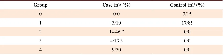
respectively. The results of immunohistochemical
staining of Smad3 in both case and control groups
are represented in table 1 and figures 1 and 2. Also,
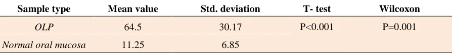
a significant difference of the expression of Smad3
was observed between normal oral mucosa and
OLP (Table 2).
Discussion
We designed the present study in order
to evaluate Epithelial-Mesenchymal Transition
(EMT) changes in oral lichen planus because
of malignant behavior of this disease and its
tendency to change to head and neck squamous
cell carcinoma (HNSCC) which was found in
previous studies.
Among the proteins which have role in EMT,
we chose Smad3 because it is involved in TGF-β
pathway and is highly expressed in several cancers
(12). The SMAD genes encode components of the
TGF-β signaling normally inhibits the cell cycle;
the loss of these genes may allow unrestrained cell
growth (4). Smad3 together with co-factor SNAIL
also acts in repression of E-cadherin and Occludin
which help tumoral progression (13). Smad3 may
have a role in apoptosis and also in inflammation,
but the results are contradictory (14-15).
Table 1. IHC staining findings of Smad3 in OLP and control group.
Group Case (n)/ (%) Control (n)/ (%)
0 0/0 3/15
1 3/10 17/85
2 14/46.7 0/0
3 4/13.3 0/0
4 9/30 0/0
Fig 1. IHC staining of normal oral mucosa showing no
expression of SMAD3 (X100).
Fig 2. IHC staining of OLP demonstrates high expression
(score 4) of SMAD3 (X400).
In this study, we found a higher expression
rate in OLP compared to normal oral mucosa which
was statistically significant (P<0.001). Different
target genes are regulated by Smad 2 and Smad 3
(16). So, the higher expression of Smad 3 in OLP
could prove that Smad 3 target genes are more
expressed in OLP. This finding supports the
malignant potential of OLP.
Karatsaidis et al. found a decreased expression
of Smad 2/3 in their study. This difference could be
due to the use of different antibodies, different IHC
methods and inclusion of both OLP and oral
lichenoid reactions in their research (10).
Danielsson et al. found an increased
expression of Smad 3 in OLP, dysplasia and tumors
compared to normal controls which is in
accordance with our findings (11).
In the present study, expression was seen in
the nucleus of epithelial cells which is explained by
Smad3 active status due to phosphorylation.
Considering normal oral mucosa, we found few
cells with Smad 3 expression which is in
accor-dance with Danielson et al.'s study results (11), but
is in contrast to the study of Karatsaidis who found
a strong expression of Smad 3 in normal oral
mucosa (10).
As a whole, our findings showed an increased
expression of Smad 3 in OLP compared to normal
oral mucosa which might be due to its probable
role in apoptosis, inflammation and EMT and
may indicate its higher potential for malignant
transition.
Acknowledgement
We would lik to thank Mr. Mohsen
Aghajanpour for his kind cooperation in sample
staining. This work has been supported by a grant
(#9031813) from Babol University of Medical
Sciences.
Conflict of interest
Authors declared no conflict of interest.
References
1. Neville BW, Damm DD, Allen CM, et al. Oral and
Maxillofacial Pathology. 3 ed. Missouri: Saunders; 2009:782-6.
2. Lu SL, Reh D, Li AG, et al. Overexpression of transforming
growth factor beta1 in head and neck epithelia results in
Table 2. Comparison of immunohistochemichal expression of Smad3 in OLP and normal oral mucosa.
Sample type Mean value Std. deviation T- test Wilcoxon
OLP 64.5 30.17 P<0.001 P=0.001
Normal oral mucosa 11.25 6.85
inflammation, angiogenesis, and epithelial hyperproliferation.
Cancer Res 2004;64:4405-10.
3. van der Meij EH, Schepman KP, van der Waal. The possible
premalignant character of oral lichen planus and oral lichenoid
lesions: a prospective study. Oral Surg Oral Med Oral Pathol
Oral Radiol Endod 2003;96:164-71.
4. Regezi JA, Sciubba JJ, Jordan RC. Oral pathology; clinical
pathologic correlations. 5 ed. Missouri: Saunders; 2008:90-1.
5. Gorsky M, Epstein JB. Oral lichen planus: malignant
transformation and human papilloma virus: a review of potential
clinical implications. Oral Surg Oral Med Oral Pathol Oral
Radiol Endod 2011;111:461-4.
6. Gonzalez-Moles MA, Scully C, Gil-Montoya JA. Oral lichen
planus: controversies surrounding malignant transformation.
Oral Dis 2008;14:229-43.
7. Shen ZY, Liu W, Feng JQ, et al. Squamous cell carcinoma
development in previously diagnosed oral lichen planus: de novo
or transformation? Oral Surg Oral Med Oral Pathol Oral Radiol
Endod 2011;112:592-6.
8. Luwor RB, Baradaran B, Taylor LE, et al. Targeting Stat3 and
Smad7 to restore TGF-beta cytostatic regulation of tumor cells
in vitro and in vivo. Oncogene 2013;32:2433-41.
9. Tannehill-Gregg SH, Kusewitt DF, Rosol TJ, et al. The roles
of Smad2 and Smad3 in the development of chemically induced
skin tumors in mice. Vet Pathol 2004;41:278-82.
10. Karatsaidis A, Schreurs O, Axell T, et al. Inhibition of the
transforming growth factor-beta/Smad signaling pathway in the
epithelium of oral lichen. J Invest Dermatol 2003;121:1283-90.
11. Danielsson K, Wahlin YB, Coates PJ, et al. Increased
expression of Smad proteins, and in particular Smad3, in oral
lichen planus compared to normal oral mucosa. J Oral Pathol
Med 2010;39:639-44.
12. Kumar V, Abbas AK, Fausto A, et al. Robbins basic
pathology. 8th ed. Philadelphia: Saunders; 2007:197.
13. Vincent T, Neve EP, Johnson JR, et al. A
SNAIL1-SMAD3/4 transcriptional repressor complex promotes TGF-beta
mediated epithelial-mesenchymal transition. Nat Cell Biol
2009;11:943-50.
14. Kim SG, Kim HA, Jong HS, et al. The endogenous ratio of
Smad2 and Smad3 influences the cytostatic function of Smad3.
Mol Biol Cell 2005;16:4672-83.
15. Li AG, Lu SL, Zhang MX, et al. Smad3 knockout mice
exhibit a resistance to skin chemical carcinogenesis. Cancer Res
2004;64:7836-45.
16. Schmierer B, Hill CS. Kinetic analysis of Smad
nucleocytoplasmic shuttling reveals a mechanism for
transforming growth factor beta-dependent nuclear accumulation
of Smads. Mol Cell Biol 2005;25:9845-58.