T
T
h
h
e
e
r
r
a
a
n
n
o
o
s
s
t
t
i
i
c
c
s
s
2014; 4(3):240-255. doi: 10.7150/thno.6914
Review
Current Progress in Gene Delivery Technology Based
on Chemical Methods and Nano-carriers
Lian Jin
1,*, Xin Zeng
2,*, Ming Liu
1, Yan Deng
1,3, and Nongyue He
1,31.
State Key Laboratory of Bioelectronics, School of Biological Science and Medical Engineering, Southeast University, Nanjing 210096,
China
2.
Nanjing Maternal and Child Health Medical Institute, Nanjing Maternal and Child Health Hospital Affiliated of Nanjing Medical
Uni-versity, Nanjing 210029, China
3.
Economical Forest Cultivation and Utilization of 2011 Collaborative Innovation Center in Hunan Province, Hunan Key Laboratory of
Green Packaging and Biomedical Nanotechnology, Hunan University of Technology, Zhuzhou 412007, China
* These two authors contribute to this paper equally.
Corresponding author: Nongyue He PhD, Sipailou No.2, Southeast University, Nanjing, 210096, China. Tel: +86-25-83790885, Fax:
+86-25-83790885. email: [email protected].
© Ivyspring International Publisher. This is an open-access article distributed under the terms of the Creative Commons License (http://creativecommons.org/ licenses/by-nc-nd/3.0/). Reproduction is permitted for personal, noncommercial use, provided that the article is in whole, unmodified, and properly cited.
Received: 2013.06.15; Accepted: 2013.11.16; Published: 2014.01.15
Abstract
Gene transfer methods are promising in the field of gene therapy. Current methods for gene
transfer include three major groups: viral, physical and chemical methods. This review mainly
summarizes development of several types of chemical methods for gene transfer
in vitro
and
in vivo
by means of nano-carriers like; calcium phosphates, lipids, and cationic polymers including
chi-tosan, polyethylenimine, polyamidoamine dendrimers, and poly(lactide-co-glycolide). This review
also briefly introduces applications of these chemical methods for gene delivery.
Key words: Non-viral; Gene delivery; Vectors; Chemical
Methods.
Introduction
A variety of genetic mutations, which alter
cel-lular proliferation, angiogenesis, metastasis, and
tu-mor immunogenicity result in human cancer, which
has become one of the biggest threats to human life
[1]. Despite increasing understanding of the
molecu-lar mechanism of cancers [2, 3], many malignancies
remain resistant to established traditional treatments
[4, 5]. However, the definition of tumor-associated
genetic mutations has heightened interest in cancer
treatment as the target for gene therapy [6, 7].
The therapeutic expectations of gene therapy are
considerable because of its significant potential for the
treatment of inherited and acquired life threatening
diseases caused by genetic deficiencies and
abnor-malities, such as cancer, acquired immune deficiency
syndrome (AIDS), cardiovascular diseases, and
cer-tain autoimmune disorders. Gene transfer, or
trans-fection, is a fundamental technique in molecular
bi-ology used to manipulate cells or study gene function
and protein expression
in vitro
. When gene transfer is
used in disease treatments, it is aimed at curbing
ge-netic diseases by introducing genes coding for
func-tional proteins to cells so as to normalize the cells and
even organs. Gene transfer is not only used to treat
genetic diseases, but also to produce large quantities
of secreted proteins for direct therapeutic application
or vaccines production. As is well known, it is difficult
for nucleic acids to diffuse directly through plasma
membrane due to their size and/or their
physico-chemical properties, for instance, hydrophilicity.
Various strategies for the transfer of nucleic acids,
especially genes, have thus been developed [8-10].
The ideal gene delivery and transfection systems
should have high transfection efficiency, low toxicity
Ivyspring
to the cells and single cell specificity, while also is able
to simultaneously treat heterogeneous systems with
many different cells.
Current gene transfection systems contain three
major groups: viral (transduction), physical (direct
micro injection) and chemical methods. Initially, the
gene transfer technique was envisioned to transform
cells utilizing various viral vectors, as listed in
Table
1
, by inserting a functional gene into a nonspecific site
within the genome. They are therefore the most
ad-vanced in development due to their efficiency and
specificity in entering cells and expressing the genes
carried in the modified viral genome using the cell’s
own biosynthetic machinery, particularly
in vivo
[22].
Unfortunately, well-known adverse effects were
ob-served while using this system, such as
immunogen-icity, difficulties in handling and large-scale
produc-tion, and limited length of the genes. Potential and
real risks were observed in a clinical trial of an HIV
vaccine developed from three weakened
adenovirus-es by Merck [23]. The failure to protect participants
from HIV was suspected to associate with the vector
itself or/and the pre-existing adenoviruse immunity
of the participants, which have been the obstacle on
the road from lab research to clinical use [24]. Physical
methods for gene transfer (
Figure 1
) include biolistics,
jet injection, ultrasound and so forth [25-29]. These
methods have been developed quickly because such
methods can directly penetrate genes into cells by
stimulations of electric impulses, fine needle puncture
or high-pressure gas, which may bypass some of the
side effects linked to viral or biochemical approaches,
such as limitation of the gene length that can be
car-ried by the physical vectors. Physical methods
medi-ate the direct penetration into the cytosol of both
small and large nucleic acid molecules, as well as any
other non-permeable molecule. Moreover, these
physical systems are effective for single or multiple
target cells at an intended location and carry little risk
of dispersion of transfection reagents. However, they
also present several drawbacks. On one hand, it is
difficult for the genes to be transported to the nucleus
because of little access in passing through the
mem-brane or enzymatic digestion of the naked DNA or
RNA, which results in the low transfection efficiency
and limits its clinical application. On the other hand,
they present damage to cells, difficulty in large-scale
manipulation, labor-intensive protocols and/or the
necessity of costly instruments [30].
Fig.1.
Primary physical methods for gene delivery.
Table 1.
Viral systems for gene delivery.
Transfection systems
Merits
Defects
References
Adenoviruses vectors
Large transgene capacity (up to 38 kb), low
host specificity
Tend to yield natural and acute immunologic
responses, short-term gene expression
11, 12
Adeno-associated
vectors
Safety, ability to integrate into a specific site on
chromosome 19 with no noticeable effects
complicated process of vector production and
the limited transgene capacity (up to 4.8 kb)
13, 14
Retroviral vectors
Ability to transfect dividing cells,
low efficiency
in vivo
, immunogenic problems, 15, 16
suitable for in situ treatment, transgene
capac-ity of 8 kb
the inability to transfect the non-dividing cells
and the risk of insertion
Lentivirus vectors
High-efficiency infection of dividing and
non-dividing cells, long-term stable
expres-sion, low immunogenicity, transgene capacity
of 8 kb
Difficult design and construction, concerns of
biosafety
17, 18
Herpes simplex virus vectors transgene capacity of up to 150 kb,
neu-ronotropic features
Difficulty to keep virus action under control
19, 20
Poxvirus vectors
high stable insertion capacity (more than 25
KB), simple construction, high expression
levels
Table 2.
Detailed information and cell types (in nine cell lines) with relatively higher luciferase gene transfection efficiency without fetal
calf serum of six commercially available transfection reagents.
Transfection reagent
Product origin
Based material
Cell types with relatively higher transfection
effi-ciency
Arrest-In
Open Biosystems, USA
Lipid-polymer
PT-30, HeLa, HepG2, 4T1, HCT116
ExpressFect
Denville, USA
Cationic polymer
HeLa, primary epidermal keratinocytes
FuGENE HD
Roche, Switzerland
non-liposomal lipid MC3T3-E1, PT-30, C3H10T1/2, C2C12, Hep G2, 4T1,
HCT116
jetPEI
Polyplus-transfection, USA
linear PEI
MC3T3-E1, MCF-7, C2C12, primary epidermal
keratinocytes
Lipofectamine 2000
Invitrogen, USA
cationic lipid
C3H10T1/2, MCF-7
SuperFect
Qiagen, USA
activated-dendrimer
Although both viral systems and physical
methods have drawn much attention of the
research-ers, they suffer from a number of drawbacks as
de-scribed above. To overcome these drawbacks, a
vari-ety of chemical transfection systems, such as calcium
phosphates, lipids, and cationic polymers including
polyamidoamine dendrimers and polyethylenimine
(PEI), etc, have been developed since late 1960s
[31-32]. Chemical gene delivery systems were
recog-nized as an alternative to viral gene vectors for their
potential in avoiding some problems associated with
the viral systems [33]. Great efforts have been made to
increase the gene transfer efficiency and to minimize
toxicity of these chemical transfection reagents by
tuning their molecular structures and other features
including size, surface potential and/or by combining
them with other bio-functional molecules [32].
Cur-rently, a variety of synthetic transfection reagents
have been commercialized for
in vitro
gene transfer
and both their detailed information and the
compari-son results of transfection efficiency are listed in
Ta-ble 2
[34].
One of the most important reasons why these
materials such as lipids and polymers can be used as
the vectors for gene delivery is that they can interact
with plasmid DNA (pDNA) to form nano-sized
com-plexes, which is the premise to pass through the cell
membrane. With the development of nanomaterials,
nano-sized lipids and polymers have shown great
potential as non-viral vectors for gene delivery.
Currently, both gene therapy and
nanotechnol-ogy are controversial topics which have and will get
much skepticism from both the general public and
researchers for their significant safety problems.
Nevertheless, considerable achievements in clinical
use have already been made in both gene therapy and
technology through the efforts of researchers [35, 36].
Moreover, the fastly developing nanotechnology and
theranostics provide the powerful support for the
development of the nanocarrier-based gene transfer
methods [37]. Though there will be a lot of difficulties
to resolve, they will be widely used with numerous
research and rigorous administration, just like the
genetically modified foods, which have be eventually
admitted by FDA (U.S. Food and Drug
Administra-tion) and widely used in the world.
Herein, we mainly summarize several types of
chemical transfection methods based on nanocarriers,
which include calcium phosphates, lipids, and
cati-onic polymers like chitosan, PEI, polyamidoamine
dendrimers, poly (lactide-co-glycolide) and so on. On
one hand, we discuss the mechanisms underlying
these approaches as well as their achievements and,
on the other hand, we compare their relative
ad-vantages and potential therapeutic applications in
research, preclinical and clinical medicine.
Calcium phosphate precipitation
phosphate gene transfer system are insufficient
effi-ciency and poor reproducibility compared with other
non-viral carriers [44-45].
To get rid of these disadvantages, a lot of new
methods based on the calcium-phosphate-DNA
pre-cipitation have been reported. First of all, nano-sized
materials have drawn increased attention because of
their intrinsic features based on the nanoscale size.
Cao
et al.
developed calcium phosphate
nanocompo-site particles encapsulating plasmid DNA
(CaP-pDNA), whose transfection efficiency was
sig-nificantly higher than that of standard calcium
phos-phate transfection [40]. Elangovan
et al.
prepared
nano-sized calcium phosphate particles, which
dis-played higher levels of biocompatibility and
transfec-tion efficiency
in vitro
[46]. Furthermore, there have
been some optimizations of the fabrication process.
Alireza
et al.
developed a new approach to prepare
calcium phosphate nanoparticles through simulated
body fluid (CaP-SBF), which indicated considerably
high transfection efficiency
in vitro
for CaP-SBF/DNA
complexes than those made in water [47].
In conclusion, the calcium phosphate
co-precipitation method is an attractive option for
their biocompatibility, biodegradability, ease of
han-dling and capacity to adsorb pDNA. Nevertheless,
there exists large space to make efforts to improve the
transfection efficiency by developing new synthesis
approach or combining calcium phosphate
nanopar-ticles with other materials or methods [48].
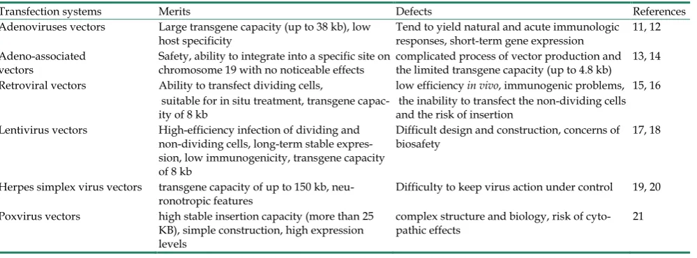
Lipids
Among the non-viral vectors, liposomes based
on cationic lipids are by far the most common gene
delivery systems and have been the subject of
con-siderable interests as non-viral delivery vectors
[49-50]. Liposomes are synthetic lipid spheres
com-posed by fatty acid on polymers with one or more
bilayered membrane structure surrounding an
aque-ous core that can be used to encapsulate small
mole-cules (
Figure 2A, 2B
). The direct complex formation of
cationic lipids with pDNA results in the self-assembly
of liposomes [51-52]. Several parameters, which
in-fluence the lipoplex formation efficacy, such as
prep-aration procedure, mixing ratio, pDNA concentration,
size of the applied cationic liposomes and ionic
strength of the buffer were investigated [53].
As known, liposomes have the distinct
ad-vantages of being both nontoxic and biodegradable
because they are composed of naturally occurring
substances. Liposomes have been shown to provide
stable encapsulation for various molecules like gene,
which can protect DNA against enzymatic
degrada-tion as well as facilitate cellular uptake and
endoso-mal escape, leading to effective gene transfer. They
have possessed not only the excellent biocompatibility
and low immunogenicity, but also the ability to
de-liver large pieces of DNA with well defined
physico-chemical composition and ease of handling and
preparation. Furthermore, they have potential to
transfect all kinds of tissue and cell types [54-57]. An
upsurge of global interest in developing efficient
cat-ionic lipids for gene delivery was therefore witnessed
in recent years [58-60].
However, due to their positive charge, cationic
liposomes may undergo non-specific interaction with
negatively charged cellular components (such as
se-rum protein and enzymes), which may result in
re-duction of cellular adhesion, hemolysis, and low
transfection efficiency [61-62]. In addition, organic
reagents such as ethylether and chloroform are
in-volved in the preparation of liposomes, which may be
harmful to both the cells and tissues.
In general, cationic liposomes are not good
enough for effective gene therapy because of their
potential cytotoxicity and low transfection efficiency
[63-66]. Therefore, it is significantly important to
de-velop novel nontoxic cationic systems with both
ef-fective gene transfection ability and good safety.
lead-ing to small particle sizes, controlled structures,
reg-ular morphology, and good stability [79].
To evaluate the potential of the lipid transfection
reagents in the clinical application, many researchers
have embarked on the transfection experiments
in vivo
with functional gene [80-81]. Nabel et al. have
devel-oped a protocol which relies upon the direct
trans-mission of human HLA-B7 gene into established
tu-mors
in vivo
, which can perform successful gene
ex-pression and show no apparent toxicity [81].
Above all, cationic nano-sized liposomes have
been investigated widely for a long time both
in vitro
and
in vivo
. Their gene delivery efficiency can be
im-proved by intergrating with varying auxiliary
meth-ods, such as the availability of light sources and
highly sensitive detection technologies [82]. But the
progress is not great enough and the transfection
effi-ciency and cellular non-toxicity have not been
quali-fied to effective gene delivery. In other words, it is a
way full of difficulties and challenges for cationic
liposomes to move from researches to clinical
appli-cations in order to contribute to gene therapy.
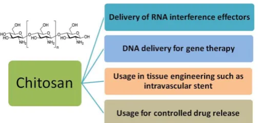
Polymeric carriers
Cationic polymers have shown promise as a safe,
predictable, biodegradable and nontoxic alternative to
viral gene therapy, relying on endocytosis of synthetic
polymer-based carriers bio-conjugated to the targeted
gene or other biological molecules [83-84]. Cationic
polymer-based gene carriers (polyplexes) showed
good biodegradability, low toxicity, triggered nucleic
acid release, structural diversity and relatively higher
transfection efficiency than liposomes [85-87], and the
gene delivery process of polymers is described in
Figure 3
. Many kinds of polymers have therefore been
investigated for gene delivery, such as chitosan, PEI,
polylysine, polyamino ester and so on [88-92].
Cati-onic segments, organelle-escape units, and
degrada-ble fragments are essential to a polymer-based vehicle
for gene delivery. The majority of these cationic
seg-ments are derived from polyamines, including
pol-ylysine, polyarginine, chitosan, polyethylenimine and
polyamidoamine dendrimers. Not only do these
cat-ionic polyamines protect DNA from degradation,
they can also promote the endocytosis of the carriers
by endosomal membranes. Degradable fragments are
necessary for the carriers to release the DNA once the
complexes enter the cytoplasm. Furthermore, the
or-ganelle-escape units are one of the key elements
in-fluencing the transfection efficiency.
Fig.2.
Schematic representation of the structure of liposomes.
However, there still exist numerous
in vitro
and
in vivo
barriers for polymer-based vehicles to achieve
ideal transfection efficiency. For
in vitro
transfection,
polymeric nanoparticles have no structure as magic as
viruses to enter cells and just depend on the
unpre-dictable endocytosis. Thus on one hand more detailed
information about the endocytosis mechanism
ob-tained by varying technical methods is needed to
in-crease the entrance into the cells. On the other hand,
the size, surface potential and N/P ratio (ratio of
ni-trogen of polymer to phosphate of DNA) were deeply
investigated and some targeting groups like nuclear
localization signal were integrated to the
nanoparti-cles. Morover, the escape of nanoparticles from the
organelle and DNA from the complexes is one of the
key elements influencing the transfection efficiency.
For example, particular interest was garnered by PEI
mainly for its organelle-escape units. Another big
problem for polymeric nanoparticles is the
cytotoxi-city, which results from poor biodegradability. So the
degradable moieties have been incorporated into
polymer, such as coating with human serum
albu-min, dextran, PEG and so forth. Out of these, the
spe-cific hurdle for transfection
in vivo
is that polymeric
nanoparticles are foreign materials whose invasion
will lead to the immune response by the body. The
most effective solution is PEGylation, which help
avoid the clearance of the reticuloendothelial system.
Many researchers are dedicated to devising
poly-mer-based vehicles for exogenous gene transfection
in
vitro
and great efforts have been made for gene
transfection
in vivo
.
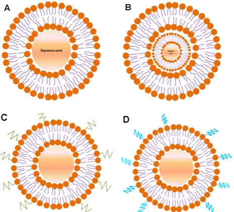
Chitosan
During the past 20 years, a good many
in vitro
and
in vivo
studies focusing on gene therapy have
been reported on chitosan for its outstanding
bio-degradability (
Figure 4
) [93-95]. Chitosan is an
attrac-tive polymer for gene delivery, showing excellent
biocompatibility, admirable biodegradability,
ecolog-ical safety, low toxicity, antimicrobial activity and low
immunogenicity [96-100]. And chitosan with
abun-dant amine and hydroxyl groups can be easily
modi-fied to enhance efficiency for gene transfer [101].
However, their application in gene delivery is limited
for their insolubility under physiological pH
condi-tion, insufficient charge and low transfection
effi-ciency [102-103].
The chitosan-DNA complexes are very easy to
synthesize and are more effective in comparison with
the commonly used polygalactosamine-DNA
com-plexes. The stability of the complexes depends on
several factors such as chitosan chain length and ratio
of chitosan/DNA. The
in vitro
transfection efficiency
was affected by factors such as the degree of
deacety-lation (DDA), molecular weight (MW) of chitosan,
nitrogen atoms (N) in the chitosan/the phosphorus
atoms (P) in DNA (N/P) ratio, physicochemical
properties of polyplexes, and so forth [104-107].
Con-sidering these important factors, great efforts have
been devoted to obtain higher transfection efficiency
[107-109]. Taking the MW for example, investigation
on the MW of the polymer revealed that on one hand
high MW chitosan offered better nucleic acid
com-plexation and stability but showed drawbacks such as
aggregation and low solubility at neutral pH
[110-112]. On the other hand, low MW chitosan allows
for a more efficient intracellular release but low
com-plexation [113]. Hence finding a balance between
these opposing effects is important to obtain a
chi-tosan carrier of optimal MW. To address the problem
of insolubility under physiological pH condition,
hy-drophobic and hydrophilic modifications have been
applied to obtain chitosan derivatives with favorable
characteristics. Hydrophobic modifications, such as
deoxycholic acid modification and thiolation, could
reinforce cell binding, alleviate serum inhibition,
protect the pDNA from enzymatic degradation, and
facilitate pDNA internalization, which have been
proven good for efficient gene transfer [114-117].
While a plenty of hydrophilic modifications, such as
PEGylation and quaternization, were also introduced
to enhance transfection efficiency due to increased
water solubility at physiological pH and improved
intracellular pDNA release [118-120]. The distinct
benefits of hydrophobic and hydrophilic
modifica-tions have promoted the possibility of obtaining
well-defined amphiphilic chitosan derivatives
[121-122]. Nevertheless, certain factors acted
differ-ently when combined with various states of other
factors. What is most important is therefore to find a
fine balance between all the tunable influencing
fac-tors on chitosan to get ideal transfection efficiency
[123-125].
The modes of administration also significantly
influence gene delivery efficiency and new methods
have been reported [126-127]. Using a novel
tech-nique, Dai et al. studied the gene delivery by
chi-tosan-DNA nanoparticles through retrograde
intra-biliary infusion (RII), which proved a possible routine
to achieve low-toxic, liver-targeted gene delivery
[127].
Currently, the optimization of chitosan-based
vectors for gene delivery includes two dominant
points, the modification of chitosan itself and
combi-nation with other vectors [128]. Consequently, the
former should be incorporated with the latter, which
may be the development tendency of chitosan-based
vectors [129]. Liu et al. synthesized amphiphilic
chi-tosan-N-octyl-N-quatenary chitosan (OTMCS)-PEI
which revealed lower cytotoxicity and the
OTMCS-PEI/DNA complexes showed higher
trans-fection efficiency
in vitro
and
in vivo
in comparison
with PEI 25 kDa, the commercially available one [130].
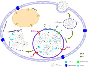
Polyethylenimine (PEI)
Among various kinds of cationic polymers, PEI
has been the most widely investigated because of not
only its strong DNA condensation capacity and
in-trinsic endosomal activity, but also its unique
buffer-ing capacity called proton sponge effect (
Figure 5
). It
facilitates the release of the gene into the cytoplasm
through osmotic swelling and burst of endosomes
[131-132].
Figure 6
shows two forms of PEI, linear PEI
(lPEI) and branched PEI (bPEI). PEI nanoparticles
have been prepared by two strategies that either by
complexation of PEI with DNA to form
nano-complexes [133] or use of cross-linkers to first
form PEI nanoparticles followed by DNA loading
onto it [134-135].
The transfection efficiency of PEI/pDNA
poly-plexes is associated with the N/P ratio [135-136]. PEI
polyplexes of N/P ratios higher than 3 contain an
excess of free PEI which is supposed to contribute to
the endosomal escape [137], which explains the
usu-ally enhanced transfection efficiency at higher N/P
ratios. The transfection efficiency of PEI/pDNA
com-plexes depends largely on the molecular weight (MW)
of PEI. Commercially available PEI 25 kDa has
excel-lent transfection efficiency in comparison with
low-molecular-weight PEI (LMW PEI) [138-140]. With
its high cationic charge density, it can effectively
condense DNA and form nanometer-sized particles
capable of being endocytosed [141]. However,
high-molecular-weight PEI (HMW PEI) (25 kDa), with
efficient gene transfection
in vitro,
lacks degradable
linkages (C-C or C-N bonds) and is too toxic for
therapeutic applications, which contributes to an
acute cytotoxicity due to cell membrane disruption
followed by induction of apoptosis [142-143]. On the
contrary, LMW PEIs possess low cytotoxicity as well
as undesirable transfection efficiency [139, 144-146]. It
is therefore a significant challenge to prepare
PEI-based vectors with high transfection efficiency as
well as low cytotoxicity [147-149]. Moreover, when it
comes to
in vivo
transfection, PEI has the tendency to
aggregate red blood cells, bind complement
compo-nents, and is not easy to be broken down and excreted
[150-151].
Fig.6.
Two forms of PEI. (A) linear PEI, (B) branched PEI.
There are two primary ways to overcome these
problems, modification of HMW PEIs to decrease the
cytotoxicity and modification of LMW PEIs to
im-prove the transfection efficiency. As for HMW PEIs,
degradable linkers are used to conjugate PEI to yield
polymers with sufficient amounts of amino groups.
Lee et al. first introduced disulfide cross-linked PEI
using dithiobis succinimidyl propionate and
dime-thyl-3,3’-0-dithiobispropionimidate as cross-linking
agents, which showed apparently reduced
cytotoxi-city [152]. Breunig et al. have reported that several cell
lines transfected with disulfide crosslinked lPEI has
shown transfection efficiencies of over 60% under
conditions that maintain a cell viability >90%
[153-154]. Studies of PEI crosslinked with other
hy-drolytically degradable groups have also been
re-ported with efficiencies near or exceeding that of
commercially available standards, as well as reduced
cytotoxicity [155-162].
To improve the transfection efficiency of LMW
PEIs, one of the main approaches is to incorporate
hydrophobic moieties into LMW PEIs. In previous
studies, some hydrophobic segments have been
in-troduced into LMW PEIs, such as polycaprolactone,
cholesterol, lipid and other hydrophobic substituents
[163-169]. Those results indicated that transfection
efficiency could be apparently improved after
intro-ducing hydrophobic chains into LMW PEIs, which
might result from the unique properties of
am-phiphilic cationic polymers. The self-assembly of
amphiphilic polymers could improve the local
cati-onic density and then facilitate nucleic acid
conden-sation. In addition, hydrophobic substituents were
expected to enhance interactions with lipophilic cell
membranes and facilitate the uptake of the complexes.
After release of the nucleic acids, self-assemblies of
amphiphilic polymers might be disassembled into
unimers, leading to relatively low cytotoxicity [170].
In addition, disulfide cross-linked PEI-SSX and
es-ter-cross-linked PEI derivatives based on different
cross-linking agents and LMW PEI have also been
investigated and exhibited higher gene transfection
efficiency and lower cytotoxicity than PEI (25 kDa) in
different types of cell lines [171-173]. Besides, the
ad-justment of particle size can also lead to effective gene
transfer. Recently, it has been reported that
copoly-mers obtained by the reaction between PCL diacrylate
and LMW PEI show effective and stable DNA
con-densation with particle sizes less than 200 nm [174].
No matter HMW PEIs or LMW PEIs,
intracellu-lar release of DNA from the complexes is of the
ut-most importance for the transfection efficiency. In
order to trigger intracellular release of therapeutic
genes from cross-linked PEI, there are two promising
methods, hydrolytic degradation and disulfide
re-duction. Disulfide cross-linked polymers were
syn-thesized to stimulate the DNA release in the
cyto-plasm by intracellular reducing environment [175].
In conclusion, although PEI has been
investi-gated widely in animals and humans for gene
deliv-ery
in vitro
and
in vivo
[176], there exist a lot of
chal-lenges such as the cytotoxicity, insufficient
transfec-tion efficiency and instability of the complexes in
complicated physiological environments, which
blocks their clinical applications. Many kinds of PEI
derivatives have shown relatively lower cytotoxicity
and higher transfection efficiency, nevertheless, it is
really a long way to go before using them for clinical
treatment. Generally, new vectors with well-marked
reduced toxicity, improved transfection efficiency and
better stability of the complexes
in vivo
, are of great
importance. That is to say, great emphasis should be
placed on the inner mechanism of toxicity, effective
targeting of cells or organs, enhanced nuclear
locali-zation and how to pass through the barriers to cells or
nuclei.
Polyamidoamine Dendrimers
Dendrimers have an increased ionic interaction
with DNA and produce very stable and highly soluble
DNA complexes [188]. However, there exist several
problems to be solved. First, although PAMAM
den-drimer-based gene transfection reagents such as
Su-perfect and Priofect have already been commercially
available, these products are more expensive than
other cationic polymers and are based on high
gener-ation dendrimers, the synthesis of which is
la-bor-consuming [189]. Second, the transfection
effi-ciency of PAMAM dendrimers is
genera-tion-dependent. Low-generation PAMAM, such as
G0-G3 show
poor gene transfection efficiency and
lower cytotoxicity, but high-generation PAMAM,
such as G4-G8 exhibit slightly better gene transfection
efficiency and higher cytotoxicity [190-192]. But ideal
gene transfection reagents should have both high
transfection effi
ciency and low cytotoxicity and thus
plenty of efforts have been made to achieve it
[193,
194]. On one hand, high generation
PAMAM dendrimers
were functionalized with different moieties as showed
in
Table 3
.
However, high
transfection effi
ciency is
usu-ally accompanied with high
cytotoxicity in these gene
delivery systems. So the paradox between transfection
efficiency and cytotoxicity makes it of crucial
im-portance to break up the correlation between them
for
PAMAM dendrimer-based gene carriers
in a near future
[210].
A promising solution to this issue is to prepare
highly effi
cient gene transfection reagents using
low
generation dendrimers. Liu et al. synthesized
disul-fide cross-linked low generation PAMAM dendrimers
for gene delivery which showed higher transfection
efficiency than G2 and G5 PAMAM dendrimers and
comparable efficiency with bPEI 25 kDa [192].
Ueka-ma et al. developed cyclodextrin-modified low
gen-eration PAMAM dendrimers for DNA delivery
[201-203, 211]. In their studies, relatively high gene
transfection effi
ciency and low
cytotoxicity of the
cy-clodextrin-dendrimer conjugates were achieved. On
the other hand, hyper-branched PAMAM dendrimers
and their modifications have been extensively
inves-tigated to increase pDNA binding [212-215]. An
in-teresting option to obtain efficient gene delivery
car-riers is to increase dendrimer flexibility using a
tri-ethanolamine core. In this case the enhanced distance
of the first generation branching points to the central
amine leads to a more flexible dendrimer and thus
increases pDNA binding [215]. Zeynep et al. prepared
Jeffamine-cored PAMAM dendrimers (JCPDs) for
gene delivery, which showed considerably improved
transfection efficiency as the number of generation
increased [216]. JCPD can therefore be considered as
an efficient transfection reagent and can be effectively
used for gene delivery applications, which is based on
the finding that partially degraded PAMAM
den-drimers show better flexibility and greater interaction
with pDNA and perform successful transfer of pDNA
[217]. Finally, the transfection efficiency of specific
dendrimer varies between different types of cells,
which is the common problem for all kinds of gene
carriers and may be closely related with different
structures of different cells [218].
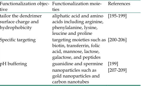
Table 3.
Functionalization of
high generation
PAMAM
den-drimers to achieve higher transfection efficiency.
Functionalization
objec-tive
Functionalization moie-
ties
References
tailor the dendrimer
surface charge and
hydrophobicity
aliphatic acid and amino
acids including arginine,
phenylalanine, lysine,
leucine and proline
[195-199]
Specific targeting
targeting moieties such as
biotin, transferrin, folic
acid, mannose, lactose,
galactose, and peptides
[200-206]
pH buffering
guanidine and spermine [199]
nanoparticles such as
gold nanoparticles and
carbon nanotubes
[207-209]
Poly (lactide-co-glycolide)
Poly (lactide-co-glycolide) (PLGA), as the name
suggests, is composed of lactic and glycolic acids,
which are linked together by ester linkages. The
polymer degrades when the free carboxylic end
groups form. The safety of PLGA-based nanoparticles
in the clinic has been well established [219].
Incorpo-ration of pDNA into PLGA particles protects pDNA
from
in vivo
degradation, triggers the controlled
re-lease of the pDNA, and acts as a transfection vector
for mammalian cells [220-223]. A PLGA based
deliv-ery system for pDNA has been widely studied and
demonstrated effectively [224-232].
nanoscale would help the protection of pDNA from
degrading enzymes [240]. Loyter et al. has also
em-phasized the importance of the nano-scaled particle
size after studying the intracellular transport of
3H-marked DNA [241]. And in some cell lines, only
the nano-sized particles are taken up efficiently other
than microparticles (e.g. Hepa1-6, HepG2, and
KLN205) [242]. Those studies revealed the importance
of the particle size in gene transfection efficiency.
Recently, Tang et al. reported a simple and
re-producible method to prepare well-defined
CaP-pDNA PLGA nanoparticles for the incorporation
of pDNA, which will enhance pDNA encapsulation
within the particle, provide an ideal release rate of
pDNA, and increase transfection efficiency
in vitro
[243]. In another study, Shau et al. concluded that a
one-step preparation of nanoparticles with PLGA
pre-modified with PEI is better in requirements for
DNA delivery compared with those prepared in a
two-step process (preformed PLGA nanoparticles and
subsequently coated with PEI) [244]. Zeng et al.
indi-cated that nanoparticles fabriindi-cated with PLGA alone
resulted in poor encapsulation of pDNA while
chi-tosan-modified PLGA nanoparticles exhibited much
higher loading efficiency than unmodified PLGA
nanoparticles [245].
Generally, PLGA-based nanoparticles have
al-ready been widely investigated in drug delivery
in
vivo
for its safety and used for clinical usage, but its
application for ideal gene delivery remains a way
filled with challenges and prospects.
Other polymeric carriers
As the polymers arouse more attention of the
researchers, more and more new polymeric carriers
have been developed for gene transfer. Some typical
polymeric carriers are listed in
Table 4
and at the
same time many other polymeric carriers such as
polypeptide and polyphosphoester have also been
reported [246-250].
Poly-L-lysine (PLL) is a kind of natural and
bio-compatible polypeptide with excellent pDNA
con-densation capacity and one of the cationic polymers
firstly used for gene delivery [251-252]. However, the
high cytotoxicity and the low transfection efficiency of
PLL severely limit its wide application in gene
deliv-ery [253-257].
Many attempts have been made to improve the
gene delivery efficiency. For example, PEG was
in-troduced to reduce the cytotoxicity [258],
target
lig-ands such as transferrin, folate, antibodies and basic
fibroblast growth factor were tethered to promote
complex cellular uptake [259-262], and histidine
de-rivatives were grafted to improve the endosomal
es-cape of the complexes [263]. Nevertheless, the
appli-cations of PLL are widely limited in comparison with
other polymers like PEI, CS and PAMAM [264-265].
Combination
Chemical vectors for gene delivery based on
only one kind of pure materials, such as lipid or
chi-tosan, have not only their special advantages but also
their intrinsic deficiencies which are difficult to
solve. To improve the transfection efficiency and
re-duce the cytotoxicity, many researchers have focused
on combining two or more kinds of materials to
en-hance the transfection efficiency and at the same time
avoiding the side effects to the cells [266].
Den-dron-bearing lipids with PAMAM G1 designated as
DL-G1-2C18 have been synthesized. In spite of less
efficient cellular uptake of the lipoplexes, they
gener-ated free pDNA molecules in the cytosol more
effec-tively than other lipoplexes did [267].
Table 4.
Summary of polymeric carriers.
Carriers
Main advantages
Main limitations
References
Chitosan
Good biocompatibility and biodegradability; low
im-munogenicity; low toxicity; antimicrobial activity;
Low insolubility under physiological pH con-
dition; low transfection efficiency;
[94, 95, 98, 100, 104,
105]
PEI
Strong DNA condensation capacity; intrinsic
endoso-mal activity; unique buffering capacity; high
transfec-tion efficiency;
Bad biodegradability; the contradiction
be-tween transfection efficiency and cytotoxicity;
[135, 138,
139, 140,
142, 143,
146]
PAMAM
surface functionality; relatively high transfection
effi-ciency; uniform size distribution; lower cytotoxicity;
Low transfection efficiency;
[177, 179,
180, 183,
186, 187]
PLGA
Safety; good biodegradability;
Low release rate and low encapsulation
effi-ciency of pDNA; acidic microenvironment
induced by it;
[224, 226,
227, 234,
235]
PLL
Excellent pDNA condensation capacity;
Relatively high cytotoxicity; low transfection
Most importantly, the core-shell nanoparticles
for gene delivery have been the hot topic in the past
few years. Although calcium-phosphate precipitation
has been used to deliver pDNA for decades, the
method is typically characterized by low transfection
efficiency relative to the other non-viral approaches,
such as liposomes and polymers. Zhou et al. have
developed a novel gene transfer vector comprising
lipid-coated nano-calcium-phosphate (LNCP) that
provides consistently efficient and satisfactory pDNA
delivery [268]. It is based on core-shell nanoparticles
comprising of a calcium-phosphate core and a cationic
lipid shell. This method, in contrast to the solution
precipitation methods used in the past, yields
colloi-dally stable calcium-phosphate nanoparticles inside
the cationic liposomes. The LNCP has shown great
potential as a novel transfection agent for gene
ther-apy. Jiang et al. have designed another cationic
core-shell lipo-nanoparticle (DLCS-NP) by
envelop-ing the plasmid-laden chitosan nanoparticle (CSNP)
into a cationic lipid shell to achieve enhanced gene
transfection efficiency for ocular eye-drop therapy
[269]. The cellular uptake of DLCS-NP is up to
1.25-fold and 5-fold higher than that of CS-NP and
lipid-coated chitosan nanoparticles (LCS-NP),
respec-tively. Moreover, it is another promising direction for
gene delivery to combine with advanced theranostic
systems and a number of liposomes and polymeric
carriers combined with approved therapeutics have
entered clinical use or are under different stages of
clinical application [270-274].
Conclusion
All the gene transfer methods (viral, physical or
chemical) must overcome two major constraints: first,
there is a need to carry the nucleic acids to the target
cells without potential risks. Viruses are ‘naturally’
equipments with ability to recognize and locate
de-fined target cells and are ‘stealthy’ with respect to the
body defense mechanisms, such as the
reticu-lo-endothelial system (RES). While chemical vectors
should combine with other technologies or conjugate
with targeting molecules to realize the specific
loca-tion. The second constraint is the penetration of the
nucleic acids into the cell through the plasma
mem-brane. While viruses achieve this purpose using
nat-ural mechanisms, chemical vectors must perturb the
plasma membrane (e.g. physical vectors) and/or
in-ternal vesicular membranes (e.g. the cationic lipids).
In order to obtain an efficient vector system and
achieve a high rate of cell transfection, these two
con-ditions must be integrated in the development of an
ideal genetic vector.
Consequently, deep investigation of the
trans-fection process incluing the endocytose of
nanocarri-ers and the release of DNA from the complexes is the
prerequisite for gene delivery research in the future.
Only with deep understanding of the mechanism can
we suit our methods to the situation. To pay more
attention to materials with excellent biodegradability
and biocompability is an important alternative to get
effective gene delivery nanocarriers in the near future.
Further, functional moieties such as cell penetration
peptides to the nanocarriers could be popular to
im-prove the transfection efficiency and at the same time
decrease the cytotoxicity. What’s more, different gene
delivery systems should be effectively combined to
make best use of the advantages and bypass the
dis-advantages, which tend to be the main stream in gene
delivery. Inorganic nanoparticles possess many
ad-vantages in gene transfection, e.g. they are not subject
to microbial attack and exhibit good storage stability
[275]. As a result of their small size, nanoparticles can
penetrate the cell membrane and deliver drugs or
biomolecules into living systems with moderate
tox-icity [276-277] or without any toxic effects on cells
[278], depending on the concentration of
nanoparti-cles used. There is a wide variety of nanopartinanoparti-cles and
many of them have been tested
in vitro
and in some
cases even in clinical trials, e.g. carbon nanotubes,
chitosan nanoparticles, calcium phosphate
nanoparti-cles and lipid nanopartinanoparti-cles [279-282]. In conclusion, it
is not only a way full of challenges and difficulties,
but also a way filled with hope and prospect in
med-ical fields.
Acknowledgments
This research was financially supported by the
National Key Program for Developing Basic Research
of China (2010CB933903 and 2014CB744501), the
NSFC (61271056 and 31201003), Hunan Science and
Technology Projects (2012SK3105), Scientific Research
Fund of Hunan Provincial Education Department
(11A030), China Postdoctoral Science Foundation
(20100471362, 2012M520980 and 2012M511660),
Jiangsu Postdoctoral Science Foundation (1301093C),
Open Research Fund by State Key Laboratory of
Bio-electronics, Southeast University, China (2013G10)
and Economical Forest Cultivation and Utilization of
2011 Collaborative Innovation Center in Hunan
Province.
Competing Interests
The authors have declared that no competing
interest exists.
References
2. Gajjar M, Candeias MM, Malbert-Colas L, Mazars A, Fujita J, Olivares-Illana V, Fåhraeus R. The p53 mRNA-Mdm2 interaction controls Mdm2 nuclear trafficking and is required for p53 activation following DNA damage. Cancer Cell. 2012; 21(1): 25-35.
3. Moon EK, Son M, Jin YW, Park S, Lee WJ. Variations of Lung Cancer Risk from Asbestos Exposure: Impact on estimation of population attributable fraction. Ind Health. 2013; 51(1): 128-133.
4. Chang HJ, Ko HL, Lee CY, Wu RH, Yeh YW, Jiang JS, Kao SJ, Chi KH. Hypofractionated radiotherapy for primary or secondary oligometastatic lung cancer using Tomotherapy. Radiat Oncol. 2012; 7: 222.
5. Henson KE, McGale P, Taylor C, Darby SC. Radiation-related mortality from heart disease and lung cancer more than 20 years after radiotherapy for breast cancer. Br J Cancer. 2013; 108(1): 179-182.
6. Gill S, Kalos M. T cell-based gene therapy of cancer. Transl Res. 2013; 161(4): 365-379.
7. Song H, Song C, Wang H, et al. Suppression of hepatocarcinoma model in vitro and in vivo by ECRG2 delivery using adenoviral vector. Cancer Gene Ther. 2012; 19(12): 875-879.
8. Mannell H, Pircher J, Fochler F, Stampnik Y, Räthel T, Gleich B, Plank C, Mykhaylyk O, Dahmani C, Wörnle M, Ribeiro A, Pohl U, Krötz F. Site di-rected vascular gene delivery in vivo by ultrasonic destruction of magnetic nanoparticle coated microbubbles. Nanomedicine. 2012; 8(8): 1309-1318. 9. Iwashita S, Hiramatsu Y, Otani T, Amano C, Hirai M, Oie K, Yuba E, Kono
K, Miyamoto M, Igarashi K. Polyamidoamine dendron-bearing lipid assem-blies: Their morphologies and gene transfection ability. J Biomater Appl. 2012; 27(4): 445-456.
10. Siu YS, Li L, Leung MF, Lee KL, Li P. Polyethylenimine-based amphiphilic core-shell nanoparticles: Study of gene delivery and intracellular trafficking. Biointerphases. 2012; 7(1-4): 16.
11. Thrasher AJ, Gaspar HB, Baum C, Modlich U, Schambach A, Candotti F, Otsu M, Sorrentino B, Scobie L, Cameron E, Blyth K, Neil J, Abina SH, Cavazzana-Calvo M, Fischer A. Gene therapy: X-SCID transgene leu-kaemologenicity. Nature. 2006; 443(7109): E5–E6.
12. Zhang Y, Gao Y, Speth RC, Jiang N, Mao Y, Sumners C, Li H. Adenoviral and adeno-associated viral vectors-mediated neuronal gene transfer to cardiovas-cular control regions of the rat brain. Int J Med Sci. 2013; 10(5): 607-616. 13. Teramato S, Ishii T, Matsuse T. Crisis of adenoviruses in human gene
thera-py. Lancet. 2000; 355(9218): 1911–1912.
14. Okada H, Iizuka T, Mochizuki H, Nihira T, Kamiya K, Inoshita A, Kasagi H, Kasai M, Ikeda K. Gene transfer targeting mouse vestibule us-ing adenovirus and adeno-associated virus vectors. Otol Neurotol. 2012; 33(4): 655-659.
15. Anson DS. The use of retroviral vectors for gene therapy-what are the risks? A review of retroviral pathogenesis and its relevance to retroviral vec-tor-mediated gene delivery. Genet Vaccines Ther. 2004; 2(1): 9.
16. Frederic D. Retroviral integration and human gene therapy. J Clin Invest. 2007; 117(8): 2083-2086.
17. Goss JR, Mata M, Goins WF, Wu HH, Glorioso JC, Fink DJ. Antinociceptive effect of a genomic herpes simplex virus-based vector expressing human proenkephalin in rat dorsal root ganglion. Gene Ther.2001; 8(7): 551-556. 18. Real G, Monteiro F, Burger C, Alves PM. Improvement of lentiviral transfer
vectors using cis-acting regulatory elements for increased gene expression. Appl Microbiol Biotechnol. 2011; 91(6): 1581-91.
19. Lachmann RH, Efstathiou S. The use of herpes simplex virus-based vectors for gene delivery to the nervous system. Mol Med Today. 1997; 3(9): 404-411. 20. Liu S, Dai M, You L, Zhao Y. Advance in herpes simplex viruses for cancer
therapy. Sci China Life Sci. 2013; 56(4): 298-305.
21. Moss B. Reflections on the early development of poxvirus vectors. Vaccine. 2013; 31(39): 4220-4222.
22. Mulligan RC. The basic science of gene therapy. Science. 1993; 260: 926-932. 23. [No authors listed]. HIV vaccine failure prompts Merck to halt trial.
Na-ture. 2007; 449(7161): 390.
24. Ledford H. HIV vaccine may raise risk. Nature. 2007; 450(7168): 325. 25. Sirsi SR, Borden MA. Advances in ultrasound mediated gene therapy using
microbubble contrast agents. Theranostics. 2012; 2(12): 1208-1222.
26. Naldini L, Blömer U, Gallay P, Ory D, Mulligan R, Gage FH, Verma IM, Trono D. In vivo gene delivery and stable transduction of nondividing cells by a len-tiviral vector. Science. 1996; 272(5259): 263-267.
27. Panje CM, Wang DS, Pysz MA, Paulmurugan R, Ren Y, Tranquart F, Tian L, Willmann JK. Ultrasound-mediated gene delivery with cationic versus neutral microbubbles: Effect of DNA and microbubble dose on in vivo trans-fection efficiency. Theranostics. 2012; 2(11): 1078-1091.
28. Gao X, Huang L. Cationic liposome-mediated gene transfer. Gene Ther. 1995; 2(10): 710-722.
29. Orio J, Coustets M, Mauroy C, Teissie J. Electric field orientation for gene delivery using high-voltage and low-voltage pulses. J Membr Biol. 2012; 245(10): 661-666.
30. Robinson HL, Pertmer TM. Nucleic acid immunizations. Curr Protoc Immu-nol. 2001; Chapter 2: Unit 2.14.
31. Vu L, Ramos J, Potta T, Rege K. Generation of a focused poly (amino ether) library: polymer-mediated transgene delivery and gold-nanorod based theranostic systems. Theranostics. 2012; 2(12): 1160-1173.
32. Khosravi-Darani K, Mozafari MR, Rashidi L, Mohammadi M. Calcium based non-viral gene delivery: an overview of methodology and applications. Acta Med Iranica. 2010; 48(3): 133-141.
33. Luten J, van Nostrum CF, De Smedt SC, Hennink WE. Biodegradable poly-mers as non-viral carriers for plasmid DNA delivery. J Control Release. 2008; 126(2): 97-110.
34. Yamano S, Dai J, Moursi AM. Comparison of transfection efficiency of nonviral gene transfer reagents. Mol Biotechnol. 2010; 46(3): 287-300. 35. Keifer OP Jr, O'Connor DM, Boulis NM. Gene and protein therapies utilizing
VEGF for ALS. Pharmacol Ther. 2013; doi: 10.1016/j.pharmthera.2013.10.009. 36. Edmundson M, Thanh NT, Song B. Nanoparticles based stem cell tracking in
regenerative medicine. Theranostics. 2013; 3(8): 573-582.
37. Li D, Liu S, Shan H, Conti P, Li Z. Urokinase plasminogen activator receptor (uPAR) targeted nuclear imaging and radionuclide therapy. Theranostics. 2013; 3(7): 507-515.
38. Graham FL, Van der Eb AJ. A new technique for the assay of infectivity of human adenovirus 5 DNA. Virology. 1973; 52(2): 456-467.
39. Orrantia E, Chang PL. Intracellular distribution of DNA internalized through calcium phosphate precipitation. Exp Cell Res. 1990; 190(2): 170-174. 40. Cao X, Deng W, Wei Y, Su W, Yang Y, Wei Y, Yu J, Xu X. Encapsulation of
plasmid DNA in calcium phosphate nanoparticles: stem cell uptake and gene transfer efficiency. Int J Nanomedicine. 2011; 6: 3335-3349.
41. Hu J, Kovtun A, Tomaszewski A, Singer BB, Seitz B, Epple M, Steuhl KP, Ergün S, Fuchsluger TA. A new tool for the transfection of corneal endo-thelial cells: Calcium phosphate nanoparticles. Acta Biomater. 2012; 8(3): 1156-1163.
42. Kingston RE, Chen CA, Okayama H. Calcium phosphate transfection. Curr Protoc Immunol. 2001; Chapter 10:Unit 10.13.
43. Olton D, Li J, Wilson ME, Rogers T, Close J, Huang L, Kumta PN, Sfeir C. Nanostructured calcium phosphates (NanoCaPs) for non-viral gene delivery: Influence of the synthesis parameters on transfection efficiency. Biomaterials. 2007; 28(6): 1267-1279.
44. Sokolova V, Radtke I, Heumann R, Epple M. Effective transfection of cells with multi-shell calcium phosphate-DNA nanoparticles. Biomaterials. 2006; 27(16): 3147-3153.
45. Pedraza CE, Bassett DC, McKee MD, Nelea V, Gbureck U, Barralet JE. The importance of particle size and DNA condensation salt for calcium phosphate nanoparticle transfection. Biomaterials. 2008; 29(23): 3384-3392.
46. Elangovan S, Jain S, Tsai PC, Margolis HC, Amiji M. Nano-sized calcium phosphate particles for periodontal gene therapy. J Periodontol. 2013; 84(1): 117-125.
47. Nouri A, Castro R, Santos JL, Fernandes C, Rodrigues J, Tomás H. Calcium phosphate-mediated gene delivery using simulated body fluid (SBF). Int J Pharm. 2012; 434(1-2): 199-208.
48. Bhakta G, Shrivastava A, and Maitra A. Magnesium phosphate nanoparticles can be efficiently used in vitro and in vivo as non-viral vectors for targeted gene delivery. J Biomed Nanotechnol. 2009; 5(1): 106-114.
49. Pezzoli D, Kajaste-Rudnitski A, Chiesa R, Candiani G. Lipid-based nanoparti-cles as nonviral gene delivery vectors. Methods Mol Biol. 2013; 1025: 269-279. 50. Zhi D, Zhang S, Wang B, Zhao Y, Yang B, Yu S. Transfection efficiency of
cationic lipids with different hydrophobic domains in gene delivery. Biocon-jug Chem. 2010; 21(4): 563-577.
51. Felgner PL, Barenholz Y, Behr JP, Cheng SH, Cullis P, Huang L, Jessee JA, Seymour L, Szoka F, Thierry AR, Wagner E, Wu G. Nomenclature for synthetic gene delivery systems. Hum Gene Ther. 1997; 8(5): 511-512. 52. Gao K, Huang L. Nonviral methods for siRNA delivery. Pharm. 2009; 6(3):
651-658.
53. Zelphati O, Nguyen C, Ferrari M, Felgner J, Tsai Y, Felgner PL. Stable and monodisperse lipoplex formulations for gene delivery. Gene Ther. 1998; 5(9): 1272-1282.
54. Liu Y, Liggitt D, Zhong W, Tu G, Gaensler K, Debs R. Cationic lipo-some-mediated intravenous gene delivery. J Biol Chem. 1995; 42(270): 24864-24870.
55. Stewart MJ, Plautz GE, Del BL, Yang ZY, Xu L, Gao X, Huang L, Nabel EG, Nabel GJ. Gene transfer in vivo with DNA-liposome complexes: safety and acute toxicity in mice. Hum Gene Ther. 1992; 3(3): 267-275.
56. Zhu N, Liggitt D, Liu Y, Debs R. Systemic gene expression after intravenous DNA delivery into adult mice. Science. 1993; 5118(261): 209-211.
57. Thierry AR, Lunardi-Iskandar Y, Bryant JL, Rabinovich P, Gallo RC, Mahan LC. Systemic gene therapy: biodistribution and long-term expression of a transgene in mice. Proc Natl Acad Sci U S A. 1995; 21(92): 9742-9746. 58. Mintzer MA, Simanek EE. Nonviral vectors for gene delivery. Chem Rev. 2009;
2(109): 259-302.
59. Bhattacharya S, Bajaj A. Advances in gene delivery through molecular design of cationic lipids. Chem Commun (Camb). 2009; 31: 4632-4656.
60. Martin B, Sainlos M, Aissaoui A, Oudrhiri N, Hauchecorne M, Vigneron JP, Lehn JM, Lehn P. The design of cationic lipids for gene delivery. Curr Pharm Des. 2005; 3(11): 375-394.
61. Escriou V, Ciolina C, Lacroix F, Byk G, Scherman D, Wils P. Cationic li-pid-mediated gene transfer: effect of serum on cellular uptake and intracellu-lar fate of lipopolyamine/DNA complexes. Biochim Biophys Acta. 1998; 2(1368): 276-288.
62. Zelphati O, Uyechi LS, Barron LG, Szoka FJ. Effect of serum components on the physico-chemical properties of cationic lipid/oligonucleotide complexes and on their interactions with cells. Biochim Biophys Acta. 1998; 2(1390): 119-133.
enhanced lipoplex stability and gene delivery. Bioconjug Chem. 2010; 2(21): 360-371.
64. Mevel M, Kamaly N, Carmona S, Oliver MH, Jorgensen MR, Crowther C, Salazar FH, Marion PL, Fujino M, Natori Y, Thanou M, Arbuthnot P, Yaouanc JJ, Jaffres PA, Miller AD. Dodag; A versatile new cationic lipid that mediates efficient delivery of pDNA and siRNA. J Control Release. 2010; 2(143): 222-232. 65. Morille M, Montier T, Legras P, Carmoy N, Brodin P, Pitard B, Benoit JP,
Passirani C. Long-circulating DNA lipid nanocapsules as new vector for pas-sive tumor targeting. Biomaterials. 2010; 2(31): 321-329.
66. Zhi D, Zhang S, Wang B, Zhao Y, Yang B, Yu S. Transfection efficiency of cationic lipids with different hydrophobic domains in gene delivery. Biocon-jug Chem. 2010; 4(21): 563-577.
67. Lv H, Zhang S, Wang B, Cui S, Yan J. Toxicity of cationic lipids and cationic polymers in gene delivery. J Control Release. 2006; 1(114): 100-109.
68. Dobbs W, Heinrich B, Bourgogne C, Donnio B, Terazzi E, Bonnet ME, Stock F, Erbacher P, Bolcato-Bellemin AL, Douce L. Mesomorphic imidazolium salts: new vectors for efficient siRNA transfection. J Am Chem Soc. 2009; 37(131): 13338-13346.
69. Nie Y, Ji L, Ding H, Xie L, Li L, He B, Wu Y, Gu Z. Cholesterol derivatives based charged liposomes for doxorubicin delivery: preparation, in vitro and in vivo characterization. Theranostics. 2012; 2(11): 1092-1103.
70. Zhu L, Lu Y, Miller DD, Mahato RI. Structural and formulation factors influ-encing pyridinium lipid-based gene transfer. Bioconjug Chem. 2008; 12(19): 2499-2512.
71. Ahmad A, Evans HM, Ewert K, George CX, Samuel CE, Safinya CR. New multivalent cationic lipids reveal bell curve for transfection efficiency versus membrane charge density: lipid-DNA complexes for gene delivery. J Gene Med. 2005; 6(7): 739-748.
72. Nie Y, Ji L, Ding H, Xie L, Li L, He B, Wu Y, Gu Z. Cholesterol derivatives based charged liposomes for doxorubicin delivery: preparation, in vitro and in vivo characterization. Theranostics. 2012; 2(11): 1092-1103.
73. Liberska A, Unciti-Broceta A, Bradley M. Very long-chain fatty tails for en-hanced transfection. Org Biomol Chem. 2009; 7(1): 61-68.
74. BajajA, Kondiah P, Bhattacharya S. Design, synthesis, and in vitro gene delivery efficacies of novel cholesterol-based gemini cationic lipids and their serum compatibility: a structure-activity investigation. J Med Chem. 2007; 50(10): 2432-2442.
75. Choi JS, Lee EJ, Jang HS, Park JS. New cationic liposomes for gene transfer into mammalian cells with high efficiency and low toxicity. Bioconjugate Chem. 2001; 1(12): 108-113.
76. Liu DL, Hu JJ, Qiao WH, Li ZS, Zhang SB, Cheng LB. Synthesis of carba-mate-linked lipids for gene delivery. Bioorg Med Chem Lett. 2005; 12(15): 3147-3150.
77. Spelios M, Kearns M, Savva M. From gene delivery to gene silencing: plasmid DNA-transfecting cationic lipid 1,3-dimyristoylamidopropane-2-[bis(2-dimethylaminoethane)] carbamate ef-ficiently promotes small interfering RNA-induced RNA interference. Bio-chemistry. 2010; 27(49): 5753-5759.
78. Yang HW, Yi JW, Bang EK, Jeon EM, Kim BH. Cationic nucleolipids as effi-cient siRNA carriers. Org Biomol Chem. 2011; 1(9): 291-296.
79. Li WJ, Szoka FC. Lipid-based nanoparticles for nucleic acid delivery. Pharm Res. 2007; 3(24): 438-449.
80. Xu XZ, Tu L, Wang LY, Fang XS, Wang DW. CYP2J3 gene delivery reduces insulin resistance via upregulation of eNOS in fructose-treated rats. Cardio-vasc Diabetol. 2011; 10: 114.
81. Nabel GJ, Nabel EG, Yang ZY, Fox BA, Plautz GE, Gao X, Huang L, Shu S, Gordon D, Chang AE. Direct gene transfer with DNA-liposome complexes in melanoma: Expression, biologicactivity, and lack of toxicity in humans. Proc Natl Acad Sci USA. 1993; 90(23): 11307-11311.
82. Leung SJ, Romanowski M. Light-activated content release from liposomes. Theranostics. 2012; 2(10): 1020-1036.
83. Zhang B, Ma XP, Murdoch W, Radosz M, Shen YQ. Bioreducible poly(amido amine)s with different branching degrees as gene delivery vectors. Biotechnol Bioeng. 2013; 3(110): 990-998.
84. Schaffert D, Troiber C, Wagner E. New sequence-defined polyaminoamides with tailored endosomolytic properties for plasmid DNA delivery. Bioconju-gate Chem. 2012; 6(23): 1157-1165.
85. Li WB, Yuan W, Xu FJ, Zhao C, Ma J, Zhan QM. Functional study of dex-tran-graft-poly ((2-dimethyl amino) ethyl methacrylate) gene delivery vector for tumor therapy. J Biomater Appl. 2013; 28(1): 125-35.
86. Wang YQ, Su J, Wu F, Lu P, Yuan LF, Yuan WE, Sheng J, Jin T. Biscarbamate cross-linked polyethylenimine derivative with low molecular weight, low cy-totoxicity, and high efficiency for gene delivery. Int J Nanomedicine. 2012; 7: 693-704.
87. Zhou J, Liu J, Cheng CJ, Patel TR, Weller CE, Piepmeier JM, Jiang Z, Saltzman WM. Biodegradable poly(amine-co-ester) terpolymers for targeted gene de-livery. Nat Mater. 2012; 1(11): 82-90.
88. Choi JS, Nam K, Park JY, Kim JB, Lee JK, Park JS. Enhanced transfection efficiency of PAMAM dendrimer by surface modification with l-arginine. J Control Release. 2004; 3(99): 445-456.
89. Pfeifer BA, Burdick JA, Little SR, Langer R. Poly(ester-anhydride): poly(beta-amino ester) micro- and nanospheres: DNA encapsulation and cel-lular transfection. Int J Pharm. 2005; 304(1-2): 210-219.
90. Anderson DG, Akinc A, Hossain N, Langer R. Structure/property studies of polymeric gene delivery using a library of poly(beta-amino esters). Mol Ther. 2005; 3(11): 426-434.
91. Hwang SJ, Bellocq NC, Davis ME. Effects of structure of be-ta-cyclodextrin-containing polymers on gene delivery. Bioconjugate Chem. 2001; 2(12): 280-290.
92. Kean T, Roth S, Thanou M. Trimethylated chitosans as non-viral gene delivery vectors: cytotoxicity and transfection efficiency. J Control Release. 2005; 3(103): 643-653.
93. Thanou M, Florea BI, Geldof M, Junginger HE, Borchard G. Quaternized chitosan oligomers as novel gene delivery vectors in epithelial cell lines. Bio-materials. 2002; 1(23): 153-159.
94. Bozkir A, Saka OM. Chitosan nanoparticles for plasmid DNA delivery: effect of chitosan molecular structure on formulation and release characteristics. Drug Deliv. 2004; 2(11): 107-112.
95. Borchard G. Chitosans for gene delivery. Adv Drug Deliver Rev. 2001; 2(52): 145-150.
96. Chandy T, Sharma CP. Chitosan - as a biomaterial. Biomater Artif Cells Artif Organs. 1990; 1(18): 1-24.
97. Rinaudo M. Main properties and current applications of some polysaccharides as biomaterials. Polym Int. 2008; 3(57): 397-430.
98. Mourya VK, Inamdar NN. Chitosan-modifications and applications: oppor-tunities galore. React Funct Polym. 2008; 6(68): 1013-1051.
99. Howard KA, Rahbek UL, Liu X, Damgaard CK, Glud SZ, Andersen MO, Hovgaard MB, Schmitz A, Nyengaard JR, Besenbacher F, Kjems J. RNA inter-ference in vitro and in vivo using a novel chitosan/siRNA nanoparticle sys-tem. Mol Ther. 2006; 4(14): 476-484.
100. Lavertu M, Methot S, Tran-Khanh N, Buschmann MD. High efficiency gene transfer using chitosan/DNA nanoparticles with specific combinations of molecular weight and degree of deacetylation. Biomaterials. 2006; 27(27): 4815-4824.
101. He XK, Yuan ZX, Wu XJ, Xu CQ, Li WY. Low molecular weight hydroxyethyl chitosan-prednisolone conjugate for renal targeting therapy: synthesis, char-acterization and in vivo studies. Theranostics. 2012; 2(11): 1054-1063. 102. Prabaharan M, Mano JF. Chitosan-based particles as controlled drug delivery
systems. Drug Deliv. 2005; 1(12): 41-57.
103. Lai WF, Lin M. Nucleic acid delivery with chitosan and its derivatives. J Control Release. 2009; 3(134): 158-168.
104. Mao SR, Sun W, Kissel T. Chitosan-based formulations for delivery of DNA and siRNA. Adv Drug Deliver Rev. 2010; 1(62): 12-27.
105. Sato T, Ishii T, Okahata Y. In vitro gene delivery mediated by chitosan. Effect of pH, serum, and molecular mass of chitosan on the transfection efficiency. Biomaterials. 2001; 15(22): 2075-2080.
106. Turan K, Nagata K. Chitosan-DNA nanoparticles: the effect of cell type and hydrolysis of chitosan on in vitro DNA transfection. Pharm Dev Technol. 2006; 4(11): 503-512.
107. Sajomsang W, Gonil P, Ruktanonchai UR, Petchsangsai M, Opanasopit P, Puttipipatkhachorn S. Effects of molecular weight and pyridinium moiety on water-soluble chitosan derivatives for mediated gene delivery. Carbohyd Polym. 2013; 2(91): 508-517.
108. Sarkar K, Kundu PP. PAMAM conjugated chitosan through naphthalimide moiety for enhanced gene transfection efficiency. Carbohydr Polym. 2013; 98(1): 495-504.
109. Wang BQ, He CB, Tang C, Yin CH. Effects of hydrophobic and hydrophilic modifications on gene delivery of amphiphilic chitosan based nanocarriers. Biomaterials. 2011; 20(32): 4630-4638.
110. Danielsen S, Strand S, Davies CD, Stokke BT. Glycosaminoglycan destabiliza-tion of DNA-chitosan polyplexes for gene delivery depends on chitosan chain length and GAG properties. Biochim Biophys Acta. 2005; 1721(1-3): 44-54. 111. Mansouri S, Lavigne P, Corsi K, Benderdour M, Beaumont E, Fernandes JC.
Chitosan-DNA nanoparticles as non-viral vectors in gene therapy: strategies to improve transfection efficacy. Eur J Pharm Biopharm. 2004; 1(57): 1-8. 112. Zhou XF, Zhang XH, Yu XH, Zha X, Fu Q, Liu B, Wan X, Chen Y, Chen Y, Shan
YM, Jin YH, Wu YG, Liu JQ, Kong W, Shen JC. The effect of conjugation to gold nanoparticles on the ability of low molecular weight chitosan to transfer DNA vaccine. Biomaterials. 2008; 1(29): 111-117.
113. Lavertu M, Methot S, Tran-Khanh N, Buschmann MD. High efficiency gene transfer using chitosan/DNA nanoparticles with specific combinations of molecular weight and degree of deacetylation. Biomaterials. 2006; 27(27): 4815-4824.
114. Chae SY, Son S, Lee M, Jang MK, Nah JW. Deoxycholic acid-conjugated chitosan oligosaccharide nanoparticles for efficient gene carrier. J Control Re-lease. 2005; 109(1-3): 330-344.
115. Liu WG, Zhang X, Sun SJ, Sun GJ, De Yao K, Liang DC, Guo G, Zhang JY. N-alkylated chitosan as a potential nonviral vector for gene transfection. Bio-conjugate Chem. 2003; 4(14): 782-789.
116. Lee D, Zhang W, Shirley SA, Kong X, Hellermann GR, Lockey RF, Mohapatra SS. Thiolated chitosan/DNA nanocomplexes exhibit enhanced and sustained gene delivery. Pharm Res. 2007; 1(24): 157-167.
117. Liu ZH, Zhang ZY, Zhou CR, Jiao YP. Hydrophobic modifications of cationic polymers for gene delivery. Prog Polym Sci. 2010; 9(35): 1144-1162.
118. Mao SR, Sun W, Kissel T. Chitosan-based formulations for delivery of DNA and siRNA. Adv Drug Deliver Rev. 2010; 1(62): 12-27.