IRANIAN JOURNAL OF VETERINARY SURGERY
(IJVS) WWW.IVSA.IR
Use of Chitosan Conduit for Bridging Small-Gap Peripheral Nerve Defect
in Sciatic Nerve Transection Model of Rat
Abbas Raisi1, DVM
SaeedAzizi∗1, DVSc
Nowruz Delirezh2, PhD
Behnam Heshmatian3, PhD
Keyvan Amini4, DVM
1
Department of Clinical Sciences, Faculty of Veterinary Medicine, Urmia University, Urmia, Iran
2
Department of Cellular and Molecular Biotechnology, Institute of Biotechnology, Urmia University, Urmia, Iran
3
Neurophysiology research center,
Urmia University of Medical Sciences, Urmia, Iran
4
Department of Veterinary Pathology, Western College of Veterinary Medicine, University of Saskatchewan, Saskatoon SK, Canada
Abstract
Objective-To evaluate effect of chitosan conduit for peripheral nerve regeneration using sciatic nerve transection model in rat
Design- Experimental in vivo study.
Animals- Sixty healthy male Wistar rats.
Procedures-The rats were divided into four experimental groups (n=15) randomly. In sham group the left sciatic nerve was exposed through a gluteal muscle incision and after careful homeostasis the wound was sutured. In transected control group the left sciatic nerve was exposed the same way, transected proximal to the tibio-peroneal bifurcation leaving a 10 mm gap and the nerve ends were sutured to the adjacent muscles. In silicone or chitosan groups the left sciatic nerve was transected the same way and proximal and distal stumps were each inserted into a silicone or chitosan tube. Each group was further subdivided into three subgroups of five animals each and were studied 4, 8, 12 weeks post operatively.
Results- Functional and electrophysiological analyses showed significant improvement of nerve function in chitosan than in silicone group (P < 0.05). Morphometric indices and
immuohistochemistry indicated that there were significant differences (P < 0.05) between
chitosan and silicone with transected control groups 12 weeks after surgery.
∗ Corresponding author: Saeed Azizi, DVM, DVSc,
Conclusion and Clinical Relevance- Chitosan conduit could be considered clinically as an effective biodegradable tube for peripheral nerve regeneration in the least harmful way that is available, easily performed and affordable. It also averts the need for foreign materials that are likely to provoke a foreign body reaction.
Key words-peripheral nerve regeneration, chitosan conduit, rat.
Introduction
Peripheral nerve injuries are common in clinical practice due to trauma or deliberate surgical resection.1 Several methods as direct neurorrhaphy, nerve grafting, neurotisation, end-to-side neurorrhaphy and tissue engineering have been used in order to bridge peripheral nerve gaps. The gaps which are not suitable to the neurorrhaphy can be bridged with autologous, heterologous (allografts) grafts or synthetic biomaterials. Autologous nerve graft is widely accepted, however, it has several disadvantages and donor graft nerves are limited.2
Therefore, studies on nerve conduits as an alternative method in nerve regeneration have been focused on using a non-bioabsorbable material such as silicone or natural bioabsorbable grafts such as artery and vein.3-5
Biodegradable nerve guides as a temporary scaffold are better than non-degradable biomaterials because latter remain in situ as a foreign body and ultimately result in limiting recovery of nerve function.6 Silicone has been extensively tested in the form of a prototype nerve tube. Nevertheless, its resistance to biodegradation can be a cause of chronic nerve compression in the long run.7 A second surgery may therefore be required for its removal. Recent studies show a benefit of using chitosan as scaffold in promoting wound healing, cartilage repair and nerve regeneration.8-10 It seems chitosan as a natural polymer has
excellent properties including biocompatibility, biodegradability, non-toxicity and adsorption properties might be a suitable functional material for peripheral nerve regeneration.10,11
The objective of this study was to evaluate the efficacy of chitosan as a conduit on peripheral nerve regeneration using a rat sciatic nerve transection model. Assessment of the nerve regeneration was based on functional (walking track analysis), electrophysiological study, muscle mass measurement, histomorphometric, and immuohistochemical (Schwann cell detection by S-100 expression) criteria 4, 8, and 12 weeks after surgery.
Materials and Methods
Experimental Design
Sixty male Wistar rats weighing approximately 210 g were divided into four experimental groups (n = 15), randomly: Sham-operation (Sham), transected control (TC), silicone conduit positive control (SIL) and chitosan conduit (Chit). Each group was further subdivided into three subgroups of five animals each. Two weeks before and during the entire experiments, the animals were housed in individual plastic cages (50 × 40 × 20 cm) with an ambient temperature of 23 ± 3 ºC, stable air humidity, and a natural day/night cycle. The animals were handled on a regular daily basis for 2 weeks prior to the study in order to acclimatize them with testing area and experiments. The rats had free access to standard rodent laboratory food and tap water. All procedures were carried out in accordance with the guidelines of the Ethics Committee and the University Research Council approved all experiments.
Chitosan solution was prepared by dissolving medium molecular weight, crab shell chitosan (~400kDa, 85% deacetylated) (Fluka, Sigma-Aldrich St. Louis, MO, USA) in an aqueous solution (1% V/V) of glacial acetic acid (Merck, Darmstadt, Germany) to a concentration of 2% (W/V) while stirring on a magnetic stirrer-hot plate. The solution was stirred with low heat (at 50oC) for 3hour. The resultant chitosan solution was filtered through a Whatman No. 3 filter paper then vacuum filtration to remove any un-dissolved particles. To overcome the fragility of chitosan, glycerol (Sigma Chemical Co., St. Louis, MO, USA) was added as 30% (W/W) of the total solid weight in solution.12Chitozan conduit was made accordingto the method described by Qiang Ao et al.,11 by gentle injection of the prepared solution into a home-made mold.
Grafting procedure
Animals were anesthetized by intraperitoneal administration of a combination of ketamine hydrochloride 5%, 90 mg/kg (Ketaset 5%; Alfasan, Woerden, The Netherlands) and xylazine hydrochloride 2%, 5 mg/kg (Rompun 2%, Bayer, Leverkusen, Germany). Following surgical preparation, in the sham-operation group the left sciatic nerve was exposed through a gluteal muscle incision and after sciatic nerve exposure the splitted muscle was sutured with 4/0 Vicryl (Ethicon, Norderstedt) and the skin with 3/0 nylon (Dafilon, B/Braun, Germany). In transected control group (TC) the left sciatic nerve was exposed the same way, transected proximal to the tibio-peroneal bifurcation where a 7 mm segment was excised, leaving a gap about 10 mm due to retraction of the nerve ends. The proximal and distal stumps were fixed in the adjacent muscle with 10/0 nylon epineurial suture and no conduit was placed between the stumps. In both silicone conduit, as positive control, and chitosan conduit, as treatment, the transected proximal and distal stumps were each inserted 2 mm into the conduit and two 10/0 nylon sutures were placed at each end of the cuff to fix the tube in place and leave a 10-mm gap between the stumps. The conduits were filled with10-µL PBS solution, and sterile Vaseline was used to seal the ends of the tubes to avoid leakage. Then, the animals were housed in groups of five per cage under the same conditions mentioned above. The animals of each group were anesthetized by intraperitoneal administration of ketamine-xylazine (see above) and were perfused via left cardiac ventricle with a fixative containing 2% paraformaldehyde and 1% glutaraldehyde buffer (pH = 7.4) at 4 (n = 5), 8 (n = 5) and 12 weeks (n = 5) after surgery.
Functional assessment of nerve regeneration
Walking track analysis was performed 4, 8 and 12 weeks after surgery based on Bain et al.,13 The lengths of the third toe to its heel (PL), the first to the fifth toe (TS), and the second toe to the fourth toe (IT) were measured on the experimental side (E) and the contra lateral normal side (N) in each rat. The Sciatic Function Index (SFI) in each animal was calculated by the following formula:
SFI= - 38.3 × (EPL-NPL)/NPL + 109.5 × (ETS-NTS)/NTS + 13.3 × (EIT-NIT)/NIT-8.8 In general, the SFI oscillates around 0 for normal nerve function, whereas around −100 SFI represents total dysfunction. The SFI was assessed based on the sham-operated group and the normal level was considered as 0. The SFI was a negative value and a higher SFI meant the better function of the sciatic nerve.
After 12 weeks, fallowing the track test, all animals were subjected to electrophysiological studies using Nacro bio system 320-3760 A trace 80 (USA). Under general anesthesia (see above), the left sciatic nerve (operated side) was re-exposed by incision of the skin at the previous surgical site. Single electrical pulses (at supramaximal intensity) were delivered via bipolar electrodes placed in turn at the proximal and distal trunk of the grafted nerve and EMG was recorded by inserting an electrode into the belly of gastronemius muscle.
The difference in latency of EMG was measured, and the distance between the proximal and distal sites of stimulation was measured to calculate the conduction velocity across the regenerated tissue cable. On the contra lateral, intact side of each animal, similar measurements were made for the determination of conduction velocity. The conduction velocity of the bridged nerve was expressed as a percentage of that on the intact side of each animal to cancel off variations between animals (% CVR).14
Muscle mass
Recovery assessment was also indexed using the weight ratio of the gastrocnemius muscles 12 weeks after surgery. Immediately after sacrificing of animals, gastrocnemius muscles were dissected and harvested carefully from intact and injured sides and weighed while still wet, using an electronic balance. All measurements were made by two blinded observers unaware of the analyzed group.
Histological preparation and morphometric studies
Graft middle cable (chit) and (SIL), midpoint of normal sciatic nerve (NC) and regenerated cable (TC) were harvested and fixed with glutaraldehyde 2.5%. The grafts were then embedded in paraplast paraffin, cut in 5 μm and were next stained with methylene blue. Morphometric analysis was carried out using an image analyzing software (Image-Pro Express, version 6.0.0.319, Media Cybernetics, Silver Springs, MD, USA). Equal opportunity, systematic random sampling and two-dimensional dissector rules were followed in order to cope with sampling-related, fiber-location-related and fiber-size-related biases.15
Immunohistochemical analysis
In this study, anti-S-100 (1:200, DAKO) was used as marker for myelin sheath. Specimens prior to immunohistochemistry were post fixed with 4% paraformaldehyde for two hours and embedded in paraffin. After non-specific immunoreactions were blocked, sections were incubated in S-100 protein antibody solution for one hour at room temperature. They were washed three times with PBS and incubated in biotynilated anti-mouse rabbit IgG solution for one hour. Horseradish peroxidase-labelled secondary antibody was developed by the diaminobenzidine method. The results of Immunohistochemistry were examined under a light microscope.
Statistical analysis
Results
Functional assessment of nerve regeneration
Table-1 shows sciatic function index (SFI) values in all four experimental groups. Prior to surgery, SFI values in all groups were near zero. After sciatic nerve transection, the mean SFI decreased to -100 due to the complete loss of sciatic nerve function in all animals. Four weeks after surgery, SFI improvement in Chit group was better than SIL group (p <0.05). Eight weeks after surgery, the improvement in SFI was observed in both Chit and SIL groups, whereas in group TC, no comparable SFI value was obtained. After 12 weeks, animals of group Chit achieved a mean value for SFI of -23.7±-3.51 i.e. an approximate improvement of 72% and in SIL group a mean value of -57± 3.5i.e. an approximate improvement of 52%, whereas in group TC, a mean value of - 94 ± - 2.8i.e. an approximate improvement of 5%, was found. The statistical analysis revealed that the recovery of nerve function was significantly (P < 0.05) different between Chit and SIL with TC groups and interposition of the chitosan and silicone conduits significantly promoted functional recovery in the course of time.
Electrophysiological measurement
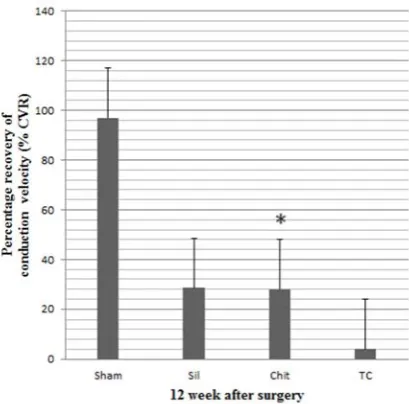
Conduction velocity along the chitosan-bridged and Silicone-bridged regenerated sciatic nerves was 28% CVR and 28.5% CVR of the intact right side, respectively. These were significantly higher than that of TC group that was 4% CVR (P <0.005) (Fig.1).
Muscle mass measurement
The mean ratios of gastrocnemius muscles weight were measured. There was statistically significant difference between the muscle weight ratios of Chit and SIL groups with TC group (P < 0.05). It showed that muscle weight ratios were bigger in Chit and SIL groups than of TC group and weight loss of the gastrocnemius muscle was ameliorated by chitosan and silicone entubulation (Fig 2).
Figure 1. Percentage recovery of conduction velocity in experimental groups. Data are presented as mean ± SD. * P< 0.005 vs TC
Histological preparation and morphometric studies
All animals of the Chit group showed nerve fibers at distal stumps 4 weeks after surgery. In the early phases of regeneration (at four weeks post operation) regenerated nerve fibers were present within the chitosan guide and regenerated nerve fibers could be confirmed after 4 weeks without any foreign body reaction. In the SIL group, the finding was similar to Chit group. In TC group, three animals presented lower number of nerve fibers at distal stumps after 8 weeks. The other two showed degenerated distal stumps. Sham-operation group presented significantly greater nerve fiber and axon diameter, and myelin sheath thickness compared to Chit, SIL and TC animals.
Both of Chit and SIL animals presented regeneration patterns. The number of nerve fibers in both of them was significantly higher than TC group. The mean diameter of the nerve fibers in the Chit (4.86±0.19) was significantly larger than that of TC (4.11 ± 0.22) (P < 0.05). The diameter of axon in Chit (3.77±0.21) was significantly larger than in TC (2.44 ± 0.63) (P<0.05)(Table 2). However, Morphometric indices in SIL group showed significantly greater values than those of Chit group (P<0.05).
Immunohistochemical analysis
Immunoreactivity to S-100 protein was extensively observed in the cross sections of regenerated nerve segments. The expression of S-100 protein signal was located mainly in the myelin sheath. The axon also showed a weak expression indicating that Schwann cell-like phenotype existed around the myelinated axons (Fig 3). In the Chit and SIL groups, the structure and function of regenerated axons and myelin sheath were far more similar than TC group to those of normal nerve. In TC, the expression of S-100 was dispersed and the findings resembled those of the histological evaluations.
Figure 3. Immunohistochemical analysis of the regenerated nerve 12 weeks after surgery from (a) midpoint of Sham (Sham), (b) middle cable (SIL), (c) middle cable Chit and (d) regenerated cable (TC). There is clearly more positive staining of the myelin sheath-associated protein S- 100 (arrow) within the periphery of nerve, indicating well-organized structural nerve reconstruction in entubulated nerve compared to transected control. (×1000)
Discussion
The assessment of sciatic nerve regeneration based on walking track analysis, electrophysiological, muscle mass, histomorphometric and immuohistochemical studies showed that chitosan as well as silicone could be used as a conduit for entubulation of transected short-gap nerve ends to enhance rat sciatic nerve regeneration.
Walking track analysis as a reliable determining functional recovery test15-17showed that improvement in recovery of sciatic nerve function in the course of time in Chit was significantly better than SIL group. Nerve conduction measurement is a direct evidence for the study of nerve transmission.18 The conduction velocity depends on the diameter of axons and the thickness of myelin sheath.19 The results of the present study showed similar conduction velocity along the chitosan-bridged and silicone-bridged regenerated sciatic nerves, therefore, the chitosan conduit could be assumed as a safe nerve guide with no nerve conduction interference.
In the present study, similar muscle mass results in both experimental groups showed again the chitosan could be as effective as silicone in entubulation of sciatic nerve defect. As the posterior tibial branch of the sciatic nerve regenerates into the gastrocnemius muscle, it will regain its mass proportional to the amount of axonal reinnervation.20,21
suggested that walking track analysis is more comprehensive than histomorphometrical methods alone. This study also supports the idea that the walking track-analysis is more comprehensive and reliable than histomorphometric methods in peripheral nerve repair studies.22-23
In the present study as observed histologically, morphometrical values did not differ significantly between 8 and 12 weeks in entubulated defects by chitosan. Also, there were no significant differences in diameter of nerve fibers and myelin thickness between Chit and TC groups.This study again supports the idea that the walking track analysis (SFI) is more comprehensive than histomorphometrical methods.22
We demonstrated in this proof of principle study that chitosan conduit can support axonal regrowth across a one cm gap in an adult rat sciatic nerve. We used well-established test systems because we aspired to examine comprehensive behavior of the conduit. Ideally, the scaffold material should last long enough for axonal regrowth and tissue repair. Previous studies shows chitosan is a biocompatible, timely biodegradable material, low toxic property, and low cost10,11and also the capacity of adherence and survival of Schwann cells on its substratum in vitro has a benefit for nerve regeneration.24,25 This natural polysaccharide, has structural characteristics similar to glycosaminoglycans and it seems to mimic the functional behavior of glycosaminoglycans and enzymatically can degrade to absorbable oligosaccharides.1 Some reports have also showed that chitosan has bacteriostatic26,27and
hemostatic28 properties which reinforcing the suitability of as a scaffold biomaterial for neural tissue engineering.
Serum lysozyme degrades chitosan29 and the degradation products are nontoxic, non-immunogenic and noncarcinogenic.30 Chitosan has been shown to physiologically stimulate the tissue repair process and favors angiogenesis.31 Chitosan in the form of a scaffold has been shown to carry angiogenic activity when implanted.32 Chitosan solution can be easily formed
due to its film-forming property and the tough and visco-elastic properties of gelled chitosan suitably provide mechanical support for nerve regeneration.33 However, initial pilot of our study showed that when chitosan fabricated into conduits it would be firm, dry and fragile enough that make it not suitable for suturing and surgical handling. Therefore, it was blended with glycerol while preserving its biocompatibility.12
In conclusion, chitosan entubulation technique has offered the hope of providing a method for achieving the peripheral nerve regeneration in the least harmful way that is available, easily performed and affordable. Using chitosan tubes in bridging of nerve defects could be promising because it is inert and does not induce extensive scarring or degeneration after implantation.
Acknowledgment
We would like to thank Dr. Mehdizadeh, Dr. Behfar and Dr. Mohammadi and Mr. Jaafary for their expert technical help.
References
1. Lin YL, Jen JC, Hsu SH, et al. Sciatic nerve repair by microgrooved nerve conduits made of chitosan-gold nanocomposites .Surg Neurol 2008; 70(1): 9-18.
3. Pfister LA, Papaloizos M, Merkle HP, et al. Nerve conduits and growth factor delivery in peripheral nerve repair. J Peripher Nerv Syst 2007; 12: 65-82.
4. Itoh S, Shinomiya K, Samejima H, et al. Experimental study on nerve regeneration through the basement membrane tubes of the nerve, muscle, and artery. Microsurgery 1996; 17: 525-534.
5. Mohammadi R, Azizi S, Delirezh N, et al. Comparison of beneficial effects of undifferentiated cultured bone marrow stromal cells and omental adipose-derived nucleated cell fractions on sciatic nerve regeneration. Muscle Nerve 2011; 43: 157-163.
6. Meek MF, Vand Der Werff JF, Nicolai JP, et al. Biodegradable P (DLLA-epsilon-CL) nerve guides versus autologous nerve grafts: electromyographic and video analysis. Muscle Nerve 2001; 24(6): 753-759.
7. Fields RD, Le Beau JM, Longo FM, et al. Nerve regeneration through artificial tubular implants. Prog Neurobiol 1989; 33: 87-134.
8. Guerra G D, Barbani N, GagliardiM, etal. Chitosan-based macromolecular biomaterials for the regeneration of chondroskeletal and nerve tissue. Int J Carbohydr Chemis doi:10.1155/2011/303708
9. Yussof S JM, Halim AS, Saad AZM, et al. Evaluation of the biocompatibility of a bilayer chitosan skin regenerating template, human skin allograft, and integra implants in rats. ISRN Materials Science 2011 doi:10.5402/2011/857483.
10. Wang X, Hu W, Cao Y, et al. Dog sciatic nerve regeneration across a 30-mm defect bridged by a chitosan/pga artificial nerve graft. Brain 2005; 128: 1897-1910.
11. Ao Q, Wang AJ, Cao WL, et al. Manufacture of multimicrotubule chitosan nerve conduits with novel molds and characterization in vitro. J Biomed Mater Res A 2006; 77: 11-18.
12. Ojagh SM, Rezaei Ma, Razavi S H, et al. Development and evaluation of a novel biodegradable film made from chitosanand cinnamon essential oil with low affinity toward water. Food Chem 2010; 122: 161-166.
13. Bian JR, Mackinnon SE and Hunter DA. Functional evaluation of complete sciatic, peroneal, and posterior tibial nerve lesions in the rat. Plast Reconstr Surg 1989; 83: 129-136
14. Di Benedetto G, Zura G ,Mazzucchelli R, et al. Nerve regeneration through a combined autologous conduit (vein plus acellular muscle grafts). Biomaterials 1998; 19: 173-181
15. Geuna A, Gigo-Benato D, Rodrigues AC. On sampling and sampling errors in histomorphometry of peripheral nerve fibers. Microsurgery 2003; 23: 72-76.
16. De MedinaceliL, Freed WJ, Wyatt RJ. An index of the functional condition of rat sciatic nerve based on measurements made from walking tracks. Exp Neurol 1982; 77: 634-643.
17. Varejão AS, Meek MF, Ferreira AJ, et al. Functional evaluation of peripheral nerve regeneration in the rat: walking track analysis. J Neurosci Methods 2001; 108: 1-9. 18. Chen CJ, Ou YC, Liao SL , et al. Transplantation of bone marrow stromal cells for
peripheral nerve repair. Exper Neurol 2007; 204: 443-453.
20. Hou Z, Zhu J. An experimental study about the incorrect electrophysiological evaluation following peripheral nerve injury and repair. Electromyogr Clin Neurophysiol 1998; 38: 301-304.
21. Evans GR, Brandt K, Widmer MS, et al. In vivo evaluation of poly (L-lactic acid) porous conduits for peripheral nerve regeneration. Biomaterials 1999; 20: 1109-1115. 22. Munro CA, Szalai JP, Mackinnon SE, et al. Lack of association between outcome
measures of nerve regeneration. Muscle Nerve 1998; 21(8): 1095-1097.
23. Kanaya F, Firrell JC, Breidenbach WC. Sciatic function index, nerve conduction tests, muscle contraction, and axon morphometry as indicators of regeneration. Plast Reconst Surg 1996; 98(7): 1264-1274.
24. Rosales-Cortes M, Peregrina-Sandoval J, Banuelos-Pineda J, et al. Immunological study of a chitosan prosthesis in the sciatic nerve regeneration of the axotomized dog. J Bio Mater Appl 2003; 18: 15-23.
25. Wang G, Lu G, Ao Q, et al. Preparation of cross-linked carboxymethyl chitosan for repairing sciatic nerve injury in rats. Bio technol Lett 2010; 32: 59-66.
26. Cuero RG. Antimicrobial action of exogenous chitosan. Exs 1999; 87: 315-333.
27. Tsai GJ, Su WH. Antibacterial activity of shrimp chitosan against escherichia coli. J Food Prot 1999; 62: 239-243.
28. Malette WG, Quigley HJ, Gaines RD, et al. Chitosan: a new hemostatic. Ann Thorac Surg 1983; 36: 55-8.
29. Tomihata K, Ikada Y. In vitro and in vivo degradation of films of chitin and its deacetylated derivatives. Biomaterials 1997; 18: 567-575.
30. Cao W, Cheng M, Ao Q, et al. Physical, mechanical and degradation properties, and Schwann cell affinity of cross-linked chitosan films. J Biomater Sci Polym Ed 2005; 16: 791-807.
31. Biagini G, Pugnaloni A, Damadei A, et al. Morphological study of the capsular organization around tissue expanders coated with N-carboxybutyl chitosan. Biomaterials 1991; 12(3): 287-291.
32. VandeVord PJ, Matthew HWT, De Silva SP. Evaluation of the biocompatibility of chitosan in mice. J Biomed Res 2001; 59(3): 585-590.
33. Cheng MY, Deng JG, Yang F, et al. Study on physical properties and nerve cell affinity of composite films from chitosan and gelatin solutions. Biomaterials 2003; 24: 2871-2880.
هﺪﻴﻜﭼ
تر
ﺐﺼﻋ
ﻊﻄﻗ
لﺪﻣ
رد
ﻚﻴﺗﺎﻴﺳ
ﺐﺼﻋ
هﺎﺗﻮﻛ
ﻪﺼﻴﻘﻧ
ﻢﻴﻣﺮﺗ
رد
ﻲﻧازﻮﺘﻴﻛ
ﺖﻴﺋوﺪﻧﺎﻛ
زا
هدﺎﻔﺘﺳا
ﻲﺴﻴﺋرسﺎﺒﻋ
1
يﺰﻳﺰﻋﺪﻴﻌﺳ،
1 ژﺮﻴﻟدزورﻮﻧ، 2
نﺎﻴﺘﻤﺸﺣمﺎﻨﻬﺑ، 3
ﻲﻨﻴﻣاناﻮﻴﻛ، 4
1
،ناﺮﻳا،ﻪﻴﻣورا،ﻪﻴﻣوراهﺎﮕﺸﻧاد،ﻲﻜﺷﺰﭙﻣادهﺪﻜﺸﻧاد،ﻲﻫﺎﮕﻧﺎﻣردمﻮﻠﻋهوﺮﮔ
2
وﻲﻟﻮﻠﺳيروﺎﻨﻓﺖﺴﻳزهوﺮﮔ ،ناﺮﻳا،ﻪﻴﻣورا،ﻪﻴﻣوراهﺎﮕﺸﻧاد،يروﺎﻨﻓﺖﺴﻳزهﺪﻜﺸﻫوﮋﭘ،ﻲﻟﻮﻜﻟﻮﻣ
3
يژﻮﻟﻮﻳﺰﻴﻓورﻮﻧﻲﺗﺎﻘﻴﻘﺤﻧﺰﻛﺮﻣ ،ناﺮﻳا،ﻪﻴﻣورا،ﻪﻴﻣوراﻲﻜﺷﺰﭘمﻮﻠﻋهﺎﮕﺸﻧاد،ﻲﻜﺷﺰﭘهﺪﻜﺸﻧاد،
4
ادﺎﻧﺎﻛ،نﻮﺗﺎﻜﺳﺎﺳ،ناﻮﭼﺎﻜﺳﺎﺳهﺎﮕﺸﻧاد،نﺮﺘﺳوﻲﻜﺷﺰﭙﻣادﺞﻟﺎﻛ،ﻲﻜﺷﺰﭙﻣادﻲﺳﺎﻨﺷﺐﻴﺳآهوﺮﮔ
. فﺪﻫ -ﺮﻴﺛﺎﺗﻲﺑﺎﻳزرا ترردﻚﻴﺗﺎﻴﺳﺐﺼﻋﻊﻄﻗلﺪﻣزاهدﺎﻔﺘﺳاﺎﺑﻲﻄﻴﺤﻣبﺎﺼﻋاﻢﻴﻣﺮﺗيورﺮﺑﻲﻧازﻮﺘﻴﻛﺖﻴﺋوﺪﻧﺎﻛ
ﻪﻌﻟﺎﻄﻣحﺮﻃ
-هﺪﻧزﻂﻳاﺮﺷردﻲﺑﺮﺠﺗﻪﻌﻟﺎﻄﻣ
تﺎﻧاﻮﻴﺣ
-60
ﻢﻟﺎﺳﺮﻧرﺎﺘﺴﻳوتر
رﺎﻛشور
-تر ﺎﻫ هوﺮﮔرﺎﻬﭼﻪﺑﻲﻓدﺎﺼﺗرﻮﻄﺑ
15
ﺪﻧﺪﺷﻢﻴﺴﻘﺗﻲﻳﺎﺗ
. شﺮـﺑزاﺲـﭘﭗﭼﻚﻴﺗﺎﻴﺳﺐﺼﻋلﺎﻣﺮﻧلﺮﺘﻨﻛهوﺮﮔرد
ﺪﻳدﺮﮔﻪﻴﺨﺑﻞﺤﻣ،يﺪﻨﺒﻧﻮﺧزاﺲﭘوهﺪﺷﻲﻳﺎﺳﺎﻨﺷﻲﻨﻳﺮﺳﻪﻠﻀﻋوﺖﺳﻮﭘ
.
ﻪﺼﻴﻘﻧﻲﻔﻨﻣلﺮﺘﻨﻛهوﺮﮔرد
لﻮـﻃﻪﺑيا
10
ﻲـﻠﻴﻣ
ﻀﻋﻪﺑهﺪﺷهﺪﻳﺮﺑيﺎﻫﺎﻬﺘﻧاﺲﭙﺳﺪﻳدﺮﮔدﺎﺠﻳاﻚﻴﺗﺎﻴﺳﺐﺼﻋيورﺮﺑﺮﺘﻣ
ﺪـﻳدﺮﮔﻪـﻴﺨﺑﻪﻴﺣﺎﻧتﻼ
. وﺖـﺒﺜﻣلﺮـﺘﻨﻛهوﺮـﮔرد
ﻪﺼﻴﻘﻧدﺎﺠﻳازاﺲﭘنﺎﻣرد
10
ﻲﻠﻴﻣ
ﻪـﻟﻮﻟﺎـﻳ ﻲﻧﻮﻜﻴﻠﻴـﺳﻪـﻟﻮﻟزاهدﺎﻔﺘـﺳاﺎـﺑﺐﺼﻋﻲﻨﻴﻳﺎﭘوﻲﻳﻻﺎﺑهﺪﺷﻊﻄﻗيﺎﻫﺎﻬﺘﻧا ،يﺮﺘﻣ
ﺪﻧﺪﺷﻂﺒﺗﺮﻣﻢﻫﻪﺑﻲﻧازﻮﺘﻴﻛ
.
هوﺮﮔﺮﻳزﻪﺳﻪﺑﺲﭙﺳهوﺮﮔﺮﻫ
5
ﻲﻧﺎﻣزﻊﻃﺎﻘﻣردوهﺪﻳدﺮﮔﻢﻴﺴﻘﺗﻲﻳﺎﺗ
4
،
8
و
12 زاﺪـﻌﺑﻪﺘﻔﻫ
ﺪﻨﺘﻓﺮﮔراﺮﻗﻪﻌﻟﺎﻄﻣدرﻮﻣﻞﻤﻋ
.
ﺞﻳﺎﺘﻧ
-ﻲﺑﺎﻳزرا ترردﻚﻴﺗﺎﻴﺳﺐﺼﻋدﺮﻜﻠﻤﻋيژﻮﻟﻮﻳﺰﻴﻓوﺮﺘﻜﻟاويدﺮﻜﻠﻤﻋيﺎﻫ رديﺎﻫ
ﺖﻓﺎﻳ يدﻮﺒﻬﺑنازﻮﺘﻴﻛونﻮﻜﻴﻠﻴﺳهﺪﻨﻨﻛ
ﻪﻈﺣﻼﻣﻞﺑﺎﻗ دادنﺎﺸﻧاريا
)
P<0.05
.(
ﺺﺧﺎﺷ ﻲﺑﺎـﻳزراوﻚﻳﺮﺘﻣﻮﻓرﻮﻣيﺎﻫ
ﻮـﻨﻤﻳايﺎـﻫ
ﻢﻴﻣﺮـﺗﺮﮕﻧﺎـﻴﺑﻲﻳﺎﻴﻤﻴـﺷﻮﺘﺴﻴﻫ
ﻞـﺑﺎﻗ
ﻪﻈﺣﻼﻣ نﺎﻳﺎﭘردﻲﻔﻨﻣلﺮﺘﻨﻛهوﺮﮔﺎﺑﻪﺴﻳﺎﻘﻣردهوﺮﮔودﺮﻫرد
12 دﻮﺑﻪﺘﻔﻫ ) P<0.05 .( ﻪﺠﻴﺘﻧ ﻲﻨﻴﻟﺎﺑدﺮﺑرﺎﻛويﺮﻴﮔ
-ﻲﻣارﻲﻧازﻮﺘﻴﻛﻪﻟﻮﻟﻲﻨﻴﻟﺎﺑﺮﻈﻧزا ﻪـﻳﺰﺠﺗﺖﻴﺋوﺪﻧﺎﻛﻚﻳناﻮﻨﻋﻪﺑناﻮﺗ
ﻢﻴﻣﺮـﺗردﻲﺘـﺴﻳزﺮﻳﺬـﭘ
ﺮﻳﺎﺳﻪﺑﺖﺒﺴﻧشورﻦﻳاﻪﻛﺖﻓﺮﮔﺮﻈﻧردﻲﻄﻴﺤﻣبﺎﺼﻋاﺮﺘﻬﺑ شور
هدﺎﺳﻲﻤﻴﻣﺮﺗيﺎﻫ
نازراوسﺮﺘـﺳدرد،ﺮـﺗﺮﻄﺧﻢﻛ،ﺮﺗ
ﺮـﺗ
ﺶﻨﻛاودﺎﺠﻳاﺮﻄﺧوهدﻮﺑ ﻲﻤﻧدﺎﺠﻳاﺰﻴﻧارﻲﺟرﺎﺧمﺎﺴﺟايﺎﻫ
ﺪﻨﻛ
.
نﺎﮔژاوﺪﻴﻠﻛ
-تر،نازﻮﺘﻴﻛﺖﻴﺋوﺪﻧﺎﻛ،ﻲﻄﻴﺤﻣﺐﺼﻋﻢﻴﻣﺮﺗ