Dorota Diakowska
A–D, Andrzej Lewandowski
A, E, F, Krystyna Markocka-Mączka
E, F,
Krzysztof Grabowski
E, FConcentration of Serum Interleukin-27 Increase
in Patients with Lymph Node Metastatic
Gastroesophageal Cancer
Zwiększenie stężenia interleukiny 27
w surowicy pacjentów chorych na raka przełyku
i raka połączenia przełykowo-żołądkowego
z przerzutami do węzłów chłonnych
Department of Gastrointestinal and General Surgery, Wroclaw Medical University, Poland
A – research concept and design; B – collection and/or assembly of data; C – data analysis and interpretation;
D – writing the article; E – critical revision of the article; F – final approval of article; G – other
Abstract
Background. Cytokines, one of the key mediators of immune response, play an important role in cancer develop-ment.
Objectives. The aim of this study was to evaluate the interleukin-27 (IL-27) concentration in the serum of patients with gastroesophageal cancer (GEC) and in patients with non-cancerous benign diseases of the upper digestive tract (NCD). We investigated the relationship between the serum IL-27 level and clinicopathological factors, and also the diagnostic utility of IL-27 as a marker of GEC presence. Additionally, we evaluated the concentrations of serum IL-27 in patients with esophageal squamous cell carcinoma before and after surgical tumor resection.
Material and Methods. Serum samples from 84 GEC patients, 39 NCD patients and 33 healthy subjects were assayed. The levels of IL-27, IL-6, IL-12 and IFN-γ were determined using ELISA kits.
Results. The serum levels of IL-27 were significantly higher in the GEC patients than in the healthy control (p < 0.0001) and in NCD patients (p = 0.006). The concentrations of serum IL-27 were related to lymph node status (p = 0.044). ROC analysis showed a significant relationship between a high level of serum IL-27 and GEC presence (AUC = 0.766, p < 0.001). The concentrations of serum IL-27 were significantly higher in patients with esophageal squamous cell carcinoma 3 months after esophagectomy than before the operation (p = 0.003).
Conclusions. Our results demonstrated that a high serum IL-27 level is associated with cancer presence and lymph node metastases in GEC. Significantly higher levels of IL-27 in patients with esophageal squamous cell carcinoma after tumor resection may imply that host immune cells are one of the important sources of circulating IL-27 (Adv Clin Exp Med 2013, 22, 5, 683–691).
Key words: squamous cell carcinoma of esophagus, cardiae cancer, interleukin-27, tumor markers.
Streszczenie
Wprowadzenie. Cytokiny, jedne z kluczowych mediatorów odpowiedzi immunologicznej, odgrywają istotną rolę w rozwoju nowotworu.
Cel pracy. Oznaczenie stężenia interleukiny 27 w surowicy pacjentów chorych na raka przełyku i raka wpustu (GEC) oraz u pacjentów z łagodnymi, nienowotworowymi schorzeniami górnego odcinka przewodu pokarmowe-go (NCD). Autorzy poszukiwali związku między stężeniem IL-27 w surowicy a parametrami klinicznymi i pato-logicznymi u chorych na GEC. Przeprowadzono analizę przydatności diagnostycznej oznaczania stężenia IL-27 w surowicy jako markera obecności GEC. Dodatkowo porównano stężenia IL-27 w surowicy pacjentów chorych na płaskonabłonkowego raka przełyku (ESCC) przed operacją usunięcia przełyku i 3 miesiące po operacji.
Materiał i metody. Zbadano surowice uzyskane od 84 pacjentów chorych na GEC, 39 pacjentów z NCD i 33
zdro-Adv Clin Exp Med 2013, 22, 5, 683–691 ISSN 1899–5276
ORIGINAL PAPERS
Esophageal and gastroesophageal junction can-cers are significant causes of cancer-related death worldwide [1, 2]. The poor outcome observed in the majority of gastroesophageal cancer (GEC) pa-tients results from the advancement of disease de-velopment at clinical diagnosis [1–3].
Squamous cell carcinoma of the esophagus and adenocarcinoma of the distal esophagus and gastroesophageal junction are related to chron-ic inflammation of differing etiologies [2]. Tumor cells cause systemic changes in the immunologi-cal profile and in cytokines and growth factors se-cretion [2, 4]. For that reason, the identification of biological markers in serum is an ideal meth-od for early determination and monitoring of pa-tients with GEC.
Interleukin-27 (IL-27) is a novel member of the IL-6/IL-12 family. It is a heterodimeric cyto-kine composed of the EBV-transformed gene 3 subunit and p28 subunit [5]. IL-27 is predomi-nantly synthesized by activated antigen-present-ing cells includantigen-present-ing monocytes, endothelial cells and dendritic cells [5, 6]. Although IL-27 can have proinflammatory effects, most studies point at the important role of IL-27 as an immunosuppres-sor [7, 8]. This cytokine is associated with early phase differentiation of Th1 lymphocytes, inhibi-tion of Th2 humoral immune response and stim-ulation of NK cells [5, 7–9]. IL-27 also synergizes strongly with IL-12 in IFN-γ production via T and NK cells (10). It has been reported that IL-27, like IL-12, has potent anti-tumor and anti-metastatic activity through its anti-angiogenic properties [5, 7–10].
Receptors for IL-27 (IL-27R) were composed of a unique WSX-1⁄TCCR and common gp130 subunits [11]. The gp130 is also a receptor subunit for IL-6 family cytokines and through this, IL-27 belongs to the IL-6 family [11].
There are some studies on the levels of cyto-kines such as IL-6 and IL-12 in gastric and esoph-ageal cancer [12–14]. However, the role of soluble
IL-27 as a possible diagnostic marker in GEC re-mains unclear. In the present study we examined the concentrations of IL-27, IL-12, IFN-γ and IL-6 in the serum of GEC patients and in patients with non-cancerous benign diseases of the upper diges-tive tract (NCD). This study was designed to in-vestigate the association of serum IL-27 concentra-tion with clinicopathological parameters in GEC patients. We also aimed for a possible diagnostic utility of serum IL-27 level in the evaluation of gas-troesophageal cancer.
Additionally, we evaluated levels of circulat-ing IL-27 in patients with esophageal squamous cell carcinoma before and after surgical tumor resection.
Material and Methods
The authors examined 84 patients with esoph-ageal squamous cell carcinoma (n = 59) and ad-enocarcinoma of the lower part of the esophagus or gastric cardia (n = 25), who were treated in the Department of Gastrointestinal and General Sur-gery from 2006 to 2011. There were 18 females and 66 males (median age: 58 years, 95% CI: 43–62) in the study group.
UICC TNM staging system was applied [15] for clinical and pathological staging. We examined two patients with clinical stage I, 14 with stage II, 28 with stage III and 40 with stage IV.
Sera from 33 blood donors were used as a control group. The group consisted of 9 females and 24 males, median age 54 years (95% CI: 38–63). Additionally, we analyzed 39 patients hospitalized due to planned treatment for benign esophageal diseases (hiatal her-nia, n = 14, cardiospasmus n = 12, burnt esophagus n = 9, non-cancerous stenosis n = 4). There were 24 females and 15 males, median age 56 years (95% CI: 49–59) in this group. In all the studied groups, none of the examined indices correlated with age.
In the 21 patients with stage I, II and III of
wych osób. Stężenia IL-27 oraz innych cytokin, jak interleukiny 6 (IL-6), interleukiny 12 (IL-12) i interferonu γ (IFN-γ) zostały zmierzone za pomocą testów ELISA. Diagnostyczna skuteczność IL-27 jako markera obecności raka została oznaczona za pomocą analizy krzywej ROC.
Wyniki. Stężenie IL-27 w surowicy chorych z GEC było istotnie większe niż w grupie kontrolnej (p < 0,0001) oraz u pacjentów z NCD (p = 0,006). Wykazano istotny związek stężenia IL-27 z przerzutami do węzłów chłonnych (p = 0,044). Analiza ROC wykazała istnienie silnego powiązania między stężeniem IL-27 w surowicy a obecnością raka u pacjentów chorych na GEC (AUC = 0,766, p < 0,001). Stężenia IL-27 były skorelowane pozytywnie ze stęże-niami IFN-γ (rho = 0,34, p = 0,013) i stężestęże-niami IL-12 (rho = 0,28, p = 0,048) w surowicy chorych na GEC. Stężenia IL-27 były istotnie większe u pacjentów chorych na płaskonabłonkowego raka przełyku (ESCC) 3 miesiące po ezo-fagektomii niż przed operacją (p = 0,003).
Wnioski. Duże stężenia IL-27 w surowicy pacjentów chorych na GEC wiążą się z obecnością nowotworu i przerzu-tami do węzłów chłonnych. Istotnie większe stężenie IL-27 u pacjentów z ESCC po operacji wskazuje, że głównym źródłem IL-27 mogą być komórki systemu obronnego organizmu (Adv Clin Exp Med 2013, 22, 5, 683–691).
esophageal squamous cell carcinoma, the serum concentrations of IL-27 were measured twice: be-fore the esophagectomy and 3 months after surgi-cal resection.
The study protocol was approved by the Medi-cal Ethics Committee of Wroclaw MediMedi-cal Univer-sity, Wrocław, Poland.
Blood samples were collected from the GEC patients and NCD patients preceding any treat-ment. Blood from the peripheral vein was collect-ed in sterile tubes, clottcollect-ed (15 min, RT) and cen-trifuged (10 min, 900 × g). The obtained sera were stored at –45ºC until assayed.
Concentrations of serum IL-27 were measured using the ELISA test (BioLegend, San Diego, USA). The sensitivity of the assay was 11 pg/mL, the in-tra-assay coefficient of variation was 4.0–5.9% and inter-assay coefficient of variation was 4.9–5.4%. All samples were run in duplicates.
Levels of IL-12 were assayed using an immu-noenzymatic test (Bender MedSystems, Vienna, Austria) accordingly to the manufacturer’s instruc-tions. IL-6 and IFN-γ were determined with Peli-Kine Compact human cytokine ELISA kits (San-quin, Amsterdam, Holland).
Distribution of data was analyzed using the Shapiro-Wilk normality test. Concentrations of all cytokines were presented as a median and 95% CI (coefficient interval). The Mann-Whitney and Kruskal-Wallis tests were used for the group com-parisons. The Spearman’s rank correlation test was used for the correlation analysis. Differences be-tween paired variables were analyzed using a Wil-coxon test. Values of p < 0.05 were considered as statistically significant. A power analysis of ap-plied statistical tests demonstrated that all values of power of the test were higher than 0.80.
The diagnostic utility of IL-27 was determined by means of a Receiver Operating Characteristics (ROC) curve analysis. The overall performance of IL-27 was expressed in terms of the area under the ROC curve (AUC) with 95% CI and p statistics for the differences between calculated AUC and AUC = 0.5. A cut-off value, sensitivity and spec-ificity, Youden’s index (sensitivity (%) + specifi-city (%) – 100%), positive and negative likelihood ratios were calculated.
The statistical analyses were performed using STATISTICA 10.0 software (StatSoft Inc., Tulsa, USA).
Results
The concentration of serum IL-27 was signifi-cantly higher in GEC patients in comparison with healthy individuals: 216.3 pg/mL (132.7–379.5) vs.
37.5 pg/mL (36.9–101.8) (p < 0.0001), respective-ly (Table 1). Also serum concentration of IL-27 in the NCD group increased significantly (89.0 pg/mL (80.2–214.5)) in comparison with the control group (p = 0.047). A comparison of IL-27 levels in GEC patients with NCD patients showed significant dif-ferences (p = 0.006) between them (Fig. 1).
The concentrations of serum IL-6, IL-12 and IFN-γ were significantly higher in GEC patients in comparison with controls (Table 1).
The associations between circulating IL-27 levels and clinicopathological variables are shown in Table 2. We observed a statistically significant relationship between serum IL-27 level and lymph node status (p = 0.044). However, the concentra-tions of serum IL-27 did not correlate with the stage of disease progression, primary tumor exten-sion and distant metastases.
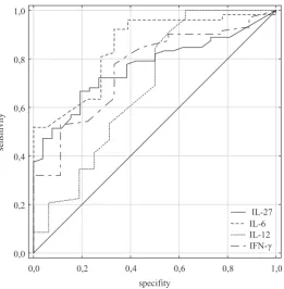
The difference in the concentrations of serum IL-27 between patients with stage I+II GEC and control was statistically significant (p = 0.007). These results suggest that serum IL-27 may be a prospective diagnostic marker of cancer pres-ence. ROC analysis revealed that serum IL-27 was a good indicator of the presence of GEC (Fig. 2). For purposes of comparison, the performances of serum IL-6, IL-12 and IFN-γ as the other potential markers of cancer presence were evaluated. The overall performance of IL-27 was better than for IL-12 and IFN-γ but slightly worse than for IL-6 (Table 3).
We evaluated the usefulness of serum IL-27 level as a marker of lymph node status (accord-ing to the clinical evaluation of lymph node me-tastases). The best cut-off value calculated from the ROC analysis was 450 pg/mL for determina-tion of lymph node involvement, AUC amount to 0.641(0.498–0.784), sensitivity was 34% and speci-ficity was 86%.
A significant positive correlation was found be-tween serum IL-27 and serum IFN-γ (rho = 0.34, p = 0.013) in GEC patients. There was also a signif-icant association between serum IL-12 and serum IL-27 (rho = 0.28, p = 0.048).
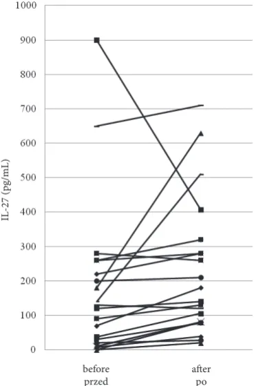
The resection of tumors in patients with esoph-ageal squamous cell carcinoma resulted in an in-crease of serum IL-27 concentration in 86% (18/21) of patients. IL-27 level increased from 120.0 pg/mL (95% CI: 72.1–324.7) before esophagectomy to 140.0 pg/mL (95% CI: 50.6–514.2) three months after surgical tumor resection (p = 0.003) (Fig. 3).
Discussion
median 25–75%
individual data dane indywidualne
GEC NCD control
0 100 200 300 400 500 600 700 800 900 1000
IL-27 (pg/mL
)
GEC vs. control p < 0.0001* NCD vs. control p = 0.047* GEC vs. NCD p = 0.006*
0,0 0,2 0,4 0,6 0,8 1,0 specifity
0,0 0,2 0,4 0,6 0,8 1,0
sensitivit
y
IL-27 IL-6 IL-12
IFN-γ
Fig. 1. Serum levels of IL-27 in GEC patients in comparison with NCD patients and healthy subjects; (* – statistically significant)
Ryc. 1. Stężenie IL-27 w surowicy pacjentów z GEC w porównaniu z pacjentami chorymi na NCD i osobami zdrowymi;
(* – istotne statystycznie)
Table 1. Serum concentration of IL-6, IL-12 and IFN-γ in GEC patients, NCD patients and healthy subjects (C)
Tabela 1. Stężenie IL-6, IL-12 and IFN-γ w surowicy pacjentów chorych na GEC, pacjentów chorych na NCD i osób zdro-wych (C)
IL-6 (pg/mL) IL-12 (pg/mL) IFN-γ (pg/mL) median
(95% CI) p-value (wartość p) median (95% CI) p-value (wartość p) median (95% CI) p-value (wartość p) GEC
(n = 84) NCD (n = 39) C (n = 33)
1.3(1.6–5.8)
0.7(0.0–6.9)
0.4(0.3–0.8)
GEC vs. C, p < 0.001* GEC vs. NCD, p = 0.003* NCD vs. C, p = 0.002*
2.7(2.8–5.7)
2.9(2.2–5.6)
2.3(1.2–3.1)
GEC vs. C, p = 0.024* GEC vs. NCD, p = 0.815 NCD vs. C, p = 0.078
8.8(8.6–9.8)
8.3(8.0–10.9)
7.6(6.7–8.5)
GEC vs. C, p = 0.011* GEC vs. NCD, p = 0.986 NCD vs. C, p = 0.083 * – statistically significant.
* – istotne statystycznie.
Fig. 2. ROC curves for serum IL-27 and IL-6, IL-12, IFN-γ as possible indicators of GEC presence
higher in GEC patients in comparison with the healthy control group. This result suggests that IL-27 overexpression may be attributed to GEC development. The levels of serum IL-27 in GEC were significantly higher than those in NCD pa-tients. This may indicate that IL-27 takes part in the inflammatory processes of the esophagus and gastric cardiae and in gastroesophageal cancer pro-inflammatory reactions.
IL-27 plays multiple roles in modulating im-mune response. This cytokine promotes inflam-mation by inducing the proliferation of naive CD4+ T cells (STAT1 and STAT3 signaling
path-ways) and early Th1 differentiation [6–9, 11]. On
Table 2. Relationship between serum IL-27 concentrations and characteristic or disease-related variables in GEC patients
Tabela 2. Związek między stężeniem IL-27 w surowicy a parametrami klinicznymi i patologicznymi pacjentów z GEC Variables (Zmienne) IL-27 (pg/mL)
median (95% CI) p-value (wartość p) Gender (Płeć)
females (kobiety) (n = 18)
males (mężczyźni) (n = 66) 280.0 (207.3–509.1)180.0 (110.4–375.8) 0.494 Age – years (Wiek – lata)
< 60 (n = 48)
> 60 (n = 36) 253.8 (64.9–322.9)341.3 (270.5–527.3) 0.361 Histology (Histologicznie)
SCC (n = 59)
ADC (n = 25) 230.0 (172.2–351.0)325.5 (245.6–502.9) 0.116 TNM Stage (Stopień TNM)
I+II (n = 16) III (n = 28) IV (n = 40)
220.0 (144.8–361.1) 255.0 (107.9–496.3) 206.3 (135.9–459.4)
0.535
Tumor (Guz pierwotny) T1+T2 (n = 18) T3 (n = 20) T4 (n = 46)
260.0 (137.1–490.0) 250.0 (150.9–398.8) 253.8 (117.1–422.1)
0.342
Lymph node metastases (Przerzuty do węzłów chłonnych) N0 (n = 28)
N1 (n = 56) 130.0 (100.6–341.2)270.3 (259.0–435.0) 0.044* Distant metastases (Przerzuty odległe)
M0 (n = 52)
M1 (n = 32) 190.0 (189.0–369.0)256.3 (239.5–481.6) 0.144 SCC – squamous cell carcinoma, ADC – adenocarcinoma, * – statistically significant.
SCC – rak płaskonabłonkowy, ADC – gruczolakorak, * – istotny statystycznie.
before
przed afterpo
IL-27 (pg/mL)
Fig. 3. Serum IL-27 concentrations in 21 patients with esopha-geal squamous cell carcinoma before and 3 months after surgical resection of tumor
the other hand, IL-27 inhibits inflammation re-sponses by suppressing Th2 and Th17 differenti-ation [16]. Analyzing the IL-27 level in NCD pa-tients, we demonstrated a statistically significant elevation of this cytokine in comparison with con-trol subjects. The increased concentration of IL-27 observed in NCD patients may be connected with the up-regulation of Th1 and down-regulation of Th2 cell activity.
The results of the present study show the highest level of serum IL-27 in GEC patients. Through ac-tive participation in inflammatory processes, IL-27 may directly or/and indirectly influence the initia-tion and development of gastroesophageal cancer. The possible role of IL-27 in GEC may be explained by the participation of this cytokine in the initia-tion of Th cell response. IL-27 generates a Th1 im-mune response, which influences the stimulation of the secretion of proinflammatory cytokines such as TNF-α, IL-1, IL-6 and IL-8 [8, 17]. IL-27 activates the production of Th1-type cytokines such as IL-12, IL-18 and IFN-γ, and inhibits secretion of Th2-type cytokines such as IL-4 and IL-10 [16, 18]. It has been shown that IL-27 plays an important role in the sup-pression of Th2 response and induction of Th1 re-sponse [5, 8, 10, 18]. Through stimulation of proin-flammatory cytokines and induction of CD8+ T and
NK cell activity, IL-27 has prospective anti-tumor and anti-angiogenic effects [7, 8, 16]. Selective us-age of these anticancer mechanisms depends on the properties of tumor cells [7, 16].
Many studies analyze biological prognostic markers, which in connection with clinicopatholog-ical parameters modify the staging systems for opti-mal therapy for patients. Our studies are the first ones which analyze serum IL-27 concentration in patients with esophageal and cardiae cancer to determine the clinicopathological and prognostic significance of this cytokine. We demonstrated a significant relationship between a high level of serum IL-27 and lymph node metastases (LNM) in GEC patients.
Early lymphatic invasion is characteristic for esophageal squamous cell carcinoma and its de-tection is significant in the course of esopha-geal cancer [19]. However, precise clinical lymph node staging is very difficult. With respect to this, a non-invasive biomarker predicting LNM may be a helpful prognostic factor in cardiae and especial-ly in esophageal cancers. However, our anaespecial-lysis of serum IL-27 level as a marker of lymph node sta-tus did not show satisfactory results.
Recently, the dependence of LNM to T tumor stage has been reported [20]. It has been observed that tumor invasion is a predictor of lymph node metastases. But in our study, IL-27 level was not significantly different in relation to the depth of primary tumor invasion and cancer stage. With respect to these results, we did not examine IL-27 concentration in GEC according to their T-LNM status.
In vitro study on multiple myeloma, colon car-cinoma tumor model and neuroblastoma tumor cells demonstrated that IL-27 could exert a po-tent anti-tumor effect [8, 21, 22]. Studies of Hu et al. [23] have demonstrated that transfection of IL-27 gene into a murine hepatocellular carcinoma cell line prevents tumor growth. In contrast, in vi-tro studies of Lo et al. [24] have shown that IL-27 has a slight influence on both innate and adap-tive lymphocytes. The exact mechanisms of these opposite effects of IL-27 are not clear and need further exploration. It is possible that IL-27, like IL-12, mediates activation of anti-tumor immuni-ty by inducing neoplastic cells to produce anti-an-giogenic factors [25].
However, on the basis of the above-mentioned observations and our data, we assume that IL-27 may possibly induce the differences in local pro-inflammatory cytokine levels in a tumor microen-vironment and simultaneously may activate host immune cells in the surrounding tissues against tumor growth and metastases.
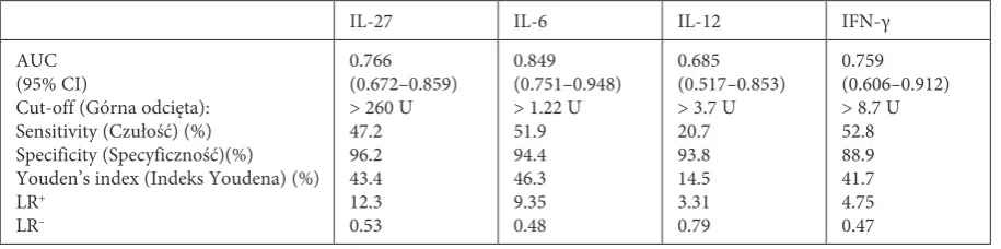
Table 3. Evaluation of serum IL-27 as a possible indicator of cancer presence in comparison with IL-6, IL-12 and IFN-γ
Tabela 3. IL-27 w surowicy jako prawdopodobny wskaźnik obecności raka w porównaniu z IL-6, IL-12 and IFN-γ
IL-27 IL-6 IL-12 IFN-γ
AUC (95% CI)
Cut-off (Górna odcięta): Sensitivity (Czułość) (%) Specificity (Specyficzność)(%) Youden’s index (Indeks Youdena) (%) LR+
LR–
0.766 (0.672–0.859) > 260 U 47.2 96.2 43.4 12.3 0.53 0.849 (0.751–0.948) > 1.22 U 51.9 94.4 46.3 9.35 0.48 0.685 (0.517–0.853) > 3.7 U 20.7 93.8 14.5 3.31 0.79 0.759 (0.606–0.912) > 8.7 U 52.8 88.9 41.7 4.75 0.47 AUC – area under ROC curve, LR+ and LR– – positive and negative likelihood ratios.
The authors observed that the level of serum IL-27 increased significantly in stage I+II of can-cer in comparison with the control, and remained high in stages III and IV of GEC. This may indi-cate that IL-27 up-regulation is associated with early neoplastic transformation and disease pro-gression, probably by activation of host immune response against cancer.
To the best of our knowledge, we are the first who have evaluated the diagnostic parameters, such as sensitivity, specificity, positive and nega-tive predicnega-tive value and ROC curve, for IL-27 in GEC patients. We showed that the IL-27 area un-der the ROC curve was higher than AUC for IL-12 and IFN-γ, but lower than AUC for IL-6. Also, prognostic sensitivity, specificity and Youden’s in-dex for IL-27 were lower than for IL-6. We com-pared our results for IL-27 with data for classic tumor markers in the serum of esophageal and gastric cancer patients [26, 27]. We revealed in our analysis that AUC for cell cancer antigen (SCC-Ag) (AUC = 0.811, p < 0.001) was higher than AUC for IL-27. Simultaneously, AUC for IL-27 was higher than AUC for carcinoembryonic anti-gen (CEA) (AUC = 0.673, p < 0.001) and carbohy-drate antigen (CA19-9) (AUC = 0.762, p < 0.001). These observations suggest that high serum IL-27 level may participate in GEC presence. Further in-vestigations are necessary to assay the role of IL-27 as a prognostic parameter in upper gastrointesti-nal tract cancers.
This is the first report about differences be-tween concentrations of IL-27 in patients with esophageal squamous cell carcinoma before and after surgical tumor resection. In the majority of patients, the level of IL-27 was higher after tumor resection than before esophagectomy. This obser-vation may be consistent with our hypothesis that host immune cells can significantly influence se-rum IL-27 concentration and that the primary tu-mor is not the only important source of circulating IL-27 in esophageal squamous cell carcinoma. It is also possible that non-detected lymph node metas-tases and micrometasmetas-tases can induce synthesis of IL-27 in patients after esophagectomy.
Further studies are necessary for better clari-fication of the main source of high IL-27 level in gastric and esophageal cancers. In our future re-search, we would like to compare IL-27 expression in tumor tissue with the tissue surrounding the tu-mor in GEC patients.
Chronic inflammation, in which many pro-in-flammatory cytokines take part, may be a risk fac-tor for cancer development [2, 3, 16]. We marked the IL-6 and IL-12 serum levels as cytokines which belong to the same family as IL-27, and IFN-γ se-rum level as well. Our data showed that sese-rum
IL-6 levels were significantly higher in GEC pa-tients than in healthy subjects. We reported sim-ilar accuracy in IL-6 detection to those described for esophageal and gastric cancer by other au-thors [26], where serum concentrations of IL-6 significantly increased. IL-6 plays an important role in host defense mechanisms. This cytokine is able to stimulate the anti-tumor activity of mac-rophages and to induce the expression of vascular endothelial growth factor [28]. However, overex-pression of IL-6 mRNA and the protective role of IL-6 from apoptosis have been observed in esoph-ageal carcinoma cell lines [18]. Recently Guzzo et al. [29] have demonstrated that IL-27 induces IL-6 expression in human monocytic cells. Our corre-lation studies did not find a significant correcorre-lation between serum IL-6 and IL-27.
The anti-tumor activity of IL-12 is highly de-pendent on primary IFN-γ produced by T and NK cells in response to IL-12 [7, 9, 10, 25]. IFN-γ plays an important role in enhancing antigen specif-ic and nonspecifspecif-ic immune responses whspecif-ich sup-port tumor rejection [25]. In the present study, we observed significantly higher levels of serum IL-12 and IFN-γ in GEC patients in comparison with the control group. These findings are in line with our previous studies, where we demonstrated that the serum level of IL-12 was significantly higher in squamous cell carcinoma of the esophagus than in healthy controls [14]. It may be possible that IL-12 and IFN-γ participate in GEC anti-tumor immu-nity by inhibition of tumor cells in proangiogenic factor production.
Because IL-27 also induces synergistic produc-tion of IFN-γ with IL-12 [10, 25] it was expected that the anti-tumor activity of IL-27 might be main-tained. In experimental studies, Shimizu et al. [7] showed the strong inhibition of neovascularization by IL-27 in chicken embryos. They said that IL-27 had a potent anti-angiogenic activity, and this ef-fect was IFN-γ independent. These results suggest that IFN-γ and IL-12 do not require IL-27 for the stimulation of anti-metastatic and anti-angiogenic activities. Our opposite results revealed that serum IL-27 levels correlate significantly with serum lev-els of IFN-γ and with serum IL-12 in GEC patients. The studies of Xu et al. [16] have also indicated that IL-27 exerts an anti-tumor effect via CD8+ T cells
and IFN-γ production in highly immunogenic tu-mors expressing MHC class I. Therefore, we sug-gest that IL-27 enhances anti-tumor and anti-an-giogenic activity through IFN-γ production in GEC patients.
Acknowledgements. The authors would like to thank dr Elżbieta Klausa from the Regional Center of Blood Donation and Therapeutics in Wrocław, Poland for supplying the serum of healthy individuals.
References
[1] Bystricky B, Okines AF, Cunningham D: Optimal therapeutic strategies for resectable oesophageal or oesopha-gogastric junction cancer. Drugs 2011, 71, 5, 541–555.
[2] Valverde C, Macarulla T, Casado E, Ramos F, Martinelli E, Tabernero J: Novel targets in gastric and esophageal cancer. Critical Rev Oncol Hematol 2006, 59, 128–138.
[3] Bird-Lieberman EL, Fitzgerald RC: Early diagnosis of oesophageal cancer. Br J Cancer 2009, 101, 1–6.
[4] Du C, Wang Y: The immunoregulatory mechanisms of carcinoma for its survival and development. J Exp Clin Cancer Res 2011, 30, 1–12.
[5] Jankowski M, Kopinski P, Goc A: Interleukin-27: biological properties and clinical application. Arch Immunol Ther Exp 2010, 58, 417–425.
[6] Matsui M, Kishida T, Nakano H, Yoshimoto K, Shin-Ya M, Shimada T, Nakai S, Imanishi J, Yoshimoto T, Hisa Y, Mazda O: Interleukin-27 activates natural killer cells and suppresses NK-resistant head and neck squamous cell carcinoma through inducing antibody-dependent cellular cytotoxicity. Cancer Res 2009, 69, 6, 2523–2530.
[7] Shimizu M, Shimamura M, Owaki T, Asakawa M, Fujita K, Kudo M, Iwakura Y, Takeda Y, Luster AD, Mizuguchi J, Yoshimoto T: Antiangiogenic and antitumor activities of IL-27. J Immunol 2006, 176, 7317–7324.
[8] Hisada M, Kamiya S, Fujita K, Belladonna ML, Aoki T, Koyanagi Y, Mizuguchi J, Yoshimoto T: Potent antitu-mor activity of interleukin-27. Cancer Res 2004, 64, 1152–1156.
[9] Owaki T, Asakawa M, Morishima N, Hata K, Fukai F, Matsui M, Mizuguchi J, Yoshimoto T: A role for IL-27 in early regulation of Th1 differentiation. J Immunol 2005, 175, 2191–2200.
[10] Hunter CA: New IL-12-family members: IL-23 and IL-27, cytokines with divergent functions. Nat Rev Immunol 2005, 5, 521–531.
[11] Liu L, Wang S, Shan B, Shao L, Sato A, Kawamura K, Li Q, Ma G, Tagawa M: IL-27-mediated activation of natural killer cells and inflammation produced antitumour effects for human oesophageal carcinoma cells. Scand J Immunol 2008, 68, 22–29.
[12] Haghazali M, Molaei M, Mashayekhi R: Proinflammatory cytokines and thrombomodulin in patients with peptic ulcer disease and gastric cancer, infected with Helicobacter pylori. Indian J Pathol Microbiol2011, 54, 103–106.
[13] Romero-Adrian TB, Leal-Montiel J, Monsalve-Castillo F:Helicobacter pylori: bacterial factors and the role of cytokines in the immune response. Curr Microbiol2010, 60, 143–155.
[14] Diakowska D, Markocka-Maczka K, Grabowski K, Lewandowski A: Serum interleukin-12 and interleukin-18 levels in patients with oesophageal squamous cell carcinoma. Exp Oncol 2006, 28, 4, 319–322.
[15] Sobin LH, Wittekind Ch: TNM classification of malignant tumors. 6th ed. Hoeboken, New Jersey: Jon Wiley& Sons 2002.
[16] Xu M, Mizoguchi I, Morishima N, Chiba Y, Mizuguchi J, Yoshimoto T: Regulation of antitumor immune responses by the IL-12 family cytokines, IL-12, IL-23, and IL-27. Clin Dev Immunol 2010 pii: 832454. Epub 2010 Sep 14.
[17] Ikeguchi M, Hatada T, Yamamoto M, Miyake T, Matsunaga T, Fukumoto Y, Yamada Y, Fukuda K, Saito H, Tatebe S: Serum interleukin-6 and -10 levels in patients with gastric cancer. Gastric Cancer 2009, 12, 2, 95–100.
[18] Leu CM, Wong FH, Chang C, Huang SF, Hu CP: Interleukin-6 acts as an antiapoptotic factor in human esophageal carcinoma cells through the activation of both STAT3 and mitogen-activated protein kinase pathways. Oncogene 2003, 22, 7809–7818.
[19] Mukai M, Sato S, Nakasaki H, Tajima T, Saito Y, Nishiumi N, Iwasaki M, Tokuda Y, Ogoshi K, Inoue H, Makuuchi H: Occult neoplastic cells in the lymph node sinuses and recurrence of primary breast, lung, esophageal, and gastric cancer. Oncol Rep 2004, 11, 1, 81–84.
[20] Gockel I, Sgourakis G, Lyros O, Polotzek U, Schimanski CC, Lang H, Hoppo T, Jobe BA: Risk of lymph node metastasis in submucosal esophageal cancer: a review of surgically resected patients. Expert Rev Gastroenterol Hepatol 2011, 5, 3, 371–384.
[21] Cocco C, Giuliani N, Di Carlo E: Interleukin-27 acts as multifunctional antitumor agent in multiple myeloma. Clin Cancer Res 2010, 16, 4188–4197.
[22] Salcedo R, Stauffer JK, Lincoln E, Back TC, Hixon JA, Hahn C, Shafer-Weaver K, Malyguine A, Kastelein R, Wigginton J: IL-27 mediates complete regression of orthotopic primary and metastatic murine neuroblastoma tumors: role for CD8+ T cells. J Immunol 2004, 173, 7170–7182.
[23] Hu P, Hu HD, Chen M: Expression of interleukins-23 and 27 leads to successful gene therapy of hepatocellular carcinoma. Mol Immunol2009, 46, 1654–1662.
metastases in GEC patients. ROC analysis showed a significant relationship between a high level of se-rum IL-27 and GEC presence. However, possible ap-plication of IL-27 as a diagnostic marker of cancer presence warrants further investigation. Significant-ly higher levels of IL-27 in patients with esophageal
[24] Lo CH, Chang CM, Tang SW, Pan WY, Fang CC, Chen Y, Wu PY, Chen KY, Ma HI, Xiao X, Tao MH:
Differential antitumor effect of interleukin-12 family cytokines on orthotopic hepatocellular carcinoma. J Gene Med 2010, 12, 423–434.
[25] DelVecchio M, Bajetta E, Canova S, Lotze MT, Wesa A, Parmiani G, Anichini A: Interleukin-12: Biological properties and clinical application. Clin Cancer Res 2007, 13, 16, 4677–4685.
[26] Łukaszewicz-Zając M, Mroczko B, Gryko M, Kędra B, Szmitkowski M: Comparison between clinical signifi-cance of serum proinflammatory proteins (IL-6 and CRP) and classic tumor markers (CEA and CA 19-9) in gastric cancer. Clin Exp Med 2011, 11, 89–96.
[27] Mroczko B, Kozłowski M, Groblewska M, Łukaszewicz M, Nikliński J, Jelski W, Laudański J, Chyczewski L, Szmitkowski M: The diagnostic value of the measurement of matrix metalloproteinase 9 (MMP-9), squamous cell cancer antigen (SCC) and carcinoembryonic antigen (CEA) in the sera of esophageal cancer patients. Clin Chim Acta 2008, 389, 1–2, 61–66.
[28] Huang SP, Wu MS, Wang HP, Yang CS, Kuo ML, Lin JT: Correlation between serum levels of interleukin-6 and vascular endothelial growth factor in gastric carcinoma. J Gastroenterol Hepatol 2002, 17, 1165–1169.
[29] Guzzo C, Che Mat NF, Gee K: Interleukin-27 induces a STAT1/3- and NF-κB-dependent proinflammatory cyto-kine profile in human monocytes. Journal of Biological Chemistry 2010, 285, 32, 24404–24411.
Address for correspondence:
Dorota Diakowska
Department of Gastrointestinal and General Surgery Wroclaw Medical University
Sklodowska-Curie 66 50-369 Wroclaw Poland
Fax: +48 71 733 26 09
E-mail: [email protected]
Conflict of interest: None declared