ABSTRACTS
The ESGE Junior Platform
Techniques and Instrumentations in Laparoscopy JP1_01
German residents’experiences in gynaecological surgery M. Stumpf, M. Klar, G. Gitsch, M. Runge, M. Foeldi
Department of Obstetrics and Gynaecology, University Medical Center, Albert-Ludwigs-University, Freiburg, Germany
Every German resident is taught in gynaecological surgery. Neverthe-less, a huge discrepancy of surgical skills among participants at the end of the residency can be observed. Gynaecological surgery is part of the 5 year residency in obstetrics and gynaecology. The mandatory residency objectives are defined by the German Medical Association. A provided log book is mandatory to record achievements. The surgical focus of this training is on performing minor operations under supervision, e.g. curettings, LLETZ or laparoscopy, as well as the assistance in more complex operations. As residents still feel unprepared for clinical practice at the completion of a residency, a teaching curriculum with skill training and assessment is highly desirable. At the Department of Obstetrics and Gynaecology at Freiburg University Hospital, Germany, a post-graduate seminar is held every week by senior consultants to impart all aspects of gynaecology to the residents of the University hospital as well as surrounding hospitals. Theoretical lessons alternate with tutorials about e.g. colposcopy, operative vaginal delivery, ultrasound or laparoscopy. Laparoscopy is trained in a skills lab under supervision of an experienced laparascopist. Video supported teaching includes all aspects of laparoscopy, from the use of a verres needel to the techniques of a LAVH. Each seminar unit closes with a formal skills assessment. Gynaecological operations are performed as teaching operations whenever possible. This system of postgraduate training seems to motivate and improve skills of future gynaecological surgeons.
JP1_02
Second-look hysteroscopy and laparoscopy following myomectomy
K. Kubinova, M. Mara, D. Kuzel
Department of Obstetrics and Gynecology, 1st Medical Faculty of Charles University. Prague, Czech Republic
Material/Methods:Patients after LM or OM for fibroid/s >4 cm in diameter were included. All myomectomies were performed in our department by one endoscopic surgeon and using unified technique. The second-look hysteroscopy and laparoscopy including adhesiolysis was performed in 3–6 months following the primary procedure.
Results:Total of 88 second-look operations were performed (63 after LM and 25 after OM). Intrauterine penetration was recorded in 32 cases (in 20 cases of LM and 12 patients with OM). Hysteroscopic finding was completely physiological in the majority of cases (77%), in 5 cases (6%) intrauterine synechiae were present. During laparoscopy we noticed a slight impression in the place of the uterine suture in 6 patients (all after LM) and 1 utero-peritoneal fistula (after OM with intrauterine penetration). Intraperitoneal adhesions were present in the total of 69 patients (78%): in 46 patients after LM (73%) and in 24 after OM (96%) (p=0.01). Adhesions were then graded using a modified American Fertility Society (mAFS) scoring method. The uterus mAFS score was applied. The mean mAFS score was 3,92 and 1,46 in the OM and LM group respectively (p=0.001). Conclusion:In contrast with the physiological hysteroscopic finding in the vast majority of cases a high percentage of intraperitoneal adhesions was present during laparoscopy after myomectomy. Patients after OM present with significantly higher extent of dense adhesions than patients after LM.
JP1_03
Expectant management of ectopic pregnancy: safe values ofβ-hCG
S.P. Rodrigues, K.J. de Burlet, E. Hiemstra, A.R.H. Twijnstra, F. W. Jansen
Department of Gynecology, Leiden University Medical Center, the Netherlands
Objective:To determine whether initial serum beta-hCG cut-off level can safely be elevated from 2000 IU/l (current standard) to 2500 IU/ l to select asymptomatic patients with suspected ectopic pregnancy (EP) eligible for expectant management.
Material and methods:A chart review was performed of all patients with suspected EP between 1991 and 2008 who visited the Leiden University Medical Centre (LUMC). Patients were either treated immediately by surgical intervention (group I: surgery within 24 hours) or managed expectantly (group II: follow up of beta-hCG and clinical signs). Some patients in the expectant group required surgery during follow-up (group IIa), while others had a spontaneous regression of trophoblast (group IIb) (figure 1).
Differences in complication rates were determined (Chi-square) for both intervention groups (I and IIa).
and the case culminated in an intrauterine gravidity. In 16 cases in group IIb (n=99 (42%)), EP remained the final diagnosis although spontaneous abortion could not be ruled out. In 17 cases of asymptomatic patients, initial beta-hCG level was between 2000 IU/ l and 2500 IU/l, of which 29.4% (n=5) was managed expectantly with success. Expectant management was the policy in 36 cases with an initial beta-hCG >2500, of which two cases were successful (5.6%). Conclusion:Initial beta-hCG cut-off can safely be raised to 2500 IU/l, for accepting expectant management in asymptomatic patients. Both intervention groups (group I and IIa) had a comparable number of cases with acute presentation and complication rate. By raising the beta-hCG cut-off to 2500 IU/l we have safely prevented unnecessary surgery in 29.4% of the asymptomatic patients with an initial beta-hCG level between 2000–2500 IU/l.
Figure 1. Groups of patients suspected of an EP
JP1_04
Intracorporeal suturing: economy of movements in a box trainer model
E. Hiemstra1, M.K. Chmarra2, J. Dankelman2, F.W. Jansen1,2 1
Department of Gynecology, Leiden University Medical Center, Leiden
2Department of BioMechanical Engineering, Faculty of Mechanical,
Maritime and Materials Engineering, Delft University of Technology, Delft, The Netherlands
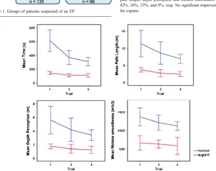
Background:Objective assessment of the suturing task is worthwhile, because this task incorporates all basic technical skills, and is a prerequisite to perform (advanced) laparoscopic procedures. The aim of this study was to determine whether motion-analysis parameters can discriminate between novice’s and experts skills during intra-corporeal suturing in a laparoscopic box trainer.
Conclusion:The construct validity has been proven for the motion-analysis parameters: time, path length, depth perception and motion smoothness for the assessment of the laparoscopic suturing task in a box trainer. The addition of economy of movement parameters has the potential to refine skills acquisition. Besides, an expert level has been set for training purposes.
JP1_05
The new concept of adhesion prevention: from the Lab to the Operating Theatre
1,2R. Corona,1K. Maylova,1M. Binda,1,3C. De Cicco,
1,2,3
P.R. Koninckx 1
Department of Obstetrics and Gynecology, University Hospital Gasthuisberg, Katholieke Universiteit Leuven, Leuven, Belgium
2
Department of Obstetrics and Gynecology, University Hospital S. Giovanni di Dio, University of Cagliari, Cagliari, Italy
3
Department of Obstetrics and Gynecology, University Hospital A. Gemelli, Università Cattolica del Sacro Cuore, Roma, Italy
The pathophysiology of adhesion formation has traditionally been considered as a local process occurring between two opposing lesions. We developed in Leuven a new concept of adhesion formation, this concept is that the entire peritoneal cavity is a cofactor in adhesion formation, and that this is quantitatively the most important aspect. This new concept was derived from animal models, especially the development of a laparoscopic mouse model, which has been fully characterized by now, has been important in demonstrating that factors from the peritoneal cavity can enhance 4 to 10 times adhesion formation following surgery. Deleterious factors identified today are mesothelial hypoxia, mesothelial hyper-oxia, dessication, and mechanical trauma. The effect of each of these deleterious factors is greatly attenuated when the temperature is lower. The mechanism involved is understood as follows. The mesothelial cells are normally large flat cells covering the entire peritoneal cavity. It was demonstrated that when‘traumatized’these large flat cells retract and bulge thus exposing between them directly the extracellular matrix.
A key observation was that these cells are more resistant to damage at lower temperatures. Finally we demonstrated that with peritoneal cavity conditioning (cooling, adding 4% of oxygen and preventing dessication) surgery adhesions can be decreased by some 75%. Combining conditioning during surgery with a local barrier after surgery adhesions decrease by over 90%.
The possibility to continue and develop this research in humans was hampered by the lack of a device permitting conditioning during surgery and until now only the effect of decreased CO2 resorbtion when 4% of oxygen is added to the CO2 pneumoperitoneum has been confirmed in the human indirectly supporting the findings of the animal models and thus the hypothesis of mesothelial retraction and bulging thus exposing the extracellular matrix following mesothelial hypoxia.
In collaboration with the engineers, we developed a prototype of a conditioning device and for the first time we “translate” the new adhesion prevention concept from the animals to the humans. Our early data confirm our concept of the entire peritoneal cavity as the most important factor in adhesion formation and confirm the feasibility of the conditioning adhesion prevention in humans.
JP1_06
A randomised controlled study comparing harmonic versus electrosurgery in laparoscopic myomectomy
1
F. Calonaci,2P. Litta,2S. Fantinato,1F. Petraglia,1P. Florio 1
Department of Pediatrics, Obstetrics & Reproductive Medicine, University of Siena; Italy,
2
Department of Gynecological Science and Human Reproduction, University of Padua, Italy
Objective: In the present study we compared the effectiveness and safety of harmonic scalpel versus electrosurgery to reduce blood loss during laparoscopic myomectomy.
Methods:A prospective randomized controlled study was based on 160 pre-menopausal women operated for symptomatic uterine leiomyomata, assigned in two treatment groups, based on the utilisation of electrosurgery devices with a vasoconstrictive solution (Group A) or harmonic scalpel (Group B). The global operative time, the time of enucleation of myoma and for suturing uterine wall defects, the degree of surgical difficulty and intraoperative blood loss were recorded.
Results:The hospital stay and the degree of pain 24 hrs after surgery were significantly (P<0.0001) lower in patients in whom the harmonic scalpel was used. The degree of surgical difficulty did not differ, but the global operative time was significantly (P=0.02) shorter in the harmonic scalpel group.
Conclusions: The use of the harmonic scalpel for laparoscopic myomectomy is associated with a low total operative time, intra-operative blood loss and, post-intra-operative pain, without increasing surgical difficulty.
JP1_07
Endometrial preparation before operative hysteroscopy in pre-menopausal women: a prospective, randomized,
double-blind, placebo controlled evaluation of oral administration of nomegestrol acetate
A. Imperatore1, A. Altomare1, S. Pinzauti1, M. Franchini2, P. Litta3, M. Gabbanini1, R. Battista1, S. Luisi1, F. Petraglia1, P. Florio1 1Department of Pediatrics, Obstetrics and Reproductive Medicine,
Section of Obstetrics and Gynecology, University of Siena, Siena, Italy;2Palagi Free-Standing Unit, Florence, Italy;
3
Department of Gynecological Science and Human Reproduction, University of Padua, School of Medicine, Padua, Italy
Objective: To evaluate the effects of 10 days treatment with nomegestrol acetate o the endometrium in order to perform office operative hysteroscopy.
Design:Prospective, randomized, clinical study. Setting:Centers for women health.
Patient(s):Eighty-six women with endometrial polyps.
Intervention(s):On day 1 of the subsequent menstrual cycle, patients were randomized to receive oral 5 mg nomegestrol acetate (n=43) or oral 4 mg folic acid (n=43) both for 10 and 30 days.
Results: Patients treated with oral nomegestrol acetate had a significantly (P<0.001) reduction in endometrial thickness compared to placebo group, both after 10 and 30 days of treatment. Moreover, the incidence of bleeding at operative hysteroscopy was significantly (P<0.01) lower in women treated with nomegestrol acetate, and the surgeon satisfaction in terms of endometrial thinning and preparation was greater than in the placebo group.
Conclusions: Oral nomegestrol acetate provides a fast, low-cost, and satisfactory preparation of the endometrium for operative hysteroscopy.
JP1_08
Additional optical feedback in a virtual reality trainer: Is it a surrogate for haptic feedback?
E. Hiemstra1, E.M. Terveer1, M.K. Chmarra2, J. Dankelman2, F.W. Jansen1,2
1
Department of Gynecology, Leiden University Medical Center, Leiden, The Netherlands
2
Department of BioMechanical Engineering, Faculty of Mechanical, Maritime and Materials Engineering, Delft University of Technology, Delft, The Netherlands
Background:Despite paramount research on the validity of various training systems, comparative research on specific features of simulators is lacking. The aim of this study was to establish the effect of naturally present haptic feedback and additional optical feedback on laparoscopic skills training.
Design:In a randomized controlled trial 50 novices were randomly assigned for 20 minutes training on one of the four training setups or for a control group (receiving no training). Training setups were a conventional VR environment (VR-I), a VR trainer with additional optical feedback (VR-II), the box trainer equivalent of VR-I (Box-I), and the box trainer equivalent of VR-II (Box-II). In each setup, the same laparoscopic task was used: piling up three cylinders. A force and hand-eye coordination requiring rubber band task was performed in a laparoscopic box trainer, as pre- and post-test. The differences between pre- and post-test results were established, taking time, path length, and depth perception into account.
VR-I
n=10
Box-II
n=10
Control
n=10
Box-I
n=10
VR-II
n=10
Pre-testPost-test
Results: The controls did not improve significantly in time, path length and depth perception. The VR-I group improved in time only, while VR-II and both box trainers groups also improved in economy of movement parameters.
Conclusion:The addition of supplementary optical feedback in VR is a promising surrogate for natural haptic feedback as present in physical box trainers.
JP1_09
Electromyographic and urodynamic study before and after tot prothesic surgery of stress incontinence
S. Caruso*, S. Bandiera*, S. Cianci*, M.G. Matarazzo*, G. Alagona†, M. Panella+.
*Department of Microbiology and Gynaecology, Santo Bambino Hospital, Catania University (95124), Italy
†Neurologic Complex Operative Unit, Azienda Ospedaliera
Canni-zzaro, Catania, Italy.
+
Department of Gynaecology and Obstetrics and Radiologic Science, Vittorio Emanuele Hospital, Catania University, Italy
Objective: To study the urethral neuromuscular function by electro-myography and urodynamic exams, before and after TOT (Trans-obturatory-tape) prosthesis surgery, in women with urodynamic stress incontinence (USI).
Study design: Urethral electromyography (EMG) data were obtained from 10 women with USI by:
urogynecologic examination-to investigate USI risk factors and to value the possible abnormal urethral mobility.
urodynamic examination-to value the pressure limits of stress incontinence;
electromyography examination-performed by 25 mm concentric needle that was put in as far as 5 mm inside internal urethral sphincter, because of the highest concentration of stripped muscular fibres; EMG data were obtained with caught, stress and rest tests.
Results: Before surgery 9 women had EMG data compatible with denervation/renervation nervous damage (tension >300 µV in com-parison with normal tension values ) in all records, and 1 woman had EMG data compatible with myogen damage (tension <100 µV in comparison with normal tension values).
After TOT prosthesis surgery, the EMG data were lower than before the treatment.
Conclusions: The EMG is a particular exam to study in deep the causes of urinary incontinence helpful to identify basis disorders of USI: the cause of this pathology is not only due to lapse support and suspension urethral structures, estimated during urogynecolog-ical examination, but also the consequence of myogen damage of urethral sphincter due to denervation and its following re-nervation.
The TOT prosthesis surgery is a modern surgical technique that by sub-urethral sling application through trans-obturatory foramen has good results. Women with USI undergoing TOT surgery have poorer urethral neuromuscolar dysfunction than before treatment.
JP1_10
Urethral sphincter electromyography in women with stress incontinence
S. Caruso*, M.G. Matarazzo*, S. Cianci*, S. Bandiera*, G. Alagona† *Department of Microbiology and Gynaecology, Santo Bambino Hospital, Catania University (95124), Italy.
†Neurologic Complex Operative Unit, Azienda Ospedaliera
Objective:The study values, through electromyography (EMG), the stripped urethral sphyncter activity in women with stress incontinence (USI)
To study the urethral neuromuscular function by electromyography and urodynamic exams, in women with stress incontinence (USI). Study design:10 women with USI and 5 healthy women (control group) underwent:
urogynecologic examination- to investigate USI risk factors and to value the possible abnormal urethral mobility.
urodynamic examination- to value the pressure limits of stress incontinence;
Urethral electromyography- (EMG) examination- performed by 25 mm concentric needle that was put in as far as 5 mm inside internal urethral sphincter, because of the highest concentration of stripped muscular fibres; EMG data were obtained with caught, stress and rest tests.
The endurance and wideness of tension were took in the consideration. Result:9 women with USI had EMG showing denervation/renervation nervous damage, supported by potential’s wideness and length higher than control group (>300 µV), either during relax or cough, and the trace’s absence of the interference with voluntary contraction. Finally, 1 woman with USI had EMG compatible with myogen damage: values <100 µV in all traces and negative anamnesis for risk factors. Conclusion:EMG is a particular exam to study in deep the causes of urinary incontinence helpful to identify basis disorders of USI: the cause of this pathology is not only due to lapse support and suspension urethral structures, estimated during urogynecological examination, but also as consequence of myogen damage of urethral sphincter due to denervation and its following renervation.
EMG is able to give information about pathophysiology of USI, so that clinicians may use the correct therapy such as rehabilitative techinique associated or not to a surgical prosthesis when urethral hypermobility is present. The limit of this method is to locate urethral sphincter.
JP1_11
Isobaric gasless myomectomy for multiple, medium or large myomas in general or combined spinal-epidural anaesthesia G. Cammareri, C.I. Brambilla, D. Rollo, F. Cirillo, E.A. Macalli, E. Ferrazzi
ICP, Hsp V. Buzzi. UO di Ostetricia e Ginecologia. Università degli Studi di Milano, Milan, Italy
Objective: To investigate the feasibility, safety and reproductive outcome of isobaric laparoscopic myomectomy using a subcutaneous lifting system (Laparotenser, Lucini L&T, Milan, Italy) and conven-tional laparotomy instruments for removing medium or large intramural and subserosal leiomyomas in general or combined spinal-epidural anaesthesia.
Design:Retrospective analysis of 142 consecutive isobaric myomectomy. Setting:University-affiliated medical center.
Patients: 142 women with 1 or more intramural or subserosal myomas measuring more than 4 cm. RESULTS: The average number of myomas was 2.2±2.0. The mean volume of the biggest fibroid was 165.3±174.8 mL with diameter of 6.9±2.4 cm. The average operating time was 112.0±45.9 minutes, blood loss was 355.2±340.1 mL. We performed 58 myomectomies in combined spinal-epidural anaesthesia (41%), with no conversion to general. The conversion to standard
laparotomy rate was 7%. The transfusion rate was 0.7%. Of the 12 women who became pregnant after operation, early miscarriage occurred in 2 cases, 3 birth took place with vaginal delivery and 7 with caesarean section.
Conclusion: Isobaric myomectomy is a safe and reliable procedure even in the presence of multiple or enlarged myomas, and it is possible to be performed in combined spinal-epidural anaesthesia.
JP1_12
Tubal sterilization by laparoscopy or hysteroscopy: which is a cost effective procedure?
A. Altomare, M. Franchini, S. Calzolari, L. Cianferoni, F. Calonaci, S. Pinzauti, M. Gabbanini, P. Florio
Section of Obstetrics & Gynecology, Pediatrics Obstetrics & Reproductive Medicine, University of Siena, Siena, Italy
Introduction: Tubal sterilization (TS) is a widely used method of birth control because of its proven safety and effectiveness. It is typically and generally performed by laparoscopy for interval procedures. However a new method for TS is based on the introduction inside both of the falloppian tubes, by hysteroscopic guidance, of a microdevice, the Essure permanent birth control system (Conceptus, San Carlos, CA) that leads to the formation within 3 months of an intratubal fibrosis created by the proliferation of connective tissue. In the present study we computed current charges of performing laparoscopic or hysteroscopic TS through the activity-based cost/management (ABC/M) system, an accounting technique that allows organizations not only to determine the actual costs associated with their services based on the resources they consume, but also to detect when, where, and why the money is spent, to use this information as a basis for cost management.
significantly reduced the total expenses for tubal sterilization through EHTO than LTS.
Conclusions:The real advantage of sterilization by EHTO respect to LTS is related to its minimal impact on women health, that allows a rapid resumption of normal activity producing a minimal discomfort. In conclusion, EHTO was less expansive tha LTS mainly for the low support before, during, and after hospital stay; EHTO expenses may be reduced further by lowering the cost of the Essure microinserts.
JP1_13
Salpingostomy or salpingectomy for ectopic pregnancy: fertility outcomes
A. Rocca, S. Angioni, M. Palomba, G.B. Melis
Department of Gynaecology and Obstetrics, University of Cagliari, Italy
Most of the ectopic pregnancies occur in young age and subsequent fertility is an important issue. Laparoscopic surgery has advantages over open surgery and results in higher rate of subsequent intrauterine pregnancies and lower rate of recurrent ectopic pregnancy.
Aim of our study was to analyze the fertility outcome after conservative or radical surgery for ectopic pregnancy and to identify any biological factors that may influence fertility after an ectopic pregnancy. A retrospective study which was carried out by collecting information from the patient’s hospital records was performed on 80 cases of confirmed ectopic pregnancy between January 2004 and January 2009 at our department. The obstetrics outcomes were obtained in 45 patients. Pregnancy rates and repeated ectopic pregnancy rates were analyzed up to 5 years after the diagnosis and surgical treatment of ectopic pregnancy. Our results showed that the term pregnancy rates were not significantly different following radical or conservative surgical treatments for ectopic pregnancy (36% in the conservative group compared with 33% in the radical group). But equally important, the risk of a further ectopic was not increased in the conservative surgery group (3% either after salpingostomy or salpingectomy). The incidence of intrauterine pregnancy rate (term pregnancy + miscarriage) was also similar. Multivariate regression analysis showed that the factors associated with higher fertility were age (less than 30 years), past history of term pregnancy and a negative history of infertility (P<0.05).
In conclusion no significant difference in intrauterine pregnancy or repeated ectopic pregnancy rates were found after radical or conservative surgical treatment for tubal pregnancy. A possible explanation of such results could be that salpingectomy is genereally choosen in cases of severely damaged fallopian tube or recurrent ectopic pregnancy in the same tube. Patient’s age, previous obstetric performance and a history of infertility significantly influenced fertility after surgery for ectopic pregnancy.
JP1_14
The Regional Expression Level of TACR1 at Specific Locations in the Peritoneum in a Rodent Animal Model
B. Kraemer1, M. Wallwiener2, C.W. Wallwiener3, I. Juhasz-Böss1, A. Hartkopf1, T.K. Rajab1
1University of Tuebingen, Tuebingen, Germany 2
University of Heidelberg, Heidelberg, Germany
3Technical University of Munich, Munich, Germany
Objective:Experimental trial in an in vivo animal model.
Materials and methods: Laboratory facilities of a university department of obstetrics and gynecology and a microarray facility. Animals: Seventeen female Wistar rats
Intervention(s):Peritoneal adhesions were induced by the placement of three unilateral ischemic buttons with 4-0 prolene. Second look analysis for adhesion scoring occurred after day 1, 3 and 5. Six tissue samples from n=3 animals were harvested at day 3 for quantitative PCR and real-time RT-PCR analysis.
Main outcome measure(s):The differential expression of TACR1 in the ischemic button compared to native peritoneum on the contralat-eral non-traumatised side.
Results:Quantitative PCR analysis after 3 days demonstrated down-regulation of TACR1 in the tip of the ischemic button compared to native peritoneum on the contralateral non-traumatised side. This difference was statistically significant (p<0.01). No TACR1 expres-sion was found beneath the tip of the button.
Conclusions:Different expression levels of TACR1 may depend on the depth of the tissue sample. We assume that the trauma site might be divided locally with different expression levels and TACR 1 might be expressed ubiquitously.
Endometriosis: Diagnosis and Surgery
JP2_01
Endometriosis and cancer A Loddo, G Benassi, B Andrei
Programma Chirurgia Ginecologica, Mininvasiva e Oncologica. Azienda Ospedaliero-Universitaria di Parma, Italy
JP2_02
Quality of sex life after laparoscopic excision of rectovaginal endometriosis
J.L. Coloma, M.A. Martínez-Zamora, F. Carmona, C. Castelo-Branco, J. Balasch
Neonatology. Hospital Clínic of Barcelona. University of Barcelona. Barcelona. Spain
Objective:To quantify the impairment of the quality of sexual life (QSL) in sexually active women with RVE and severe deep dyspareunia (SDD) and to assess the effect of the laparoscopic excision of RVE on the QSL 4–6 months after surgery.
Methods: 20 premenopausal women with RVE and 20 controls. Patients underwent laparoscopic excision of RVE. Patients with RVE, before surgery and 4–6 months afterwards, and controls, answered 3 self-administered questionnaires to assess the QSL (The Female Sexual Distress Scale (FSDS) which evaluates the sexual disfunction; The Sexual Quality Of Life Questionaire-Female (SQOL-F) which quantifies the quality of sexual life; The Brief Profile of Female Sexual Function (B-PFSF) which diagnoses a hypoactive sexual desire disorder).
Results: There were no recurrences during the study period. The comparison of patients before surgery and controls showed a statistically significant impairment of QSL in patients with RVE. Patients after surgery had a statistically significant improvement of the QSL. There were no statistically significant differences between controls and patients after surgery, showing a normalization of the QSL after surgery.
Conclusions: Patients with RVE and SDD have an impairment of QSL which improves to normality after 4–6 months after surgery.
JP2_03
Value of three-dimensional ultrasonography for diagnosing of rectovaginal septal endometriosis
P. Barri-Soldevila1, R. Fernandez1, M.A. Pascual1, L. Hereter1, B. Graupera1, F. Tresserra2, I. Rodriguez1
Department of Obstetrics, Gynaecology and Reproduction1;
Department of Pathology2Institut Universitari Dexeus, Spain
Background:Patients with deep infiltrating endometriosis (DIE) of the rectum often benefit from surgical treatment, in terms of pain relief and treatment of infertility. The aim of the present study was to evaluate the diagnostic accuracy of three-dimensional transvaginal sonography (3D TVS) for preoperative detection of rectovaginal septal endometriosis.
Methods:This prospective study included 31 women (mean age 36; range 25–44) with suspect of rectovaginal endometriosis. All patients underwent 3D TVS for the evaluation of the rectovaginal septum, before undergoing laparoscopic radical resection of endometriosis. Rectovaginal endometriosis was defined as hypoechoic areas, nodules or anatomic distortion of this specific location. Ultrasonographic results were compared to surgical and histological findings. Sensitiv-ity, specificSensitiv-ity, positive and negative predictive values (PPV and NPV) and test accuracy were then calculated.
Results:Rectovaginal septum endometriosis was confirmed in 19 out of 31 (61.3%) cases. The sensitivity, specificity, PPV, NPV, and test accuracy on rectovaginal septum infiltration were 71.4%, 88.9%,
90.9%, 66.7% and 78.26%, respectively. Best accuracy was observed in the last patients
Conclusions: Three-dimensional US appears an effective mean of detecting endometriosis of the rectovaginal septum and should be included in the imaging process for patients presenting with clinically suspected rectovaginal endometriosis.
JP2_04
Deep pelvic endometriosis: from diagnosis to wellness
A. Cocco, A. Borghero, C. Saccardi, G. Guidetti, L. Conte, P. Litta
Department of Gyanecological Sciences and Human Reproduction, Padua University, Italy
Objective: To determine efficacy of laparoscopic excision of deep pelvic endometriosis.
Methods:102 high symptomatic women with deep pelvic endome-triosis underwent clinical examination, transvaginal ultrasound, NMR and sonovaginography. Among of the 102 women, 70 patients, with severe symptoms, underwent laparoscopic excision of deep pelvic endometriosis. Endoscopic surgery was performed with complete separation of rectovaginal space and resection of the node. In case of vaginal involvement was performed vaginal exeresis, in case of rectal wall involvement more than 50%, segmental bowel resection was performed. Operative data as well as dysmenorrhoea, dyspareunia, chronic pelvic pain and dyschezia before and 6 and 12 month after surgical treatment were recorded.
Results: Mean operative time was 126.4±34.7, mean blood loss was 76.2±22 ml. In 17 (34%) cases we perform excision of posterior vaginal fornix cause vaginal wall involvement. In 6 cases (12%) cases we performed excision of rectal wall. At 12 month follow up 39 (78%) women revealed absent or mild dysmenorrhoea, 45 (90%) women revealed absent or mild dyspareunia, 46 (92%)) women revealed absent or mild chronic pelvic pain, 48 (96%) women revealed absent or mild dyschezia. Conclusions: Surgical management of deep pelvic endometriosis could be conservative, ensuring good improvement of symptoms, performing vaginal or rectal exeresis only when strictly necessary.
JP2_05
Presurgical diagnosis of posterior DIE
R. Locci, S. Sanna, E. Stochino, S. Pirarba, S. Angioni, G.B. Melis
Department of Obstetrics and Gynaecology, University of Cagliari, Cagliari, Italy
perfor-mance (accuracy, sensitivity, specificity, positive and negative predictive value) for predicting retrocervical and/or rectovaginal endometriosis were assessed. Multiple logistic regression analysis to select the best combination of these parameters for predicting posterior DIE was calculated. Our results suggest that bimanual pelvic examination showed a high accuracy, sensitivity and specificity in the diagnosis of DIE. A thorough evaluation of symptoms and signs allowed the diagnosis of posterior DIE in the majority of cases before surgery.
JP2_06
Dysmenorrhoea after cesarean section. Not always endometriosis
N. Molin Pradel, G. Maricosu, G.B. Melis, S. Angioni
Department of Obstetrics and Gynaecology, University of Cagliari, Cagliari, Italy
Dysmenorrhoea is common, and in up to 20% of women it may be severe enough to interfere with daily activities. Dysmenorrhoea may begin soon after the menarche, after which it often improves with age, or it may originate later in life after the onset of an underlying causative condition. We describe a case of secondary dysmenorrhoea after caesarean section. A 39 years old women referred to our attention after two years of menstrual pain. She reported two caesarean sections. Instrumental controls (transvaginal ultrasound and MRI), blood tests and CA125 were normal. Since her symptoms persisted after medical treatment she underwent a diagnostic laparoscopy. The surgical examination of the pelvis evidenced the absence of endometriosis lesions and the presence of post obstetric surgery strong adhesions between the uterus and the anterior abdominal wall. The anterior part of the uterus until its fundus was stucked to the abdomen. Cold knife adhesiolysis was performed and anti-adherence device (Hyalobarrier) was used to prevent the recurrence. This case suggest that the occurrence of secondary dysmenorrhoea after uterine surgeries could be related even to the fixation of the uterus to the abdominal wall not only to endometriosis.
JP2_07
Prevalence of DIE in patients with ovarian endometrioma I. Arena, S. Pirarba, S. Sanna, E. Stochino-Loi, S. Angioni, G.B. Melis
Dept. of Gynaecology and Obstetrics, University of Cagliari, Italy
Three types of endometriosis have been described: peritoneal, endometrioma and deep infiltrating endometriosis (DIE).
Aim of our study was to investigate the role of endometrioma as a marker of DIE. 822 consecutive surgical reports of patients with endometriosis have been collected and the association of the presence of endometrioma(s) with DIE has been detailed.
254 patients had ovarian endometriosis and in 128/254 (50,39%) it was associates to DE. Left side was involved in 40,63% of the cases, right side in 33,65% while the bilateral involvement of the ovaries was present in 36,72% of the cases. In the 126 patients with endometrioma (s) but without DE the involvement of both ovaries was only in 19,05%. This study indicates that left side lesions are more frequent than right ones. The observation that left lesions tended to be more severe than right-hand ones gives some support to the idea of a different anatomical
distribution of endometriotic lesions. From an anatomical point of view, these findings support the transplantation therapy in the pathogenesis of endometriosis. The moderately high positive likelihood ratios reported in this study shows that in this cohort of women with endometriosis, the presence of endometrioma is a good indicator of deep infiltrating endometriosis in particular when both ovaries are involved. Patients with evidence of endometriomas should be carefully studied before surgery in order to be referred to centres that can offer the appropriate advice and treatment including the potential to manage the DE with a careful and complete laparoscopic excision of the lesions.
JP2_08
Laparoscopic ureterocystoneostomy in severe endometriosis G. Maricosu, M. Melis, G.B. Melis, S. Angioni
Department of Gynaecology and Obstetrics, University of Cagliari, Italy
Endometriosis has been estimated to affect 4%–15% of all women with child-bearing potential. The pelvis is the most common location. Cases have also been reported with distant locations such as the lungs and gastrointestinal tract. Urinary tract involvement is uncommon, with a variable incidence reported. Deep infiltrating endometriosis may involve the ureters in some cases. Most of them have an extrinsic compression that can be solved with careful excision of the disease. The symptoms of ureteral compression are non-specific at clinical presentation and preoperative diagnosis is very important. A case of ureterectomy, ureterocystoneostomy and bladder fixation on ileopsoas muscle using the laparoscopic approach will be described. The patient presented chronic pelvic pain and signs of ureteral stenosis. Histological examination, confirmed active ureteral endometriosis, which was found to be intrinsic. No ureteral endometriosis relapses occurred within the follow-up. Ureteral endometriosis is marked by non-specific symptoms, making preoperative diagnosis sometime difficult. Therefore, an ultrasound or urographic examination of the urinary tract in case of pelvic endometriosis is mandatory. Laparoscopic terminal ureterectomy with ureterocystoneostomy has provided long-term favourable results. It is feasible and presents the well known advantages of the mini-invasive approach.
JP2_09
Psychological aspects of surgery for endometriosis I. Melis, I. Arena, G.B. Melis, S. Angioni
Department of Gynaecology and Obstetrics, University of Cagliari, Italy
depres-sive mood. Even in cases in which surgery is able to resolve the anatomical causes of dyspareunia it, sometimes, persists representing the cognitive schema of a long lasting mechanism the link intercourse to pain. Taking into count these aspects allows considering all the variables that could interfere on surgical results and on the coping with the disease. Our clinical experience on endometriosis patients suggests the need of a psychological support in every step of the medical intervention from diagnosis, to surgery, from the follow-up to the coping with important problems like infertility.
* Finanziamento: Regione Autonoma della Sardegna. Programma Master and Back. Beneficiaria: D.ssa Irene Melis
Hysteroscopy: from Office to Resectoscope JP3_01
Local anaesthesia for reducing pain and vasovagal reactions during outpatient hysteroscopy: a systematic review
N.A.M. Cooper, K.S. Khan, T. J. Clark
University of Birmingham, Birmingham Womens Hospital, UK
Aim: In recent years the ‘classical’ in-patient hysteroscopy’ has evolved into a diagnostic and therapeutic outpatient procedure. The RCOG has recently published standards of care stating that‘ outpatient-based diagnostic services should be available in the community and hospital setting, including operative procedures for carefully selected cases’.1 For this to be possible the procedure needs to be acceptable to patients. The aim of this systematic review is to look at how the use of local anaesthetic affects the patient’s pain experience during out-patient hysteroscopy and also the incidence of vasovagal attacks. 1. Royal College of Obstetricians and Gynaecologists. Standards for Gynaecol-ogy. Report of a working party. RCOG Press. London 2008. P28. Method: A Systematic review and meta-analysis of all published randomised controlled trials that look at the use of local anaesthetic for pain relief during out-patient hysteroscopy. EMBASE, Medline, CINAHL and the Cochrane library were searched for relevant studies. The abstracts of all the resultant studies were read by two authors independently and studies that met the inclusion criteria were selected. The full articles were then obtained and further irrelevant or non RCT studies were excluded. The references of all selected studies were searched to ensure that any relevant studies had been not been overlooked. Where possible meta-analysis was performed using RevMan.
Results:Intracervical and paracervical injection of local anaesthetic reduce the pain of outpatient hysteroscopy. Transcervical and topical application of anaesthetic to the ectocervix do not. There is no significant reduction in the number of vasovagal episodes when local anaesthetic is used for the procedure.
Conclusions:Clinically relevant conclusions can be drawn from the results indicating the best use of local anaesthetic for outpatient hysteroscopy.
JP3_02
Endometrial preparation for hysteroscopic surgery
M. Maksimović, S. Spremović, G. Lazović Radonjić, A. Gudov, T. Dosev, S. Milićević
Institute of Gynecology and Obstetrics, Clinical Center of Serbia
Introduction: Method of endometrial preparation influences the visibility during the hysteroscopy interventions. Aim of the study is
to compare three standard methods of endometrial preparation for hysteroscopy: a gonadotropin-releasing hormone analogue therapy, an oral contraceptive therapy and non-medicament planning according to menstrual cycle.
Patients and Methods: We examined 182 patients who had hysteroscopy interventions in the period of one-year. Preoperatively, 15 patients were treated by oral contraceptives, 147 of them were treated by gonadotropin-releasing hormone analogue (Diphereline) and 20 patients were not treated by any medicaments for endometrial preparation. We performed biopsy and histological analysis of endometrial quality in order to compare effectiveness of endometrial preparation methods.
Results: According to histological examinations, intraoperative condition of endometrium was described as atrophic, early prolifera-tive, or late proliferative. All the Diphereline-treated patients had atrophic endometrium, except two of the patients with early proliferative endometrium, with BMI 35 and 40, separately. All the patients treated with oral contraceptives, had early prolifera-tive endometrium, except two of the patients with atrophic and 4 of the patients with late proliferative endometrium. Patients who did not undergo medicament preparation had early proliferative endometrium.
Conclusion: Hysteroscopy planning according to the end of menstrual bleeding is difficult for work organization in hospitals with a number of patients. The best hysteroscopic conditions were achieved using the gonadotropin-releasing hormone analogue for preoperative preparation. Weakness of this method is high cost of these medicaments and necessity of hormone therapy after the intervention.
JP3_03
Diagnostic hysteroscopy and pain. Conventional CO2
vs. vaginoscopic approach using saline S. Sanna, A.M.B. Vacca, G.B. Melis, S. Angioni
Department of Gynaecology and Obstetrics, University of Cagliari, Italy
Diagnostic hysteroscopy is not widely used in the office setting due to the discomfort produced by the procedure, especially in nulliparous women.
We performed a prospective, randomised trial designed to assess the roles of uterine distension medium (CO2or saline) and the technique (use of speculum and tenaculum or vaginoscopy) on patients discomfort. Fifty women attending the infertility clinic were randomly assigned to undergo conventional diagnostic hysteroscopy using CO2 (group A,n=25) or diagnostic hysteroscopy (group B,n=25) using saline and the vaginoscopic approach. All procedures were performed by the same surgeon using the same instrument. Patient discomfort was analysed throughout the visual analogue score (VAS) at the following steps: 1) vagina, 2) penetration inside the uterus, 3) observation of the cavity and 4) 15′after the procedure. Group B patients showed significantly less pain in particular in steps 1,2 and 4 (p<0.01).
JP3_04
Office hysteroscopy in the IVF Center: our experience
M. Ruggiero, G. Viana, N. Pluchino, P. Monteleone, V. Valentino, V. Cela, A.R. Genazzani
Department of Reproductive Medicine and Child Development, Division of Obstetric and Gynaecology, University of Pisa, Itay
Aim:The role of office hysteroscopy in the management of infertility is still controversial. The aim of this study was to analyse the results obtained from diagnostic hysteroscopy performed routinely during the investigation of the infertile women, to evaluate the effectiveness of the procedure in the diagnosis of uterine causes of female infertility. Methods:We evaluate the hysteroscopic findings in 100 consecutive infertile female admitted at the IVF Center of the Department of Gynaecology and Obstetrics, University of Pisa, using a office hysteroscopy 2,9 mm with operative canal and bipolar electrode. Results:The hysteroscopy was abnormal in 53% of cases: endome-trial polyps (11%), uterine fibroids (9%), endometritis (9%), uterine synechias (3%), substenosis cervical canal (13%), endometrial dysfunctions (4%), and uterine anatomical variants (4%). The operating hysteroscopy procedure was performed during the same procedure with“see and treat”techniques in 40% of patients and in a secondary procedure with monopolar or bipolar resettoscope in 60%. Conclusion:Our data suggest that hysteroscopy presents an important role in the management of female infertility, and in particular the office hysteroscopy offers the possibility of diagnosis as well as treatment in the same session. Consequently, in view of the low complication rates, minimal time requirement, and a significant effect on the diagnostic trial, hysteroscopy could be routinely performed on all infertile patients.
JP3_05
Risk factors related with recidivals of endometrial polyps after histeroscopy polyp resection
M. Aubá, B. Olartecoechea, D. Díaz, M. García Manero, J.A. Mínguez
Clínica Universidad de Navarra, Spain
Aim of the study:Endometrial polyps are glands and endometrial stroma hiperplasic growth, which create projections through uterine cavity. It is a very frequent entity, that can recidivate after a first treatment. The aim of our study is to evaluate the different risk factors that are involved after a histeroscopic polyp resection.
Methods: A retrospective study of 204 patients in whom a polipectomy by hysteroscopy was realized from January 2002 to December 2004 with a up until April 2008( minimum follow-up of 40 months and maximum follow-follow-up of 76 months)in order to detect patients who present recidival and estimate the principal risk factors involved.Of the 204 patients, 67 are premenopausic and 137 postmenopausic.The main risk factors studied were: age, menopause, menopause timing, symptoms, Hormonal replacement therapy, Ta-moxifen, antihypertensives, polyp size and body mass index. Results:In the 204 cases that were analized we have found 14% of recurrrences. We found a greater percentage of recidivals ( 32%) among patients who took tamoxifen, in comparison with the patients who did not took it( 9.9%). Neither the age, menopause or the size of the polyp conditions a recurrence of the sickness.
Conclusions: With our data, and assuming that our sample size is small for the risk factors studied, only the patients taking tamoxifen have a major risk of recurrence after the polipectomy.
JP3_06
Endometrial preparation before operative hysteroscopy in pre-menopausal women: a randomized, double-blind, placebo controlled comparison of vaginal and oral administration of danazol
M. Gabbanini1, A. Altomare1, M. Franchini2, S. Pinzauti1, A. Imperatore1, F. Petraglia1, P. Florio1
1
Department of Pediatrics, Obstetrics and Reproductive Medicine, Section of Obstetrics and Gynecology, University of Siena, Siena, Italy;
2
Palagi Free-Standing Unit, Florence, Italy
Objective: Hysteroscopy is the “gold standard” investigation for visualizing and treating any focal lesion, however the operative technique is performed at its best only after having a flat and atrophic endometrium at the time of the procedure. In the present study we the usefulness and the efficacy of a dose of 600μg of vaginal versus 600μg orally administered danazol treatment was compared in women undergoing endometrial preoperative preparation for hysteroscopic surgery.
Design:Prospective, randomized, controlled clinical study. Setting:University department of women care.
Patients:Ninety-one patients with endouterine pathologies (endome-trial polyps, submucous myoma, septate uterus).
Interventions: Patients treated with oral and vaginal danazol (600 mg/daily) underwent transvaginal sonography and operative hysteroscopy
Main outcome measures: Endometrial response to the medical treatment, side effects, procedure time, intraoperative bleeding, infusion volume, surgeon satisfaction.
Results:Endometrial response was higher in patients treated by vaginal danazol administration, that also had less side effects. Cervical dilatation did not differ between groups, however the length of the operating time and the volume needed for infusion were significantly (P<0.001, for both) higher in patients treated by oral than vaginal danazol administration. Surgeon satisfaction in terms of endometrial preparation was also statistically significantly (P<0.001) higher for women treated by vaginal danazol, because of the endometrial appearance.
Conclusions:Both administration routes are good ways to prepare the endometrium for operative hysteroscopy, however, the data suggest that vaginal administration is preferable to the oral one.
JP3_07
A case series of patients undergoing concomitant Novasure™ endometrial ablation and Essure™sterilisation in the outpatient setting
N.A.M. Cooper1, T. Bingham2, T.J. Clark2
1University of Birmingham, Edgbaston, Birmingham, UK 2
Birmingham Women’s Hospital, Edgbaston, Birmingham, UK
concom-itant outpatient endometrial ablation and hysteroscopic sterilisation (Essure™) between March 2006 and June 2009 from a prospective electronic database which recorded patient characteristics and procedural information. Identified patients were followed up by a standardised telephone interview to enquire about their menstrual bleeding symptoms, treatment side effects, continued use of Essure™for contraception, return to activities of daily living and satisfaction with the treatments. Results: Seven women had undergone Novasure™ endometrial ablation followed immediately by Essure™ hysteroscopic sterilisa-tion during the same appointment. All of the procedures were completed successfully and safely with no serious side effects or complications reported. For the seven successful procedures, the initial three patients underwent hysterosalpingogram (HSG) although only one could be successfully completed because of problems instrumenting the uterus. These two patients, along with the remaining four underwent ultrasound scan to confirm correct placement of the Essure™ inserts. Satisfactory placement of the Essure™was confirmed in all cases except one patient in whom an HSG was indicated (a unilateral intrauterine trail length of 14 coils) but failed due to technical factors. She subsequently underwent a laparoscopic sterilisation at a later date. Six patients are relying on Essure™for contraception (as one woman later underwent a hysterec-tomy for persistent vaginal discharge) and all have reported satisfaction and significant improvement in their menstrual symptoms (range 3 months to 3 years). One woman underwent the procedure very recently and has only had one period since; therefore she is unable to comment on her bleeding symptoms. The median number of days taken to resume normal daily activities after the procedures was 4 (range 1–7). Conclusions:Concomitant Novasure™endometrial ablation followed by immediate placement of Essure™hysteroscopic sterilisation in the outpatient setting appears to be safe, feasible and effective for the treatment of dysfunctional uterine bleeding and permanent birth control. An ultrasound scan is the test of choice to confirm satisfactory placement. Studies are needed to confirm these findings and to evaluate the optimal sequence of the procedures.
JP3_08
Learning curve and feasibility of hysteroscopic endometrial polypectomy in the office setting: Results of the initial sample of sixty patients
N. Pluchino, V. Cela, A.R. Genazzani
Department of Reproductive Medicine and Child Development, Division of Gynecology and Obstetrics, University of Pisa, Pisa, Italy
Study Objective:Analysis of the learning curve and feasibility of office hysteroscopic surgery for endometrial polyps in terms of patient compliance and require of analgesia/anesthesia.
Design:The study was a case series of sixty patients who underwent surgeries for endometrial polyps.
Setting:Tertiary Gynecology Endoscopy Center of University of Pisa. Interventions: All procedures were performed using a 5.2 mm continuous flow office hysteroscope (Bettocchi Office hysteroscope; Karl Storz GmbH & Co., Tuttlingen, Germany) equipped with an operative channel of 1.8 mm in which coaxial bipolar electrode may be inserted (Versapoint Bipolar Electrosurgical System-Gynecare; Ethicon Inc., NJ, USA). All surgeons had a large experience in office diagnostic hysteroscopy.
Measurements & Main Results: Patient characteristics, polyp size and hystological findings were recorded. All patients with polyps
measuring more than 3 cm were excluded from the analysis. Surgical time was measured and patient procedure compliance was assessed by means of a 10 cm visual analogue scale (VAS) after the end of the procedure. All procedure started without analgesia/anesthesia and patient require of pain-reducing treatment was assed for each hysteroscopy. To evaluate the effects of learning curve all patients were divided into three chronological groups of 20 patients each. Time procedure and patient compliance (VAS) resulted significantly improved with surgical experience. In particular, the rate of analgesia/ anesthesia requirement was significant reduced just after twenty surgeries, thus increasing the number of patient that completed endometrial polypectomy in the office setting with a good compliance. However, patient characteristics (mainly age) and polyp diameter are two independent factors affecting patient compliance
Conclusion: Office hysteroscopic polypectomy is a feasible surgery with a relatively short learning curve to obtain well-tolerated procedures. However the selection of patients appears essential to increase compliance, at least for an initial sample of sixty patients.
JP3_09
Septate uterus and infertility: accuracy of the combination of diagnostic hysteroscopy and doppler sonography in diagnosing uterine anomalies
S. Arena, S. Canonico, G. Luzi, G.F. Brusco, G. Affronti
S.C. Ostetricia e Ginecologia Azienda Ospedaliera di Perugia, Perugia, Italy
Objective:The aim of our study was to evaluate the effectiveness of diagnostic hysteroscopy and Doppler sonography in the correct management of septate uterus.
Materials and methods: We enrolled 108 patients diagnosed with septate uterus and scheduled them to diagnostic hysteroscopy and Doppler sonography. Hysteroscopic metroplasty was performed only in cases negative for vascular flow of the fundus at Doppler sonography. From January 2007 to December 2008 we submitted to diagnostic hysteroscopy 504 patients for infertility; 108 (21.42%) of them were diagnosed with a septate uterus, thus submitted to Doppler sonography. In all cases in which the fundus was not vascularized, (64.81%), a hysteroscopic metroplasty was performed.
Results:After the intervention, the conception rate was 40%, becoming 28 patients pregnant. The abortion rate was 2.85%. The term pregnancy rate was 37.1%. Septate uterus reduces pregnancy and conception rate. Conclusion:According to our experience not all septate uteri should be treated but only those negative at Doppler sonography. Combining these two diagnostic steps we increased significantly our pregnancy rate, probably reducing useless metroplasty of vascularized tissue.
JP3_10
Hysteroscopic diagnosis of stromomyoma
C. Coppola*- A. Di Spiezio Sardo*- M. Spinelli - B. Zizolfi, G. Mansueto** - C. Nappi*
*Department of Gynaecology and Obstetrics, and Pathophysiology of Human Reproduction, University of Naples“Federico II”, Naples, Italy **Department of Biomorphologic and Functional Sciences, Pathology Section, University of Naples“Federico II”, Naples, Italy
cells. It is important that these tumours be diagnosed early, since they are likely to behave according to their stromal component, which may easily metastasise.
Materials and methods: We report the case of a 56-year-old postmenopausal woman, referred to our Department for postmeno-pausal bleeding. Since trans-vaginal pelvic scan showed an unhomo-geneous sub-mucosal lesion, the patient underwent office hysteroscopy with vaginoscopic approach, using a 4-mm continuous-flow office hysteroscope. Two contiguous intra-cavitary lesions were detected: the first was cystic, hypervascularized, 1.0 cm-sized, in the fundal area; the second was solid, whitish, 1.5 cm sized, in the anterior wall of the fundal area, with polypoid aspect and hard consistency. Multiple targeted biopsies were performed by means of bipolar Twizzle electrode and 5Fr grasping forceps.
Results: Histological examination revealed both endometrial stro-mal and smooth muscle tissues as specifically related to stromo-myomas. The patient underwent total abdominal hysterectomy. Pathological findings confirmed the lesion to be extended throughout the thickness of miometrium. When hysteroscopic examination reveals a lesion with both cystic and solid features, multiple biopsies should be performed since this finding may be suggestive of a stromomyoma.
Conclusions:The choice among conservative and radical treatment should be made in light of histological findings, symptoms and family planning needs of the patient.
JP3_11
Office Operative Hysteroscopy: an update of the evidence (2002–2009)
M. Spinelli, A. Di Spiezio Sardo, S. Bettocchi, L. Nappi, M. Guida, C. Nappi
Pathophysiology of Human Reproduction, University of Naples
“Federico II”, Naples, Italy
Objective: The aim of our review article was to provide a comprehensive survey of the newest advancements of office operative hysteroscopy (O.O.H) in the treatment of those gynaecological pathologies/conditions, which have not been included in the scheme of treatment indication for office procedure proposed in 2002. Materials and methods:We searched MEDLINE, EMBASE and the Cochrane Database of Systematic Reviews in order to identify papers published from 2002 to 2009. We identified 20 articles: 10 were related to hysteroscopic sterilization; 1 was on metroplasty; 1 was on hysteroscopic embryo implantation; 8 were related to the office-based treatment of some uncommon gynaecological pathologies/conditions (i.e cases of hematometra, diagnosis and treatment of vaginal lesions, treatment of uterine cystic neo-formations, bleeding from cervical stump, diagnosis and treatment of endo-cervical ossification, removal of utero-vaginal packing).
Results: Most of the procedures were carried out safely and successfully in the office setting, with high patients’ compliance. The success of the procedures has been confirmed by resolution of the symptomatology as well as by a follow-up instrumental examination.
Conclusions: Currently, thanks to technological advancement and increased operator experience, more and more gynaecological pathol-ogies/conditions, traditionally treated in the operating room, may be treated safely and effectively by O.O.H. This represents the“new
frontiers of office operative hysteroscopy”which may further promote the spreading of such philosophy.
Key words: bipolar electrode, mechanical instruments, miniatur-ized hysteroscopes, office operative hysteroscopy, see and treat hysteroscopy.
JP3_12
Anatomical impediments at Office Hysteroscopy:“Tip and Tricks”
for getting over them!
M. Sorrentino, A. Di Spiezio Sardo, M. Guida, M. Spinelli, D. Canzaniello, C. Nappi
Pathophysiology of Human Reproduction, University of Naples
“Federico II”, Naples, Italy
Objective:The aim of our study was to describe“tips and tricks”for over-passing anatomical impediments (AI) at office hysteroscopy (OH).
Materials and methods:We conduced a retrospective study on 3756 office procedures performed from 2003 to 2008, in 1007 patients diagnosed at OH as having a variety of cervical scarring on the external and/or the internal uterine ostium. Several“tip and tricks”
were used to get-over such AI, including the avoiding of the speculum, the use of oval-profile hysteroscopes, the adhesiolysis with the tip of the hysteroscope, the use of mechanical instruments and the radial incision of the uterine ostium with 5Fr bipolar electrodes (BE). AI were over-passed in 92.2% of cases. The vaginoscopic approach, a liquid distension medium and oval-profile hysteroscopes with continuous-flow and operative sheaths were used in all cases.
Results:Cervical stenosis were overcome with mechanical adhesiol-ysis with the tip of hysteroscope and with grasping forceps in 88.8% and 7% of cases, respectively. BE and scissors were used in 2.3% and 1.9% of cases, respectively. Minimal and mild stenosis were overcome by gently pushing the tip of the hysteroscope or by stretching the fibrous tissue with grasping forceps with teeth. While, moderate and severe AI were over-passed using sharp scissors and BE, the latter mostly used when an access to the cervical canal could not be detected.
Conclusions: Recent technical advances, as well as the increased operator’s experience, have made it possible to treat even severe AI at OH, reducing significantly the rate of failed procedures.
JP3_13
Conservative hysteroscopic treatment of atypical polypoid adenomyoma: our experience
L.M. Sosa Fernandez, A. Di Spiezio Sardo, M. Scognamiglio, M. Guida, M. Spinelli, C. Nappi
Department of Gynecology, Obstetrics and Pathophysiology of Human Reproduction, University of Naples“Federico II”, Italy
removal of endometrium adjacent to the APA (2), removal of myometrium underlying the APA (3) and multiple random endometrial biopsies (4). Each step was completed by a patholog-ical analysis. An intrauterine device releasing levonorgestrel (Mirena) was inserted in patient I, as she had required for contraception. In both cases, conservative surgery was effective as histological examination of all specimens confirmed the APA to be confined to the endometrium, without any premalignant or malignant lesion. Trans-vaginal ultrasound and O.H. with targeted biopsies at 1 and 6 months after surgery were negative for malignancy. Patient I is currently under a 2-year-treatment with Mirena, while patient II achieved a pregnancy 9 months after surgery, after a IVF cycle. In both cases, follow-up biopsies performed every 6 months were negative for malignancy. Our technique, eventually associated with Mirena, under a close postoperative surveillance, may represent a good therapeutic option for those women with APA wishing to preserve their fertility.
JP3_14
Role of resectoscopic endometrial exeresis in the treatment of atypical endometrial hyperplasia
E. Marra1, D. De Angelis1, F. Armillotta1, A.M. Perrone1, M.C. Scifo1, G. Formelli1, G. Gubbini2, A. Di Spiezio Sardo3, P. Casadio1
From Department of Gynaecology and Obstetrics, S.Orsola-Malpighi, University of Bologna, Italy1, Department of Gynaecology of Hospital “Madre Fortunata Toniolo”, Bologna, Italy2 and Department of Obstetrics and Gynaecology and Pathophisiology of Human Repro-duction, University of Naples, Italy3
Objective: To assess the diagnostic accuracy of Resectoscopic Endometrial Exeresis (TCRE) in the cases of Atypical Endometrial Hyperplasia (AEH), and to study their potential in the management of patients at high surgical risk (ASA IV).
The need of a diagnostic technique in the pre-operative phase, more accurate than ones currently available, results from the possibility of the AEH to progress to type 1 endometrial carcinoma in approx-imately 30% of cases, or to coexist with endometrial carcinoma in 25% cases already at the time of diagnosis.
Materials and methods:Between 2003 and 2006, 60 patients aged between 46 and 68 years, with a diagnosis of AEH obtained by endometrial biopsy in course of“office”hysteroscopy, were under-gone TCRE. The resection involved the entire surface of the uterine cavity with removal of the endometrium and the first 2–3 mm of underlying miometrium, except the isthmic endometrium.
Results: The histological examination confirmed the preoperative diagnosis of AEH in 54 cases (90%). In 5 patients (8.3%) microfoci of endometrioid adenocarcinoma in situ were found. In 1 case (1.7%) adenocarcinoma well differentiated micro-invasive was diagnosed. Therefore, in the last 6 cases total hysterectomy was performed, without, however, evidence of residual neoplastic endometrial tissue at histological examination.
Of the remaining 54 patients, 44 (ASA I-III) have undergone hysterectomy, the histological examination did not show residual atypical hyperplastic endometrial tissue. The remaining 10 patients at high surgical risk (ASA IV) have been selected for a clinical-instrumental follow-up (“office”hysteroscopy with biopsy after 3, 6, 12, 18 and 36 months; pelvic transvaginal ultrasound every six months) for 3 years, without spotting.
The uterine cavity were explorable in all cases. The presence of atypical hyperplastic changes in biopsies performed in course of “office”hysteroscopy was never detected. We had the drop out of a patient.
Conclusions: In our experience, the TCRE was proved as valid diagnostic method in case of AEH, showing the coexistence of microfoci of adenocarcinoma in 10% of cases.
In near future, also a therapeutic role for the patients at high surgical risk, for whom the radical intervention would result a unfavorable risk-benefit, is desirable for this technique.
JP3_15
Patients selection for hysteroscopic metroplasty
E. Marra1 MD, A. Di Spiezio Sardo3, P. Casadio1, D. Nascetti2, M. Spinelli3, E. Greco3, T. Ghi1, C. Nappi3, G. Gubbini2
From Department of Gynaecology and Obstetrics, S.Orsola-Malpighi, University of Bologna, Italy1, Department of Gynaecology of Hospital
“Madre Fortunata Toniolo”, Bologna, Italy2 and Department of
Obstetrics and Gynaecology and Pathophisiology of Human Repro-duction, University of Naples, Italy3
Objective: To establish criteria for selecting patients with dysmor-phic uterus (uterus septum and subseptum) for the hysteroscopic metroplasty.
Materials and methods:The diagnosis of uterine malformations can be made by different disciplines: hysteroscopy, laparoscopy, trans-vaginal ultrasound, and hysterosalpingography.
The metroplasty can be“office”, by mechanical instruments (scissors 5 Fr) or bipolar electrodes, or resectoscopic, by equatorial loop and pure cutting monopolar current. The type of treatment should be personalized for each woman, carefully selecting patients according to uterine morphological characteristics and their reproductive needs.
The intervention is proposed to any fertile age women who desires a pregnancy. In particular, it is suggested to patients with previous adverse obstetric history (repeated miscarriage, premature or preterm delivery), but it can be extended also to infertile patients, especially if they are candidates for medically assisted procreation (MAP) intervention, or with previous failures of MAP, or if they are“older”, with active and not waiting conduct.
Although the gold standard for diagnosis and treatment of uterine anomalies is represented by combined hysteroscopy and laparoscopy, in the last years it is possible to select patients for the intervention by the integration of the hysteroscopy with the three-dimensional trans-vaginal ultrasound (3D) for the study of uterine fundus.
From the combined hysteroscopy and 3D ultrasound it is possible to obtain adequate information regarding the thickness, the external contour and the morphology of the uterine fundus, using the inter-ostial line as reference line on coronal scan of the uterus. Thus, the laparoscopy can be reserved for cases in which you want to study tubal patency or to treat any concomitant adnexal diseases.
defines precisely each uterine anomaly on the basis of the combination of the two variables.
Conclusions:It is necessary to identify univocal criteria to make an appropriate surgical option. However, there are many patients and several uterine dysmorphisms, so the metroplasty must be adjusted on the basis of anatomical, clinical and anamnestical aspects. The ideal uterine morphology and the result of metroplasty will be different for each specific patient.
In addition, there are several hysteroscopic surgeons and differences in terminology and classification regarding to müllerian malformations. It is therefore necessary to make the metroplasty a methodologically repro-ducible intervention for clinical purposes, safety, comparison and research. Our proposed subclassification provides unambiguous criteria for presur-gical evaluation, allowing us to customize and modular the intervention on the basis of the uterine morphology of each patient.
Furthermore, this system allows a clinical comparison not only in the patients selection but also in the results analysis, and in the reproductive follow-up.
In conclusion, it should be noted that the metroplasty is not a cosmetic surgery but a functional surgery.
Case Report JP4_01
Disseminated leiomyomatosis associated with adenomyosis and endometriosis after laparoscopic supracervical hysterectomy: a case report
H. Krentel, J. Hucke
Bethesda Hospital Wuppertal, Germany
Disseminated leiomyomatosis associated with adenomyosis and endome-triosis after laparoscopic supracervical hysterectomy: a case report. Disseminated leiomyomas have so far rarely been reported after laparoscopic hysterectomy or myomectomy. We present the case of a 42-year-old women who had undergone laparoscopic supracervical hysterectomy because of hypermenorrhoe and uterine myomas. Seven month post-hysterectomy she presented with pelvic pain, cervical bleedings and various pelvic masses that were removed laparoscopically with conversion to laparotomy as a large mass reached from the supracervical region to the deep right retroperitoneal space. The histopathologic examination revealed a pelvic leiomyomatosis associated with adenomyosis and peritoneal endometriosis. Five months later performing preventively a laparoscopic bilateral oophorectomy we found disseminated peritoneal leiomyomas that were completely removed. In the next 14 months she had undergone several surgical resections due to disseminated leiomyomas and endometriosis including resection of a part of the right ureter. Interestingly the serum estradiol that presurgically remained in a premenopausal level (102,9 pg/ml) decreased five days after complete resection of the leiomyomas ( 8,1 pg/ml). Conclusion: High levels of female gonadal steroids are considered to play an important role in the pathogenesis of disseminated leiomyomatosis. The possibility that the tumor produces estrogen in situ in an autocrine or paracrine way should be of interest in further investigation considering a possible treatment with aromatase-inhibition. If the intraperitoneal dispersion of the uterine tissue by morcellation causes the disseminated leiomyomatosis it will be interesting to see if the incidence of this rare disease will increase with the very popular use of electric morcellation.
JP4_02
Migrated intrauterine device removal with laparoscopy in a pregnant woman
B. Olartecoechea, M. Aubá, M. García Manero
Clínica Universidad de Navarra, Spain
A 33 year old patient with an IUD placed at Douglas cul de sac, which was removed with laparoscopy. After the surgery, we realised she was pregnant. (video)
Mrs. MLB is a 33 year old patient, with a previous pregnancy and eutocic delivery in our hospital. Then, she had an intrauterine device (IUD) put in another institution, as birth control. Three months after its placement, she felt some hipogastric discomfort, and she was diagnosed of IUD migration to Douglas cul de sac. We offered her the IUD removal with laparoscopy. According to the patient’s agenda, we fixed the surgery: it would be on a 26th day of the patient’s cycle. Three days before the surgery (day 23rd) we made a pregnancy test in urine, which was negative.
We made an exploratory laparoscopy, saw a normal abdominal cavity, and found the IUD placed at Douglas cul de sac, under the peritoneal sheet. To help the surgery, we placed a uterine movilizer. Carefully, we opened the peritoneum, took the IUD, and removed it through one of the ports. She was given of discharge the following day.
Six weeks after the surgery, she came for the first control, referring genital bleeding and dismenorrea like pain. The pelvic ultrasound discovered an 11-weeks intrauterine pregnancy: when the patient was operated, she was on the 26th day of her cycle, so the embryo was in the process of endometrial implantation. Despite the pneumoperito-neum and the uterine movilizing, the embryo was able to survive! Thus the pregnancy started with the diagnosis of a threatened miscarriage, all the further controls were normal (every 4 weeks). Unfortunately, during the 30th gestational week, the patient felt no fetal movements; the cardiac activity was negative. The necropsy showed no macro nor microscopic alteration that could explain the intrauterine death.
Hysterectomy and related Techniques JP5_01
Implementation of laparoscopic hysterectomy: Maintenance of skills after mentor-traineeship
ARH Twijnstra1, MD Blikkendaal1, W Kolkman1, MJGH Smeets2, JPT Rhemrev2, FW Jansen1
1
Department of gynaecology, Leiden University Medical Center, Leiden, The Netherlands
2
Department of gynaecology, Bronovo Hospital, The Hague, The Netherlands
Objective: To evaluate the implementation and maintenance of laparoscopic skills after a structured mentortraineeship in Laparoscop-ic Hysterectomy (LH) in a teaching hospital.
Design and setting:Cohort retrospective analysis of successive LHs performed by two gynaecologists during and after mentor-traineeship (Canadian Task Force classification II-2).