CD10, BCL6, and MUM1 Expression in Diffuse Large
B-Cell Lymphoma on FNA Samples
Immacolata Cozzolino, MD, PhD1; Valeria Varone, MD2; Marco Picardi, MD3; Carlo Baldi, MD4; Domenico Memoli, MD4; Giuseppe Ciancia, MD2; Carmine Selleri, MD4; Gaetano De Rosa, MD2;
Antonio Vetrani, MD1; and Pio Zeppa, MD, PhD4
BACKGROUND:Gene expression profiling has divided diffuse large B-cell lymphoma (DLBCL) into 2 main subgroups: ger-minal center B (GCB) and non-GCB type. This classification is reproducible by immunohistochemistry using specific anti-bodies such as CD10, B-cell lymphoma 6 (BCL6), and multiple myeloma oncogene 1 (MUM1). Fine-needle aspiration (FNA) plays an important role in the diagnosis of non-Hodgkin lymphoma, and in some cases FNA may be the only available pathological specimen. The objectives of the current study were to evaluate CD10, BCL6, and MUM1 immunostaining on FNA samples by testing the CD10, BCL6, and MUM1 algorithm on both FNA cell blocks (CB) and conventional smears (CS), evaluating differences in CB and CS immunocytochemical (ICC) performance, and comparing results with histologi-cal data.METHODS:Thirty-eight consecutive DLBCL cases diagnosed by FNA were studied. Additional passes were used to prepare CB in 22 cases and CS in 16 cases; the corresponding sections and smears were immunostained using CD10, BCL6, and MUM1 in all cases. The data obtained were compared with histological immunostaining in 24 cases.RESULTS: ICC was successful in 33 cases (18 CB and 15 CS) and not evaluable in 5 cases (4 CB and 1 CS). The CD10-BCL6-MUM1 algorithm subclassified DLBCL as GCB (9 cases) and non-GCB (24 cases). ICC data were confirmed on histologic staining in 24 cases.CONCLUSIONS:CD10, BCL6, and MUM1 ICC staining can be performed on FNA samples. The results herein prove it is reliable both on CB and CS, and is equally effective and comparable to immunohistochemistry data.Cancer (Cancer Cytopathol)2016;124:135-43.VC 2015 American Cancer Society.
KEY WORDS:B-cell lymphoma 6 (BCL6); CD10; cell block; diffuse large B-cell lymphoma (DLBCL); fine-needle aspiration (FNA); non-Hodgkin lymphoma; multiple myeloma oncogene 1 (MUM1).
INTRODUCTION
Diffuse large B-cell lymphoma (DLBCL) is the most common type of non-Hodgkin lymphoma (NHL); it may arise de novo or from the transformation of a former NHL, and accounts for approximately 40% of NHL cases.1,2DLBCL is a heterogeneous disease with a variable clinical course, and patients currently are treated with a combination of immunotherapy and chemotherapy.3The clinical presentation and outcome are remarkably variable, reflecting biologic and pathogenetic heterogeneity.4 On the basis of gene expression profiling (GEP), DLBCL can be divided into distinct subgroups that differ in terms of molecular features and reflect the origin from different stages of B-cell differentiation during germinal center maturation.5This classification based on the cell of origin divides DLBCL into at least 3 different groups: the germinal center B cell-like (GCB), the activated B cell-like (ABC), and the unclassifiable DLBCL. This subclassification appears to have prognostic and predictive
Corresponding author:Immacolata Cozzolino, MD, Department of Public Health, University Medical Center “Federico II,” via Pansini n. 5, 80131 Naples, Italy; Fax: (011) 39-081-7463679; [email protected]
1
Department of Public Health, University of Naples “Federico II, ” Naples, Italy;2
Department of Advanced Biomedical Sciences, University of Naples “Federico II, ” Naples, Italy;3
Department of Medicine and Surgery, University of Naples “Federico II, ” Naples, Italy;4
Department of Medicine and Surgery, University of Salerno, Salerno, Italy.
We thank Ms. Michela Renna for her assistance with editing.
Received:May 2, 2015;Revised:September 2, 2015;Accepted:September 2, 2015 Published online September 28, 2015 in Wiley Online Library (wileyonlinelibrary.com)
value, because patients with ABC and unclassifiable DLBCL have worse overall survival compared with patients with GCB DLBCL and respond less effectively to the current therapeutic regimens; an overall cure rate of approximately 40% is reported.4,6GEP may not be used in routine clinical practice because of high costs and the need for specific tech-nologies; therefore, different immunohistochemical (IHC) algorithms have been proposed within the last decade to classify DLBCL subgroups by means of specific phenotypic profiles.7–13 These algorithms include various antibodies, with CD10, B-cell lymphoma 6 (BCL6), and multiple myeloma oncogene 1 (MUM1) being the most frequently used.7–10 The combination of CD10, BCL6, and MUM1 may classify DLBCL into GCB DLBCL and non-GCB DLBCL (ABC and unclassified subgroup), with approxi-mately 80% concordance with GEP.3,7
Fine-needle aspiration (FNA) plays an important role in the diagnosis and management of NHL and may repre-sent the only source of diagnostic material in specific clini-cal contexts.14Therefore, an accurate FNA diagnosis with prognostic/predictive information is advisable, with or with-out subsequent histological examination. Immunocyto-chemical (ICC) assessment is commonly used in FNA on different cytological samples, such as conventional smears (CS), cytospin preparations, ThinPrep, and cell blocks (CB), with different fixation of the samples and varying procedures.15–18 Because the 2 institutions involved in the current study routinely use CB and CS, respectively, for ICC, this difference was examined to assess whether there might be differences in terms of ICC performance.
The objectives of the current study were the evalua-tion of CD10, BCL6, and MUM1 ICC on FNA samples; testing of the CD10-BCL6-MUM1 algorithm on FNA CB and CS preparations; and identification of differences in CS and CB ICC performance in comparison with histo-logical data.
MATERIALS AND METHODS
A prospective study of DLBCL FNA was performed at the cytopathology services of the university hospitals of the “Federico II” University of Naples and the University of Salerno, both in Italy. The study design was approved by the Campania Sud Ethics Committee ([email protected]). Forty-one consecutive DLBCL cases, diagnosed by FNA over a 2-year period (January 2013-December 2014), were retrieved from the files of the 2
institutions. Three of these cases had been histologically diagnosed as high-grade follicular lymphoma and were not considered in the current study, leaving 38 proven DLBCL cases.
FNA Procedures and Enrollment Criteria FNA and enrollment criteria were similar in the 2 institu-tions. FNA procedures and related risks were discussed with patients and informed consent was obtained. FNA generally was performed with a 23-gauge needle under ultrasound guidance, with the exception of 2 axillary lymph nodes that were aspirated under direct palpation and 1 para-aortic lumbar lymph node in which a 22-gauge, 20-cm long Chiba needle was used with a probe adaptor. In all the cases, rapid on-site evaluation (ROSE) of Diff-Quik-stained smears was performed to evaluate the adequacy of the sample and for a primary diagnosis. Flow Cytometry Procedure
A second pass was flushed in phosphate-buffered saline and used for flow cytometry (FC) analysis using the follow-ing fluoresceinated antibodies: CD3, CD5, CD19, CD23, FMC7, and CD10; kappa and lambda light chains; and a 3-color analysis technique on a Becton Dickinson FACS scan (BD Biosciences, San Jose, Calif) as previously described.19,20FC was considered not effective (NE) when fluoresceinated antibodies, including those targeting light chains, were not expressed or evaluable.19,21When DLBCL was suspected at the time of ROSE, additional passes were then flushed in 5 mL of buffered formalin to prepare paraffin-embedded CB using the Shandon Cytoblock CB preparation system (Thermo Fisher Scientific, Waltham, Mass), according to the manufacturer’s instructions; alter-natively,3 additional CS were fixed in 95% alcohol for ICC analysis. CB were used to analyze 22 cases and CS were used in 16 cases.
ICC Procedure
An ICC study was performed on 4-mm sections of dew-axed and dehydrated formalin-fixed, paraffin-embedded CB and on alcohol-fixed CS. After heat-induced antigen retrieval, slides were processed using the BenchMark Autostainer (Ventana Medical Systems Inc, Tucson, Ariz) using the iVIEW 3,3’-diaminobenzidine detection kit (Ventana Medical Systems Inc) according to the manufac-turer’s instructions. The following prediluted monoclonal antibodies were used in all cases: CD10 (clone SP67;
Ventana), BCL6 (clone GI191/A8; Cell Marque Corpora-tion, Rocklin, Calif), and MUM1 (clone MRQ-43; Cell Marque Corporation). CD3, CD15, CD30, and ALK-1 were also used in 4 cases, in which a differential diagnosis of T-cell lymphoma, Hodgkin lymphoma (HL), and anaplastic (ALK-1 positive or negative) lymphoma was considered.
Evaluation Criteria for ICC
To determine CD10, BCL6, and MUM1 expression, ICC evaluation was performed through the identifica-tion of nuclear (BCL6 and MUM1) or cytoplasmic (CD10) membrane positivity. According to the litera-ture,7,11,13,22the corresponding antibodies were consid-ered positive when expressed in at least 30% of the diagnostic cells. Smaller or indeterminate DLBCL cells were not considered in cell counting; only large and definitively atypical DLBCL cells were incorporated into the count. ICC was considered NE when insuffi-cient cells were present on the CB or none of the cells on the CS was immunostained, and negative when DLBCL cells were not immunostained, apart from background positivity (namely granulocytes, medium-sized follicular center cells, occasional stromal cells, and plasma cells). The intensity of the signal was not con-sidered.7,11,13,22 For ICC evaluation on CB sections, routine histological or hematopathological samples were used as positive controls and tested with the same antibodies during the same IHC run. Negative controls were obtained by omitting the primary antibody. With regard to CS, negative and positive cells in the back-ground of the corresponding smears were used as inter-nal controls. Quantification was then performed by counting the number of positive cells in 5 to 10 fields at 3430 high-power fields. Cases were then classified as positive, negative, and NE for the corresponding anti-bodies. The cases from each institution were blindly and reciprocally reviewed by 2 of the authors (I.C. and P.Z), who confirmed the original diagnoses, ICC evaluation, and quantification. The data obtained were compared with the histological data in 24 of 38 cases. To evaluate ICC performance on FNA DLBCL, a linear regression analysis was performed between ICC and IHC data on the 24 cases by plotting the 6 possible combinations generated by the expression of CD10, BCL6, and MUM1 in a scatter plot graph. The six categories are the following: 1) CD10-, BCL6-,
Figure 1.(A) Cytological features of diffuse large B-cell lym-phoma (DLBCL) showing large isolated cells with an irregular nuclear shape; small lymphocytes are present in the back-ground. Note the expression of nuclear fragility indicated by the nuclear strips (Diff-Quik stain,3430). (B) DLBCL smear showing large irregular nuclei with coarse, granular, dispersed chromatin and 1 nucleoli; erythrocytes and small lympho-cytes are present in the background (Papanicolaou stain,
3430). (C) Cell block appearance of DLBCL showing large, isolated, irregular cells with dispersed chromatin and large nucleoli (Papanicolaou stain,3430).
MUM11; 2) CD101, BCL6-, MUM1-; 3) CD101, BCL61, MUM1-; 4) CD10-, BCL6-, MUM1-; 5) CD10-, BCL61, MUM1-; 6) CD10-, BCL61, MUM11.
RESULTS
FNA samples from 38 DLBCL cases were used. Lymph nodes were lateral cervical in 18 cases, inguinal in 8 cases,
submandibular in 5 cases, supraclavicular in 4 cases, axil-lary in 2 cases, and para-aortic lumbar in 1 case. The male-to-female ratio was 20:18, and the median age of the patients was 62.4 years (range, 36-91 years). DLBCL smears were generally highly cellular. The predominant cell population was represented by large isolated cells with irregular nuclear membranes and coarse, granular chroma-tin with an overall pale appearance when compared with small lymphoid cells (Figs. 1A-1C). One or 2 large, Figure 2.Immunostaining with CD10, B-cell lymphoma 6 (BCL6), and multiple myeloma oncogene 1 (MUM1) in diffuse large B-cell lymphoma cell blocks. In positive cases,>30% of the diagnostic cells show nuclear (BCL6 and MUM1) and cytoplasmic (CD10) positivity. In negative cases, occasional diagnostic cells or small cells in the background represent positive internal controls (CD10-BCL6-MUM1 immunostain,3430). - indicates negative;1, positive.
irregular, eccentric nucleoli were often present (Figs. 1B and 1C). Cytoplasm was thin, often ill-preserved, and occasionally completely absent, giving the cells the appear-ance of naked large nuclei. Nuclear fragility was often pres-ent, with nuclear disruption or strips observed when compared with cell integrity in the background (Figs. 1A and 1B) of small and medium-sized lymphocytes, granulo-cytes, occasional eosinophils, and macrophages. In 2 cases
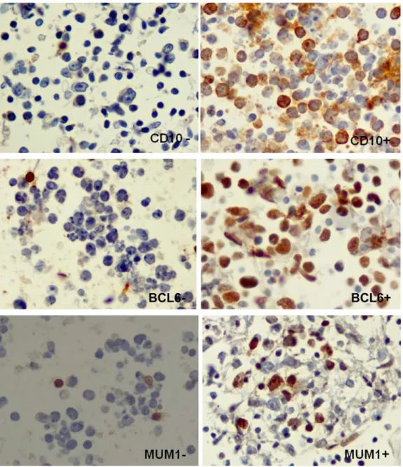
with scant diagnostic cells and relatively numerous eosino-phils, the differential diagnosis of HL was considered. In such cases (cases 12 and 35), ICC for CD15 and CD30 on the CB and CS was negative, thus excluding the diag-nosis of HL. In another 2 cases with more marked nuclear atypia (cases 25 and 22), the differential diagnosis with anaplastic lymphoma or T-cell NHL was considered and excluded by FC and by CD30 and ALK ICC negativity, Figure 3.Immunostaining with CD10, B-cell lymphoma 6 (BCL6), and multiple myeloma oncogene 1 (MUM1) in diffuse large B-cell lymphoma conventional smears. In positive cases,>30% of the diagnostic cells show nuclear (BCL6 and MUM1) and cytoplasmic (CD10) positivity. In negative cases, occasional diagnostic cells or small cells in the background represent positive internal con-trols (CD10-BCL6-MUM1 immunostain,3430). - indicates negative;1, positive.
respectively. FC was effective in 26 cases, demonstrating the following phenotypes: CD191/CD5-/CD10-/CD23-/ FMC7- in 10 cases; CD191/CD5-/CD101/CD23-/ FMC7- in 11 cases; CD191/CD5-/CD101/CD23-/ FMC71 in 2 cases; and CD191/CD5-/CD10-/CD23-/ FMC71in 3 cases. Kappa and lambda light chain restric-tion was observed in 18 and 8 cases, respectively. In 12 cases in which FC was NE, the FNA diagnosis of DLBCL was basically morphological and confirmed by the clinical history in cases of DLBCL recurrence; by using CD3, CD15, CD30, and ALK in selected cases; by the CD10-BCL6-MUM1 algorithm; and by the follow-up histological controls when available. Therefore, the diagnosis of DLBCL was performed on the basis of morphological, cytological, FC, and ICC data. Histological controls were available in 24 of 38 cases, and confirmed the FNA diagno-sis of DLBCL in 24 cases. Eight cases were DLBCL recur-rences, which had been previously diagnosed histologically. At the time of recurrence, the FNA diagnosis was consistent with the clinical presentation, and histology was not required by clinicians. In case 20 (para-aortic lymph node) and cases 10 and 29 (patients aged 85 years and 91 years, respectively), lymph nodes were not removed for the histo-logical evaluation for clinical reasons and because the FNA diagnosis matched with the clinical data. Finally, cases 2, 21, and 32 were histologically confirmed at other institu-tions, but IHC data were not available for the algorithm evaluation. ICC for CD10, BCL6, and MUM1 was suc-cessfully performed on 18 CB and 15 CS, and was NE in
5 cases (Figs. 2 and 3). ICC combinations were CD10-/ BCL6-/MUM11 (20 cases), CD101 /BCL6-/MUM1-(1 case), CD101/BCL61/MUM1- (8 cases), CD10-/ BCL6-/MUM1- (2 cases), CD10-/BCL61 /MUM1-(1 case), and CD10-/BCL61/MUM11 (1 case) (Figs. 2 and 3). The CD10-BCL6-MUM1 algorithm classified DLBCL as GCB in 9 cases (27%; 2 CB and 7 CS) and non-GCB in 24 cases (73%; 16 CB and 8 CS). The histo-logical controls confirmed the ICC data in 23 of the 24 available IHC cases, being discordant in only 1 case (case 6) that was found to be negative for BCL6 on CB and positive on IHC lymph node section. NE ICC results were reported in 4 CB cases and 1 CS case (Table 1). No significant dif-ferences were detected in terms of ICC quality when com-paring ICC performance on CS and CB; the higher number of NE findings among CB in comparison with CS (4 cases vs 1 case) were caused by the scant cellularity of CB sections in the corresponding cases.
The linear regression analysis obtained by grouping the 6 combinations of CD10, BCL6, and MUM1 expres-sion and plotting the ICC against the corresponding IHC revealed a correlation coefficient value equal to 0.9819, thus demonstrating a strong concordance between ICC versus IHC data (Fig. 4).
DISCUSSION
Using the CD10-BCL6-MUM1 ICC algorithm applied to CS and CB, we were able to subclassify DCBLC into GCB and non-GCB in a series of 38 lymph node FNA cases. The original algorithm proposed by Hans et al7was based on the expression of CD10, BCL6, and MUM1 and was effective in the classification of DLBCL with a GEP con-cordance in approximately 80% of the cases. Additional studies have proposed other antibodies (such as BCL2, Ki-67, and human leukocyte antigen [HLA])8–12 or new antibodies (such as Forkhead box-P [FoxP], GCET1 (cen-terin),22and LMO210,13) to be used in addition to the 3 original ones (CD10, BCL6, and MUM1) or by replacing BCL6.23,24 The algorithm by Choi et al22introduced the germinal center B cell-expressed transcript 1 (GECT1) as the first discriminator of DLBCL cells, and FOXP1 as a discriminator of CD10-/BCL61 cases achieving a 93% concordance with GEP. In other recently proposed algo-rithms, BCL6 was replaced by BCL224or included CD10, GCET, FOXP1, and MUM1 antibodies, which appear to be the most specific for GCB and non-GCB, respectively.25 Figure 4.Immunocytochemistry (ICC)/immunohistochemistry
(IHC) scatter plot graph. The graph shows a scatter plot of 6 ICC immunophenotypes plotted versus the corresponding IHC immunophenotypes among 24 patients analyzed. The correlation coefficient value of 0.9819 revealed a strong con-cordance between ICC and IHC data.
The most recently proposed algorithm by Visco et al10 based on the expression of CD10, FOXP1, GCET-1, MUM1, and BCL610 gained a 92.6% concordance with GEP. All the proposed algorithms have advantages and lim-itations, and none appears to dramatically improve either the DLBCL subclassification or the concordance with GEP. Moreover, in terms of predictive implications, the retrospective evaluation of patients treated with different protocols might determine different biologic behaviors and survival rates in individuals affected by the same DLBCL subtype (GCB or non-GCB).11,25 The CD10-BCL6-MUM1 algorithm, as originally proposed,7 may classify DLBCL into GCB and non-GCB (ABC and unclassifi-able), with approximately 80% concordance with GEP.7 The new algorithms suggested in the literature,10,22,25such
as the algorithm by Visco et al,10might improve the con-cordance with GEP significantly, but such algorithms require the use of antibodies that are not used routinely.
The use of cytological material, which is quantita-tively limited by definition, necessitates a more limited number of antibodies. We chose CD10, BCL6, and MUM1, which are available and routinely used in our lab-oratories. Moreover, CD10, BCL6, and MUM1 were rou-tinely tested on the corresponding histological data with the same procedure. FNA is generally used as the first diag-nostic step or in the follow-up of lymph node and extra lymphoproliferative processes, and it is followed by histo-logical examination in the majority of cases. Therefore, an accurate cytological subclassification of DLBCL might be considered to an objective that goes beyond the limits of TABLE 1. Clinical and Phenotypic Classification of 38 Cases of DLBCL Diagnosed by FNA
Case No. Localization Age, Years Sex CB/CS CD10 BCL6 MUM1 DLBCL Subtype
Histological Concordance
1 Left lateral cervical 43 F CB - - 1 Non-GCB Yes
2 Bilateral lateral cervical 66 F CB - - 1 Non-GCB Yesa
3 Right submandibular 47 F CB - - 1 Non-GCB Yes
4 Right lateral cervical 49 M CB NE NE NE NP NA
5 Left supraclavicular 52 F CB - - 1 Non-GCB Yes
6 Left lateral cervical 46 F CB 1 - - GCB No BCL61
7 Left lateral cervical 80 F CB - - 1 Non-GCB NA
8 Left supraclavicular 55 F CB - - 1 Non-GCB NA
9 Left supraclavicular 68 F CB - - - Non-GCB Yes
10 Left lateral cervical 85 M CS NE NE NE NP NA
11 Left inguinal 72 M CB 1 1 - Non-GCB NA
12 Right lateral cervical 77 M CB - - 1 Non-GCB Yes
13 Right lateral cervical 55 M CS - - 1 Non-GCB Yes
14 Right lateral cervical 43 M CB NE NE NE NP NA
15 Right lateral cervical 65 F CS 1 1 - GCB NA
16 Right submandibular 76 M CS 1 1 - GCB Yes
17 Left inguinal 81 M CS - - 1 Non-GCB Yes
18 Right inguinal 58 F CS 1 1 - GCB Yes
19 Right lateral cervical 49 M CS - - 1 Non-GCB Yes
20 Para-aortic lumbar 72 M CB NE NE NE NP NA
21 Right inguinal 51 F CS 1 1 - GCB Yesa
22 Left supraclavicular 66 F CB - - 1 Non-GCB Yes
23 Left lateral cervical 69 M CB - 1 - GCB Yes
24 Left axillary 69 M CB - 1 1 Non-GCB Yes
25 Left submandibular 39 F CB - - 1 Non-GCB Yes
26 Left lateral cervical 60 M CB - - 1 Non-GCB Yes
27 Left axillary 69 M CB - - - Non-GCB Yes
28 Left inguinal 54 F CB - - 1 Non-GCB Yes
29 Left lateral cervical 91 M CB - - 1 Non-GCB NA
30 Left lateral cervical 65 M CS - - 1 Non-GCB NA
31 Right submandibular 76 M CS 1 1 - GCB Yes
32 Left lateral cervical 72 F CS 1 1 - GCB Yesa
33 Left lateral cervical 53 M CS - - 1 Non-GCB Yes
34 Left submandibular 62 M CB NE NE NE NP NA
35 Left inguinal 36 M CS - - 1 Non-GCB Yes
36 Left lateral cervical 53 F CS - - 1 Non-GCB Yes
37 Right inguinal 65 F CS 1 1 - GCB Yes
38 Right inguinal 81 F CS - - 1 Non-GCB Yes
Abbreviations: -, negative;1, positive; BCL6, B-cell lymphoma 6; CB, cell block; CS, conventional smear; DLBCL, diffuse large B-cell lymphoma; F, woman; FNA, fine-needle aspiration; GCB, germinal center B; M, man; MUM1, multiple myeloma oncogene 1; NA, not available; NE, not effective; NP, not performed.
FNA. Nonetheless, in specific clinical situations, FNA may be the only diagnostic procedure, and the diagnosis of DLBCL with an accurate subclassification may be useful, providing additional clinical information. The main diffi-culties in DLBCL subclassification on FNA samples include the management of the diagnostic material, the choice of an effective and limited panel of antibodies, and the interpretation of cytological results and their reproduci-bility when compared with histological data.
In our experience, as well as the experience of others,15,26the management of diagnostic material is deter-mined by ROSE. In fact, after the routine preparation of CS and cell suspension for FC, when DLBCL was consid-ered in the differential diagnosis, additional passes and residual material were used to prepare CB and additional CS for ICC assessment. DLBCL diagnostic cells are often intermingled with other inflammatory, nonlymphomatous cells; as a result, antigen expression has to be evaluated in relation to the cytological features. Therefore, ICC, per-formed either on CS or CB, was appropriate for compar-ing the obtained results with the histological data and was the ancillary technique used for the current study. The panel of CD10, BCl6, and MUM1 was chosen because it was specific and available in both institutions, as well as in all the IHC controls, and left extra material for further diagnostic tests. In this respect, CD3, CD15, CD30, and ALK were initially tested in 4 cases to exclude T-cell NHL; HL; and anaplastic, ALK1 lymphoma, respectively, and diagnostic material was still available to include corre-sponding cases in the current study. When the final FNA diagnosis of DLBCL was achieved, the corresponding CB and CS were tested by the CD10-BCl6-MUM1 algorithm. As reported earlier, ICC was successful in 33 of the 38 cases and classified as positive or negative for the corre-sponding antibodies. ICC was NE in 5 of the 38 cases (in 4 CB due to the lack of cells and in 1 CS because of defec-tive fixation). The same criteria were used to evaluate histo-logical data, and a general concordance was achieved between FNA data and the 24 cases of corresponding his-tological data. Moreover, in the current series, CD10 and MUM1 expression were mutually exclusive, helping to dis-tinguish between the GCB and non-GCB samples.
The results of the current study suggest that the CD10-BCL6-MUM1 ICC algorithm may be hampered mainly by insufficient cells on CB but, when performed, is effective and reproducible when compared with histologi-cal data. With regard to CB and CS efficacy as a technihistologi-cal
support for ICC, CS appears to be more effective than CB because of the possible scant cellularity of the latter, being ICC quality equally effective on CB and CS. The clinical value of the DLBCL classification is not accepted unani-mously, and the current series was too small to have any clinical and predictive value. However, we believe it is large enough to assess the reproducibility of the algorithm when compared with the corresponding histological data.
The CD10-BCL6-MUM1 ICC algorithm is reliable for FNA specimens and equally effective on CB and CS, with cell adequacy being the main technical limitation. Therefore, ICC data are comparable to the histological data. FNA subclassification of DLBCL may provide addi-tional prognostic information that might be useful when histological data are not available.
FUNDING SUPPORT
No specific funding was disclosed.
CONFLICT OF INTEREST DISCLOSURES
The authors made no disclosures.
REFERENCES
1. Swerdlow SH, Campo E, Harris NL, et al. WHO Classification of Tumours of Haematopoietic and Lymphoid Tissues. 4th ed. Lyon, France: IARC Press; 2008.
2. Flowers CR, Sinha R, Vose JM. Improving outcomes for patients with diffuse large B-cell lymphoma.CA Cancer J Clin.2010;60:393-408. 3. Schneider C, Pasqualucci L, Dalla-Favera R. Molecular
pathogene-sis of diffuse large B-cell lymphoma.Semin Diagn Pathol.2011;28: 167-177.
4. Fowler LJ, Lachar WA. Application of immunohistochemistry to cytology.Arch Pathol Lab Med.2008;132:373-383.
5. Alizadeh AA, Eisen MB, Davis RE, et al. Distinct types of diffuse large B-cell lymphoma identified by gene expression profiling.
Nature.2000;403:503-511.
6. Rosenwald A, Wright G, Chan WC, et al; Lymphoma/Leukemia Molecular Profiling Project. The use of molecular profiling to pre-dict survival after chemotherapy for diffuse large-B-cell lymphoma.
N Engl J Med.2002;346:1937-1947.
7. Hans CP, Weisenburger DD, Greiner TC, et al. Confirmation of the molecular classification of diffuse large B-cell lymphoma by immuno-histochemistry using a tissue microarray.Blood.2004;103:275-272. 8. Barrans SL, Fenton JA, Banham A, Owen RG, Jack AS. Strong
expression of FOXP1 identifies a distinct subset of diffuse large B-cell lymphoma (DLBCL) patients with poor outcome. Blood.
2004;104:2933-2935.
9. Montes-Moreno S, Roncador G, Maestre L, et al. Gcet1 (centerin), a highly restricted marker for a subset of germinal center-derived lymphomas.Blood.2008;111:351-358.
10. Visco C, Li Y, Xu-Monette ZY, et al. Comprehensive gene expres-sion profiling and immunohistochemical studies support applica-tion of immunophenotypic algorithm for molecular subtype classification in diffuse large B-cell lymphoma: a report from the International DLBCL Rituximab-CHOP Consortium Program Study.Leukemia.2012;26:2103-2113.
11. van Imhoff GW, Boerma EJ, van der Holt B, et al. Prognostic impact of germinal center-associated proteins and chromosomal breakpoints in poor-risk diffuse large B-cell lymphoma. J Clin Oncol.2006;24:4135-4142.
12. de Jong D, Rosenwald A, Chhanabhai M, et al; Lunenburg Lym-phoma Biomarker Consortium. Immunohistochemical prognostic markers in diffuse large B-cell lymphoma: validation of tissue microarray as a prerequisite for broad clinical applications–a study from the Lunenburg Lymphoma Biomarker Consortium. J Clin Oncol.2007;25:805-812.
13. Morton LM, Cerhan JR, Hartge P, et al. Immunostaining to iden-tify molecular subtypes of diffuse large B-cell lymphoma in a population-based epidemiologic study in the pre-rituximab era.Int J Mol Epidemiol Genet.2011;2:245-252.
14. Nunez AL, Jhala NC, Carroll AJ, et al. Endoscopic ultrasound and endobronchial ultrasound-guided fine-needle aspiration of deep-seated lymphadenopathy: analysis of 1338 cases.Cytojournal.2012; 9:14.
15. Vigliar E, Cozzolino I, Picardi M, et al. Lymph node fine needle cytology in the staging and follow-up of cutaneous lymphomas.
BMC Cancer.2014;14:8.
16. Schmitt F, Cochand-Priollet B, Toetsch M, Davidson B, Bondi A, Vielh P. Immunocytochemistry in Europe: results of the European Federation of Cytology Societies (EFCS) inquiry. Cytopathology.
2011;22:238-242.
17. Pinheiro C, Roque R, Adriano A, et al. Optimization of immuno-cytochemistry in cytology: comparison of two protocols for fixation and preservation on cytospin and smear preparations.Cytopathology.
2015;26:38-43.
18. Beraki E, Olsen TK, Sauer T. Establishing a protocol for immuno-cytochemical staining and chromogenic in situ hybridization of
Giemsa and Diff-Quick prestained cytological smears.Cytojournal.
2012;9:8.
19. Zeppa P, Marino G, Troncone G, et al. Fine-needle cytology and flow cytometry immunophenotyping and subclassification of non-Hodgkin lymphoma: a critical review of 307 cases with technical suggestions.Cancer.2004;102:55-65.
20. Zeppa P, Vigliar E, Cozzolino I, et al. Fine needle aspiration cytol-ogy and flow cytometry immunophenotyping of non-Hodgkin lymphoma: can we do better?Cytopathology.2010;21:300-310. 21. Bertram HC, Check IJ, Milano MA. Immunophenotyping large
B-cell lymphomas. Flow cytometric pitfalls and pathologic correla-tion.Am J Clin Pathol.2001;116:191-203.
22. Choi WW, Weisenburger DD, Greiner TC, et al. A new immuno-stain algorithm classifies diffuse large B-cell lymphoma into molec-ular subtypes with high accuracy. Clin Cancer Res. 2009;15: 5494-5502.
23. Natkunam Y, Farinha P, Hsi ED, et al. LMO2 protein expression predicts survival in patients with diffuse large B-cell lymphoma treated with anthracycline-based chemotherapy with and without rituximab.J Clin Oncol.2008;26:447-454.
24. Nyman H, Jerkeman M, Karjalainen-Lindsberg ML, Banham AH, Leppa S. Prognostic impact of activated B-cell focused classification in diffuse large B-cell lymphoma patients treated with R-CHOP.
Mod Pathol.2009;22:1094-1101.
25. Meyer PN, Fu K, Greiner TC, et al. Immunohistochemical methods for predicting cell of origin and survival in patients with diffuse large B-cell lymphoma treated with rituximab.J Clin Oncol.
2011;29:200-207.
26. da Cunha Santos G, Ko HM, Saieg MA, Geddie WR. “The petals and thorns” of ROSE (rapid on-site evaluation). Cancer (Cancer Cytopathol).2013;12:4-8.