Internal limiting membrane peeling versus no peeling for idiopathic full thickness macular hole: A pragmatic randomised controlled trial
Noemi Lois,1*MD, PhD; Jennifer Burr2, MD; John Norrie2,3, MSc; Luke Vale2,4, PhD;
Jonathan Cook2, PhD; Alison McDonald2, MSc;Charles Boachie2, MSc;Laura Ternent,2,4
MSc; and Gladys McPherson2, MSc; for the Full-thickness macular hole and Internal
Limiting Membrane peeling Study (FILMS) Group
*Corresponding author and reprint requests
1Ophthalmology Department, Grampian University Hospitals-NHS Trust, Aberdeen, AB25
2ZN, UK. Phone: 00-44-1224-553217, FAX: 00-44-1224-559231; Email: [email protected]
2Health Services Research Unit, University of Aberdeen, Aberdeen, AB25 2ZD, UK. 3Robertson Centre for Biostatistics, University of Glasgow, Glasgow, G12 8QQ, UK. 4Health Economics Research Unit, University of Aberdeen, Aberdeen, AB25 2ZD, UK.
The work presented here has been submitted, as a paper, to the XXVIIth Meeting of the Club Jules Gonin, Kyoto, Japan, November 3-6, 2010.
Abstract
Purpose: To determine whether internal limiting membrane (ILM) peeling is effective and cost-effective compared with no peeling for patients with idiopathic stage 2 or 3 full-thickness macular hole (FTMH).
Methods: Pragmatic, multicenter randomized controlled trial. Eligible participants from nine centres were randomized to ILM peeling or no peeling (1:1 ratio) in addition to phaco-vitrectomy, including detachment and removal of the posterior hyaloid and gas tamponade. The primary outcome was distance visual acuity (VA) at six months post-surgery. Secondary outcomes included hole closure, distance VA at other time points, near VA, contrast
sensitivity, reading speed, re-operations, complications, resource use and participant reported health status, visual function and costs.
Results: Of 141 participants randomized in nine centres, 127 (90%) completed six-month follow up. Non-statistically significant differences in distance visual acuity at six-months were found between groups [Mean difference 4.8, 95% CI (-0.3, 9.8); p=0.063]. There was a statistically significantly higher rate of hole closure in the ILM peel group [56 (84%) versus 31 (48%)] at one month [Odds ratio (OR):6.23, 95% CI (2.64 to 14.73); p<0.001] with fewer re-operations [8 (12%) versus 31 (48%)] performed by six months [OR:0.14, 95% CI (0.05, 0.34); p<0.001]. Peeling the ILM was likely to be cost-effective.
Conclusion: There was no evidence of a difference in distance VA following ILM peel and no-ILM peel techniques. An important benefit in favour of no ILM peeling was ruled out. Given the higher anatomical closure and lower re-operation rates in the ILM peel group, ILM peeling would seem the treatment of choice for idiopathic stage 2-3 FTMH.
An idiopathic full-thickness macular hole (FTMH) represents a defect in the area of maximal vision of the retina, the fovea; if left untreated often leads to severe central visual loss.1
FTMH are common, with an estimated incidence of 7.8 persons / 100,000 population per year.2 Macular hole surgery, first developed by Kelly and Wendell in 1991,3 represents one
of the most common procedures performed by vitreo-retinal surgeons.4
Four stages (1-4) of FTMH have been described;5 randomized controlled clinical trials
(RCTs) conducted in the 1990s showed that macular hole surgery was effective for stages 2, 3 and 4.6,7
Peeling the internal limiting membrane (ILM) of the retina was introduced as an additional manoeuvre in macular hole surgery in order to improve anatomical and functional outcomes following surgery.8 Several observational studies suggested a benefit of peeling the
ILM (reviewed by Abdelkader and Lois9). Furthermore, recent data from two small RCTs
undertaken in China and Denmark, which included 49 and 75 patients, respectively, suggested a potential beneficial effect of this operation.10,11
The Full-thickness macular hole and Internal Limiting Membrane peeling Study (FILMS), herein reported, was designed as a “pragmatic” RCT to test the hypothesis that surgical peeling of the ILM for people with idiopathic FTMH (stages 2 or 3), compared with surgery without peel, improves vision, the likelihood of hole closure, participant reported health related quality of life and is cost-effective. “Pragmatic” RCTs aim to determine the effects of an intervention under the usual circumstances (i.e. normal clinical practice); as opposed to “explanatory” RCTs which are primarily designed to determine the effects of an intervention under ideal circumstances. 12 The advantages of a pragmatic design are that if the
treatment is effective it can be implemented into clinical practice.
Methods
The FILMS study was a multicenter, pragmatic RCT. Patients were enrolled at nine centres, seven in UK and two in the Republic of Ireland, by local clinical investigators. Eligible participants were those with stage 2 or 3 idiopathic FTMH of < 18 months duration and visual acuity (VA) < 20/40 in the study eye. At each participating centre, vitreo-retinal surgeons followed their usual clinical practice [including slit-lamp biomicroscopy, colour and red-free photographs, optical coherence tomography (OCT), fluorescein angiography and fundus autofluorescence (AF), as required] to stage FTMH and to confirm eligibility. Individuals with stage 1 or 4 FTMH, idiopathic FTMH stages 2-3 of > 18 months duration, with other causes of decreased vision, and those with FTMH related to high myopia or trauma were excluded. Individuals that did not understand English and/or unable to give informed consent were also excluded. The Medical Research Council Good Clinical Practice Guidelines, 1998, and the UK Data Protection Act, 1998 were followed. Approval by the Multicentre Research Ethics Committee, Local Ethics Committees and local hospital trusts of each participating centre and participants’ written informed consent were obtained prior to the initiation of the study.
During the recruitment phase, two RCTs of ILM peeling versus no ILM peeling presented their results. 10,11 In light of this, an independent Data Monitoring Committee was
established (Jan 2007) and reviewed the external and accumulating FILMS trial evidence and recommended continuing recruitment.
Meetings, involving all individuals participating in this RCT (including vitreo-retinal surgeons, optometrists and photographers, in addition to those responsible for the running of the trial) were held prior to the initiation of the study to discuss the methodology of this RCT and issues regarding data collection.
All participants received a combined phacoemulsification and pars plana vitrectomy,
including detachment and removal of the posterior hyaloid, followed by a fluid-air exchange and air-gas [12% perfluoropropane (C3F8)] exchange. Participants were randomized 1:1 to
the above procedure alone or combined with ILM peeling using a central telephone IVR system at the trial office (Centre for Healthcare Randomised Trials (CHaRT) – UK NIHR trials unit. In the latter, following ILM staining with trypan blue (0.15% trypan blue under air for 3 minutes) removal of the ILM was performed in an area of around 1-2 disc diameters around the hole. Re-staining with trypan blue was permitted at the discretion of the
vitreoretinal surgeon. Each vitreo-retinal surgeon judged whether the peeling of the ILM was “complete”, as defined by a removal of the ILM all around the hole within a minimal
extension of 1 disk diameter centred on the centre of the hole or “incomplete” if otherwise. A minimisation algorithm (according to Taves, with P=1)13 was used which allocated treatment
with respect to minimising imbalance in trial centre, distance vision in the study and fellow eye (20/40-20/160; 20/200-20/500; <20/500), stage of the macular hole (2 or 3), duration of symptoms (≤1 year; >1 year) and lens status (phakic, aphakic or pseudophakic). Participants were followed up at one, three and 6 months post-surgery.
Further surgical intervention was allowed in either group according to standard clinical practice if the macular hole remained opened following surgery, including ILM peeling in participants initially randomized to no peel. All participants were instructed to posture face down for a period of five-seven days post-surgery and received a posturing chart to record the time postured.
Outcomes
The primary outcome was the mean difference between treatment groups in the Early Treatment Diabetic Retinopathy Study (ETDRS) distance VA score at six months.
Secondary outcomes included ETDRS distance VA at three months, near VA (Bailey-Lovie) at three and six months, contrast sensitivity (Pelli-Robson) at six months, reading speed (MNRead) at six months, anatomical closure of the macular hole at each time point (one, three and six months), participant related outcomes, as determined by the EQ-5D and VFQ-25 questionnaires, at six months, costs to the health service and the participant, incremental costs per quality adjusted life year (QALY) and adverse events.
The macular hole was considered to be closed when the hole was no longer visible clinically (open holes with or without subretinal fluid around them were considered to be open). At each participating centre, vitreo-retinal surgeons followed their usual clinical practice [including slit-lamp biomicroscopy, colour and red-free photographs, optical
coherence tomography (OCT), fluorescein angiography and fundus autofluorescence (AF) as required] to determine whether the macular hole was open or closed.
Sample size and statistical analysis
Assuming a common standard deviation of 12 ETDRS points in the two randomised groups, to detect a 6 ETDRS score difference (an effect size of 0.5) using a two sample two sided t-test at a 5% level of significance and 80% power, it was estimated that 64 participants would be required in each group. This calculation was based on data from published studies.14,15
significance. The principal analysis was based upon available case data with no imputation of missing values. Sensitivity analyses of the primary and macular hole closure outcomes which assessed the impact of missing data by imputing extreme values (lowest and highest/open and closed respectively) was also undertaken. Additionally, the primary outcome was analysed according to the pre-specified subgroups (lens status, duration and stage of the macular hole) by including the corresponding interaction term(s) in the regression model using stricter criteria for statistical significance (2P ≤ 0.01). Macular hole closure, complications and further surgery were analysed using logistic regression model with adjustment for the minimisation covariates (except study centre). Analyses of health related quality of life measures (EQ-5D and VFQ-25) and secondary measures of visual function (contrast sensitivity, reading speed and distance and near visual acuity) were undertaken using linear regression adjusted for baseline score in addition to the minimisation variables (bar centre). A mean difference was reported for a continuous outcome and an odds ratio (OR) for a binary outcome. The corresponding confidence interval (CI) and p-value were reported.
Economic Evaluation
A cost per participant for each arm of the trial was calculated based upon the use of primary and secondary care services. Resource utilisation per participant was estimated using patient questionnaires and information recorded in the three and six month case report forms. Unit costs/prices were obtained using information from the National Health Service, published estimates for health care services and/or interventions,16-18 as well as study specific estimates.
QALYs were calculated from responses to the EQ-5D using the area under the curve method with EQ-5D responses valued using UK population tariffs.19 Point estimates for mean costs
minimisation factors and baseline EQ-5D for QALYS. These data were subsequently used to estimate an incremental cost per QALY gained.
The results were presented as point estimates of mean incremental costs, QALYS, and incremental cost per QALY. Measures of variance for these outcomes were estimated by bootstrapping costs, QALYs, and incremental cost per QALY. The incremental effectiveness data obtained by the bootstrapping exercise were presented in terms of cost-effectiveness acceptability curves (CEACs).
Further details of the methodology used in FILMS have been published elsewhere.20 As
stated in this published protocol, in addition to the above, in all participating centres colour photographs and red free images were obtained at each time point (baseline, 1, 3 and 6 months) and evaluated, in a masked fashion and using a computerised programme, to objectively determine the size of the hole prior to surgery and the status of the hole post-operatively. These data, not presented here, will form part of a separate study evaluating the effect of the size of the macular hole on outcomes following surgery. Furthermore, a long-term 24 month follow-up evaluation is also included.
Results
Study Participants
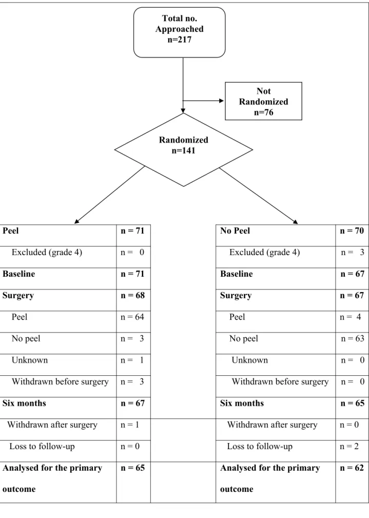
Between July 1st 2005 and January 31st 2009, 217 patients were approached; of these 76 were
Surgical details and intraoperative complications
Table 2 summarizes details of the surgery and intraoperative complications. More
participants in the no ILM peel group had surgery performed under general anaesthesia. On average, the surgery took longer if the ILM was peeled than if it was not removed (72.6 versus 62.6 minutes). The ILM was completely removed in 64 of 67 participants receiving surgery in the ILM peel group; in three participants in this group the ILM was not removed; for one participant it was unknown whether they had surgery or not (Figure 1). Four
participants in the no ILM peel group received ILM peeling and represented protocol deviations though retained in the analyses. No statistically significant differences in the number of intraoperative complications were found between groups [38 (57%) versus 28 (42%) OR: 1.73, 95% CI (0.86, 3.50); p = 0.166]. For both groups, the most common intraoperative complication was the occurrence of retinal tears.
Primary and Secondary Outcomes
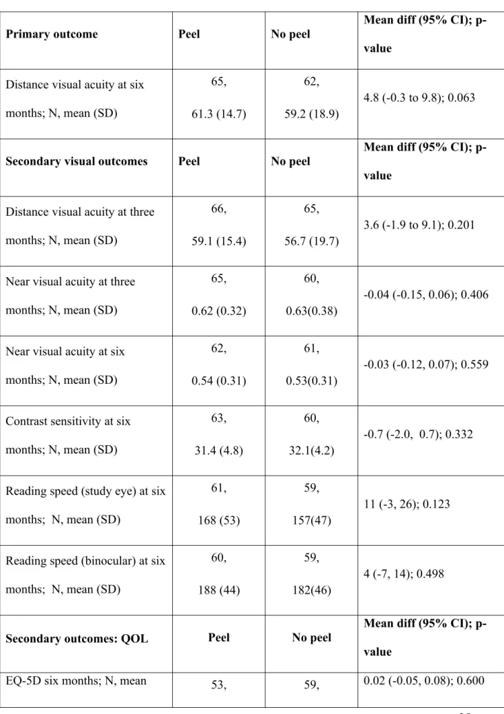
No statistically significant differences in distance visual acuity at six months (primary outcome) were detected between groups [4.8, 95% CI (-0.3, 9.8); p=0.063] (Table 3). The result was not sensitive to missing data based upon the sensitivity analyses (results not shown). However, an adjusted mean difference of 5 ETDRS letters was observed in favour of the ILM peel group. None of the sub-group analyses of the primary outcome were found to be statistically significant (data not shown). No statistically significant differences were observed in other functional outcomes at any of the time points studied between groups (Table 3).
statistically significant difference in rates of hole closure between ILM peel and no ILM peel groups was observed at three and six months. The sensitivity analysis results for macular hole closure did not change the above findings. A post-hoc subgroup analysis of macular hole closure by stage of the hole showed no significant difference [OR: 0.46, 95% CI (0.04, 4.99); p = 0.400]. No statistically significant differences in responses to the EQ-5D and VFQ-25
scores were observed between groups.
Postoperative complications
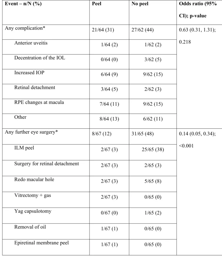
No statistically significant differences in the number of complications after initial surgery were found between groups [21 (31%) versus 27 (41%); OR: 0.63, 95% CI (0.31, 1.31); p= 0.218]. Rhegmatogenous retinal detachment occurred in 5% and 3% of cases in ILM peel and no ILM peel groups, respectively. Post-operative complications have been summarized in Table 4. Eight (12%) in the ILM peel group compared with 31 participants (48%) in the no ILM peel group required at least one further surgery [OR: 0.14, 95% CI (0.05, 0.34); p<0.001] (Table 4). This difference was driven by 25 (38%) participants in the no ILM peel group undergoing peeling of the ILM within the follow-up period. There was one re-opening of the macular hole in the no ILM peel group and none in the ILM peel group.
Economic evaluation
was not statistically significant at the 5% level. However, based upon the bootstrapped estimates of cost-effectiveness it is probable that ILM peeling would be cost-effective at typical threshold values for society’s willingness to pay for a QALY (90% chance of being considered cost-effective when society’s willingness to pay for a QALY is £20,000).
Discussion
FILMS found no evidence of a difference between groups in distance visual acuity. The observed difference, which favoured ILM peeling, was however of a clinically relevant magnitude (5 ETDRS letters). Furthermore, an important benefit in favour of no ILM peeling was ruled out. This, together with the higher rates of macular hole closure and corresponding fewer re-operations needed in the ILM peel group suggests that ILM peeling is the treatment of choice for patients with stage 2-3 FTMH.
Participants randomized to no-ILM peel received ILM peeling if the macular hole remained open; this most likely explains the lack of differences in macular hole closure rates at three and six months. The difference in the number of re-operations was the main driver of differences in cost between the two procedures. Indeed, the results suggest that there is over 90% chance that ILM peeling is less costly than the no peeling technique. Importantly, data from this study support a lack of a deleterious effect of ILM peeling in visual function, one of the main concerns raised of performing ILM peeling.21 The functional
and anatomical outcomes achieved in FILMS are consistent with those from two other smaller trials comparing macular hole surgery with or without ILM peeling recently
published.10,11 The benefit of ILM peeling has been also suggested by the results of one other
RCT very recently completed, which included only participants (n=80) with large holes (>400 microns) and which has been published only, to date, in an abstract form.22
in FILMS may seem lower than those reported in previously published observational studies (summarized by Abdelkader and Lois).9 Case series, however, are highly prone to selection
bias and reporting better outcomes than those seen in standard clinical practice. Most vitreo-retinal surgeons (>80%) participating in FILMS had extensive experienced in vitreo-vitreo-retinal surgery, including macular hole surgery. Interestingly, anatomical results in FILMS are comparable to those obtained in one of the recently published RCTs with similar inclusion criteria, definition for hole closure and surgical protocol than FILMS, in which macular hole closure at three months was achieved in 44% and 89% in the no ILM peel group and in the trypan-blue assisted ILM group, respectively.10 Thus, it is likely that these lower rates of
closure are representative of real clinical practice.
Cataract surgery was performed in FILMS at the time of the macular hole repair. This would have avoided progression of pre-existing cataracts to act as a possible confounder in the evaluation of the functional results. Furthermore, as complications reasonably accredited to the combined procedure seemed to be few and had no deleterious effects in vision (Table 5), phaco-vitrectomy for FTMH appears to be a reasonable and convenient option for patients and may be a more efficient use of resources.
FILMS randomised 141 participants (90% completed the 6 month follow-up) in 9 centres in the UK and Eire. The study was intentionally designed as a pragmatic, as opposed to a more explanatory, trial thereby increasing the external validity (generalisability) of its findings by reflecting decision making in clinical practice. Vitreo-retinal surgeons determined the diagnosis and staging of FTMH and its status following surgery and the need for further surgery based on their own routine clinical practice. Participating surgeons used their own routine techniques to induce a posterior hyaloid detachment, to peel the ILM (with the exception of the use of trypan blue to stain the ILM) and to achieve an adequate gas exchange.
In conclusion, data from FILMS suggests that ILM peeling is a safe, clinically superior and more cost-effective approach than no ILM peeling to treat patients with
idiopathic stage 2 or 3 FTMH and, thus, may be considered the treatment of choice for patients with this common vitreo-retinal disease.
Acknowledgment
We are indebted to all of the participants and to the Chief Scientist Office of the Scottish Government Health Directorates for funding the study. Jonathan Cook holds a Medical Research Council UK fellowship. The Health Services Research Unit and the Health Economics Research Unit are both core funded by the Chief Scientist Office of the Scottish Government Health Directorates. We thank the independent Data Monitoring Committee members: Professor Gordon Murray (chair), Mr Bill Aylward and Mr Tom Williamson. We wish to thank Mary Kilonzo and Robyn de Verteuil for their assistance in the economic evaluation of this trial, to Daniel Barnett for programming support and Diana Collins for data entry.
Clinical investigators: Hatem Atta, Stephen Beatty, Catherine Cleary, Andrew Dick, John Ellis, John Forrester, Carl Groenewald, Richard Haynes, Henrich Heimann, Muhammad Irfan Khan, Dara Kilmartin, Noemi Lois, Asif Orakzai, CK Patel, Ian Pearce, Tarik Saddik, David Steel, David Wong. Optometrists and local co-ordinators: Charles Cottriall, Cherry Daly, Laura Duncan, Karon McEwing, Sarah Muir, Anita Murphy, Stan Keys, Lynda Lindsell and Valerie Tompkin. Photographers: Terri Ainley, Victor Beatty, Gillian Bennerson, Anne Bolton, Jon Brett, Alison Farrow, Ronnie Jackson, Tony Johnston, Marie Kinsella, Stephen Neilson, Hugh Nolan, Sarah Stanley and Jim Talbot. Medical imaging: Ayyakkawnu Manivannan. Data Monitoring Committee: Prof Gordon Murray (Chair), Mr Bill Aylward and Mr Tom Williamson.
Authors’ contributions
NL conceived the study, participated in its design, coordination and interpretation of its results and drafted this manuscript. JB participated in the design, coordination and
interpretation of its results and contributed to this manuscript. JN participated in the design of the study and its statistical plan and contributed to the manuscript. LV participated in the design, coordination and interpretation of its results, and contributed to this manuscript. JC participated in the design, drafted the statistical plan, supervised the analysis of the data, contributed to the interpretation of the results and contributed to the manuscript. CB
References
1. Casuso LA, Scott IU, et al. Long-term follow-up of unoperated macular holes.
Ophthalmology 2001;108:1150-5.
2. McCannel CA, Ensminger JL, Diehl NN, Hodge DN. Population-based incidence of macular holes. Ophthalmology 2009;116:1366-9.
3. Kelly NE, Wendel RT. Vitreous surgery for idiopathic macular holes. Results of a pilot study. Arch Ophthalmol 1991;109:654-9.
4. Ah-Fat FG, Sharma MC, Majid MA, McGalliard JN, Wong D. Trends in vitreoretinal surgery at a tertiary referral centre: 1987 to 1996. Br J Ophthalmol 1999, 83:385-6.
5. Gass JD. Idiopathic senile macular hole. Its early stages and pathogenesis. Arch Ophthalmol.1988;106:629-39.
6.Kim JW, Freeman WR, Azen SP, el-Haig W, Klein DJ, Bailey IL. Prospective randomized trial of vitrectomy or observation for stage 2 macular holes. Vitrectomy for Macular Hole Study Group. Am J Ophthalmol 1996;121:605-14.
7. Freeman WR, Azen SP, Kim JW, el-Haig W, Mishell DR III, Bailey I. Vitrectomy for the treatment of full-thickness stage 3 or 4 macular holes. Results of a multicentered randomized clinical trial. The Vitrectomy for Treatment of Macular Hole Study Group. Arch Ophthalmol 1997;115:11-21.
9. Abdelkader E, Lois N. Internal limiting membrane peeling in vitreo-retinal surgery. Surv Ophthalmol 2008;53:368-96.
10. Christensen UC, Kroyer K, Sander B, et al. Value of internal limiting membrane peeling
in surgery for idiopathic macular hole stage 2 and 3: a randomised clinical trial. Br J
Ophthalmol 2009;93:1005-15.
11. Kwok AK, Lai TY, Wong VW. Idiopathic macular hole surgery in Chinese patients: a
randomised study to compare indocyanine green-assisted internal limiting membrane
peeling with no internal limiting membrane peeling. Hong Kong Med J 2005;11:259-66.
12.Thorpe KE, Zwarenstein M, Oxman AD, Treweek S, Furberg CD, Altman DG, Tunis S,
Bergel E, Harvey I, Magid DJ, Chalkidou K. A pragmatic-explanatory continuum indicator
summary (PRECIS): a tool to help trial designers. J Clin Epidemiol. 2009 May;62(5):464-75.
13. Taves DR. Minimization: A new method of assigning patients to treatment and control groups. Clinical pharmacology and therapeutics, 1974;15:443-453.
14. Brooks HL, Jr. Macular hole surgery with and without internal limiting membrane peeling. Ophthalmology. 2000;107:1939-48.
15. Paques M, Chastang C, Mathis A, et al. Effect of autologous platelet concentrate in surgery for idiopathic macular hole: results of a multicenter, double-masked, randomized trial. Platelets in Macular Hole Surgery Group. Ophthalmology 1999;106:932-8.
16. British National Formulary 57 [website on the Internet]. London: BMJ Group and RPS
17. ISD Scotland [database on the Internet]. Edinburgh: Information Services Division, NHS National Services Scotland; 2009 [accessed June 2009]. Available from: URL:
http://www.isdscotland.org/isd/1.html.
18. Curtis L Curtis L. Unit Costs for Health and Social Care [document on the Internet].
University of Kent: Personal Social Services Research Unit; 2008 [accessed June 2009].
Available from: URL: http://www.pssru.ac.uk/uc/uc2008contents.htm.
19. Dolan P, Gudex C, Kind P, et al. A social tariff for Euroqol: results from a UK general
population survey. Discussion Paper 138 [document on the Internet]. University of York:
Centre for Health Economics; 1995 [accessed June 2009]. Available form: URL:
http://www.york.ac.uk/inst/che/pdf/DP138.pdf.
20. Lois N, Burr J, Norrie J, Vale L, Cook J, McDonald A, for the Full-thickness macular hole and internal limiting membrane peeling study (FILMS) Group. Clinical and
cost-effectiveness of internal limiting membrane peeling for patients with idiopathic full thickness macular hole. Protocol for a Randomised Controlled Trial: FILMS (Full-thickness macular hole and Internal Limiting Membrane peeling Study). Trials. 2008 Nov 3;9:61.
Figure 1: Consort diagram
Peel n = 71 No Peel n = 70
Excluded (grade 4) n = 0 Excluded (grade 4) n = 3 Baseline n = 71 Baseline n = 67 Surgery n = 68 Surgery n = 67
Peel n = 64 Peel n = 4
No peel n = 3 No peel n = 63
Unknown n = 1 Unknown n = 0
Withdrawn before surgery n = 3 Withdrawn before surgery n = 0 Six months n = 67 Six months n = 65 Withdrawn after surgery n = 1 Withdrawn after surgery n = 0
Loss to follow-up n = 0 Loss to follow-up n = 2
Analysed for the primary outcome
n = 65 Analysed for the primary
outcome
n = 62 Total no.
Approached n=217
Not Randomized
n=76 Randomized
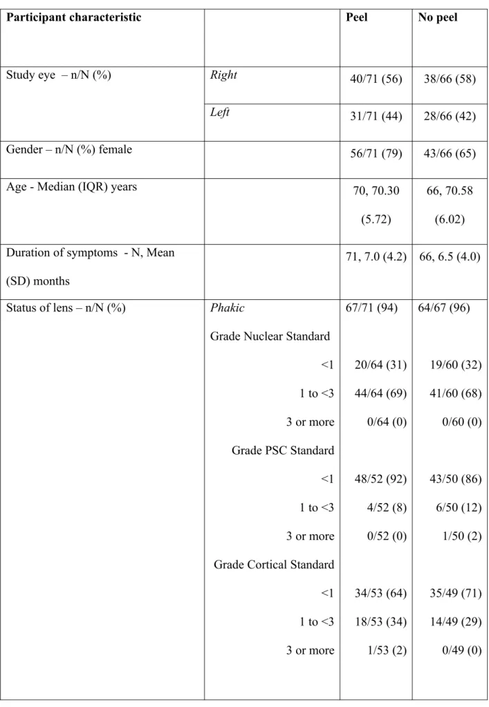
Table 1. Baseline characteristics
Participant characteristic Peel No peel
Study eye – n/N (%) Right 40/71 (56) 38/66 (58)
Left 31/71 (44) 28/66 (42)
Gender – n/N (%) female 56/71 (79) 43/66 (65)
Age - Median (IQR) years 70, 70.30
(5.72)
66, 70.58 (6.02) Duration of symptoms - N, Mean
(SD) months
71, 7.0 (4.2) 66, 6.5 (4.0)
Status of lens – n/N (%) Phakic
Grade Nuclear Standard <1 1 to <3 3 or more Grade PSC Standard <1 1 to <3 3 or more Grade Cortical Standard <1 1 to <3 3 or more
Pseudophakic 4/71 (6) 3/67 (4)
Stage of macular hole – n/N (%) (Clinical classification)
II 29/71 (41) 25/66 (38)
III 42/71 (59) 41/66 (62)
EQ-5D – N, mean (SD) 65,
0.80 (0.21)
63, 0.88 (0.13)
VFQ-25 – N, mean (SD) 66,
79.9 (15.5)
64, 80.3 (17.5) Distance visual acuity (ETDRS) score
– N, mean (SD)
Study eye 71,
48.1 (13.8)
66, 50.0 (10.6)
Fellow eye 71,
76.1 (13.5)
66, 75.7 (17.1) Near visual acuity (Bailey-Lovie) – N,
mean (SD))
Study eye 67,
0.86 (0.30)
65, 0.81 (0.31)
Fellow eye 67,
0.32 (0.34)
64, 0.27 (0.32) Contrast Sensitivity (Pelli-Robson) –
N, mean (SD)
Study eye 66,
30.8 (5.0)
65, 30.8 (5.0) Reading speed MN Read – N, mean
(SD)
Study eye 68,
137.76 (55.7)
Binocular 66, 174.4 (44.5)
63, 174.2 (45.1)
IQR = interquartile range, PSC = posterior subcapsular cataract, SD=standard deviation, ETDRS = early
Table 2. Surgical details and intra-operative complications
Surgical detail Level Peel No peel
Surgeon grade – n/N (%) Consultant 55/67 (82) 51/65 (78) Fellow 12/67 (18) 14/65 (22) Assisting surgeon – n/N (%) Consultant 8/67 (12) 10/63 (16) Fellow 10/67 (15) 10/63 (16) Registrar 25/67 (37) 27/63 (43)
SHO 4/67 (6) 3/63 (5)
Associate 0/67 (0) 1/63 (2)
None 20/67 (30) 12/63 (19)
Type of anaesthesia – n/N (%) General 28/67 (42) 35/65 (54) Local 39/67 (58) 30/65 (46) Operation time – N, mean
(SD)
Minutes 62, 72.6 (19.0)
63, 62.6 (21.7)
ILM peeling – n/N (%) 64/67 (96) 4/67 (6%)
Any intraoperative complication* – n/N (%)
38/67 (57) 28/65 (43)
Retinal tears – n/N (%) 18/67 (27) 21/65 (32)
Retinal detachment – n/N (%)
4/67 (6) 1/65 (2)
Retinal haemorrhages – n/N (%)
Retinal whitening – n/N (%) 9/67 (13) 1/65 (2)
Choroidal/Suprachoroidal haemorrhages – n/N (%)
2/67 (3) 0/65 (0)
Other 7/67 (10) 6/65 (9)
Table 3. Primary and secondary outcomes
Primary outcome Peel No peel
Mean diff (95% CI); p-value
Distance visual acuity at six months; N, mean (SD)
65, 61.3 (14.7)
62, 59.2 (18.9)
4.8 (-0.3 to 9.8); 0.063
Secondary visual outcomes Peel No peel
Mean diff (95% CI); p-value
Distance visual acuity at three months; N, mean (SD)
66, 59.1 (15.4)
65, 56.7 (19.7)
3.6 (-1.9 to 9.1); 0.201
Near visual acuity at three months; N, mean (SD)
65, 0.62 (0.32)
60, 0.63(0.38)
-0.04 (-0.15, 0.06); 0.406
Near visual acuity at six months; N, mean (SD)
62, 0.54 (0.31)
61, 0.53(0.31)
-0.03 (-0.12, 0.07); 0.559
Contrast sensitivity at six months; N, mean (SD)
63, 31.4 (4.8)
60, 32.1(4.2)
-0.7 (-2.0, 0.7); 0.332
Reading speed (study eye) at six months; N, mean (SD)
61, 168 (53)
59, 157(47)
11 (-3, 26); 0.123
Reading speed (binocular) at six months; N, mean (SD)
60, 188 (44)
59, 182(46)
4 (-7, 14); 0.498
Secondary outcomes: QOL Peel No peel Mean diff (95% CI); p-value
(SD) 0.85 (0.20) 0.87 (0.15)
VFQ-25 six months; N, mean (SD)
55, 85.7 (11.6)
59, 85.8 (13.2)
0.4 (-3.3, 4.1); 0.847
Secondary outcome: macular hole status – n/N (%) closed
Peel No peel
Odds ratio (95% CI);p-value
one month 56/67 (84) 31/64 (48) 6.23 (2.64 to 14.73);
<0.001
three months 61/66 (92) 52/63 (83) 2.65 (0.84 to 8.36); 0.097
Table 4. Complications and further eye surgery reported during the follow-up (up to six months)
Event – n/N (%) Peel No peel Odds ratio (95%
CI); p-value
Any complication* 21/64 (31) 27/62 (44) 0.63 (0.31, 1.31);
0.218
Anterior uveitis 1/64 (2) 1/62 (2)
Decentration of the IOL 0/64 (0) 3/62 (5)
Increased IOP 6/64 (9) 9/62 (15)
Retinal detachment 3/64 (5) 2/62 (3)
RPE changes at macula 7/64 (11) 9/62 (15)
Other 8/64 (13) 6/62 (11)
Any further eye surgery* 8/67 (12) 31/65 (48) 0.14 (0.05, 0.34); <0.001
ILM peel 2/67 (3) 25/65 (38)
Surgery for retinal detachment 2/67 (3) 2/65 (3)
Redo macular hole 2/67 (3) 5/65 (8)
Vitrectomy + gas 2/67 (3) 0/65 (0)
Yag capsulotomy 0/67 (0) 1/65 (2)
Removal of oil 1/67 (1) 0/65 (0)
Epiretinal membrane peel 1/67 (1) 0/65 (0)