Review
1
Applications of the FIV Model to Study HIV
2
Pathogenesis
3
Craig Miller 1,*, Zaid Abdo 2, Aaron Ericsson3, John Elder 4, and Sue VandeWoude 2
4
1 Department of Veterinary Pathobiology, Oklahoma State University, Stillwater, OK
5
2 Department of Microbiology, Immunology, and Pathology, Colorado State University, Fort Collins, CO
6
3 Department of Veterinary Pathobiology, University of Missouri, Columbia, Missouri
7
4 Department of Immunology and Microbiology, The Scripps Research Institute, La Jolla, CA
8
9
* Correspondence: [email protected]; Tel.: 1+405-744-2219
10
11
Abstract: Feline immunodeficiency virus (FIV) is a naturally-occurring retrovirus that infects
12
domestic and non-domestic feline species, producing progressive immune depletion that results in
13
an acquired immunodeficiency syndrome (AIDS). Much has been learned about FIV since it was
14
first described in 1987, particularly in regard to its application as a model to study the closely related
15
lentivirus, human immunodeficiency virus (HIV). In particular, FIV and HIV share remarkable
16
structure and sequence organization, utilize parallel modes of receptor-mediated entry, and result
17
in a similar spectrum of immunodeficiency-related diseases due to analogous modes of immune
18
dysfunction. This review summarizes current knowledge of FIV infection kinetics and mechanisms
19
of immune dysfunction in relation to opportunistic disease, specifically in regard to studying HIV
20
pathogenesis. Furthermore, we present data which highlight changes in the oral microbiota and oral
21
immune system during FIV infection, and outline the potential for the feline model of oral AIDS
22
manifestations to elucidate pathogenic mechanisms of HIV-induced oral disease. Finally, we discuss
23
advances in molecular biology, vaccine development, neurologic dysfunction, and the ability to
24
apply pharmacologic interventions and sophisticated imaging technologies to study experimental
25
and naturally occurring FIV, which provide an excellent, but often overlooked resource for
26
advancing therapies and management of HIV/AIDS.
27
Keywords: Feline immunodeficiency virus; FIV; human Immunodeficiency virus; HIV; animal
28
models, opportunistic disease, lentiviral pathogenesis; molecular biology
29
30
1. Feline immunodeficiency virus
31
Feline immunodeficiency virus (FIV) is a naturally-occurring retrovirus that infects domestic
32
and non-domestic feline species. In domestic cats, FIV produces progressive immune depletion that
33
eventually results in an acquired immunodeficiency syndrome (AIDS) [1-10]. As a consequence, FIV
34
infection is associated with a variety of clinical syndromes, including cachexia, anterior uveitis,
35
chronic rhinitis, gingivostomatitis and periodontitis, encephalitis and neurologic dysfunction, and
36
lymphoma [1,4,9,11-21]. The acute phase of FIV infection, lasting approximately 4-8 weeks, is
37
characterized by a sharp increase in CD4+ T lymphocytes that are accompanied by high levels of FIV
38
viral RNA and proviral DNA in circulation [4,8,22]. These hematologic changes are typically
39
accompanied by mild to moderate clinical signs which include pyrexia, lethargy, and peripheral
40
lymphadenopathy [4,22,23]. Following a prolonged asymptomatic phase, during which the levels of
41
circulating virus remains stable and integrated provirus establishes a reservoir of latently infected
42
target cells, there is progressive decline of CD4+ T lymphocytes and other immunocytes, resulting in
43
functional immunodeficiency and susceptibility to opportunistic infections [6,24-26].
44
During FIV infection, loss of CD4+ T lymphocytes is directly attributable to a viral-induced
45
cytopathic effect, in addition to an increase in FIV-specific CD8-mediated programmed cell death,
46
lack of thymic regeneration, and spontaneous apoptosis in response to decreased cytokine support
47
[10,25,27,28]. The most frequent clinical disease syndromes associated with FIV infection manifest as
48
a consequence of immune dysfunction, such as oral opportunistic infection (gingivitis, stomatitis, and
49
periodontitis), immune-mediated glomerulonephritis, chronic rhinitis, and dermatitis
50
[15,16,19,20,29,30]. Oral opportunistic infections are prevalent in a high proportion of FIV-infected
51
cats, and frequently present as erythematous, inflammatory lesions along the gingival margin
52
(gingivitis), multifocal areas of necrotizing inflammation within the gingival sulcus or periodontal
53
ligament (periodontitis), or ulcerative inflammatory lesions along the buccal mucosa, hard palate, or
54
soft palate (stomatitis) [20,31-33]. Changes in the salivary/oral microbiota have been increasingly
55
associated with FIV infection, and shifts in the proportion of opportunistic pathogens in saliva of
FIV-56
infected cats have been associated with the development of oral inflammatory lesions [31,34].
57
Similarly, FIV-infected cats frequently present with severe, necrotizing and/or ulcerative
58
inflammatory lesions (dermatitis) due to opportunistic infection with various bacterial, fungal,
59
protozoal, and parasitic etiologies, including mycobacteriosis, leishmaniasis, toxoplasmosis, and
60
dermatophytosis [16,29,35,36]. Upper respiratory disease is also a frequently finding in FIV-infected
61
cats, and may occur in conjunction with concurrent viral, bacterial, or fungal infections [4,15,37,38].
62
Interestingly, FIV is also associated with the occurrence of neoplastic diseases, most frequently
63
demonstrated by the development of lymphoma in a large proportion of infected cats [7,39]. This
64
association has been described in both naturally and experimentally infected animals, and
65
predominately manifests as high-grade B-cell neoplasms that are remarkably similar to
HIV-66
associated diffuse large B-cell lymphoma (DLBCL) (Figure 1) [7,40-42]. Also similar to HIV, direct
67
viral-mediated oncogenesis related to proviral integration within oncogenes is an uncommon feature
68
of FIV infection, and neoplastic transformation has been attributed to indirect consequences of
viral-69
induced immune dysfunction that arise in response to prolonged viral infection [7,42-44]. Specifically,
70
recent studies have shown that clonal proviral integration sites are not typically detected during FIV
71
infection and that proviral loads are lower in neoplastic tissues, indicating neoplastic growth of cells
72
lacking provirus [7]. Conversely, FIV and other lentiviral infections are strongly associated with
73
polyclonal B-cell expansion, immunoglobulin production, and cytokine expression of proliferative
74
mediators in response to immune activation and dysregulation [45,46]. It is proposed that such
75
infection kinetics provide opportunities for somatic rearrangements associated with generation of
B-76
cell receptor diversity, or mutations in immunological cells during rapid expansion that disrupt or
77
activate oncogenes; thus resulting in neoplastic transformation [7,42]. However, the causal
78
relationship of FIV and lymphoma has not been fully elucidated, and further studies are necessary to
79
evaluate the specific role that viral infection and immune function play during tumorigenesis.
80
FIV-induced renal disease is also observed in both experimentally and naturally infected cats,
81
and includes pathologic changes which include glomerulonephritis, proteinuria, protein tubular
82
casts and tubular microcysts, as well as diffuse interstitial inflammatory infiltrates [30,47]. Mesangial
83
widening with glomerular and interstitial amyloidosis is also observed in kidneys of FIV-infected
84
cats, and when evaluated in the context of another frequent finding during FIV infection,
85
hypergammaglobulinemia, indicate the potential for immune complex deposition to occur within the
86
glomerulus as a result of chronic antigenic stimulation and immune activation [30,48,49].
87
Neurologic disease is an important manifestation of FIV infection, and affected cats may present
88
with either central nervous system (CNS) or peripheral nervous system (PNS) involvement
89
[14,17,18,50,51]. In the PNS, FIV induces significantly increased numbers of CD3+ T cells and
90
macrophages in dorsal root ganglia, and infected cats exhibit pronounced changes in epidermal nerve
91
fiber densities [50,52]. FIV enters the CNS during the acute stages of infection and is present within
92
the brain and cerebral spinal fluid [14,17,53]. The primary neuropathogenic effect of FIV infection
93
within the CNS manifests as infiltration and accumulation of perivascular lymphocytes and
94
macrophages (encephalitis), activation of microglial cells and astrocytes (gliosis), and occasional
95
consequences associated with immune activation within the CNS frequently results in clinically
97
apparent neurologic deficits and gradual decline in CNS function, functionally manifesting as
98
abnormal stereotypic motor behaviors, anisocoria, increased aggression, prolonged latencies in
99
brainstem evoked potentials, delayed righting and pupillary reflexes, decreased nerve conduction
100
velocities, and deficits in cognitive-motor functions [55-58].
101
102
Figure 1. Immunohistochemistry from a FIV-infected cat with primary B cell lymphoma.
103
(A) Mesenteric lymph Node; 40x and 4x (inset). Normal amounts of interlobular T lymphocytes
104
(arrows) are present throughout the lymph node. Anti-CD3 (IHC) with DAB as chromogen and
105
hematoxylin counterstain. (B) Mesenteric lymph Node; 40x and 4x (inset). Neoplastic B
106
lymphocytes (arrows) multifocally expand the normal lymph node architecture. Anti-CD79a
107
IHC with DAB as chromogen and hematoxylin counterstain.
108
2. FIV as a molecular analogue to HIV
109
FIV is a member of the Lentivirus genus within the Retroviridae family, and much has been
110
learned about FIV since it was first described in 1987, particularly in regard to its application as a
111
model to study the closely related lentivirus, human immunodeficiency virus (HIV) [8-10,59,60]. The
112
FIV virion is approximately 100nm in diameter, spherical, and contains two identical strands of
113
positive-sense RNA in its 9400-base genome, which is tightly associated with the nucleocapsid
114
protein (NC, p7) and a t-RNAlys bound to each RNA molecule, which serves as a primer for negative
strand transcription [25,59-61]. This protein complex, along with viral enzymes involved with
116
replication and maturation (protease, reverse transcriptase, integrase, and dUTPase), are enclosed
117
within a core of capsid protein (CA, p24), and surrounded by a shell of matrix protein (MA, p14)
118
[25,59,60]. Viral envelope glycoproteins (gp) are embedded within an outer lipid bilayer surrounding
119
the matrix coat, and include the surface (SU, gp95) and transmembrane (TM, gp40) subunits, which
120
are cleaved from a 130-150kDa membrane-bound precursor protein, glycosylated, and
non-121
covalently anchored within the envelope in a trimeric form [25,59,60,62].
122
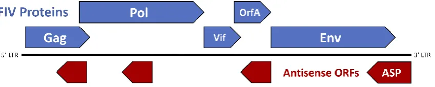
The genomic structure of FIV consists of three primary open reading frames (ORFs), gag, pol, and
123
env, which are flanked by two long-terminal repeats (LTR) and accompanied by numerous small
124
ORFs containing regulatory and accessory genes such as vif, rev, and orfA (Figure 2). FIV gag encodes
125
the Gag polyprotein, which is cleaved by the protease to form the three mature proteins (MA, CA,
126
and NC) and is necessary to achieve formation of mature virus particles [59,63,64]. Pol polyprotein,
127
the primary product of the FIV pol gene, contains 4 important enzymes involved in virus replication
128
and maturation: protease, reverse transcriptase (RT), integrase (IN), and dUTPase (DU) [59]. Viral
129
protease (PR) facilitates the cleavage of Gag and Pol polyproteins into functional enzymatic or
130
structural proteins; DU catalyzes hydrolysis of dUTP to dUMP in effort to minimize
131
misincorporation of potentially mutagenic dUTP into host DNA [59,65,66]; FIV RT is an
RNA-132
dependent DNA polymerase involved in the reverse transcription of viral genomic RNA into a
133
double-stranded copy of proviral DNA (cDNA). Once synthesized, cDNA is integrated into the host
134
genome by a mature IN containing three functional domains: an N-terminal domain, a central
135
catalytic core, and a C-terminal domain [67-69]. The FIV Env polyprotein, a 130-150 kDa product of
136
the env gene, is glycosylated and trimmed within the Golgi apparatus, and proteolytically cleaved
137
into two mature, glycosylated proteins prior to virion budding at the cell surface: SU (gp95) and TM
138
(gp40), both of which play critical roles in virion attachment and entry into target cells [59,60].
139
140
Figure 2. Genomic organization of FIV. The genomic structure of FIV consists of three primary
141
open reading frames (ORFs), gag, pol, and env, which are flanked by two long-terminal repeats
142
(LTR) and accompanied by numerous small ORFs containing regulatory and accessory genes
143
such as vif and orfA. Potential short ORFs (antisense ORFs - shown in red) may be translated
144
from a negative strand message.
145
FIV requires an initial interaction with a primary binding receptor for infection, and binds to
146
host cells through a high-affinity interaction of the envelope SU protein (gp95) with the CD134
147
surface molecule present on CD4+ lymphocytes and monocytes/macrophages [70-74]. This
148
interaction induces a conformational change in the SU protein, which then exposes a cryptic epitope
149
in the V3 loop of Env; the binding site necessary for binding with the entry (co-) receptor CXCR4
150
[26,73,74]. Binding of the V3 loop exposes the serpentine region of TM (gp40), which results in the
151
formation of a hairpin structure that allows fusion with the cell membrane and subsequent cell entry
152
[26,74,75]. However, as infection progresses, the production of neutralizing antibodies by the host
153
increases the need for FIV to escape selective pressures. New viral variants arise which exhibit a
154
decreased dependence on CD134 and increased ability to infect cells that express CXCR4 with limited
155
CD134 expression, such as naïve B cells and CD8+ T cells [2,3,60,76-79]. This expanded cell tropism
156
results in an increase in the number of target cells susceptible to infection, which subsequently causes
157
The structural and sequence organization of FIV is very similar to HIV, which is also a member
159
of the lentivirus genus [59]. HIV is morphologically characterized by a spherical virion that is roughly
160
120nm in diameter, and contains a diploid genome composed of two copies of single stranded,
161
positive-sense RNA that is packaged with nucleocapsid (p7) and accessory proteins (protease, reverse
162
transcriptase, integrase) [80]. Like FIV, the ribonucleoprotein complex at the heart of the HIV virion
163
is contained within a dense core of Capsid protein (CA, p24) and surrounded by a spherical shell of
164
Matrix protein (MA, p17)[80]. Mature Env glycoproteins, SU (gp120) and TM (gp 41), are anchored
165
within the external lipid bilayer, and play significant role in cell entry through binding to host cell
166
receptors. HIV also requires an initial interaction with a primary binding receptor for infection, and
167
utilizes analogous modes of receptor-mediated entry as FIV utilizing chemokine co-receptors [81-83].
168
However, in lieu of CD134, HIV utilizes CD4 as primary binding receptor and CCR5 as its primary
169
entry receptor, although HIV is also able to utilize CXCR4 [81,82]. Much like FIV, HIV binds to CD4+
170
target cells through a high-affinity interaction with the CD4 receptor that induces a conformational
171
change in the envelope glycoprotein gp120, subsequently exposing the binding sites necessary for
172
chemokine co-receptor binding (CXCR4 or CCR5) and subsequent fusion with the cell membrane.
173
The HIV genome encodes three primary polyproteins, Gag, Pol, and Env, as well as the
174
regulatory protein, Rev, and accessory protein, Vif – all of which exhibit similar functions to FIV
175
[59,60,80]. However, in addition to these, HIV also contains genes that encode additional accessory
176
proteins involved in viral maturation, replication, and survival [80]. These include: Tat (p16/p14), a
177
viral transcriptional activator; Vpr (p10-15), a promoter of nuclear localization and inhibitor of cell
178
division (cell cycle arrest at G2/M); Vpu (p16); a promotor of extracellular release of viral particles;
179
Nef (p27-25), a downregulator of CD4 and MHC I expression; Vpx (p12-16), a Vpr homolog present
180
in HIV-2 (absent in HIV-1); and Tev (p28), a tripartite tat-env-rev protein [80].
181
The FIV genome contains one regulatory gene (rev) and two accessory genes (vif and orfA). FIV
182
rev encodes Rev, a nucleolar polyprotein that binds to the Rev Response Element (RRE) to allow
183
export of partially spliced and unspliced viral RNA transcripts out of the nucleus with the help of the
184
nuclear export protein, exportin-1 [59,60,84]. The FIV Vif protein, is crucial to FIV replication and is
185
involved in counteraction of host defense mechanisms such as APOBEC3, a cellular protein that
186
exerts an antiviral effect by deamination of cytosine to uracil during viral replication, resulting in
187
degradation of synthesized minus-strand DNA [59,60,85]. FIV Vif counteracts APOBEC3 by
188
targeting the host protein to the E3 ubiquitin ligase complex, which is subsequently degraded by the
189
proteasome [59,60,85].
190
The FIV OrfA protein is encoded by the accessory gene orfA (Figure 2), and was originally
191
considered a transactivator of transcription due to a role in increasing the net translation of proteins
192
expressed from genes under transcriptional control of the FIV LTR. The localization of the orfA gene
193
in the viral genome also roughly coincides with the location of the gene encoding the HIV
194
transactivator, Tat [86]. However, studies have failed to show increase in transcription directed by
195
OrfA and there is no trans-activation response (TAR) element, as acted on by HIV Tat. Thus, increase
196
in net protein translation facilitated by OrfA must be by other means and may be involved in late
197
steps of virion formation and the early steps of virus infectivity, although the precise role of OrfA is
198
still undetermined [59,60,87-90]. OrfA localizes in the nucleus and causes cell cycle arrest at G2 in
199
infected cells, reminiscent of effects caused by the Vpr protein in HIV-1. Also, OrfA has been shown
200
to downregulate expression of the viral receptor for FIV (CD134) on the surface of cells, as well as E2
201
ubiquitin-conjugating enzymes and a ubiquitin-protein ligase [60,86,91], similar to effects ascribed to
202
the Nef protein on CD4 downregulation during HIV-1 infection. These potential functions of OrfA
203
may have implications which aid in viral dissemination by preventing surface interactions with
204
budding virions, and limit degradation of viral proteins by host cell ubiquitin ligase mechanisms.
205
In 1988, Miller [92] made the observation that there was also potential to encode a peptide
206
product from an RNA transcribed from the minus strand of the provirus. Since then, there have been
207
a number of reports providing evidence for predicted RNA and protein products from the minus
208
strand in HIV-1 [93-102], SIV [103], FIV [104], and in the deltaretrovirus, BLV [105]. In FIV, there are
209
(Figure 2). However, the major potential reading frame in the negative strand of both FIV and HIV
211
coincides with the Env coding region in the plus strand RNA, in the region underlying the Rev
212
Responsive Element (RRE) encoded on the plus strand. A recombinant protein transcribed and
213
translated from the ASP open reading frame has been used to screen both naturally and
214
experimentally FIV infected cats for antibodies to the protein and a small percentage (<10%) do show
215
some level of positivity (manuscript in preparation) (Figure 2). Furthermore, knocking out the
216
putative start codon for ASP resulted in a dramatic reduction in viral protein production, suggesting
217
a critical role in the virus life cycle. Immunohistochemistry shows a non-nuclear localization of the
218
protein, suggestive of some post-transcription event. Further studies will be required to define the
219
role of ASP, but it may contribute to the ability of the virus to replicate by counteracting some innate
220
anti-viral response in the cell.
221
3. FIV as a model to study HIV pathogenesis
222
3.1. Immune dysfunction
223
The primary immunodeficiency of FIV, a gradual and progressive decline in CD4+ T
224
lymphocytes, is a hallmark feature of both natural and experimental infection, and the most obvious
225
fundamental feature to parallel HIV infection. During both FIV and HIV infection, CD4+ lymphocyte
226
numbers decline over an extended asymptomatic phase, and is associated with an increase in
227
activated CD8+ lymphocytes that have antiviral activity [106-109]. The net effect of this event is a
228
decrease in the ratio of CD4+ cells to CD8 + cells (CD4:CD8), and is used as a clinical indicator of
229
immunosuppression in both FIV and HIV infected patients [108-110]. Additionally, several studies
230
have shown that FIV induces defects in immune function similar to HIV, such as a decreased
231
proliferation response of T lymphocytes in response to mitogens, a deficit in the humoral immune
232
response, and dysregulation of cytokine expression [10,24,59].
233
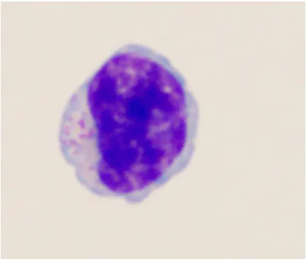
Large granular lymphocytes (LGLs) are a lymphoid subset comprising 10-15% of peripheral
234
mononuclear blood cells (PBMCs) (Figure 3), and consist of either CD3- NK cells or CD3+ T-cells that
235
mediate antibody-dependent cytotoxicity [111-114]. Analysis of LGL populations during HIV
236
infection have been hampered by the low percentage of these cells in circulation, and has typically
237
only been reported in association with neoplasia [114-116]. However, recent studies have shown that
238
LGLs are detectable and elevated during HIV infection in humans, and may represent
viral-239
suppressive CD8+ T cells [114,117]. Interestingly, studies in FIV-infected cats have determined that
240
similar elevations in LGL phenotypes may represent polyclonal T-cells with viral suppressive
241
properties, indicated by increased interferon- (IFN-) expression and decreased PBMC proviral
242
loads in correlation with LGL lymphocytosis [114,118].
243
244
Figure 3. Cytological morphology of a large granular lymphocyte (LGL). Recent studies have
245
determined that elevations in LGL phenotypes during both FIV and HIV infection may represent
246
Conversely, recent studies have shown that CD4+ CD25+ T regulatory (Treg) cells are
248
responsible for inhibition of CD8+ IFN- production during both FIV infection [119] and HIV
249
infection [120], highlighting potential mechanisms by which these viruses exhibit an
250
immunosuppressive effect on the CD8+ immune response. Furthermore, additional studies have
251
shown that FIV directly infects and activates CD4+ CD25+ Treg cells, which are then able to suppress
252
CD4+ CD25- T helper (Th) cells [121]. While this relationship and the potential mechanisms of Treg
253
cell activation during HIV infection is still unclear, such comparative studies in FIV may offer
254
potential to help our understanding of CD8+ T cell function in HIV infection.
255
3.2. Neurologic dysfunction
256
Previous studies have shown that both FIV and HIV enter the central nervous system (CNS) at
257
acute stages of infection, either via trafficking of infected monocytes and lymphocytes, or by
258
penetration of free virus across the blood-brain or blood-CSF barriers [17,122-127]. Once present in
259
the CNS, both FIV and HIV infection spread to microglia and astrocytes, which then serve as a
260
reservoir for latent viral persistence [13,17,126-128]. Although multinucleated giant cells are rarely
261
observed in the CNS during FIV infection, the fundamental neuropathologic finding of encephalitis
262
is well-documented in both HIV and FIV infected patients, and resultant proliferation and activation
263
of these cells (gliosis) is associated with neurodegenerative processes such as myelin degradation and
264
neuronal injury/loss [14,17,51,54,129]. Thus, the clinical manifestations associated with
265
neuropathology of FIV are likewise observed in HIV infection, and because of this, FIV has been
266
repeatedly used as a model to investigate the pathogenesis of dementia and cognitive-motor
267
processing deficits in AIDS patients. In vitro models of FIV have been useful to expand our
268
understanding of role of calcium dysregulation and neural dysfunction during lentiviral infection,
269
and have provided a unique system for the development neuroprotective treatments such as
270
neurotrophin ligands, which prevent the delayed accumulation of intracellular calcium and
271
decreased cytoskeletal damage of neuronal dendrites [17,130]. Furthermore, because of the low
272
natural prevalence and slow clinical course associated with lentiviral-induced neurologic
273
dysfunction, experimental in vivo studies have been developed in the FIV model which accelerate
274
neuropathogenesis (neonatal inoculation, inoculation with neurovirulent strains, direct intracranial
275
inoculation), allowing increased opportunity to evaluate viral kinetics of CNS infection,
276
neurovirulence determinants, and the potential for novel treatments designed to decrease
277
neurocognitive defects during HIV infection [53,57,130,131].
278
The use of neurovirulent strains of FIV has also allowed for the investigation of neuropathogenic
279
effects on the peripheral nervous system (PNS) as a model of HIV distal symmetric polyneuropathy
280
(DSP), demonstrating rapid onset of peripheral neuropathy in FIV infected cats with axonal injury,
281
macrophage activation, and detection of virus within the nerve [50,132]. Indeed, FIV infection results
282
in pathological events in the PNS that are very similar to HIV, including increased numbers of CD3+
283
T lymphocytes and activated macrophages in skin and dorsal root ganglia (DRGs) that are associated
284
with increased expression of the pro-inflammatory cytokines, as well as changes in epidermal nerve
285
fiber densities, indicative of axonal and myelin degeneration [50,52]. FIV has also been useful in the
286
evaluation of the neurotoxicity of antiretroviral toxic neuropathy (ATN), due to mitochondrial
287
dysfunction associated with nucleoside analogue reverse transcriptase (NRTI) inhibitor treatment.
288
Thus, FIV has the potential to expand our understanding of the role of the immunopathology and
289
progression of neuropathy in FIV-infected cats.
290
SIV models of neuropathogenesis have been used to study HIV-associated neurologic
291
dysfunction (HAND), and has resulted in elucidation of many mechanisms of neuroAIDS
292
development, such as acute CNS infection and the importance of monocyte/macrophage activation
293
in driving CNS lesions [133-136]. Recently, the SIV model of neuroAIDS has been adapted to study
294
peripheral neuropathy, and significant advances have been made that have implicated macrophages
295
within dorsal root and trigeminal ganglia as a source of viral maintenance, in addition to their role in
296
neuronal loss and neuronophagia [137,138]. These findings are coupled with additional studies that
297
have defined impaired mitochondrial function in distal axons which are more pronounced in
ART-298
However, the SIV model of HAND is most commonly employed in rhesus macaques using SIV
300
strains that arose via nosocomial infections or lab adaptation of African monkey strains [140]. SIV
301
neurologic disease is therefore chiefly manifested as rapid progression to AIDS with hallmarks of
302
CNS inflammation which amplify pathology compared to HIV-infected humans [135,136].
303
Furthermore, NHP studies are also limited by increased zoonotic risk to researchers, high cost
304
associated with animal care and housing, the low number of animals available for research, and the
305
potential for co-infection with a wide array of other pathogens, including rhesus rhadinovirus (RRV),
306
lymphocryptovirus (LCV), simian cytomegalovirus (CMV), simian foamy virus (SFV), simian virus
307
40 (SV40), and rhesus papillomavirus (RhPV) [141,142].
308
In mechanistic studies of HIV-associated neurologic dysfunction, interaction of CXCR4 with
309
viral envelope has been shown to enhance neuronal apoptosis via Ca2+-regulating systems and
310
NMDA receptors (NMDARs) in the synaptic membrane [143-149]. This neurotoxic pathway is known
311
to involve Ca2+ influx through NMDARs, nitric oxide (NO) production, and subsequent activation
312
cGMP-dependent protein kinase II, however, the precise cellular mechanisms by which this occurs
313
are unknown and difficult to assess in chronically infected human patients [150-155]. Because FIV
314
binds to CXCR4 on the neuronal membrane in a similar non-infectious interaction as HIV, the feline
315
model may provide answers particularly in regard to the viral envelope-receptor interaction and
316
synaptic activity-mediated neurotoxicity in HAND [156,157]. Given these similarities (and
317
limitations of the SIV model), FIV represents an adjunct lentiviral model that can accurately
318
recapitulate neuroAIDS progression in HIV-infected humans for applications such as evaluation of
319
ART-induced neurotoxicity, neurofibrillary tangle development, and calcium homeostasis during
320
viral infection [14,17].
321
3.3. Vaccine development
322
Considerable effort has been directed at the development of an anti-HIV vaccine strategy that
323
can produce protective immunity in humans, and this effort has been paralleled in regard to FIV. A
324
commercially available, whole inactivated virus vaccine containing two FIV subtypes (Fel-O-Vax
325
FIV® ) is currently licensed for use in the United States, and various reports have described virus
326
neutralization and cellular immunity in a significant proportion of study animals [158-160].
327
However, the efficacy of this vaccine is still under debate, as recent studies and field evaluations have
328
reported that the vaccine does not confer immunity against certain FIV strains (ie: FIVGL8), and that
329
the neutralizing antibody response and protective rate may be low in certain cat populations (i.e.
330
protection is not conferred to certain virulent recombinant strains of FIV) [161-164]. Other attempts
331
at FIV vaccine development have either failed to induce protective immunity against FIV infection,
332
or have resulted in increased susceptibility to infection via antibody-dependent enhancement or
333
general immune activation [165-170].
334
The development of an anti-HIV vaccine has been impeded by a wide variety of similar
335
complications, such as lack of efficacy or unanticipated side effects, as well as increased susceptibility
336
to infection via analogous mechanisms of FIV vaccine enhancement (antibody-dependent viral
337
enhancement or general immune activation) [171-177]. Indeed, vaccine-induced enhancement of
338
viral infection has been previously reported in a large number of HIV studies [178-181], and has been
339
shown to occur via antibody-dependent or antibody-independent mechanisms of complement
340
activation [182-189], as well as an increase in general immune activation and/or expansion of
341
lymphoid target cells [190-194]; features that have also been observed in FIV studies [165-170].
342
However, despite these setbacks in lentiviral vaccine development, there are many similarities in the
343
disease course of HIV and FIV infection, and the use of the FIV model to circumvent these roadblocks
344
may have great potential to provide a translational model for the development of novel
345
immunotherapies to protect from HIV infection in humans.
346
Traditionally, non-human primate (NHP) models have been at the forefront of anti-HIV vaccine
347
development due to the similarities of SIV and HIV, and have revealed several promising vaccine
348
targets such as nef-deleted SIV (which protects from wild-type SIV infection) and broad neutralizing
349
antibodies utilizing chimeric SHIVs that express the HIV-1 envelope glycoprotein [195-198].
350
significantly impeded by various causes, such as restrictions on the use of live-attenuated HIV-1 in
352
humans, as well as difficulty in producing a sufficiently efficacious neutralizing antibody response
353
by vaccination [196]. Alternatively, various humanized mouse models have played a vital role in
354
elucidating key aspects of the immune response to HIV, primarily through use of generally
355
immunocompromised mice engrafted with reconstituted human immune system tissues such as
356
human fetal thymus and liver (scid-hu-Thy/Liv) or peripheral blood lymphocytes (scid-hu-PBL) [199].
357
These models have been used for key studies in HIV immunopathogenesis, including mechanisms
358
of CD4+ T-cells loss, antiretroviral therapy response, and passive immunization with monoclonal
359
antibodies to HIV envelope protein (and testing of Env-based vaccines) [142,199-203]. However,
360
because only certain parts of the human immune system can be reconstituted in humanized mouse
361
models, interactions between the introduced human cells and the murine immune system cannot be
362
evaluated in these hosts, nor the effects of HIV infection in non-hematopoietic tissues [199]. Although
363
FIV lacks certain molecular similarities to HIV, it induces similar immunopathologies in its natural
364
host, and therefore represents an important yet underutilized animal model for full evaluate the
365
immune response during natural lentiviral infection. Furthermore, the availability of a
commercially-366
available vaccine in cats with efficacy against at least a subset of FIV may provide important clues to
367
improving the efficacy of anti-HIV vaccines, and the elucidation of the mechanisms associated with
368
vaccination failure in analogous FIV and HIV models of immunotherapy may provide key insights
369
into improving the efficacy of lentiviral vaccines.
370
3.4. HIV-induced oral disease
371
Oral manifestations of HIV are exhibited through various disease syndromes such as Oral
372
Candidiasis (OC, “thrush”), Linear Gingival Erythema (LGE), Necrotizing Ulcerative Gingivitis
373
(NUG), and Necrotizing Ulcerative Periodontitis (NUP) [204-206]. Despite the success of
374
combinational antiretroviral therapy (cART) in diminishing HIV viral replication and prolonging
375
immune function, lesions associated with systemic and local immune activation and opportunistic
376
oral infections persist in HIV-infected patients [204,207-209]. Previous studies have demonstrated
377
that CD4+ T-cells are rapidly and severely depleted from the intestinal mucosa following HIV
378
infection due to direct effects of targeted virus infection and virus-induced Fas-mediated apoptosis,
379
resulting in loss of mucosal integrity and a reduced capacity to control potential pathogens at
380
mucosal surfaces - thereby triggering local and systemic pro-inflammatory responses [210-213].
381
Based upon the analogous microenvironments of the oral and gastrointestinal mucosa, the same
382
effects of viral-induced immunosuppression is predicted to occur in the oral cavity, resulting in a
383
chronic cycle of immune stimulation, leukocyte recruitment, and target cell infection that produces
384
HIV-induced oral disease lesions [204,214].
385
The FIV model is particularly well suited for studies of HIV-associated oral disease, as it not
386
only parallels HIV in its structural, biochemical, and immunological properties, but it is also the only
387
naturally occurring lentivirus to predictably induce oral lesions in its natural host, the domestic cat
388
[1,4,9,10,31,32]. Non-human primate (NHP) models of HIV do not reliably cause oral disease and are
389
limited by zoonotic risk to researchers, high cost associated with animal care and housing, the low
390
number of animals available for research, while humanized mouse models of HIV lack both the
391
prevalence of oral lesions and the presence of tonsillar structures similar to humans [142,215-217]. In
392
contrast, FIV oral manifestations are common in naturally and experimentally-infected cats [20,31,32],
393
and the range of lesions seen include gingivitis, periodontitis, and feline chronic gingivostomatitis
394
[32], with striking similarities to LGE, NUG, and NUP lesions noted in untreated HIV patients
395
[1,4,107,204,218-221]. Furthermore, opportunistic infections detected in HIV-positive individuals are
396
paralleled in feline oral disease syndromes [35,222-231], and feline tonsillar tissues (palatine,
397
pharyngeal, and lingual tonsils) are analogous to those in humans [216]. Coupled with recent
398
advances in new generation cART protocols available for use in cats [232-235], the domestic cat model
399
of FIV presents an easily manipulated animal model to evaluate drivers of immune dysfunction and
400
microbial dyscrasias during HIV infection using a controlled in vivo study design.
401
Thus, in order to assess in vivo mechanisms contributing to oral disease during lentiviral
402
cats (12-14 month-old) and examined samples by 16S rRNA metagenomics analysis to detect
404
differences in the oral microbiota of naïve and age-matched cats infected with FIV(PPR strain) of 8
405
months duration (n=5/group). FIVPPR is a relatively apathogenic strain of FIV that typically results in
406
mild self-limiting gingivitis and/or periodontitis during acute infection [236], and animals did not
407
have overt, visual signs of clinical periodontitis at the time of sampling. FIV-infected and naïve SPF
408
animals were maintained on a similar diet, and similar anatomic regions were swabbed from all
409
animals at the same time of day. DNA was extracted [237], and amplicon sequencing was performed
410
using illumina MiSeq to generate paired-end 2x250bp sequences of the hyper-variable region 4 (V4)
411
of the 16S rDNA. Data were normalized using cumulative sum scaling [238], and used to construct a
412
nonmetric multidimensional scaling 3D plot (Figure 4A).
413
414
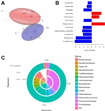
Figure 4. Salivary microbiome alterations during FIV infection. (A) 3D Nonmetric
415
Multidimensional Scaling (NMDS) separates clusters of FIV- and FIV+ cat microbiome samples.
416
Ovals represent the 90% confidence ellipsoids around the centroid of the clusters (FIV+ = red;
417
FIV- = blue). (B) OTUs with significant log-fold change in abundance between FIV+ and FIV-
418
cats at the 0.1 level of significance (after correcting for multiple testing). The list on the left shows
419
the genera of each of these OTUs. Red indicates over representation of that OTU in the FIV+ cats.
420
(C) FIV+ cat with clinical gingivitis/periodontitis with near monoculture of Moraxellaceae (outer
421
circle) compared to the mean microbial community structure of cats that are FIV + (middle circle)
422
Significant differences were detected in the oral microbiota composition of FIV-infected cats
424
relative to naïve animals (Figure 4A). Normalized data were tested using the Zero Inflated Gaussian
425
model implemented in the R package metagenomeSeq [239] to identify the putative OTUs driving
426
differences between FIV+ and FIV- cats. Significant log-fold change in abundance in 12 genera were
427
noted between groups at the 0.1 level of significance after correction for multiple testing (Figure 4B).
428
One FIV-positive cat developed moderate to severe erythematous gingivitis during the course of
429
infection and saliva was collected and analyzed as described above. Upon analysis of saliva, this
430
individual demonstrated a dramatically altered microbiome population with >95% operational
431
taxonomic units (OTUs) corresponding to the genus Moraxellaceae as compared to the FIV+ cats with
432
no lesions and the FIV- cats (Figure 4C).
433
Collectively, these results demonstrate that similar to HIV, FIV infection of domestic cats is
434
associated with oral microbiota dysbiosis and marked loss of microbial diversity during
lentiviral-435
associated periodontitis. The persistence of HIV infection and periodontitis in patients on cART
436
indicates that ancillary treatments specifically directed at restoring the normal oral microbiota in
437
conjunction with cART may improve HIV periodontal progression and decrease systemic immune
438
activation [225,240-242]. Feline dental disease is currently managed by comprehensive dental
439
treatment consisting of hand and ultrasonic scaling, identical to techniques used in humans [243,244].
440
Probiotic supplementation has been successful in early studies as an adjuvant for treating
441
periodontitis in people, and similar commercial oral probiotics products are available for
442
management of feline oral conditions [245-247]. Thus, the application of comprehensive dental
443
cleaning with probiotic treatments in the feline model has the potential to assess the impact of local
444
therapy for restoring oral homeostasis during lentiviral infection, and may increase our
445
understanding of the progression and/or resolution of FIV-induced oral lesions and oral microbiome
446
in the presence and absence of cART.
447
4. Conclusions
448
Our understanding of FIV infection of cats has progressed remarkably over the last 3 decades,
449
yet much remains to be learned from this widespread lentiviral infection. Correspondingly, many
450
aspects of HIV pathogenesis and mechanisms of immune dysfunction are still poorly understood.
451
Most notably, complete elimination of HIV from the host and restoration of immune function has not
452
yet been achieved, nor has the means to provide protective immunity from infection. In regard to the
453
future of HIV research, a precise understanding of the mechanisms for immunodeficiency, especially
454
in the face of co-infections, viral-associated disease, and in the presence and absence of antiretroviral
455
therapy will be necessary for the development of restorative or immuno-protective therapies and
456
prophylaxis.
457
While genetically divergent, FIV shares remarkable overlap with HIV in regard to molecular
458
biology and function. Coupled with the flexibility of working with a small animal model, FIV
459
represents a useful system to assess in vivo aspects of lentiviral pathogenesis. As noted above,
460
comparative pathogenesis of lentiviral immune dysfunction, neurologic and oral disease in the feline
461
model could aid understanding of HIV AIDS. Further, the successful deployment of an FIV vaccine
462
provides great opportunities for evaluation of lentiviral prophylaxis leading to sterilizing immunity.
463
The application of investigations in the molecular biology and function of genetic elements is
464
another area which affords great potential to understand mechanisms of lentiviral infection via the
465
FIV model. For example, contemporary studies in FIV have recently used the 3D structure of FIV
466
reverse transcriptase to uncover the mechanistic basis of viral resistance to non-nucleoside inhibitor
467
drugs [248]. These studies are now uncovering crucial elements in RT structure that can be used as a
468
template for the development of novel compounds that target conventional sites of drugs resistance,
469
providing increased efficacy against drug-resistant strains of HIV [248].
470
Finally, the FIV model holds significant potential as a tractable vehicle to assess efficacy of novel
471
anti-retroviral therapies. Recent studies employing progressive cART regimen composed of
472
nucleoside reverse transcriptase inhibitors (emtricitabine, tenofovir) and integrase inhibitors
473
235]. Additionally, immuno-restorative therapies employing recombinant feline interferon omega
475
(rFeIFN-) have resulted in improvement of clinical symptoms in FIV-associated oral disease and
476
feline chronic gingivostomatitis [249-251]. IFN-ω has also been reported to be a potent inhibitor of
477
HIV infection in vitro, but in vivo therapeutic potential in human patients has not been evaluated
478
[252]. Because IFN-ω exerts strong immunomodulatory effects by stimulating Natural Killer cell
479
activity, enhancing expression of MHC-I, and inhibiting lymphocyte proliferation, testing outcomes
480
of IFN-ω therapy on FIV-associated disease may therefore elucidate anti-inflammatory mechanisms
481
and offer significant potential for adoption as an agent to treat HIV-associated diseases [253].
482
Improvements in molecular technology and available diagnostic analyses for domestic cats, as
483
well as the ability to apply pharmacologic interventions and sophisticated imaging technologies to
484
the study of experimental and naturally occurring FIV provide an excellent, but often overlooked
485
resource for advancing therapies and management of HIV/AIDS.
486
Acknowledgments: Work in this article is supported by National Institute of Allergy and Infectious Diseases
487
and the National Institute of Dental and Craniofacial Research of the National Institutes of Health under award
488
numbers R01AI25825 and F32DE026679-01. The content is solely the responsibility of the authors and does not
489
necessarily represent the official views of the National Institutes of Health. Special thanks to Dr. Wendy Sprague
490
for the large granular lymphocyte (LGL) image used in figure 3.
491
Conflicts of Interest:
492
The authors declare no conflict of interest.
493
494
References
495
496
1. Siebelink, K.H.; Chu, I.-H.; RIMMELZWAAN, G.F.; Weijer, K.; van Herwijnen, R.; Knell, P.;
497
EGBERINK, H.F.; BOSCH, M.L.; OSTERHAUS, A.D. Feline immunodeficiency virus (fiv)
498
infection in the cat as a model for hiv infection in man: Fiv-induced impairment of immune
499
function. AIDS research and human retroviruses 1990, 6, 1373-1378.
500
2. Dean, G.A.; Himathongkham, S.; Sparger, E.E. Differential cell tropism of feline
501
immunodeficiency virus molecular clones in vivo. Journal of virology 1999, 73, 2596-2603.
502
3. English, R.V.; Johnson, C.M.; Gebhard, D.H.; Tompkins, M.B. In vivo lymphocyte tropism of
503
feline immunodeficiency virus. Journal of virology 1993, 67, 5175-5186.
504
4. Pedersen, N.; Yamamoto, J.K.; Ishida, T.; Hansen, H. Feline immunodeficiency virus
505
infection. Veterinary immunology and immunopathology 1989, 21, 111-129.
506
5. Torten, M.; Franchini, M.; Barlough, J.E.; George, J.W.; Mozes, E.; Lutz, H.; Pedersen, N.C.
507
Progressive immune dysfunction in cats experimentally infected with feline
508
immunodeficiency virus. Journal of virology 1991, 65, 2225-2230.
509
6. Hosie, M.J.; Addie, D.; Belák, S.; Boucraut-Baralon, C.; Egberink, H.; Frymus, T.;
Gruffydd-510
Jones, T.; Hartmann, K.; Lutz, H.; Marsilio, F. Feline immunodeficiency: Abcd guidelines on
511
prevention and management. Journal of Feline Medicine and Surgery 2009, 11, 575-584.
512
7. Magden, E.; Miller, C.; MacMillan, M.; Bielefeldt-Ohmann, H.; Avery, A.; Quackenbush, S.L.;
513
VandeWoude, S. Acute virulent infection with feline immunodeficiency virus (fiv) results in
514
lymphomagenesis via an indirect mechanism. Virology 2013, 436, 284-294.
515
8. Pedersen, N.C.; Ho, E.W.; Brown, M.L.; Yamamoto, J.K. Isolation of a t-lymphotropic virus
516
from domestic cats with an immunodeficiency-like syndrome. Science 1987, 235, 790-794.
517
9. Elder, J.H.; Lin, Y.-C.; Fink, E.; Grant, C.K. Feline immunodeficiency virus (fiv) as a model
518
10. Burkhard, M.; Dean, G.A. Transmission and immunopathogenesis of fiv in cats as a model
520
for hiv. Current HIV research 2003, 1, 15-29.
521
11. Bęczkowski, P.M.; Litster, A.; Lin, T.L.; Mellor, D.J.; Willett, B.J.; Hosie, M.J. Contrasting
522
clinical outcomes in two cohorts of cats naturally infected with feline immunodeficiency
523
virus (fiv). Veterinary microbiology 2015, 176, 50-60.
524
12. Colitz, C.M. Feline uveitis: Diagnosis and treatment. Clinical techniques in small animal practice
525
2005, 20, 117-120.
526
13. Dow, S.W.; Poss, M.L.; Hoover, E.A. Feline immunodeficiency virus: A neurotropic
527
lentivirus. JAIDS Journal of Acquired Immune Deficiency Syndromes 1990, 3, 658-668.
528
14. Fletcher, N.F.; Meeker, R.B.; Hudson, L.C.; Callanan, J.J. The neuropathogenesis of feline
529
immunodeficiency virus infection: Barriers to overcome. The Veterinary Journal 2011, 188,
260-530
269.
531
15. Hopper, C.; Sparkes, A.; Gruffydd-Jones, T.; Crispin, S.; Muir, P.; Harbour, D.; Stokes, C.
532
Clinical and laboratory findings in cats infected with feline immunodeficiency virus. The
533
Veterinary record 1989, 125, 341-346.
534
16. Lappin, M. In Opportunistic infections associated with retroviral infections in cats, Seminars in
535
veterinary medicine and surgery (small animal), 1995; pp 244-250.
536
17. Meeker, R.B.; Hudson, L. Feline immunodeficiency virus neuropathogenesis: A model for
537
hiv-induced cns inflammation and neurodegeneration. Veterinary Sciences 2017, 4, 14.
538
18. Miller, C.; Bielefeldt-Ohmann, H.; MacMillan, M.; Huitron-Resendiz, S.; Henriksen, S.; Elder,
539
J.; VandeWoude, S. Strain-specific viral distribution and neuropathology of feline
540
immunodeficiency virus. Veterinary immunology and immunopathology 2011, 143, 282-291.
541
19. Miller, C.; Boegler, K.; Carver, S.; MacMillan, M.; Bielefeldt-Ohmann, H.; VandeWoude, S.
542
Pathogenesis of oral fiv infection. PloS one 2017, 12, e0185138.
543
20. Tenorio, A.P.; Franti, C.E.; Madewell, B.R.; Pedersen, N.C. Chronic oral infections of cats and
544
their relationship to persistent oral carriage of feline calici-, immunodeficiency, or leukemia
545
viruses. Veterinary immunology and immunopathology 1991, 29, 1-14.
546
21. Yamamoto, J.; Hansen, H.; Ho, E.; Morishita, T.; Okuda, T.; Sawa, T.; Nakamura, R.; Pedersen,
547
N. Epidemiologic and clinical aspects of feline immunodeficiency virus infection in cats from
548
the continental united states and canada and possible mode of transmission. Journal of the
549
American Veterinary Medical Association 1989, 194, 213-220.
550
22. Pedersen, N. In Feline immunodeficiency virus infection, Animal Models in AIDS: International
551
TNO meeting, Maastricht, Netherlands, 23-26 October 1989., 1990; pp 165-183.
552
23. Del Fierro, G.; Meers, J.; Thomas, J.; Chadwick, B.; Park, H.; Robinson, W. Quantification of
553
lymphadenopathy in experimentally induced feline immunodeficiency virus infection in
554
domestic cats. Veterinary immunology and immunopathology 1995, 46, 3-12.
555
24. Bendinelli, M.; Pistello, M.; Lombardi, S.; Poli, A.; Garzelli, C.; Matteucci, D.;
Ceccherini-556
Nelli, L.; Malvaldi, G.; Tozzini, F. Feline immunodeficiency virus: An interesting model for
557
aids studies and an important cat pathogen. Clinical Microbiology Reviews 1995, 8, 87-112.
558
25. Lecollinet, S.; Richardson, J. Vaccination against the feline immunodeficiency virus: The road
559
26. Taniwaki, S.A.; Figueiredo, A.S.; Araujo Jr, J.P. Virus–host interaction in feline
561
immunodeficiency virus (fiv) infection. Comparative immunology, microbiology and infectious
562
diseases 2013, 36, 549-557.
563
27. Beatty, J.A.; Willett, B.J.; Gault, E.A.; Jarrett, O. A longitudinal study of feline
564
immunodeficiency virus-specific cytotoxic t lymphocytes in experimentally infected cats,
565
using antigen-specific induction. Journal of virology 1996, 70, 6199-6206.
566
28. Guiot, A.-L.; Rigal, D.; Chappuis, G. Spontaneous programmed cell death (pcd) process of
567
lymphocytes of fiv-infected cats: Cellular targets and modulation. Veterinary immunology and
568
immunopathology 1997, 58, 93-106.
569
29. Hughes, M.; Ball, N.; Love, D.; Canfield, P.; Wigney, D.; Dawson, D.; Davis, P.; Malik, R.
570
Disseminated mycobacterium genavense infection in a fiv-positive cat. Journal of Feline
571
Medicine and Surgery 1999, 1, 23-29.
572
30. Poli, A.; Tozon, N.; Guidi, G.; Pistello, M. Renal alterations in feline immunodeficiency virus
573
(fiv)-infected cats: A natural model of lentivirus-induced renal disease changes. Viruses 2012,
574
4, 1372-1389.
575
31. Diehl, L.J.; Mathiason-Dubard, C.K.; O'Neil, L.L.; Obert, L.A.; Hoover, E.A. Induction of
576
accelerated feline immunodeficiency virus disease by acute-phase virus passage. Journal of
577
virology 1995, 69, 6149-6157.
578
32. Kornya, M.R.; Little, S.E.; Scherk, M.A.; Sears, W.C.; Bienzle, D. Association between oral
579
health status and retrovirus test results in cats. Journal of the American Veterinary Medical
580
Association 2014, 245, 916-922.
581
33. de Rozières, S.; Mathiason, C.K.; Rolston, M.R.; Chatterji, U.; Hoover, E.A.; Elder, J.H.
582
Characterization of a highly pathogenic molecular clone of feline immunodeficiency virus
583
clade c. Journal of virology 2004, 78, 8971-8982.
584
34. Weese, S.J.; Nichols, J.; Jalali, M.; Litster, A. The oral and conjunctival microbiotas in cats with
585
and without feline immunodeficiency virus infection. Veterinary research 2015, 46, 21.
586
35. Mancianti, F.; Giannelli, C.; Bendinelli, M.; Poli, A. Mycological findings in feline
587
immunodeficiency virus-infected cats. Journal of Medical and Veterinary Mycology 1992, 30,
257-588
259.
589
36. Pennisi, M.G. Leishmaniosis of companion animals in europe: An update. Veterinary
590
parasitology 2015, 208, 35-47.
591
37. Sparkes, A.; Hopper, C.; Millard, W.; Gruffydd‐Jones, T.; Harbour, D. Feline
592
immunodeficiency virus infection clinicopathologic findings in 90 naturally occurring cases.
593
Journal of veterinary internal medicine 1993, 7, 85-90.
594
38. Hartmann, K. Feline immunodeficiency virus infection: An overview. The Veterinary Journal
595
1998, 155, 123-137.
596
39. Callanan, J.; Jones, B.; Irvine, J.; Willett, B.; McCandlish, I.; Jarrett, O. Histologic classification
597
and immunophenotype of lymphosarcomas in cats with naturally and experimentally
598
acquired feline immunodeficiency virus infections. Veterinary pathology 1996, 33, 264-272.
599
40. English, R.; Nelson, P.; Johnson, C.M.; Nasisse, M.; Tompkins, W.A.; Tompkins, M.B.
600
Development of clinical disease in cats experimentally infected with feline
601
41. Gabor, L.; Love, D.; Malik, R.; Canfield, P. Feline immunodeficiency virus status of australian
603
cats with lymphosarcoma. Australian veterinary journal 2001, 79, 540-545.
604
42. Beatty, J. Viral causes of feline lymphoma: Retroviruses and beyond. The Veterinary Journal
605
2014, 201, 174-180.
606
43. Endo, Y.; Cho, K.-W.; Nishigaki, K.; Momoi, Y.; Nishimura, Y.; Mizuno, T.; Goto, Y.; Watari,
607
T.; Tsujimoto, H.; Hasegawa, A. Molecular characteristics of malignant lymphomas in cats
608
naturally infected with feline immunodeficiency virus. Veterinary immunology and
609
immunopathology 1997, 57, 153-167.
610
44. Shiramizu, B.; Herndier, B.G.; McGrath, M.S. Identification of a common clonal human
611
immunodeficiency virus integration site in human immunodeficiency virus-associated
612
lymphomas. Cancer Research 1994, 54, 2069-2072.
613
45. Beatty, J.; Lawrence, C.; Callanan, J.; Grant, C.; Gault, E.; Neil, J.; Jarrett, O. Feline
614
immunodeficiency virus (fiv)-associated lymphoma: A potential role for immune
615
dysfunction in tumourigenesis. Veterinary immunology and immunopathology 1998, 65, 309-322.
616
46. Yamamoto, H.; Umemura, T.; Inoshima, Y.; Nakamura, M.; Adachi, I.; Miyazawa, T.; Mikami,
617
T. Immunological and histological disorders in cats experimentally infected with feline
618
immunodeficiency virus subtype b (tm2 strain). Veterinary microbiology 1997, 57, 313-324.
619
47. Poli, A.; Abramo, F.; Taccini, E.; Guidi, G.; Barsotti, E.; Bendinelli, M.; Malvaldi, G. Renal
620
involvement in feline immunodeficiency virus infection: A clinicopathological study.
621
Nephron 1993, 64, 282-288.
622
48. MATSUMOTO, H.; TAKEMURA, N.; SAKO, T.; KOYAMA, H.; MOTOYOSHI, S.; INADA,
623
Y. Serum concentration of circulating immune complexes in cats infected with feline
624
immunodeficiency virus detected by immune adherence hemagglutination method. Journal
625
of veterinary medical science 1997, 59, 395-396.
626
49. Poli, A.; Falcone, M.; Bigalli, L.; Massi, C.; HOFMANN‐LEHMANN, R.; Lombardi, S.;
627
Bendinelli, M.; Lutz, H. Circulating immune complexes and analysis of renal immune
628
deposits in feline immunodeficiency virus‐infected cats. Clinical & Experimental Immunology
629
1995, 101, 254-258.
630
50. Burdo, T.H.; Miller, A.D. Animal models of hiv peripheral neuropathy. Future virology 2014,
631
9, 465-474.
632
51. Podell, M.; March, P.A.; Buck, W.R.; Mathes, L.E. The feline model of neuroaids:
633
Understanding the progression towards aids dementia. Journal of Psychopharmacology 2000,
634
14, 205-213.
635
52. Zhu, Y.; Antony, J.; Liu, S.; Martinez, J.A.; Giuliani, F.; Zochodne, D.; Power, C. Cd8+
636
lymphocyte-mediated injury of dorsal root ganglion neurons during lentivirus infection:
637
Cd154-dependent cell contact neurotoxicity. Journal of Neuroscience 2006, 26, 3396-3403.
638
53. Power, C.; Buist, R.; Johnston, J.; Del Bigio, M.; Ni, W.; Dawood, M.; Peeling, J.
639
Neurovirulence in feline immunodeficiency virus-infected neonatal cats is viral strain
640
specific and dependent on systemic immune suppression. Journal of virology 1998, 72,
9109-641
9115.
642
54. ABRAMO, F.; BO, S.; CANESE, M.G.; POLI, A. Regional distribution of lesions in the central
643
nervous system of cats infected with feline immunodeficiency virus. AIDS research and human
644
55. Steigerwald, E.S.; Sarter, M.; March, P.; Podell, M. Effects of feline immunodeficiency virus
646
on cognition and behavioral function in cats. JAIDS Journal of Acquired Immune Deficiency
647
Syndromes 1999, 20, 411-419.
648
56. Maingat, F.; Vivithanaporn, P.; Zhu, Y.; Taylor, A.; Baker, G.; Pearson, K.; Power, C.
649
Neurobehavioral performance in feline immunodeficiency virus infection: Integrated
650
analysis of viral burden, neuroinflammation, and neuronal injury in cortex. Journal of
651
Neuroscience 2009, 29, 8429-8437.
652
57. Phillips, T.; Prospero-Garcia, O.; Wheeler, D.; Wagaman, P.; Lerner, D.; Fox, H.; Whalen, L.;
653
Bloom, F.; Elder, J.; Henriksen, S. Neurologic dysfunctions caused by a molecular clone of
654
feline immunodeficiency virus, fiv-ppr. Journal of neurovirology 1996, 2, 388-396.
655
58. Phipps, A.J.; Hayes, K.A.; Buck, W.R.; Podell, M.; Mathes, L.E. Neurophysiologic and
656
immunologic abnormalities associated with feline immunodeficiency virus molecular clone
657
fiv-ppr DNA inoculation. Journal of acquired immune deficiency syndromes (1999) 2000, 23, 8-16.
658
59. Sparger, E.E. Fiv as a model for hiv: An overview. In In vivo models of hiv disease and control,
659
Springer: 2006; pp 149-237.
660
60. Kenyon, J.C.; Lever, A.M. The molecular biology of feline immunodeficiency virus (fiv).
661
Viruses 2011, 3, 2192-2213.
662
61. Steinman, R.; Dombrowski, J.; O'Connor, T.; Montelaro, R.C.; Tonelli, Q.; Lawrence, K.;
663
Seymour, C.; Goodness, J.; Pedersen, N.C.; Andersen, P.R. Biochemical and immunological
664
characterization of the major structural proteins of feline immunodeficiency virus. Journal of
665
General Virology 1990, 71, 701-706.
666
62. Hu, Q.-Y.; Fink, E.; Hong, Y.; Wang, C.; Grant, C.K.; Elder, J.H. Fine definition of the
cxcr4-667
binding region on the v3 loop of feline immunodeficiency virus surface glycoprotein. PLoS
668
One 2010, 5, e10689.
669
63. Egberink, H.F.; Ederveen, J.; Montelaro, R.C.; Pedersen, N.C.; Horzinek, M.C.; Koolen, M.J.
670
Intracellular proteins of feline immunodeficiency virus and their antigenic relationship with
671
equine infectious anaemia virus proteins. Journal of General Virology 1990, 71, 739-743.
672
64. Elder, J.; Schnölzer, M.; Hasselkus-Light, C.; Henson, M.; Lerner, D.; Phillips, T.; Wagaman,
673
P.; Kent, S. Identification of proteolytic processing sites within the gag and pol polyproteins
674
of feline immunodeficiency virus. Journal of virology 1993, 67, 1869-1876.
675
65. Von der Helm, K. Retroviral proteases: Structure, function and inhibition-from a
non-676
anticipated viral enzyme to the target of a most promising hiv therapy. BIOLOGICAL
677
CHEMISTRY HOPPE SEYLER 1996, 377, 765-774.
678
66. Gadsden, M.H.; McIntosh, E.; Game, J.C.; Wilson, P.J.; Haynes, R. Dutp pyrophosphatase is
679
an essential enzyme in saccharomyces cerevisiae. The EMBO journal 1993, 12, 4425.
680
67. Khan, E.; Mack, J.P.; Katz, R.A.; Kulkosky, J.; Skalka, A.M. Retroviral integrase domains:
681
DNA binding and the recognition of ltr sequences. Nucleic acids research 1991, 19, 851-860.
682
68. Vink, C.; van der Linden, K.H.; Plasterk, R. Activities of the feline immunodeficiency virus
683
integrase protein produced in escherichia coli. Journal of virology 1994, 68, 1468-1474.
684
69. North, T.W.; Cronn, R.C.; Remington, K.M.; Tandberg, R.T.; Judd, R.C. Characterization of
685
reverse transcriptase from feline immunodeficiency virus. Journal of Biological Chemistry 1990,