ABSTRACT
DENNING, STEFANIE CLAIRE. Dual Strategy for Characterizing foxq1b. (Under the direction of Dr. Antonio Planchart).
Dual Strategy for Characterizing foxq1b
by
Stefanie Claire Denning
A thesis submitted to the Graduate Faculty of North Carolina State University
in partial fulfillment of the requirements for the degree of
Master of Science
Zoology
Raleigh, North Carolina
2015
APPROVED BY:
_______________________________ ______________________________
Dr. Antonio Planchart Dr. Nanette Nascone-Yoder
Co-Chair of Advisory Committee Co-Chair of Advisory Committee
ii
DEDICATION
iii
BIOGRAPHY
iv
ACKNOWLEDGMENTS
I would like to thank my advisor Antonio Planchart for giving me the opportunity to work on this research project. Also for all of his support and guidance along the way. I have learned so much from him over the past two years.
I am very grateful for my committee members for all of their advice and
encouragement: Dr. Nanette Nascone-Yoder and Dr. Beth Hawkins. I sincerely appreciate all of Carolyn Mattingly, David Chris Cole, and Norma Escalante's help in the lab. My project would not have gone as smoothly without them. Special thanks to Elizabeth Cook and Lindsey St. Mary for being very helpful and encouraging in the lab, and to all of our good times.
v
TABLE OF CONTENTS
LIST OF TABLES ... vi
LIST OF FIGURES ... vii
I. LITERATURE REVIEW ... 1
Introduction ... 1
Upstream Regulators of FOXQ1... 3
FOXQ1 in Development ... 9
FOXQ1 induces Epithelial-Mesenchymal Transition (EMT) ... 12
Other Roles of FOXQ1 in Carcinoma ... 16
Conclusion ... 20
II. DESIGN, CONSTRUCTION AND PRELIMINARY CHARACTERIZATION OF A MYC-TAGGED ALLELE OF THE ZEBRAFISH FORKHEAD BOX GENE, foxq1b ... 23
Abstract ... 23
Introduction ... 24
Materials and Methods ... 29
Results ... 44
Discussion... 56
Conclusion ... 58
III. FOXQ1B GENETIC KNOCKOUT UTILIZING CRISPR-CAS TECHNOLOGY ... 59
Abstract ... 59
Introduction ... 59
Materials and Methods ... 64
Results ... 71
Discussion... 82
Conclusion ... 84
vi
LIST OF TABLES
Table 1: Forkhead Box (Fox) Gene Subfamily Members. ... 21
Table 2: Knockdown of foxq1b increases expression of genes involved in the
apoptotic pathway ... 28
Table 3: Primer names and sequences used to create foxq1b-myc construct ... 42
vii
LIST OF FIGURES
Figure 1: Cloning strategy of the foxq1b-myc construct in the pBluescriptII/SK+
plasmid. ... 47
Figure 2: Intermediate steps in the cloning strategy as visualized by gel electrophoresis. ... 48
Figure 3: Identification of foxq1b-myc potential founders. ... 49
Figure 4: Transmission of foxq1b-myc to offspring by female founder #2.. ... 50
Figure 5: Transgene-positive offspring transcribe foxq1b-myc mRNA. ... 51
Figure 6: Foxq1b-myc protein is nuclearly localized to cells in the periphery of the otic vesicle and jaw region. ... 52
Figure 7: RT-qPCR of 48 hpf foxq1b-myc zebrafish exposed to DMSO or 3nM TCDD. ... 55
Figure 8: In situ hybridization using a foxq1b probe. ... 62
Figure 9: Knockdown of foxq1b expression using a morpholino. ... 63
Figure 10: Tyrosinase-gRNA injected vs. uninjected zebrafish. ... 74
Figure 11: Foxq1b-gRNA design. ... 75
Figure 12: Foxq1b-gRNA injected zebrafish. ... 76
Figure 13: Abnormal foxq1b-gRNA injected zebrafish. ... 77
Figure 14: qPCR melting curves of foxq1b-gRNA injected compared to uninjected zebrafish. ... 78
1
I. LITERATURE REVIEW
Introduction
The prototype forkhead box gene (fkh) was discovered in a mutagenesis screen of Drosophila melanogaster homeotic transformants (Jürgens and Weigel 1988). Forkhead box (Fox) genes are a family of transcription factors whose name derives from the phenotype: mutations in Drosophila fkh cause the appearance of a two-spiked head structure, resembling a fork (Carlsson and Mahlapuu 2002). At present, there are 19 subfamilies, FoxA through FoxS, in mammals; within each subfamily, there can be multiple genes, each of which is identified by a number (Table 1 and Jonsson and Peng 2005). Fox genes contain an 80 to 110 amino acid DNA binding domain called the “forkhead box” or "winged helix" domain, the latter named from its X-ray crystallography structure that is comprised of three helices linked by two loops (Carlsson and Mahlapuu 2002). This domain allows them to bind to and regulate certain genes contributing to the growth and formation of an organism’s body structure (Wotton and Shimeld 2011). Fox proteins identified in mice and humans are found to play an important role in not only development but also metabolism and
immunoregulation. Although Fox genes are critical for many cellular functions,
misregulation of these genes can contribute to aging and cancer (Jonsson and Peng 2005). Adequate regulation of Fox genes is essential for maintaining fundamental processes that are required for cell growth and survival.
2 Caenorhabditis spp. contain 15, Drosophila spp. contain 20, and mammals contain over 40 Fox gene family members (Carlsson and Mahlapuu 2002, Hannenhalli and Kaestner 2009); therefore, the number of Fox genes an organism possesses appears to be directly proportional to the structural complexity of that organism.
Throughout evolution, many genome duplications have occurred resulting in a gain, loss, or change in genetic information (Crow and Wagner 2006). In the animal kingdom, vertebrates underwent two genome duplications prior to the divergence of fish and mammals. After the branching of the mammalian linage, a third genome duplication occurred in fish. Since zebrafish contain two paralogs for many mammalian genes, it is thought that this third genome duplication resulted in polyploidization, which is a multiplication of a whole
chromosome (Postlethwait 1998). When paralogs are generated from a genome duplication, they may diverge: one paralog may acquire a new function (neofunctionalization);
alternatively, the ancestral function may be split between the two paralogs
(subfunctionalization); and lastly, one paralog may retain the original function subject to selection whereas the other may degenerate (Presgraves 2005). The Fox gene family has multiplied and acquired new members with distinct roles throughout evolution (Lynch and Conery 2000). Many of the Fox genes and their specific functions have been studied in various organisms, including zebrafish, and compared to their human orthologs.
3 species (Bieller 2001, Hong 2001). The zebrafish paralogs, foxq1a and foxq1b, are also structurally similar to mammalian FOXQ1 (Planchart and Mattingly 2010). The role of FOXQ1 in development and disease, as well as, how it is regulated and how it regulates gene expression will be discussed in the following sections.
Upstream Regulators of FOXQ1
FOXQ1 interacts with signaling pathways and gene families that are crucial for growth and development. Important pathways and genes that involve FOXQ1 are: hedgehog and Wnt signaling pathways, transforming growth factor beta (TGFβ), Aryl hydrocarbon receptor (AhR), Homeobox (Hox) genes, and the retinoic acid pathway. The hedgehog signaling pathway and the Wnt signaling pathway are essential during embryonic
development and promote cell proliferation, differentiation, migration, and growth (Yang 2010 and Le 2014). TGFβ is involved in cell growth and differentiation (Zhao 2014). AhR is a ligand-activated transcription factor that is necessary for xenobiotic metabolism and has been shown to be an upstream activator of murine Foxq1 and zebrafish foxq1b (Planchart and Mattingly 2009). During developmental processes, FOXQ1 interacts with the Hox gene family, which participates in organogenesis and developmental patterning (Martinez-Ceballos 2005). FOXQ1 has also been shown to be a downstream target of the retinoic acid pathway. Retinoids are natural analogues of vitamin A, and are necessary for proper
4 functions. The following paragraphs summarize what is known about the interactions between FOXQ1 and these pathways and genes.
Hedgehog Signaling: The hedgehog signaling pathway plays an important role in cell growth and maintenance during development and throughout the lifetime of an organism. The transmembrane protein receptor Patched (PTCH1) regulates hedgehog signaling by repressing the G-protein coupled receptor Smoothened (SMO) until the hedgehog protein (Hh) binds to PTCH1, initiating signaling. SMO activation stimulates a downstream signal transduction event that inactivates the intracellular hedgehog pathway regulator SUFU, causing the transcriptional effector GLI to translocate from the cytoplasm to the nucleus where it activates or represses transcription of target genes (Katoh 2008). In silico analysis of the rat genome discovered that GLI2 potentially activates many genes in the Fox family, including Foxq1 (Katoh 2008). However, the significance of this potential interaction remains unknown.
Wnt Signaling: The Wnt signaling pathway is activated throughout embryonic development, but also plays a role in cancer development and progression. The Wnt protein activates signaling by binding to the G-protein coupled receptor Frizzled (Logan and Nusse 2004). The resulting signal transduction cascade stabilizes the effector molecule, β-catenin, and releases it from an intracellular destruction complex. β-catenin then translocates to the nucleus where it binds to the TCF/LEF family of transcription factors and activates
5 Since both the Wnt signaling pathway and FOXQ1 are upregulated in colorectal cancer, among others, studies have been performed to determine the cause and effect of this upregulation. It was concluded that FOXQ1 and Wnt signaling are highly correlated: an increase in Wnt signaling in human colorectal cancer cells leads to an increase in FOXQ1 mRNA and protein levels, signifying that FOXQ1 is a downstream target of the Wnt signaling pathway (Christensen 2013), which is supported by the finding that FOXQ1 contains a TCF-4 binding motif in its promoter region. Chromatin immunoprecipitation (ChIP-seq) experiments, which analyze protein interactions with DNA, found that β-catenin binds to this region of the FOXQ1 promoter, thus indicating that Wnt signaling directly regulates the expression of FOXQ1 (Christensen 2013). In Wnt signaling, the protein Groucho mediates transcriptional activity by acting as a corepressor. As discussed below, it appears that FOXQ1 also regulates transcription primarily through repression. Interestingly, FOXQ1 contains two engrailed homology (eh1)-like motifs, which are motifs known to mediate interactions with Groucho (Yaklichkin 2007). These results indicate that the Wnt signaling pathway is a direct upstream activator of FOXQ1 transcription by the binding of β-catenin to FOXQ1's promoter, and that FOXQ1 may interact with the Wnt corepressor
Groucho through its eh1-like motifs. Further studies would have to be conducted to verify the relationship between FOXQ1 and Groucho.
6 Smad4 and translocate to the nucleus where they regulate transcription of target genes (Zhao 2014). Both TGFβ1 and FOXQ1 have been shown to play a role in epithelial plasticity (that is, the ability of epithelial cells to alter their phenotypic state), and are involved in epithelial-mesenchymal transitions (EMT). During EMT, epithelial cells lose their polarity and become invasive and migratory (Feuerborn 2011 and Zhang 2011). An increase in TGFβ1 signaling in mouse epithelial cells results in an increase in Foxq1 expression, demonstrating a positive correlation. The transcription factors Zeb1 and Zeb2, which are known targets of the TGFβ1 signaling pathway, enhance EMT during cancer progression (Feuerborn 2011). siRNA knockdown of Foxq1 expression results in decreased expression of Zeb1 and Zeb2, suggesting Foxq1 may play a role in TGFβ1 by directly regulating target genes, or by regulating upstream activators or repressors of TGFβ1 targets. Since a decrease in Foxq1 signaling inhibits TGFβ1-induced EMT in carcinoma, this pathway may be very useful in studying possible cancer therapies (Zhang 2011). These results indicate a clear relationship between TGFβ and FOXQ1 in both development and carcinogenesis.
Aryl Hydrocarbon Receptor (AhR) Pathway: AhR is a ligand-activated Per-Arnt-Sim (PAS) transcription factor that functions in xenobiotic metabolism, liver development, and hepatocarcinogenesis (Stevens 2009 and Faust 2013). Once a ligand binds, AhR is released from its chaperone complexes in the cytoplasm, where it translocates to the nucleus of the cell. The PAS domain of AhR mediates heterodimerization of AhR to ARNT (aryl
7 activation of genes that are involved in xenobiotic metabolism (Stevens 2009). Potential DREs have been discovered within the promoter region of FOXQ1 in zebrafish, mice, and humans (Planchart and Mattingly 2010). AhR activity has been shown to increase foxq1b expression in zebrafish, the rat hepatoma cell line, H4IIE, and the mouse hepatoma cell line, Hepa-1c1c7, upon exposure to the polycyclic aromatic hydrocarbon
2,3,7,8-tetrachlorodibenzo-p-dioxin (TCDD) (Planchart and Mattingly 2010, Dere 2011, Thornley 2011). In 48 hours post fertilization (hpf) TCDD exposed zebrafish, an upregulation of foxq1b was localized to the jaw structure known as Meckel's cartilage, which is derived from the first pharyngeal arch (Planchart and Mattingly 2010). In WB-F344 cells, TCDD-activated AhR increases the expression of Foxq1, and exposure to the AhR inhibitor CH223191
reduces Foxq1 mRNA levels (Faust 2013), further demonstrating Foxq1 is a downstream target of AhR.
Foxq1-8 luc are co-transfected into NIH3T3 cells or C2C12 myofibroblasts, Hoxc13 upregulates the expression of Foxq1 (Potter 2006). Although these results are contradictory, which could be due to cell type or in vitro versus in vivo studies, luciferase assays indicate that Hoxc13 binds directly to the Foxq1 promoter (Potter 2006). Another Hox gene, Hoxa1, which is a target of retinoic acid, is an essential gene for correct hindbrain and skull formation. Foxq1 expression levels increase in an engineered Hoxa1 double knockout embryonic stem cell line (Martinez-Ceballos 2005), indicating Hoxa1 is a repressor of Foxq1 transcription. FOXQ1 is important for craniofacial development, and its regulation by HOXA1 could be necessary for proper formation of the craniofacial region.
Retinoic Acid Pathway: Like Hoxa1, Foxq1 has been identified as a downstream target of the retinoic acid (RA) pathway. Retinoic acid binds and activates retinoic acid receptors (RARs α, β, and γ) and retinoid X receptors (RXRs α, β, and γ). The RARs then regulate transcription by binding to retinoic acid response elements (RAREs) in the promoter or enhancer regions of target genes (Zhuang 2003). The RARβ gene contains four isoforms: β1, β2, β3, and β4, where β2 is the most abundant and the major RA-inducible isoform. In
wildtype F9 teratocarcinoma cells, Foxq1 mRNA levels increased upon exposure to RA; however, when a homozygous knockout of RARβ2 was generated in F9 cells, Foxq1 mRNA
9 FOXQ1 in Development
Embryonic development requires a vast number of genes working together to create every aspect of a properly functioning organism. Early in development, structures called pharyngeal or branchial arches appear in the head region of vertebrate embryos. The arches are populated by migrating neural crest cells that give rise to diverse cell lineages. The evolution of the pharyngeal arches has been highly conserved throughout the development of vertebrate animals. In jawless chordates, such as the lamprey, the pharyngeal skeleton is comprised of cartilage that forms an unjointed branchial basket which supports the gills (Yao 2011). In jawed chordates, such as zebrafish, the first pharyngeal arch forms a jointed jaw (Cerny 2010). The posterior pharyngeal pouches become the thyroid, parathyroid, and thymus, which are structures of great importance to tetrapods (Graham 2005). The conservation of foxq1 expression in the pharyngeal pouches has been seen throughout evolution.
In situ hybridization using a foxq1 riboprobe was performed on the jawless chordate model, Lampetra planeri (lamprey), for a better understanding of its mRNA localization throughout different stages of development. Foxq1 mRNA is initially located in the
10 formed by the continuation of the lips that leads to the mouth, and the branchial basket
(Wotton and Shimeld 2011) (for more information on lamprey staging refer to (Piavis 1960)). Other chordates, such as the invertebrates Amphioxus and Ciona, express foxq1 in the
endostyle, which is a structure that aids in transporting food to the esophagus (Mazet 2005, Ogasawara and Satou 2003).
In the African clawed frog, Xenopus laevis, foxq1 expression appears in the gastrula stage, but is not fully defined until neurulation (Choi 2006) (For more information on Xenopus staging, refer to Nieuwkoop and Faber 1994). As Xenopus embryos develop, foxq1 is expressed in the gastrointestinal tract and pharyngeal pouches. In situ hybridization of stage 44 Xenopus embryos shows foxq1 is specifically expressed in the tongue, as well as part of the stomach and intestines (Choi 2006). Comparing the role of foxq1 in the agnatha (jawless) model and the gnathostome (jawed vertebrate) model provides evidence that its restriction to the endoderm and expression in the gastrointestinal system is evolutionarily conserved.
11 mucin (Verzi 2008). These results show that Foxq1 plays an important role in gastric function through the regulation of acid and mucin secretion.
Craniofacial development is also regulated by Foxq1, as seen in the Foxq1 mouse knockout. The Foxq1 deficient mutant mouse embryos displayed head abnormalities due to a smaller brain vesicle, which could have been the result of decreased cerebrospinal fluid volume. Transverse sections of the forebrain that were stained with hematoxylin-eosin revealed shrinkage of the third and lateral ventricles (Goering 2008). Foxq1 deficient mice also have a greater chance of mortality; however, upon examining the deceased embryos, the liver, heart, and placenta were all normal compared to wild-type and Foxq1 hemizygotes. The cause of lethality is unknown but restricted to Foxq1 deficient offspring from crosses between C57BL/6J X 129/Sv (Goering 2008). Preliminary data in our lab has shown that foxq1b knockdown in zebrafish also causes craniofacial abnormalities, specifically in the developing jaw.
Foxq1 plays a role in hair follicle development, as described previously. A deletion in the Foxq1 gene results in the satin hair mutation (Hong 2001). Satin hair is shinier and thinner than that of wild type mice, and its appearance is due to increased fibrous material and the inability to keratinize correctly. Since Foxq1 acts directly on the matrix cells, which differentiate into the outgrowing hair shaft and inner root sheath (Driskell 2011), its
12 Foxq1 is involved in smooth muscle development by repressing the transcription of smooth muscle-specific telokin, smooth muscle α, and smooth muscle myosin by directly binding to the forkhead consensus sites located on their promoters (Hoggatt 2000). Since Foxq1 expression is mostly found in epithelial cells, it was suggested that its role is to negatively regulate the transcription of smooth muscle genes in cell types other than smooth muscle, thereby restricting their expression to smooth muscle cells (Hoggatt 2000). FOXQ1's involvement in gastric acid secretion, hair follicle formation, craniofacial patterning, and smooth muscle regulation indicates its vital role in various aspects of development.
FOXQ1 induces Epithelial-Mesenchymal Transition (EMT)
Epithelial cells form a protective barrier against bacteria, prevent substances from leaving their internal environment, and regulate absorption and secretion of fluids in the gut (McConnell 2005). Their properties include, cell adhesion, polarity, and stationary
13 E-cadherin is an adherens junction protein that is an important component of the epithelium because of its involvement with cell adhesion and maintenance of epithelial cell polarity (Gumbiner 1996). β-catenin and γ-catenin are proteins that also play roles in cell adhesion. Epithelial cells that downregulate E-cadherin, β-catenin, and γ-catenin exhibit a fibroblast-like phenotype characterized by detachment and dispersion from the basement membrane (Zhang 2011 and Qiao 2011). Fibronectin, Vimentin, and N-cadherin, on the other hand, are known markers of mesenchymal cells. When ectopic FOXQ1 expression is induced in the human mammary cell line, HMLE, the cells appear spindle-like compared to control cells, and E-cadherin, β-catenin, and γ-catenin are downregulated, whereas
Fibronectin, Vimentin, and N-cadherin are upregulated, signaling the beginning of EMT. However, when FOXQ1 is overexpressed in the MDCK cell line, E-cadherin expression decreases but no other epithelial or mesenchymal markers are affected (Zhang 2011). The varying epithelial and mesenchymal cell marker expression seen in the HMLE and the MDCK cells could be due to different cell properties. However, these results indicate
14 the standpoint of cancer therapy, pharmacologic inhibition of FOXQ1 could be used as a strategy in the fight against cancer.
Several transcription factors such as Snail1, Snail2, Twist1, E47, Zeb1, and Zeb2 are overexpressed in carcinomas and induce EMT by inhibiting E-cadherin expression (Zhu 2013). These transcription factors bind to the E-box region of the E-cadherin promoter, downregulating its transcription and promoting EMT (Zhu 2013). Since FOXQ1 overexpression also decreases the expression of E-cadherin, a study was performed to
determine if FOXQ1 and E-cadherin directly interact. Using ChIP-seq, FOXQ1 was found to bind the E-box region of the E-cadherin promoter (Zhang 2011). These results imply that FOXQ1 overexpression activates EMT in a similar manner as the EMT transcription factors by directly repressing E-cadherin transcription.
The EMT transcription factors, Twist1 and Zeb2, have been identified as downstream targets of FOXQ1. Three potential binding sites for FOXQ1 have been discovered in the Twist1 promoter, and two in the Zeb2 promoter through in silico analysis (Meng 2014). Although FOXQ1 promotes EMT by acting as a repressor of E-cadherin, it also may promote EMT by acting as an activator of Twist1 and Zeb2 (Abba 2013 and Xia 2014).
15 either PDGFRα or β is silenced in HMLE/FOXQ1 cells (where FOXQ1 is overexpressed), there is a decrease in FOXQ1-induced cell migration and invasion in a Transwell migration assay; however, EMT is not reversed (Meng 2014). This indicates that PDGFRα and β may play a role in the maintenance of EMT, but other genes are involved in the activation of EMT.
16 marker Vimentin and the E-cadherin repressors Slug and Snail. BITC increases cell adhesion in breast cancer cells compared to control cells. However, the overexpression of FOXQ1 makes it more difficult for BITC to increase E-cadherin protein levels or suppress Vimentin (Sehrawat 2013). These results further demonstrate the impact FOXQ1 has on the resistance against chemotherapies. Therefore, repressing expression of FOXQ1 could be beneficial in these treatments by increasing cancer cell apoptosis, and reverting mesenchymal cells back to an epithelial-like state in order to regain cell adhesion and epithelial cell properties.
Other Roles of FOXQ1 in Carcinoma
According to the American Cancer Society, there were approximately 1.7 million new cancer cases that led to almost 600,000 deaths in the United States in 2014 (Siegel 2014). FOXQ1 expression is necessary for proper embryonic development, but research has shown that its aberrant expression in adulthood plays a role in cancer formation and
progression, as described in the preceding section.
Overexpression of FOXQ1 is present in colorectal cancer, breast cancer, pancreatic cancer, and many others. FOXQ1 has been shown to play a role in the cell cycle, which can influence cell proliferation (Gao 2012). It can also affect caspase activity, leading to a decrease in apoptosis (Kaneda 2010). A few microRNAs have been shown to be
17 cycle, interaction with the immune system, regulation of miRNAs, and relationship with other cellular factors may contribute to the formation, growth, and metastasis of cancer.
The in vitro overexpression of FOXQ1 increases migration and invasion of bladder cancer, breast cancer, lung cancer, and colorectal cancer (Zhu 2013, Zhang 2011, Feng 2014, and Abba 2013). In colorectal cancer, FOXQ1 upregulation caused an increase in VEGFA, WNT3A, RSPO2, and BCL11A genes, which are all known to play a role in tumor growth (Kaneda 2010). RSPO2 has also been shown to be an upstream activator of Wnt signaling and play a role in craniofacial patterning (Jin 2011), further signifying a relationship with FOXQ1. Although overexpression leads to angiogenic and anti-apoptotic properties in
colorectal cancer, FOXQ1 does not affect tumor stage, grade, localization or metastatic status of this particular cancer (Kaneda 2010, Christensen 2013). Interestingly, overexpression of FOXQ1 in colorectal cancer was not associated with EMT (Christensen 2013). In
glioblastoma, FOXQ1 represses neurexin (NRXN3), which is important for cell adhesion and recognition. The repression of NRXN3 caused an increase in proliferation and migration of tumors called gliomas in the central nervous system (Sun 2013). FOXQ1 is also upregulated in nasopharyngeal cancer, and its expression was lowest in clinical stage I and tumor stage T1, but increased in clinical stage II-IV and T2-T4 (clinical stage indicates degree or spread of cancer and tumor stage (T) indicates size and growth of tumor) (Peng 2014). This
18 to non-metastatic HCC (Xia 2014). FOXQ1 has the highest expression in gastric, colorectal, and lung cancer, and its overexpression has been shown to influence aggressive cell
properties in a variety of cancer types (Kaneda 2010).
The cell cycle, which consists of a series of events that lead to cell replication and division, is maintained by checkpoints, called G1 and G2, which halt the cell cycle if any DNA damage is detected. FOXQ1 is important for cell proliferation by assisting in the G1-phase of the cell cycle (Gao 2012). When FOXQ1 is repressed, cells are more likely to remain in the G0 and G1 phases, and fewer cells are seen transitioning to S-phase, G2, and Mitosis (Feuerborn 2011). Cyclin-dependent kinases CDK4 and CDK6 are required for movement of cells from G1 to S-phase. When FOXQ1 is repressed in mammary gland cells, the protein levels of CDK4 and CDK6 decrease (Feuerborn 2011), and cyclin-dependent kinase inhibitors p27Kip1 and p21Cip1 increase, consistent with the hypothesis that FOXQ1
19 Numerous caspases are activated in cells undergoing apoptosis (Chen and Wang 2002). Caspase activity was measured in ovarian carcinoma SKOV3 cells in order to study FOXQ1's role in apoptosis. The activity of caspase 3 and 7 increases 48 hours after FOXQ1 expression is knocked down indicating FOXQ1 may have anti-apoptotic functions (Gao 2012). Similar results are seen in colorectal cells, where the overexpression of FOXQ1 leads to a decrease in caspase 3 expression (Kaneda 2010). Ectopic FOXQ1 expression also
diminished polyADP ribose polymerase (PARP), which detects single stranded DNA breaks, as well as signals for repair (Qiao 2011), both of which may contribute to the anti-apoptotic characteristics of FOXQ1. These results indicate that decreasing FOXQ1 expression might be useful at increasing apoptosis driven by chemotherapy drugs.
20 When miRNAs are aberrantly expressed, they can either play a role in tumor
suppression or tumor progression. Several miRNAs have been found to regulate FOXQ1. For example, direct regulation of FOXQ1 mRNA by miR-124 resulting in reduced FOXQ1 expression levels was observed in nasopharyngeal carcinoma (NPC), which decreased migration and invasion of NPC cells (Peng 2014). A similar study was conducted on the relationship between miRNA-422a and FOXQ1 in HCC, where miR-422a overexpression was shown to diminish FOXQ1 expression. On the other hand, FOXQ1 overexpression decreased miR-422a levels via a negative feedback loop. ChIP-PCR showed no interaction between FOXQ1 and the promoter region of miR-422a, indicating that this effect is indirect (Zhang 2015). These results demonstrate that miR-124, miR-422a, and potentially other miRNAs, are tumor-suppressors and their activation could be useful targets in chemotherapy by downregulating genes, like FOXQ1, that are overexpressed in cancer.
Conclusion
21
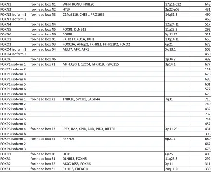
Table 1: Forkhead Box (Fox) Gene Subfamily Members. Each subfamily symbol, approved name, synonyms, human chromosome location, and protein size are listed. If a subfamily member contains more than one isoform it is also listed along with its protein size. This collection was compiled using the National Center for Biotechnology Information website.
Approved Symbol Approved Name Synonyms
Human Chromosome Location
Number of Amino Acids
FOXA1 forkhead box A1 HNF3A, TCF3A 14q21.1 472
FOXA2 isoform 1 forkhead box A2 HNF3B, TCF3B 20p11 463
FOXA2 isoform 2 457
FOXA3 forkhead box A3 HNF3G, FKHH3, TCF3G 19q13.32 350
FOXB1 forkhead box B1 HFKH-5, FKH5 15q22.2 325
FOXB2 forkhead box B2 bA159H20.4 9q21.2 432
FOXC1 forkhead box C1 ARA, IGDA, IHG1, FKHL7, IRID1, RIEG3, FREAC3, FREAC-3 6p25 553
FOXC2 forkhead box C2 FKHL14, MFH-1, MFH1, LD 16q24.1 501
FOXD1 forkhead box D1 FKHL8, FREAC4, FREAC-4 5q13.2 465
FOXD2 forkhead box D2 FKHL17, FREAC9, FREAC-9 1p34-p32 495
FOXD3 forkhead box D3 Genesis, HFH2, AIS1, VAMAS2 1p31.3 478
FOXD4 forkhead box D4 FKHL9, FREAC5, FREAC-5, FOXD4A 9p24.3 439
FOXD5 forkhead box D5 FOXD4L1, bA395L14.1 2q13 408
FOXD6 forkhead box D6 FOXD4L3 9q21.11 417
FOXE1 forkhead box E1 FKHL15, TITF2, FOXE2, TTF2, TTF-2, HFKH4, HFKL5 9q22 373
FOXE3 forkhead box E3 FKHL12, FREAC8 1p32 319
FOXF1 forkhead box F1 FKHL5, FREAC1, ACDMPV 16q24 379
FOXF2 forkhead box F2 FKHL6, FREAC2, FREAC-2 6p25.3 444
FOXG1 forkhead box G1
BF1, BF2, QIN, FKH2, HBF2, HFK1, HFK2, HFK3, KHL2, FHKL3, FKHL2, FKHL3, FKHL4, HBF-1, HBF-2, HBF-3, FOXG1A, FOXG1B, FOXG1C, HBF-G2
14q13 489
FOXH1 forkhead box H1 FAST1, FAST-1 8q24.3 365
FOXI1 isoform a forkhead box I1 HFH3, FKH10, HFH-3, FKHL10, FREAC6, FREAC-6 5q34 378
FOXI1 isoform b 283
FOXI2 forkhead box I2 FLJ46831 10q26.2 318
FOXJ1 forkhead box J1 FKHL13, HFH-4, HFH4 17q25.1 421
FOXJ2 forkhead box J2 FHX 12p13.31 562
FOXJ3 isoform 1 forkhead box J3 KIAA1041 1p34.2 622
FOXJ3 isoform 2 588
FOXK1 forkhead box K1 FOXK1L 7p22.1 733
FOXK2 forkhead box K2 ILF, ILF1, ILF-1 17q25 660
FOXL1 forkhead box L1 FKHL11, FREAC7, FKH6 16q24 345
FOXL2 forkhead box L2 BPES, BPES1, PFRK, POF3, PINTO 3q23 376
FOXM1 isoform 1 forkhead box M1 MPP2, TGT3, HFH11, HNF-3, INS-1, MPP-2, PIG29, FKHL16,
FOXM1B, HFH-11, TRIDENT, MPHOSPH2 12p13 801
FOXM1 isoform 2 763
FOXM1 isoform 3 748
FOXM1 isoform 4 748
22
Table 1 Continued
FOXN1 forkhead box N1 WHN, RONU, FKHL20 17q11-q12 648
FOXN2 forkhead box N2 HTLF 2p22-p16 431
FOXN3 isoform 1 forkhead box N3 C14orf116, CHES1, PRO1635 14q31.3 490
FOXN3 isoform 2 468
FOXN4 forkhead box N4 12q24.11 517
FOXN5 forkhead box N5 FOXR1, DLNB13 11q23.3 292
FOXN6 forkhead box N6 FOXR2 Xp11.21 311
FOXO1 forkhead box O1 FKHR, FOXO1A, FKH1 13q14.11 655
FOXO3 forkhead box O3 FOXO3A, AF6q21, FKHRL1, FKHRL1P2, FOXO2 6p21 673
FOXO4 isoform 1 forkhead box O4 MLLT7, AFX, AFX1 Xq13.1 505
FOXO4 isoform 2 450
FOXO6 forkhead box O6 1p34.2 492
FOXP1 isoform 1 forkhead box P1 MFH, QRF1, 12CC4, hFKH1B, HSPC215 3p14.1 677
FOXP1 isoform 2 114
FOXP1 isoform 3 676
FOXP1 isoform 4 693
FOXP1 isoform 5 601
FOXP1 isoform 6 577
FOXP1 isoform 8 679
FOXP2 isoform 1 forkhead box P2 TNRC10, SPCH1, CAGH44 7q31 715
FOXP2 isoform 2 740
FOXP2 isoform 3 432
FOXP2 isoform 4 732
FOXP2 isoform 5 714
FOXP2 isoform 6 457
FOXP3 isoform a forkhead box P3 IPEX, JM2, XPID, AIID, PIDX, DIETER Xp11.23 431
FOXP3 isoform b 396
FOXP4 isoform 1 forkhead box P4 hFKHLA 6p21.1 680
FOXP4 isoform 2 667
FOXP4 isoform 3 678
FOXQ1 forkhead box Q1 HFH1 6p25 403
FOXR1 forkhead box R1 DLNB13, FOXN5 11q23.3 292
FOXR2 forkhead box R2 MGC21658, FOXN6 Xp11 311
23
II. DESIGN, CONSTRUCTION AND PRELIMINARY CHARACTERIZATION OF A
MYC-TAGGED ALLELE OF THE ZEBRAFISH FORKHEAD BOX GENE, foxq1b
Abstract
Colorectal, pancreatic, and breast cancer are among the top five most deadly cancers. The transcription factor FOXQ1 is significantly upregulated in these cancers, yet many of the genes directly regulated by FOXQ1 are still unknown. Through preliminary research, our lab uncovered several genes upregulated in a foxq1b morphant, in which foxq1b was knocked down using a gene-specific morpholino. Since there are discrepancies with data generated through the use of morpholinos, a myc-tag sequence was engineered onto the C-terminus of the zebrafish foxq1b open reading frame in order to evaluate the previously acquired data utilizing chromatin immunoprecipitation by way of an anti-myc antibody. A transgenic fish, Tg(foxq1b:foxq1b-myc), was created that successfully transmitted the myc-tagged foxq1b allele, under the control of the endogenous foxq1b promoter, to its offspring. We confirmed that the transgenic DNA was transcribed successfully and that the resulting mRNA was translated into protein. This transgenic zebrafish line will be useful for characterizing genes directly regulated by foxq1b and assist in the analysis of other datasets, including those generated from morpholino knockdown of foxq1b. Tg(foxq1b:foxq1b-myc) will also be useful in characterizing AhR binding sites within the foxq1b promoter. AhR directly
24 benefit the scientific community by providing greater insight into the interactions of genes associated with known developmental pathways and cancer.
Introduction
One in four deaths in the United States is due to cancer, making cancer an enormous public health concern (Siegel 2014). Cancer was the leading cause of death for both men and women ages 40 and above in 2010, and it is estimated that there are over 4,500 new cancer diagnoses every day (Siegel 2014). Colorectal, pancreatic, and breast cancer are among the top five cancers that led to death in 2010. Although cancer death rates have continued to decline over the past 20 years (Siegel 2014), much is still unknown about the molecular and cellular mechanisms involved in cancer development and progression. Learning more about these mechanisms could aid in the discovery of new treatments and improved therapies for this disease.
Human forkhead box Q1 (FOXQ1) is a 403 amino acid transcription factor that contains a winged helix DNA binding domain, similar to other Fox gene family members (Carlsson and Mahlapuu 2002, Bieller 2001 and Hong 2001). FOXQ1 plays an important role in embryonic developmental processes including craniofacial development (Goering 2008), hair follicle formation (Driskell 2011), and gastrointestinal development (Verzi 2008). Overexpression of FOXQ1 has been found in many cancer types, including colorectal
25 the cell cycle (Kaneda 2010 and Feuerborn 2011). However, the genes directly regulated by FOXQ1 are still largely unknown.
Zebrafish have become a useful model organism for studying human development and disease. During its evolutionary history, zebrafish underwent a genome duplication resulting in two paralogs, called foxq1a and foxq1b, orthologous to mammalian FOXQ1. In zebrafish, foxq1b is upregulated through the AhR pathway when exposed to
2,3,7,8-tetrachlorodibenzo-p-dioxin (TCDD). However, no significant changes in foxq1a expression were observed upon TCDD exposure (Planchart and Mattingly 2010). In the mouse derived cell line, Hepa-1C1C7, foxq1 mRNA expression also significantly increased when exposed to TCDD (Thornley 2011). These results appear to suggest that zebrafish foxq1b more closely resembles the ancestral form of Foxq1, while foxq1a may have undergone
neofunctionalization. The foxq1b protein is 312 amino acids in size, and its protein sequence is similar to human FOXQ1.
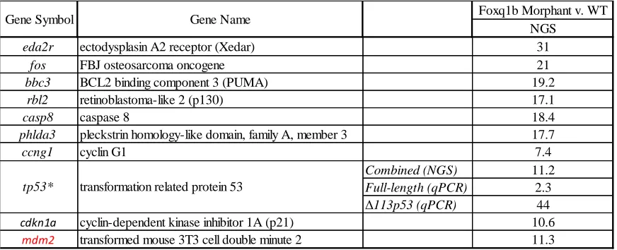
Preliminary research using foxq1b morphant zebrafish, in which foxq1b was knocked down using antisense morpholinos, indicates that downregulating foxq1b results in
upregulation of several genes involved in apoptosis and the cell cycle (Table 2). A few of the genes upregulated by knock down of foxq1b are Rbl2, Tp53, Eda2r, and Mdm2. Rbl2, also known as p130, is a gene associated with the regulation of the G0 to G1 transition of the cell
cycle (Jackson 2005). Rbl2 is also important for the transcriptional regulation of genes involved in the G2 phase of the cell cycle (Jackson 2005). Another gene upregulated in the
26 well as apoptosis in response to DNA damage (Agarwal 1995). Tp53 acquires a spectrum of mutations in the presence of cancer, which interfere with its regulatory mechanisms (Bennett 1999). Tp53 contains numerous isoforms, one of which is Δ133p53, found in humans and zebrafish. The full-length tp53 has been shown to transactivate the Δ113p53 isoform, which then antagonizes tp53-induced apoptosis creating a negative feedback loop (Chen 2009). Interestingly, in the foxq1b morphant data the Δ113p53 isoform is most significantly upregulated (44 fold) when foxq1b is knockdown, while the full-length tp53 is not strongly affected (2.3-fold upregulation). Eda2r, a gene significantly upregulated in the foxq1b morphant, is important during hair and sweat gland development in mammals (Kere 1996), regulation of ectodermal function, and induction of cell death (Brosh 2010). Eda2r is a known downstream target of p53, and is activated by p53 in cancer cells where it induces anoikis (Brosh 2010). Anoikis is a type of apoptosis in epithelial cells in response to detachment from the surrounding matrix (Wazir 2015), which occurs during EMT. The oncogene Mdm2, which is upregulated by the knockdown of foxq1b, is known for its anti-apoptotic effect through negative regulation of p53 (Momand 1992). The morphant data reveal that foxq1b is a negative regulator of the transcription of these genes, which correlates with the hypothesis that foxq1b is primarily a repressor of transcription.
27 further analysis of data derived from the foxq1b morphant, as well as characterize genes that are regulated directly by foxq1b.
The foxq1b-myc construct can also be useful in characterizing AhR binding site location on the foxq1b promoter. 2,3,7,8-tetrachlorodibenzo-p-dioxin (TCDD), also known as dioxin, is a polychlorinated compound that has been shown to activate the aryl hydrocarbon receptor (AhR). Zebrafish exposed to TCDD causes upregulation of foxq1b mRNA. A program called MatInspector (Genomatrix) was used to analyze 7 kilobases (kb) upstream from the zebrafish foxq1b transcription start site (TSS), and found seven potential AhR binding sites, called dioxin response elements (DRE) (Planchart and Mattingly 2010), which are located at -371, -1330, -1349, -4627, -5637, -7,060, and -7,425. DREs contain a core binding site consisting of the sequence: 5'-CGTG-3' (Lusska 1993). Since the
Tg(foxq1b:myc) zebrafish only contain the 5kb foxq1b promoter region, the foxq1b-myc expression level upon exposure to TCDD will further characterize the location of AhR binding to the foxq1b promoter.
28
Table 2: Knockdown of foxq1b increases expression of genes involved in the apoptotic pathway. Next generation sequencing (NGS) and RT-qPCR were used to generate a list of genes that increased expression in foxq1b morphants compared to wild-type zebrafish. The majority of the genes are proapoptotic. tp53*: The induction of full length tp53 and its isoform Δ113p53 from qPCR analysis is shown. The induction of both the full length and isoform combined from NGS analysis is shown. mdm2 (in red): only anti-apoptotic gene generated by NGS. (Preliminary research conducted by the Planchart Lab).
Foxq1b Morphant v. WT NGS
eda2r ectodysplasin A2 receptor (Xedar) 31
fos FBJ osteosarcoma oncogene 21
bbc3 BCL2 binding component 3 (PUMA) 19.2
rbl2 retinoblastoma-like 2 (p130) 17.1
casp8 caspase 8 18.4
phlda3 pleckstrin homology-like domain, family A, member 3 17.7
ccng1 cyclin G1 7.4
Combined (NGS) 11.2
Full-length (qPCR) 2.3
Δ113p53 (qPCR) 44
cdkn1a cyclin-dependent kinase inhibitor 1A (p21) 10.6
mdm2 transformed mouse 3T3 cell double minute 2 11.3 Gene Symbol
29
Materials and Methods
DNA preparation from Glycerol Stock
A fosmid (CH1073-460D17) was propagated in E. coli from a glycerol stock by streaking it on an agar plate supplemented with chloramphenicol (SIGMA C-0857) (12.5μg/ml) and incubating it for 20 hours at 37° C. CH1073-460D17 harbors an
approximately 6 kb fragment containing the promoter region (~5 kb) and open reading frame of zebrafish foxq1b. A single colony was cultured in a 15ml culture tube containing 2ml Lysogeny Broth (LB; 10g NaCl, 10g Tryptone, and 5g yeast extract in 1L of water adjusted to pH 7.5 and autoclaved) supplemented with 12.5μg/ml chloramphenicol for 20 hours at 37o C and 250 rpm. A mini-prep of the culture was performed using the QIAGEN Plasmid Mini kit (Cat. 12123). A restriction digest using HindIII (NEB Cat. R0104S) was performed in order to confirm that the fosmid carried the desired insert. Briefly, 3μl 10X buffer, 21μl water, 5μl mini-prep DNA (776ng/μl) and 1μl of enzyme were gently mixed by pipetting and incubated in a 37o C water bath for 4 hours. The digest was analyzed on a 0.8% agarose gel.
Engineering and cloning of foxq1b promoter sequence
All primers were purchased from Integrated DNA Technologies (IDT) and sequences are shown in Table 3. Forward (zf_foxq1b_KpnI_Prom_L) and reverse
(zf_foxq1b_NotI_3UTR_R) primers were designed to flank the 5.2kb foxq1b promoter region with KpnI and NotI restriction enzyme sites on the 5' and 3' ends, respectively. PCR
30 of mini-prep DNA (77.6ng/μl), 0.3μl Phusion DNA Polymerase (NEB Cat. M0530S), and 0.6μl dNTPs (10mM stock) were assembled in PCR reaction tubes. The thermal cycling conditions used were: 93o C x 2 minutes; 30 cycles at 93o C x 30 seconds, 55o C x 30 seconds, 72o C x 5 minutes; 72o C x 10 minutes. Amplification products were visualized on a 0.8% agarose gel and the correct amplicon was gel-extracted using a QIAGEN QIAquick Gel Extraction kit (Cat. 28704).
31 (2.1ng/μl), 1μl T4 Ligase (NEB Cat M0202S), 1.5μl 10X T4 Buffer, and 5.5μl water. The ligation reactions were incubated in the thermal cycler at 15o C for 16 hours. A
transformation was carried out by thawing 50μl of C2925 cells (NEB Cat C2925I) on ice for 10 minutes, then adding 1μl of the ligation reaction to the cell tube and flicking gently to mix. The tubes were placed on ice for 30 minutes, heat shocked for 45 seconds in a 42o C water bath and immediately placed back on ice for 5 minutes. 200μl of room temperature S.O.C. Outgrowth Medium (NEB Cat. B9020S) was added to each tube, and the tubes were incubated for 1 hour at 37o C and 250 rpm. 200μl of each tube were spread on pre-warmed agar plates supplemented with 100μg/ml Ampicillin (SIGMA Cat. A5354), and incubated at 37o C for 16 hours. Single colonies were placed in culture tubes containing 2ml LB
supplemented with 100μg/ml Ampicillin, and incubated for 20 hours at 37o C and 250 rpm.
Mini preps were performed to isolate the plasmid DNA following the QIAGEN mini kit protocol, and digested with KpnI HF and NotI HF, as previously described. One clone with the correct insert was selected and production was scaled up by inoculating 500mL of LB supplemented with ampicillin and culturing as described. A glycerol stock was made
containing 700μl of the bacterial culture, 150μl LB, and 150μl glycerol (Fisher Scientific Cat. 56-81-5), and stored at -80o C. Plasmid was purified from the remaining culture using the
QIAGEN maxi kit (QIAGEN Cat. 12163) as described by manufacturer. We confirmed the sequence of the foxq1b promoter inserted into pBluescriptII/SK+ (referred to as
32 Engineering and cloning of foxq1b open reading frame and myc-tag sequence Forward (zf_foxq1b_NotI_m-2ORF_L) and reverse
(zf_foxq1b_SacI_m-2ORFmyc_R) primers were designed to flank the 864bp foxq1b open reading frame, with NotI and SacI restriction sites, respectively. The reverse primer contained a myc-tag
sequence upstream of the SacI site. A PCR was performed as described above using 390ng of fosmid DNA and the following thermal cycling conditions: 93o C x 2 minutes; 30 cycles of 93o C x 30 seconds, 56o C x 30 seconds, 72o C x 2 minutes; 72o C x 10 minutes. The product
was analyzed on a 1.25% agarose gel to confirm size. A phenol-chloroform extraction was performed on the PCR amplicon as previously described. The foxq1b-myc open reading frame amplicon and the pBS_foxq1bP vector were digested with NotI HF and SacI (NEB Cat. R0156S), as previously described. Restriction enzymes were inactivated by incubating digests at 65o C for 20 minutes. Vector linearization was confirmed on a 1% agarose gel. The
digested pBS_foxq1bP vector and foxq1b-myc open reading frame amplicon were ligated and transformed as previously described. Single colonies were cultured in 15ml culture tubes containing 2ml LB and 100μg/ml Ampicillin, and incubated for 16 hours at 37o C and 250
rpm. Mini preps using the QIAGEN mini kit were performed on bacterial cultures in order to isolate the plasmid DNA, which was digested using NotI HF and SacI, as described
33 to as pBS_foxq1bP_foxq1b-myc) was sequenced by the GSL to confirm the myc-tagged foxq1b open reading frame sequence.
Engineering and cloning of foxq1b polyA signal sequence Forward (zf_foxq1b_SacI_polyAsignal_L) and reverse
(zf_foxq1b_SacI_polyAsignal_R) primers were designed to amplify the 600bp foxq1b polyA signal region flanked by SacI restriction enzyme sites. A PCR was performed on the fosmid DNA using the PCR and thermal cycle conditions previously stated. Correct product size was confirmed on a 1% agarose gel. A phenol-chloroform extraction was performed on the PCR product, as previously described. The foxq1b polyA signal DNA amplicon and
pBS_foxq1bP_foxq1b-myc vector were digested using SacI, for 2 hours at 37o C. Linearization of the vector was confirmed on a 1% agarose gel. An unexpected extra fragment was produced, and further analysis revealed that there was an additional SacI site located in the center of the polyA signal sequence. A PCR was performed on the fosmid DNA using HotMaster Buffer and Taq Polymerase (5 Prime Cat. 2200300). PCR and thermal cycling condition used were previously described. The amplicon was cloned into the TOPO TA vector (Invitrogen Cat. K4575J10) using 4 μl of amplicon, 1 μl of TOPO vector, and 1μl salt solution and incubating at room temperature for 10 minutes. Transformation was
performed using 50μl C2566 cells (NEB Cat. 2566I) following previously stated protocol. 50μl of the transformation solution was streaked on pre-warmed agar plates supplemented with 100μg/ml Ampicillin and incubated at 37o C for 20 hours. Single colonies were cultured
34 hours at 37o C and 250 rpm. Mini preps were performed following the QIAGEN mini kit to isolate the plasmid DNA from the cultures. The mini prep DNA was digested with SacI at 37o C for 3 hours. A 1% agarose gel identified mini preps that contained the TOPO vector and foxq1b polyA signal insert, and GSL confirmed proper DNA sequence.
Mutagenesis was performed in order to remove the internal SacI site. Forward
(zf_foxq1b_polyAsignal_mut_L) and reverse (zf_foxq1b_polyAsignal_mut_R) primers were designed to change the first guanine in the SacI sequence to a thymine. Mutagenesis was performed utilizing the QuikChange II XL Site-Directed Mutagenesis Kit (Agilent Technologies Cat. 200523) using 26ng of TOPO/polyA signal DNA, and 125ng of the
forward and reverse primers. The PCR conditions, DpnI restriction digest, and transformation of XL10-Gold Ultracompetent cells were carried out following the kit protocol. Cultures were made containing single colonies of bacteria, 2ml LB, and 100μg/ml Ampicillin, and incubated for 20 hours at 37o C and 250 rpm. Plasmid DNA was isolated by mini prep using the QIAGEN kit. The mini preps were digested using SacI, and incubated in a 37o C water bath for 3 hours. Successful mutagenesis was visualized on a 1% agarose gel and confirmed by DNA sequencing.
35 for 1 hour at 37o C and 5 minute at 70o C, in order to prevent self re-ligation. Ligation was performed using the dephosphorylated pBS_foxq1bP_foxq1b-myc and the foxq1b polyA signal insert. A transformation was carried out from the ligation reaction following the protocol previously described. Single colonies were cultured in culture tubes containing 2ml LB and 100μg/ml Ampicillin. Mini preps were performed on the bacterial cultures to isolate the plasmid DNA using the QIAGEN kit. The mini preps were digested using SacI, and incubated for 16 hours at 37o C. A 1% gel was made to confirm polyA insertion into the
vector, and renamed pBS_foxq1bP_foxq1b-myc_polyA.
36 Injection of foxq1b myc-tag construct
An I-SceI (NEB Cat. R0694S) restriction digest was performed using
pBS_foxq1bP_foxq1b-myc_polyA DNA at 37o C for 1 hour to liberate the targeting cassette from the remainder of the cloning vector. 1μl 1% phenol red (SIGMA P5530) was added to 10μl of the restriction digest, and 1.5μl of the injection mixture was loaded into a needle made from borosilicate glass capillaries (Sutter Instrument, Item #:BF120-94-10, Lot: 161356-3), which were pulled on a Sutter Instrument Flaming/Brown micropipette puller (Model P-87) with the following settings: P= 400, T= 605, pull= 45, velocity= 110, and time= 195. The tip of the needle was clipped with forceps so that 1.5nl of DNA was injected into the 1-cell stage of wild type (Wik x Wik) zebrafish embryos. Injected embryos were placed in a Petri dish containing 0.5X E2 Medium (1L of 0.5X E2 Medium includes 7.5ml NaCl (1000mM), 0.5ml KCl (500mM), 0.5ml MgSO4 (1000mM), 0.075ml KH2PO4
(1000mM), 0.05ml Na2HPO4 (500mM), 1ml CaCl2 (500mM), 0.35ml Na2HCO3 (1000mM),
990.025ml autoclaved H2O, and 0.5ml of 0.1% methylene blue) and incubated at 28o C. At
37 Foxq1b myc-tag potential founders
At three months, potential founders were genotyped. First, DNA was extracted from fin clips. Briefly, fish were anesthetized in individual cups filled with water and 500μl of buffered Tricaine (Western Chemical Inc. Cat. 200-226; 4mg/ml). A corner of the caudal fin of each zebrafish was sliced with a razor blade and placed in a PCR tube containing lysis buffer (2.5μl 10X Hot Master Buffer, 1.25μl 10mg/ml proteinase K (SIGMA Cat. P2308), and 21.25μl water). The fish was placed back in fresh water to recover. DNA was extracted from the fin clippings by vortexing, briefly centrifuging, and incubating the tubes at 55o C for 1 hour, followed by re-vortexing and centrifuging, and placing them back in the thermal cycler at 55o C for 30 minutes and 95o C for 15 minutes. PCR was performed as previously described, with HotMaster Buffer and Taq Polymerase using zf_foxq1b_ORFmyc_L (10μM stock) and 1μl zf_foxq1b_polyA_R (10μM stock) primers. The thermal cycler conditions used: 95o C x 45 seconds; 30 cycles at 95o C x 30 seconds, 55o C x 30 seconds, 72o C x 1 minute; 72o C x 10 minutes. PCR product was analyzed on a 2.5% agarose gel, wherein a 100bp would identify the endogenous foxq1b allele and a 150bp band would identify the foxq1b-myc.
Inheritance of myc-tag foxq1b DNA
Potential founders identified as described above were spawned with wildtype zebrafish. Pools of 15 embryos (48 hpf) were used for DNA extraction using 100μl NTES Mod2 (10mM Tris pH 8.0, 25mM EDTA, 50mM NaCl, and 0.5% SDS) per pool,
38 rocking until embryos dissolved (~1 hour). A phenol-chloroform extraction was performed following previous protocol. The pellets were resuspended in 70μl 1X TE buffer. PCR was conducted on extracted DNA using a forward primer (zf_foxq1b_ORFmyc_L) designed within the open reading frame of foxq1b and a reverse primer (zf_foxq1b_polyA1_R) designed within the polyA signal. As a control, -actin was amplified using Zf-BactinF and Zf-BactinR primers (10μM stock). Amplicons were analyzed on a 2.5% agarose gel to determine if the embryos inherited the myc-tag foxq1b construct. A 100bp band represented endogenous foxq1b, and a 150bp band represented myc-tag foxq1b if present.
Expression of myc-tag foxq1b RNA in founder offspring
39 5μl of 3M Sodium Acetate (pH 5.0) was added. The solution was vortexed and 125μl of 100% Ethanol was added. The tube was vortexed and placed at -20o C for 45 minutes.
Afterwards, the tube was placed in a 4o C centrifuge and centrifuged at 14K rpm for 10 minutes. The supernatant was removed, 500μl of 70% Ethanol was added to the pellet, and the tube was re-centrifuged at 14K rpm for 5 minutes at 4o C. The pellet was dried and resuspended in 15μl water. cDNA was made by adding 2μl Oligo d(T) (IDT; 50μM ), 2.8μl DNase I treated RNA (720.8ng/μl), and 7.2μl water in a PCR tube. The tube was placed in the thermal cycler at 80o C for 3 minutes, followed by the addition of 2μl 10X M MMLV Reverse Transcriptase (RT) Buffer, 1μl dNTPs (10mM stock), 0.25μl RNase Inhibitor Murine (NEB Cat. M0314S), 0.50μl M-MuLV Reverse Transcriptase (RT; NEB Cat.
M0253S), and 4.25μl water. The contents were gently mixed and placed in the thermal cycler at 42o C for 1 hour, followed by a 10-minute incubation at 92o C. The RT reactions were
supplemented with 40μl, and 2μl were used for PCR following the PCR and thermal cycler conditions described in the Foxq1b myc-tag potential founders section, but using 40 cycles instead of 30. A 2.5% agarose gel was made to confirm the presence of foxq1b-myc RNA.
40 4 hours, and then rinsed in 1ml of 1X PBST (Phosphate Buffered Saline (Fisher Scientific Cat. BP665-1), 0.1% Tween-20 (Fisher Scientific Cat. 9005-64-5)) at room temperature. The PBST was removed and the embryos were washed twice with methanol, after which they were stored at -20o C in 1ml of fresh methanol. The embryos were serially rehydrated in 95% MeOH + 5% PBST, 75% MeOH + 25% PBST, 50% MeOH + 50% PBST, and 25% MeOH + 75% PBST, followed by washing four times in 1ml PBST for 5 minutes. Embryos were permeabilized by adding 1ml of ice-cold acetone (Fisher Scientific Cat. 67-64-1) and
incubating for 8 minutes at -20o C. The embryos were again washed four times in 1ml PBST for 5 minutes and blocked in 1ml block (PBST + 5% Sheep Serum (SIGMA Cat. S3772)) by rocking for 1 hour at room temperature. Anti-myc primary antibody (Invitrogen, Cat. 46-0603) was placed on the embryos at a 1:100 dilution in block, and rocked at 4o C overnight. The next day, the antibody was removed and embryos were washed five times at room temperature in 1ml block for 10 minutes per wash, followed by three washes in 1ml PBST for 10 minutes. A 1:1000 dilution of Alexa Fluor 594 donkey anti-mouse secondary
41 with1X PBS once the agarose solidified. Imaging was performed using confocal microscopy on a Zeiss LSM-720 located at North Carolina State University Cellular and Molecular Imaging Facility.
AhR regulation of foxq1b-myc
DMSO or 3nM TCDD was exposed to 6 hpf foxq1b-myc embryos for 24 hours. RNA was extracted from foxq1b-myc embryos (4 replicates of 15 pooled embryos) at 48 hpf, quantified using the Agilent Bioanalyzer, normalized, and reverse transcribed to make cDNA. qPCR analysis was performed using GAPDH left and GAPDH right primers for normalization, cyp1a1_L and cyp1a1_R primers for amplification of cyp1a (a known AhR target), zf_foxq1b_qpcrL2 and zf_foxq1b_qpcrR2 primers for amplification of foxq1b (both endogenous and foxq1b-myc), and zf_foxq1b_ORF1_L and zf_foxq1b_myc _R primers for the amplification of foxq1b-myc. The qPCR reaction included 5.125μl water, 12.5μl 2X Brilliant II SYBR Green QPCR Master Mix, 0.375μl ROX (1:500), 1μl forward primer (10μM stock), 1μl reverse primer (10μM stock), and 5μl cDNA. A Comparative Quantitation (Calibrator) qPCR experiment using MxPro software was performed using the following thermal parameters: 95o C x 10 minutes; 50 cycles of 95o C x 30 seconds, 55o C x 30 seconds,
42
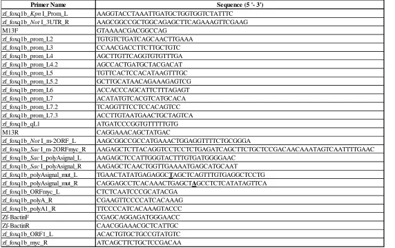
Table 3: Primer names and sequences used to create foxq1b-myc construct. List of all the primer names and sequences that are mentioned in the methods section. *Bold and underlined nucleotides are the nucleotides mutated through mutagenesis.
Primer Name Sequence (5 '- 3')
zf_foxq1b_KpnI_Prom_L AAGGTACCTAAATTGATGCTGGTGGTCTATTTC zf_foxq1b_NotI_3UTR_R AAGCGGCCGCTGGCAGAGCTTCAGAAAGTTCGAAG M13F GTAAAACGACGGCCAG zf_foxq1b_prom_L2 TGTGTCTGATCAGCAACTTGAAA zf_foxq1b_prom_L3 CCAACGACCTTCTTGCTGTC zf_foxq1b_prom_L4 AGCTTGTTCAGGTGTGTTTGA zf_foxq1b_prom_L4.2 AGCCACTGATGCTACGACAT zf_foxq1b_prom_L5 TGTTCACTCCACATAAGTTTGC zf_foxq1b_prom_L5.2 GCTTGCATAACAGAAAGAGTCG zf_foxq1b_prom_L6 ACCACCCAGCATTCTTTAGAGT zf_foxq1b_prom_L7 ACATATGTCACGTCATGCACA zf_foxq1b_prom_L7.2 TCAGGTTTCCTCCACAGTCC zf_foxq1b_prom_L7.3 ACCTTGTAATGAACTGCTAGTCA zf_foxq1b_qL1 ATGATCCCGGTGTTTTTGTG M13R CAGGAAACAGCTATGAC
zf_foxq1b_NotI_m-2ORF_L AAGCGGCCGCCATGAAACTGGAGGTTTTCTGCGGGA
zf_foxq1b_SacI_m-2ORFmyc_R AAGAGCTCTTACAGGTCCTCCTCTGAGATCAGCTTCTGCTCCGACAACAAATAGTCAATTTTGAAC zf_foxq1b_SacI_polyAsignal_L AAGAGCTCCATTGGGTACTTTGTGATGGGGAAC
zf_foxq1b_SacI_polyAsignal_R AAGAGCTCAACTGGTTGAAAATGAGCATGCAAT
43
Table 3 Continued
Primer Name Sequence (5' - 3')
44
Results
A 6.7 kb fragment of the locus encoding the zebrafish transcription factor, foxq1b (Chr. 2) consisting of the complete ORF, polyA signal, and 5.2kb directly upstream of the transcription start site, was successfully cloned into the multiple cloning site (MCS) of pBluescriptII/SK+. A myc-tag was engineered in frame with the 3’end of the ORF, directly before the stop codon (Figure 1) to facilitate downstream applications, including
immunohistochemistry and ChIP-seq analysis. Intermediate steps of the cloning strategy are summarized in Figure 2.
The pBluescriptII/SK+ plasmid was previously modified to contain two I-SceI meganuclease restriction enzyme sites flanking the MCS (kind gift from Dr. Jochen
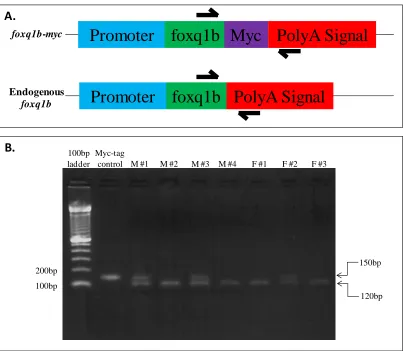
Wittbrodt, Heidelberg). The purpose of these sites is to allow excision of the cassette – in this case the engineered foxq1b-myc gene – from the vector before injecting the construct into embryos. Meganuclease restriction sites are not found in zebrafish; therefore, the digest can be injected directly into the embryo without the need to inactivate the I-SceI enzyme. 1.5nl of the digest was injected into 500, 1-cell stage, wild-type zebrafish embryos. The fish were reared to adulthood and genotyped via fin clip to verify that the cassette had been integrated in the genome (Figure 3). The primer strategy in Figure 3A was utilized to identify 9
potential founders that harbored the transgene, representing about an 18% success rate since many of the injected embryos did not survive till adulthood.
45 endogenous foxq1b and foxq1b-myc in the offspring. Of the nine potential founders only one, a female, transmitted the transgene to its offspring (Figure 4). The F1 embryos from this founder were reared to adulthood.
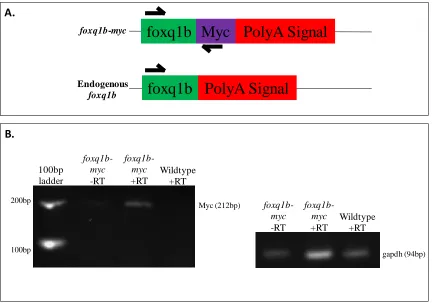
In order to confirm that the transgene-positive F1 embryos expressed the transgene, 90 embryos were pooled for RNA extraction and reverse transcribed. A forward primer was designed within the foxq1b open reading frame and a reverse primer designed within the myc sequence to validate transcription of the transgene (Figure 5).
Translation of the transgene mRNA was assessed in 48 hpf embryos by
immunohistochemistry, using a mouse anti-myc monoclonal primary antibody detected by a donkey anti-mouse secondary antibody labeled with Alexa Fluor 594. DAPI was used to counter-stain nuclei. Since foxq1b is a transcription factor and presumably located within the nucleus, DAPI enabled us to visualize nuclei and ascertain if foxq1b localized to the nucleus (Figure 6). The embryos were imaged at 40X magnification by confocal microscopy. We observed that foxq1b-myc indeed localized to several nuclei near the otic vesicle and within the jaw region (Figure 6).
47
Figure 1: Cloning strategy of the foxq1b-myc construct in the pBluescriptII/SK+ plasmid. The cloning strategy is described in Materials and Methods.
Sacl
Myc
Sacl
Kpnl Notl
Promoter
foxq1b
PolyA Signal
5,228bp 864bp 30bp 645bp
I-SceI
48
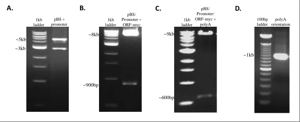
Figure 2: Intermediate steps in the cloning strategy as visualized by gel electrophoresis. A. Restriction digest with KpnI and NotI confirms the insertion of the ~5kb foxq1b promoter into the ~3kb plasmid. B. Restriction digest with NotI and SacI confirms the insertion of the ~900bp foxq1b open reading frame modified to carry a myc-tag into the ~3kb plasmid containing the ~5kb promoter (~8kb total). C. Restriction digest with SacI confirms the insertion of the ~600bp foxq1b polyA signal into the ~3kb plasmid containing the ~5kb promoter and ~900bp open reading frame (~9kb total). D. PCR amplification using a forward primer that resides in the foxq1b open reading frame and a reverse primer that resides in the polyA signal confirmed proper polyA signal orientation due to the production of a 977bp fragment.
A.
B.
C.
49
Figure 3: Identification of foxq1b-myc potential founders.A. Primer strategy: Forward primer was designed within the open reading frame of foxq1b and the reverse primer was designed within the polyA signal. The primers are designed to amplify a 150bp product from foxq1b-myc and a 120bp product from the endogenous locus. B. Genotypes are confirmed by gel electrophoresis PCR products derived from nine fish. The presence of two fragments represent potential founders containing both foxq1b-myc and endogenous foxq1b. Plasmid DNA containing foxq1b-myc (see Figure 1) was diluted and used as a positive control.
foxq1b-myc
Endogenous foxq1b
Promoter
foxq1b Myc PolyA Signal
PolyA Signal
foxq1b
Promoter
A.
B. 100bp
ladder
Myc-tag control
150bp
120bp 100bp
200bp
50
Figure 4: Transmission of foxq1b-myc to offspring by female founder #2.Embryos were pooled into three pools of 15 embryos each. Genomic DNA was extracted from each pool and used for PCR to confirm the transmission of the foxq1b-myc transgene from female #2. The strategy used to distinguish between the endogenous and transgenic loci is described in Figure 3B.
100bp
ladder
Myc-tag
control
foxq1b-myc
founder embryo pool:
1 2 3
100bp 200bp
150bp
51
Figure 5: Transgene-positive offspring transcribe foxq1b-myc mRNA. A. Primer
strategy: Forward primer was designed within the foxq1b open reading frame, reverse primer was designed within the myc-tag sequence. If the foxq1b-myc mRNA is present, a band at 212bp will be produced by RT-PCR. If the foxq1b-myc RNA is not present, no band will be produced. B. Gel electrophoresis of RT-PCR products. RNA was DNase-treated prior to reverse transcriptase reaction as described in Materials and Methods. –RT: no reverse transcriptase. +RT: reverse transcriptase; gapdh was used as a positive control.
foxq1b-myc
+RT
foxq1b-myc
Endogenous
foxq1b
foxq1b Myc PolyA Signal
52
Figure 6: Foxq1b-myc protein is nuclearly localized to cells in the periphery of the otic vesicle and jaw region. Immunohistochemistry was utilized as described in Materials and Methods to detect foxq1b-myc protein in 48 hpf embryos at 40X magnification using
53
Eye
Eye
Otic
Vesicle
Otic
Vesicle
A.
54
Ventral jaw
Ventral jaw
55
Figure 7: RT-qPCR of 48 hpf foxq1b-myc zebrafish exposed to DMSO or 3nM TCDD.
Each bar represents the average of four biological replicates consisting of 15 embryos each. Foxq1b primers would amplify both the endogenous, as well as foxq1b-myc. Foxq1b-myc reverse primer included the myc-tag in order to only amplify the foxq1b-myc transgene. Cyp1a1 was upregulated 128-fold, and endogenous foxq1b 4-fold, whereas foxq1b-myc showed no significant change.
DMSO TCDD DMSO TCDD DMSO TCDD
56
Discussion
A whole genome duplication occurred in the evolutionary history of zebrafish, which most likely explains the existence of two paralogs of the foxq1 transcription factor, namely foxq1a and foxq1b. The expression profile of mammalian FOXQ1 and the effects caused by inactivating it (Goering 2008) are closely mirrored in zebrafish foxq1b, whereas this is not the case for foxq1a. In fact, foxq1a is maternally deposited and generates no phenotype when knocked down using morpholinos. In addition, its expression domain has been largely undetectable (A. Planchart, personal communication). Therefore, based on these observations, foxq1b has been the primary focus of our lab since it appears to be more functionally similar to the human FOXQ1 gene.
An engineered allele of foxq1b, under the control of the foxq1b promoter, designed to encode a modified protein in which the C-terminus is tagged with the myc epitope, is
hypothesized to be expressed in the same cells as the endogenous foxq1b gene. The myc-tag was engineered to not interfere with the function of foxq1b, since the C-terminal tail plays no known role in the binding and regulation of foxq1b. This engineered allele of foxq1b was successfully integrated into the zebrafish genome and intergenerationally transmitted from founder to offspring, in which it was transcribed, and translated. This transgenic line will be useful for further studies of the role FOXQ1 plays in both embryonic development and cancer.
57 development and cancer. Initial transcriptomic analysis of the foxq1b morphant yielded a list of genes that are potential downstream targets of foxq1b and play significant roles in both development and carcinogenesis; however, whether they are direct or indirect targets remains a subject of speculation. Thus, the foxq1b-myc transgenic line may prove valuable for further characterization of these genes, especially by chromatin immunoprecipitation sequencing (ChIP-seq). Using this procedure, genomic DNA cross-linked to foxq1b-myc protein can be isolated using anti-myc antibodies, reverse cross-linked and sequenced, thus helping to validate morpholino data by determining if foxq1b regulates these genes directly by binding to their promoter regions.
A common thread tying several genes within the morphant dataset is the observation that they play a role in apoptosis, which is necessary for proper embryonic development (Brill 1999). Disruption of this pathway can lead to abnormalities during development, as well as contribute to cancer formation and progression later in life, presumably because cells with mutations arising early in development are not eliminated but remain and cause havoc later in life. Determining if foxq1b directly binds and regulates these genes could provide greater evidence about its function in development and cancer.