Western University Western University
Scholarship@Western
Scholarship@Western
Electronic Thesis and Dissertation Repository
1-31-2014 12:00 AM
Surface Functionalization and Bioconjugation of Nanoparticles
Surface Functionalization and Bioconjugation of Nanoparticles
for Biomedical Applications
for Biomedical Applications
Longyan Chen
The University of Western Ontario
Supervisor Jin Zhang
The University of Western Ontario
Graduate Program in Chemical and Biochemical Engineering
A thesis submitted in partial fulfillment of the requirements for the degree in Doctor of Philosophy
© Longyan Chen 2014
Follow this and additional works at: https://ir.lib.uwo.ca/etd
Part of the Analytical Chemistry Commons, Biochemical and Biomolecular Engineering Commons,
Bioimaging and Biomedical Optics Commons, Biological Engineering Commons, Biomaterials Commons, and the Materials Chemistry Commons
Recommended Citation Recommended Citation
Chen, Longyan, "Surface Functionalization and Bioconjugation of Nanoparticles for Biomedical Applications" (2014). Electronic Thesis and Dissertation Repository. 1903.
https://ir.lib.uwo.ca/etd/1903
This Dissertation/Thesis is brought to you for free and open access by Scholarship@Western. It has been accepted for inclusion in Electronic Thesis and Dissertation Repository by an authorized administrator of
SURFACE FUNCTIONALIZATION AND
BIOCONJUGATION OF NANOPARTICLES FOR
BIOMEDICAL APPLICATIONS
(Thesis format:
Integrated Article
)
by
Longyan
Chen
Graduate Program in Chemical and Biochemical Engineering
A thesis submitted in partial fulfillment
of the requirements for the degree of
Doctor of Philosophy
The School of Graduate and Postdoctoral Studies
The University of Western Ontario
London, Ontario, Canada
ii
Abstract
Colloidal inorganic nanoparticles (NPs) have been attracting considerable interest in
biomedicine, from drug and gene delivery to imaging, sensing and diagnostics. It is
essential to modify the surface of nanoparticles to have enhanced biocompatibility and
functionality for the in vitro and in vivo applications, especially in delivering locally and
recognizing biomolecules. Herein, the goal of this research work is to develop advanced
NPs with well-tailored surface functionalities and/or bio-functionality for the applications
in cell tracking and analytes detection.
In the first project, quantum dots incorporating with gelatin nanoparticles (QDs-GNPs)
have been developed for bioimaging applications. Two different approaches have been
developed, i.e. directly encapsulating QDs with gelatin polymer (QDs-GNP1) and
layer-by-layer (LBL) adsorption of QDs approach (QDs-GNP2), respectively. The special
hybrid structures of two QDs-GNPs nanosystems were investigated by transmittance
electron microscopy, scanning electron microscopy, X-ray energy dispersion, Fourier
Transform Infrared spectroscopy, fluorometer, and confocal fluorescent microscopy. Both
nanosystems exhibit high intensive luminescence and good biocompatibility. Compared
to free QDs, QDs-GNP2 shows improved quantum yield and longer lifetime, due to
multiple layers of polyelectrolytes protection. Furthermore, QDs-GNP2 demonstrates the
proton-resistant properties in term of PL intensity and lifetime. The bright and stable
photoluminescence (PL) allows the QD-GNP2 for labeling living 3T3 cells in vitro,
which may indicate the QDs-GNP2 are able to be a suitable candidate for bio-imaging
application.
In the second project, fluorescent magnetic nanoparticles (FMNPs) were bioconjugated
with gentamicin (Gm) for rapid capture, detection and decontamination of bacteria. The
Gm-FMNPs consist of a fluorescent silica shell and an iron oxide magnetic core. Initially,
iii
found 20 % higher than that of the free antibiotic. We further improved NPs stability and
capture efficiency by a two-step thermal decomposition method to produce the
fluorescent magnetic core-shell nanoparticles. It is noted that one mg of gentamicin
conjugated FMNPs are able to capture both gram-negative bacteria Escherichia coli and
gram-positive bacteria Staphylococcus aureus as low as 1x10⁴ colony-forming unit/mL
(cfu/mL) in less than one minute. It is expected that the Gm-FMNPs could be a promising
multifunctional platform for disease control in clinic and wastewater treatment.
In addition, a nanosensor for detecting human thrombin has been designed and
developed. A recombinant luciferase was covalently conjugated to gold nanoparticles (Au
NPs) through 1-Ethyl-3-(3-dimethylaminopropyl) carbodiimide (EDC) mediated reaction.
The conjugation enables Au NPs to quench the bioluminescence produced by luciferase.
Recovery of bioluminescence has been studied as a function of the concentration of
thrombin. The results indicate that the designed nanosensor can efficiently detect a
protease thrombin in both buffer and human urine spiked buffer. A linear assay response
is found between 300 ng/mL to 300 µg/mL, with limit of detection (LOD) of 3 ng/mL in
both buffer and human urine spiked samples. The assay time can be less than 15 min. The
nanosensor can thus be a promising tool for clinical diagnostic of thrombin related
diseases.
In summary, different strategies have been explored to engineer the surface of hybrid NPs
with good water stability, biocompatibility and enhanced chemical and physical
properties. The thesis addresses that engineered hybrid NPs in biomedicine and how the
biofunctionalized NPs can find applications in imaging and diagnostics indistinctly.
iv
Co-Authorship Statement
Chapters 1 and 2 entitled “Introduction” and “Background and Literature Review”,
respectively, were written by Longyan Chen, with suggestions from Dr. Jin Zhang. Figure
2.1 is adapted with permission from (Kelly et. al., Journal of Physical Chemistry B,
107(3): P. 668-677). Copyright (2002) American Chemical Society."
Chapter 3, 4 and 5 encompass research studies have been published or are in preparation
for publication. Individual contributions of the author of each article are stated below.
Chapter 3:
The experiment work and draft of manuscript related in preparation of QDs-GNP1 was
conducted by co-operation with Adrienne Willoughby (a former undergraduate student
supervised by Dr. Jin Zhang at Chemical and Biochemical Engineering Department, the
University of Western Ontario). The manuscript was drafted by the author and reviewed
several times by Dr. Jin Zhang. This work was supervised by Dr. Jin Zhang.
This chapter encompass two research studies have been published or
submitted.
•
The experiment work of QDs-GNP2 was achieved by co-operation of Dr. Alex
Siemiarczuk (Photon Technology International Inc, London, Canada), Dr. Hong Hai, Dr.
Yi Chen, and Guobang Huang (Chemical and Biochemical Engineering Department, the
University of Western Ontario). The manuscript of this part was drafted by the author and
reviewed by Dr. Jin Zhang extensively. This work was supervised by Dr. Jin Zhang. Longyan Chen, Adrienne Willoughby and Jin Zhang (2013) Luminescent Gelatin
Nanospheres by Encapsulating CdSe Quantum Dots, Luminescence, 29, 74-79 ..
• Longyan Chen, Alex Siemiarczuk, Hong Hai, Yi Chen, Guobang Huang and Jin
Zhang, Development of Biocompatible and Proton-resistant Quantum Dots
Assembled on Gelatin Nanospheres, Langmuir (2014) Accepted.
v
In the part of the experiment work relating to one-pot preparation of Gm-FMNP, the
experiment work was achieved by the author with the co-operation with Dr. Fereidoon S.
Razavi (Brock University), Abdul Mumin (Chemical and Biochemical Engineering
Department, the University of Western Ontario), Xiaoxuan Guoand Dr
supervised by Dr. Jin Zhang.
•
Capture, Detection, and Decontamination, RSC Advances, 3, 2390-2397.
In the work relating to the preparation of Gm-FMNPs through two-step method and study
of their bacterial capture efficiency, the experiment work, analysis of data and manuscript
were prepared by the author. It was reviewed by Dr. Jin Zhang, who also provided a
series of revision steps for improvement.
• Longyan Chen and Jin Zhang (2012) Bioconjugated Magnetic Nanoparticles for Rapid Capture of Gram-positive Bacteria, Journal of Biosensor and Bioelectronics.
S11:005.
Chapter 5:
•
The experiment work was conducted by Longyan Chen under supervision of
Dr. Jin Zhang. Both authors analyzed the data. Various drafts of the manuscript were
reviewed by Dr. Jin Zhang. Mr. Yige Bao provided urine samples and advices for
pretreatment of urine samples. This work was supervised by Dr. Jin Zhang.
Longyan Chen
Chapter 6 was written by Longyan Chen, with the suggestions from Dr. Jin Zhang.
, Yige Bao and Jin Zhang (2013) “Luciferase Conjugated Gold
vi
To my wife, Shan
for her love, patience and encouragement.
vii
Acknowledgements
This thesis would not have been in the current form without the help and guidance from
several people. I would, therefore, like to offer my sincere thanks to all of them.
I would like to extend my heartfelt gratitude to my supervisor Dr. Jin Zhang, for her
enthusiastic support and continued guidance throughout the course of my PhD research.
The encouragement and support she has provided me over the past years have been and
will continue to be indispensable for my professional development.
I would like to thank my examination committee: Drs. Amarjeet Bassi, Charles
(Chunbao) Xu, Lyudmila Goncharova and Yuning Li for their guidance and feedback
throughout this endeavour. I would like to express my thanks to my committee board Drs.
Anand Prakash and Tim A. Newson for their time in reviewing my annual progress report
and providing useful suggestions for my research.
My special thanks to Dr. Richard Gardiner and Ms. Nygard Karen in Biotron center for
their assistants in using of the facilities. I appreciate very much for the advices from Dr
David Litchfield, Dr Susan Koval, and Ms. Judy Sholdice for discussing part of the work
in Chapter 3.
Special gratitude is extended to all of my current and previous colleagues at
Multifunctional Nanocomposite Lab, Dr. Yi Chen, Guobang Huan, Andrew Tse, Dr.
Robert Bi, Pei Yin, Abdule Md. Mumin, Dr. Hong Hai, Kazi Farida Akhter, Anu Alice
Thomas, Adrienne Willoughby and Kim Hyung, for their continuous support,
encouragement, and nice time that we have had together.
Finally, I would dearly like to thank my family for their support and encouragement. My
wife, Shan, deserves much credit for her immeasurable love, support, and prayer for me.
This work has been supported by funding from Natural Science and Engineering
Research Council of Canada (NSERC) through a Discovery Grant and Western
viii
Table of Content
Abstract ... ii
Co-Authorship Statement... iv
Acknowledgements ... vii
Table of Content ... viii
List of Figures ... xiii
List of Tables ... xvi
List of Abbreviations ... xvii
CHAPTER 1 ... 1
GENERAL INTRODUCTION AND MOTIVATION ... 1
1.1 Overview of nanoparticles in biomedical applications ... 2
1.1.1 Nanoparticles as imaging agents and biosensing transducer ... 4
1.1.2 Fluorescence resonance energy transfer as readout ... 5
1.2 Limitations of using NPs in biomedical fields ... 7
1.3 Aims and Objectives ... 8
1.4 References ... 9
CHAPTER 2 ... 13
BACKGROUND AND LITERATURE REVIEW ... 13
2.1 Colloidal Inorganic Nanoparticles for Biomedical Application ... 14
2.1.1 Noble metal NPs ... 14
2.1.2 Semiconductor quantum dots ... 16
2.1.3 Magnetic Nanoparticles ... 18
2.2 Synthesis of colloid NPs for biomedical use ... 21
2.2.1 Synthesis of NPs in water ... 22
ix
2.3 Surface Functionalization of NPs ... 23
2.3.1 Ligand exchange ... 24
2.3.2 Ligand modification ... 26
2.3.3 Ligand addition ... 27
2.4 Bioconjugation ... 29
2.4.1 Direct absorption ... 29
2.4.2 Covalent coupling ... 30
2.4.3 Biological approaches... 33
2.5 Summary ... 35
2.6 References ... 36
CHAPTER 3 ... 48
DEVELOPMENT OF BIOCOMPATIBLE LUMINESCENT NANOPARTICLES FOR BIOIMAGING APPLICATIONS ... 48
3.1 Introduction ... 49
3.2 Experimental procedures ... 50
3.2.1 Materials and Reagents ... 50
3.2.2 Preparation of hydrophilic CdSe QDs ... 51
3.2.3 Preparation of gelatin NPs ... 51
3.2.4 Preparation of QDs-GNP1 by direct encapsulation of QDs in GNPs. ... 52
3.2.5 Preparation of QDs-GNP2 ... 53
3.2.6 Characterization ... 54
3.2.7 Analysis of pH effect in photostability ... 54
3.2.8 Cell viability/cytotoxicity study ... 55
3.2.9 In vitro fluorescence imaging ... 55
3.3 Results and Discussion (I) ... 56
x
3.3.2 Cytotoxicity ... 59
3.4 Results and Discussion (II) ... 60
3.4.1 Materials characterization ... 61
3.4.2 Absorbance and PL spectra ... 62
3.4.3 The pH effects on photostability ... 63
3.4.4 Cytotoxicity analysis of QDs-GNP2 ... 65
3.4.5 In vitro bio-imaging application ... 66
3.5 Conclusions and Prospects ... 67
3.6 References ... 67
CHAPTER 4 ... 72
DEVELOPMENT OF BIOCONJUGATED MAGNETIC LUMINESCENT NANOMATERIALS FOR BACTERIAL CAPTURE, DETECTION AND ANTIBACTERIAL APPLICATIONS ... 72
4.1 Introduction ... 73
4.2 Experimental ... 75
4.2.1 Preparation of FMNP1 by one-pot method ... 76
4.2.2 Preparation of FMNP2 by two-step coating method ... 77
4.2.3 Bioconjugation of FMNPs with Gm ... 78
4.2.4 Characterization ... 79
4.2.5 Bacteria Capture ... 79
4.2.6 Analysis of the antibacterial effect ... 80
4.2.7 Determine the concentration of gentamicin on the FMNP1 ... 80
4.2.8 Preparation of thin section samples for TEM analysis ... 81
4.3 Results and Discussion ... 82
4.3.1 Characterization ... 82
xi
4.3.3 Ubiquitous capture of bacteria by Gm-FMNP2 ... 93
4.4 Conclusion ... 94
4.5 References ... 95
CHAPTER 5 ... 100
DEVELOPMENT OF LUCIFERASE CONJUGATED GOLD NANOPARTICLES IN DETECTION OF THROMBIN ... 100
5.1 Introduction ... 101
5.2 Experimental section ... 104
5.2.1 Design of the pRluc-Au NPs nanosensor ... 104
5.2.2 Dihydrolipoic acid (DHLA) synthesis ... 105
5.2.3 Synthesis and functionalization of Gold Nanoparticles ... 105
5.2.4 Plasmid constructions ... 106
5.2.5 Protein Expression and Purification ... 106
5.2.6 Bioconjugation of Au NPs by pRluc protein ... 107
5.2.7 Characterization ... 107
5.2.8 Thrombin assay... 108
5.3 Results and Discussion ... 108
5.3.1 Characterization of the pRluc-Au NP conjugate ... 108
5.3.2 Assay optimization ... 109
5.3.3 Determination of thrombin concentration in both buffer and urine samples 113 5.4 Conclusions and Prospects ... 114
5.5 References ... 114
CHAPTER 6 ... 118
SUMMARY AND RECOMMENDATIONS... 118
6.1 Summary and Conclusion ... 119
xii
6.3 Limitations of the Research and Suggestions for Future Studies ... 125
6.4 References ... 126
Appendices ... 129
Appendix 1. Quantification of QD coated onto GNs ... 130
Appendix 2. pH value effects on fluorescence decay of QDs and core-shell QDs-GNP2 ... 131
Appendix 3. XANES spectra of FMNPs ... 136
Appendix 4. Determine the concentration of gentamicin on Gm-FMNP1. ... 137
Appendix 5. Plasmid map of recombinant protein pRluc ... 138
Appendix 6. TEM image of Au NPs and SDS-PAGE of pRluc ... 139
Appendix 7. Copyright permission ... 140
xiii
List of Figures
Figure 1.1 Schematic illustration of FRET process 6
Figure 1.2 Strategy for bridging the unique feature of nanoparticles to
biomedical applications
8
Figure 2.1 Schematic illustration of the SPR effect. 15
Figure 2.2 Size dependent photophysical properties of semiconductor
quantum dots
17
Figure 2.3 The responses of materials to magnetic field. 18
Figure 2.4 Schematic illustration of common surface functionalization
strategies
24
Figure 2.5 Common ligands for stabilizing NPs in water 25
Figure 2.6 Representive bioconjugation protocols 31
Figure 2.7 Reaction scheme for (a) EDC-NHS crossliking and (b) SMCC
mediated conjugation
33
Figure 2.8 Mechanism of intein mediated bioconjugation 34
Figure 3.1
Figure 3.2
Schematic illustration of preparation of QD-GNP1
Schematic illustration of preparation of QDs-GNP2 by LBL
technique.
52
53
Figure 3.3 TEM micrograph of (a) MUA-CdSe QDs and (b) QD-GNP1. 56
Figure 3.4 FTIR spectra of MUA-QDs, GNPs and QD-GNP1 57
Figure 3.5 Fluorescent spectra of CdSe QDs, GNPs and QD-GNP1 58
Figure 3.6 Confocal fluorescent of microscopy images of QD-GNP1 59
Figure 3.7 Cytotoxicity analysis of GNPs, MUA-QDs and QD-GNP1 60
Figure 3.8 Characterization of QDs-GNP1 by electron microscopy 61
Figure 3.9 FTIR spectra of bare (i) GNPs, (ii) PE coating GNPs, (iii) single
layer QDs coating GNPs, (iv) multilayer QDs coating GNPs, and
(v) free MUA-QDs.
62
Figure 3.10 (a) Absorbance and (b) PL spectra for MUA-QDs and
corresponding QDs-GNP2
63
xiv
Figure 3.12 Cytotoxicity profile of MUA-QDs and corresponding
QDs-GNP2 after 24 h incubation with 3T3 cells.
65
Figure 3.13 Confocal microscopy images of (a) GNPs (0.3 mg/mL), (b)
MUA-QDs (0.1 mg/mL), and (c) QDs-GNP2 (0.4 mg/mL) after
co-incubation with NIH/3T3 mouse fibroblast cells.
66
Figure 4.1 Schematic illustration of the synthesis of FMNPs used for E. coli
capture and magnetic separation in solution
76
Figure 4.2 TEM micrograph of the core–shell structures of (a) FMNP1 and
(b) FMNP2 .
83
Figure 4.3 XRD profiles of the core-shell structured (a) FMNP1 and (b)
FMNP.
84
Figure 4.4 Characterization of magnetic property. VSM measurement of
hysteresis loops of (a) FMNP1 and (b)FMNP2; SQUID analysis
of zero-field-cooled (ZFC) magnetization curve of (c) FMNP1
and (d) FMNP2 at 50 Oe
85
Figure 4.5 FTIR absorption spectra of FMNP1, glutaraldehyde (Glu)
modified FMNP1 (Glu-FMNP1), and Gm-FMNP
86
Figure 4.6 Magnetic capture of bacterium E. coli under external magnetic
field
86
Figure 4.7 Fluorescent spectra of free FITC, FMNP1, and the Gm-FMNP1
before/after detecting E. coli cells
88
Figure 4.8 Analysis of the interaction between NPs and bacterium E. coli. 90
Figure 4.9 Antimicrobial efficiency of Gm-FMNP1 (0.1 mg/mL) with E.
coli
92
Figure 4.10 TEM micrographs of capture of bacteria E. coli and S. aureus by
Gm-FMNP2.
93
Figure 5.1 Schematic diagram of detection of thrombin via pRluc
conjugated gold nanoparticles
104
Figure 5.2 (a) UV-visible spectra and (b) FTIR spectra of bare Au NPs,
alkanoethiol acid functionalized Au NPs and pRluc conjugated
Au NPs
xv
Figure 5.3 Bioluminescent spectra of pRluc-Au NPs conjugates 111
Figure 5.4 The effect of (a) ligand length, (b) the molar ratio of Au NPs and
pRluc, (c) the molar ratio of Au NPs and EDC, and (d) digestion
time to the relative BL ratio
112
Figure 5.5 Detection of thrombin by using pRluc-Au NPs nanosensor in (a)
TBS buffer and (b) TBS buffer spiked with human urine.
113
Figure A1 Fluorescence decay of QDs-GNP2 (a-d) and CdSe QDs (e-h) when
the pH value of aqueous media is 1, 4, 7, and 9, respectively;
130
Figure A2 Standard curve of the emission intensity of the QDs as a function of
their concentration.
131-135
Figure A3 Fe K-edge XANES spectra of FMNPs (black line) and model
compound, Fe3O4 (red line)
136
Figure A4 UV-vis spectra of the o-phthalalehyde derived gentamicin
products from standard gentamicin solutions
137
Figure A5 Map of plasmid containing recombinat protein pRluc 138
xvi
List of Tables
Table 1.1 Typical inorganic nanoparticles and their general applications in
biomedicine
3
Table A1 Absorption values at 292 nm of o-phthalalehyde -derived
xvii
List of Abbreviations
NPs ̶ Nanoparticles
GNPs ̶ Gelatin Nanoparticles
QDs ̶ Quantum dots
MNPs ̶ Magnetic Nanoparticles
FMNPs ̶ Fluorescent magnetic nanoparticles
Au NPs ̶ Gold nanoparticles
LBL ̶ Layer-by-layer
FRET ̶ Förster (or Fluorescent) resonance energy transfer
BRET ̶ Bioluminescence resonance energy transfer
Gm ̶ Gentamicin
E. coli ̶ Escherichia coli
S. aureus ̶ Staphylococcus aureus
FITC ̶ Fluorecein isothiocyanate
BSA ̶ Bovine serum albumin
CFU ̶ Colony forming unit
MUA ̶ 11-Mercaptoundecanoic acid
DHLA ̶ Dihydrolipoic acid
Rluc
pRluc
̶
̶
Renilla luciferase
Recombinant protein containing Rluc
PBS ̶ Phosphate buffered saline
TBS ̶ Tris buffered saline
TEOS ̶ Tetraethyl orthosilicate
CTAB ̶ Hexadecyltrimethylammonium bromide
TOP ̶ Trioctylphosphine
1
CHAPTER 1
2
1.1
Overview of nanoparticles in biomedical applications
The term “nanotechnology” is defined as the creation and utilization of materials, devices
and systems through the control of matter at the length scale of 1 to 100 nanometer (nm),
i.e. at atomic, molecular or supermacromolecular level.1 Yet, materials or structures with
at least one dimension below 100 nm also enter into this definition. Materials built at this
size scale (i.e. nanomaterials) often exhibit distinctive physical and chemical properties
due to quantum mechanical effects and/or large surface-to-volume ratio.
Since last decades, a variety of nanomaterials has been developed for both research and
industrial applications. For example, zinc oxide nanocrystals have been fabricated and
incorporated to sunscreens for blocking ultraviolet light; and silver nanoparticles have
been coated or embedded in bandages to kill microbes and prevent post-surgery infection.
Possibilities of application of nanomaterials are numerous, from producing energy saving
battery for vehicle, to manufacturing materials with anti-corrosion and scathing surface;
from fabricating chemical and biological sensors for health care, to environmental safety
solar-energy panels.
In recent, nanomaterials, in particular nanoparticles (NPs), offer immense potential for
various biomedical applications from drug and gene delivery to imaging, sensing and
diagnostics (Table 1.1), depending upon the nature of the materials and their chemical
and physical properties. Of all biomedical applications, the development of nanoparticles
based imaging agents for cancer therapy and biosensor for diagnostics stand out as the
3
Table 1.1 Typical inorganic nanoparticles and their general applications in biomedicine
Category Examples Intrinsic properties Biomedical applications
Metallic NPs Gold or Silver NPs Surface plasmon resonance (SPR), Surface reactivity, catalysis. Chemical/biochemical sensing, disease diagnostics; Optical imaging, Raman probe; photothermal cancer therapy; drug delivery; antimicrobial agents. Semiconduct NPs, or/ Quantum dots CdSe, CdS, CdTe Fluorescence, Luminescence
High-resolution and sensitive cellular imaging; long-term cell trafficking; diagnostics and sensing based on energy transfer techniques.
Magnetic NPs
Fe3O4, Co,
FePt NPs
Superparamagnetic, Large magnetic moments in a magnetic field
Magnetic separation for
4
1.1.1 Nanoparticles as imaging agents and biosensing transducer
Molecular imaging and image-guided therapy is now emerging as a most promising basic
tool for monitoring disease. A variety of imaging modalities such as optical imaging,
magnetic resonance imaging (MRI), X-ray and ultrasound has been established for
diagnose disease and cancer therapy. Yet, these techniques are generally not sensitive
enough to detect tissue changes at molecular level that are usually the first signs of
evolution of healthy body to diseased ones. Further improvement of imaging involves the
design of novel contrast agents. The discovery and understanding of nanomaterials with
unique size dependent physical and chemical features has drawn the attention of research
in using nanoparticles as new contrast agents.
The current types of NPs widely used as imaging agents are luminescent nanoprobes for
optical imaging and magnetic nanoparticles for MRI. For example, the plasmon
absorption and scattering properties make the use of gold nanoparticles in targeting and
imaging of cells and cancer makers.2 Quantum dots (QDs) have been demonstrated to be
excellent agents for optical imaging, because of their bright and stable
photoluminescence. In particular, the tunable emission spectrum allows QDs emission at
near-infrared light (700 nm -900 nm) which could effectively penetrate in deep tissue.
Superparamagnetic properties of magnetic NPs provides contrast enhancement by
facilitating MRI signal strengths for non-invasive imaging.3, 4
Likewise, research in development of NPs based biosensors has showed highly
fascinating and promising results in the early and accurate diagnosis of clinical
conditions. For example, high surface to volume ratio of the nanomaterials enables the
detection of biomolecules under extremely low concentration by using optical,5 electrical
or electrochemical signal.6 Size-dependent physical and chemical properties make the use
of NPs for high throughput labelling and detection of bioanalytes.7, 8 Furthermore,
superparamagnetic NPs based systems that can be automated and miniaturized, which
provide enormous advantages over others for their potentials use in field situations.9, 10
Due to these great advantages, a number of nanosensors have now been developed in
5
A sensor generally consists of two components: a recognition element for target
recognizing and a transducer to convert the biological events into measurable signal
(typically optical signal). Bio-labelled NPs usually have been demonstrated to be
excellent transducers. In this case, NPs with unique optical property can be directly
labelled with a biological recognition element such as nucleic acid probe, antibody,
enzyme, aptamer or small bioactive ligands for detection.13 The concentrations of the
analytes are thus linked directly to the changes of optical signal.
1.1.2 Fluorescence resonance energy transfer as readout
Apart from providing signals for directly labelling in sensing and imaging applications,
NPs can also provide fluorescent signals in the form of Förster/fluorescence resonance
energy transfer (FRET), which is a process that nonradiative energy transfer occurs from
an excited fluorophore (donor) to a proximal ground state fluorophore (acceptor). This
process results in reduction of donor fluorescence intensity and increasing of acceptor
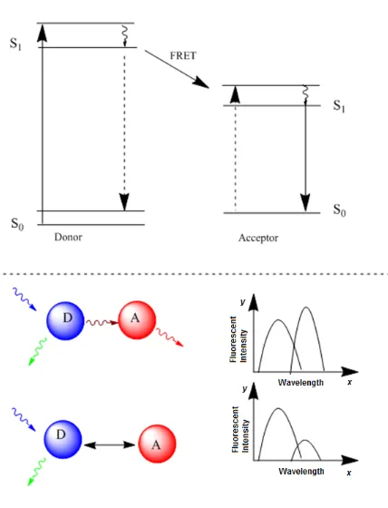
emission intensity (Figure 1.1).14, 15 The efficiency of energy transfer is strongly
dependent on the distance between the donor and acceptor. By measuring the efficiency
of energy transfer (e.g. through the changes of fluorescent signal from either donor and/or
acceptor), one can determine the distance between two fluorophore-partners. Based on
this, FRET has shown as a powerful tool for probing biological interations, sensing
analytes and imaging applications.
The pair of donor and acceptor in conventional FRET system can be organic dyes pair
(Cy3-Cy5), fluorescent proteins pair (e.g. cyan fluorescent protein and red fluorescen
protein pair), or dye-fluorescent proteins pair. Fluorescent NPs, such as QDs can also be
used in FRET system. For example, by taking the advantage of broad absorption spectra
and tunable emission profile, QD is particularly suitable as a donor in the FRET
system.16-18Numerous investigators have utilized the QD-based FRET system for various
applications, such as detection of pathogenic DNA and DNA point mutation,19, 20
immunoassay,21, 22 tracking and quantifying enzyme activity,23-26 protein conformation
changes,27, 28 sensing pH 29 and ion changes,30 and interaction between of biomolecules.28
6
QDs can also be energy acceptor, particularly while incorporated into bioluminescence
resonance energy transfer (BRET) 32 and chemiluminescence resonance energy transfer
(CRET),33 whereby non-radiative energy transferring from substrate catalyzed by enzyme
and chemiluninescent donor to QDs, respectively. The major advantages for those
systems are low background noise and high excitation efficiency, as there is no need for
an extra excitation source, typically high-energy laser. In this way, in particular, QD
based BRET system has showed great potential in deep tissue imaging with low harmful
effect on bodies.32
Figure 1.1 Schematic illustration of FRET process
Nanomaterials with special surface chemical properties can be used as a quenching
energy acceptor incorporating in the FRET system. Au NP was used as a quencher for
QD based FRET probe in a multiplexed assay in detection of the activity of enzymes and
7
1.2
Limitations of using NPs in biomedical fields
Despite that colloidal inorganic NPs offer immense promise for biomedical applications,
several concerns remain to be addressed.
The primary concern probably is the stability of NPs in water, as most biochemical
process occurs in aqueous environment. However, generally used methods usually
provide nanoparticles with hydrophobic ligands, meaning they are unstable in aqueous
solution. Another important concerning is the potential health and safety issues while
exploring nanomaterials to human body and environment. The determinants of particle
toxicity are known to be the large surface area and chemical reactivity in relation to small
size (and thus the ability to generate reactive oxygen species) and the capability to
penetrate tissues and cells.34 Thus, particles in nano-scale are likely to be more hazardous
than their bulk compartments, and free particles more toxic than fixed ones.35 One of the
examples to be considered is the potential cytotoxicity of QDs, because the heavy metals
core is toxicity, and particularly at high concentrations they could cause harmful effects
on embryo development and cell viability and function.36 Lastly, a nanoparticle must be
conjugated to a well-defined biological molecule, such as antibody, receptor, enzyme or
nucleic acid, for targeting application. Hence, there exists a gap between the nature of
NPs and their uses in biomedicine.
One method to bridge such a gap is the surface functionalization of nanoparticles (Figure
1.2). A proper surface coating can stabilize particles and avoid agglomeration, which
hence may increase the sensitivity of NPs based sensor. In addition, a proper surface
coating enables the nanoparticles in response specifically to biological species and avoids
non-specific interactions with components in the complex matrix. Coating is also an
effective manner of preventing the dissolution and release of core materials that may
cause toxicity to biological system.37 Furthermore, the steric hindrance of coating can
affect the fate of NPs in biological system, such as cellular uptake and accumulation,
circulation and clearance from body.38-40 In addition, the surface can affect the
8
behaviour. Moreover, appropriate surface functionality is the perquisite for conjugating
biomolecules to NPs for biomedical applications.
Figure 1.2 Strategy for bridging the unique feature of nanoparticles to biomedical
applications.
1.3
Aims and Objectives
In view of the above overview, the overall objective of current research projects is to
design and develop advanced NPs with suitable surface for cellular imaging and
biosensing applicatoins. Several goals are set up to fulfill the objective as showed below.
(1) To develop hybrid fluorescent nanoparticles with unique proton resistant property
for cellular imaging application.
(2) To synthesize and prepare bioconjugated magnetic fluorescent nanoparticles for
simultaneous capture, detection and deactivation of bacteria for infectious disease
control.
(3) To develop sensitive and fast responsible luminescent nanosensors for disease
early diagnostics
To achieve these goals, the following specific objectives are sought in various chapters of
the thesis:
•
This chapter provides an overview of biomedical application of NPs, the outline
of objectives and the layout of the thesis. Chapter 1: Introduction
•
This chapter presents a general review of strategies for surface functionalization
9
•
This chapter presents preparation of biocompatible polymer-quantum dots hybrid
nanocomposites and their potential application in bioimaging. Two approaches
were developed to produce the nanocomposites. In particular, multiple layer
coating NPs prepared by the second approach shows its advantages in stabilizing
QDs luminescence in term of intensity and lifetime among different pH range. Chapter 3: Development of biocompatible luminescent nanoparticles for
bioimaging applications.
•
This chapter describes preparation of antibiotic gentamicin functionalized
fluorescent magnetic nanoparticles. The nanoparticles have a fluorescent
silica/iron oxide core/shell structure. The gentamicin conjugated fluorescent
magnetic nanoparticles shows an all-in-one method for capture, detection and
deactivation of both Gram-negative and positive bacteria.
Chapter 4: Development of bioconjugated magnetic luminescent nanoparticles for
bacterial capture, detection and antibacterial applications.
•
Following the success of my developed advance nanoparticles, in this chapter, I
extended the application of bioconjugated fluorescent nanoparticles into
nanosensors. The detection manner is based on energy transfer from
bioluminescence from enzyme catalytic reaction to gold nanoparticles. We found
the nanosensor could detect thrombin at nano-gram range in 15 min from both
buffer and human urine samples.
Chapter 5: Luciferase conjugated nanoparticles for biosensing applications.
•
This chapter provides a general conclusion of the above studies and
recommendations for future work on the surface modification and conjugation of
the nanoparticles.
Chapter 6: General discussion and recommendation.
1.4
References
1. National Science and Technology Council, National Nanotechnology Initiative Strategic Plan In 2011.
10
3. Shen, T.; Weissleder, R.; Papisov, M.; Bogdanov, A.; Brady, T. J., Monocrystalline iron oxide nanocompounds (MION): Physicochemical properties. Magnetic Resonance in Medicine 1993, 29, (5), 599-604.
4. Weissleder, R.; Bogdanov, A.; Neuwelt, E. A.; Papisov, M., Long-circulating iron oxides for MR imaging. Advanced Drug Delivery Reviews 1995, 16, (2-3), 321-334.
5. Nam, J.-M.; Thaxton, C. S.; Mirkin, C. A., Nanoparticle-Based Bio-Bar Codes for the Ultrasensitive Detection of Proteins. Science 2003, 301, (5641), 1884-1886. 6. Luo, X.; Morrin, A.; Killard, A. J.; Smyth, M. R., Application of Nanoparticles in
Electrochemical Sensors and Biosensors. Electroanalysis 2006, 18, (4), 319-326. 7. Stroh, M.; Zimmer, J. P.; Duda, D. G.; Levchenko, T. S.; Cohen, K. S.; Brown, E.
B.; Scadden, D. T.; Torchilin, V. P.; Bawendi, M. G.; Fukumura, D.; Jain, R. K., Quantum dots spectrally distinguish multiple species within the tumor milieu in vivo. Nature Medicine 2005, 11, (6), 678-682.
8. Resch-Genger, U.; Grabolle, M.; Cavaliere-Jaricot, S.; Nitschke, R.; Nann, T., Quantum dots versus organic dyes as fluorescent labels. Nature Methods 2008, 5, (9), 763-775.
9. Gijs, M. M., Magnetic bead handling on-chip: new opportunities for analytical applications. Microfluidics and Nanofluidics 2004, 1, (1), 22-40.
10. Sung Kim, K.; Park, J.-K., Magnetic force-based multiplexed immunoassay using superparamagnetic nanoparticles in microfluidic channel. Lab on a Chip 2005, 5, (6), 657-664.
11. Doria, G.; Conde, J.; Veigas, B.; Giestas, L.; Almeida, C.; Assunção, M.; Rosa, J.; Baptista, P. V., Noble metal nanoparticles for biosensing applications. Sensors 2012, 12, (2), 1657-1687.
12. El-Ansary, A.; Faddah, L. M., Nanoparticles as biochemical sensors. Nanotechnology, science and applications 2010, 3, 65-76.
13. Syed, M. A., Advances in nanodiagnostic techniques for microbial agents. Biosensors and Bioelectronics 2014, 51, (0), 391-400.
14. Tran, P. T.; Anderson, G. P.; Mauro, J. M.; Mattoussi, H., Use of Luminescent CdSe–ZnS Nanocrystal Bioconjugates in Quantum Dot-Based Nanosensors. Physica Status Solidi B 2002, 229, (1), 427-432.
15. Willard, D. M.; Van Orden, A., Quantum dots: Resonant energy-transfer sensor. Nature Materials 2003, 2, (9), 575-576.
16. Medintz, I. L.; Uyeda, H. T.; Goldman, E. R.; Mattoussi, H., Quantum dot bioconjugates for imaging, labelling and sensing. Nature Materials 2005, 4, (6), 435-446.
17. Goldman, E. R.; Clapp, A. R.; Anderson, G. P.; Uyeda, H. T.; Mauro, J. M.; Medintz, I. L.; Mattoussi, H., Multiplexed Toxin Analysis Using Four Colors of Quantum Dot Fluororeagents. Analytical Chemistry 2003, 76, (3), 684-688.
18. Dennis, A. M.; Bao, G., Quantum Dots-Fluorescent Protein Pairs as Novel Fluorescence Resonance Energy Transfer Probes. Nano Letters 2008, 8, (5), 1439-1445.
11
20. Freeman, R.; Girsh, J.; Willner, I., Nucleic Acid/Quantum Dots (QDs) Hybrid Systems for Optical and Photoelectrochemical Sensing. ACS Applied Materials & Interfaces 5, (8), 2815-2834.
21. Goldman, E. R.; Medintz, I. L.; Whitley, J. L.; Hayhurst, A.; Clapp, A. R.; Uyeda, H. T.; Deschamps, J. R.; Lassman, M. E.; Mattoussi, H., A Hybrid Quantum Dots-Antibody Fragment Fluorescence Resonance Energy Transfer-Based TNT Sensor. Journal of the American Chemical Society 2005, 127, (18), 6744-6751. 22. Nikiforov, T. T.; Beechem, J. M., Development of homogeneous binding assays
based on fluorescence resonance energy transfer between quantum dots and Alexa Fluor fluorophores. Analytical Biochemistry 2006, 357, (1), 68-76.
23. Kim, G. B. K., Y-P., Analysis of Protease Activity Using Quantum Dots and Resonance Energy Transfer. Theranostics 2012, 2, (2), 11.
24. Choi, Y.; Lee, J.; Kim, K.; Kim, H.; Sommer, P.; Song, R., Fluorogenic assay and live cell imaging of HIV-1 protease activity using acid-stable quantum dot-peptide complex. Chemical Communications 2010, 46, (48), 9146-9148.
25. Kim, Y.-P.; Oh, Y.-H.; Oh, E.; Ko, S.; Han, M.-K.; Kim, H.-S., Energy Transfer-Based Multiplexed Assay of Proteases by Using Gold Nanoparticle and Quantum Dot Conjugates on a Surface. Analytical Chemistry 2008, 80, (12), 4634-4641. 26. Kimura, R. H.; Steenblock, E. R.; Camarero, J. A., Development of a cell-based
fluorescence resonance energy transfer reporter for Bacillus anthracis lethal factor protease. Analytical Biochemistry 2007, 369, (1), 60-70.
27. Krusinski, T.; Ozyhar, A.; Dobryszycki, P., Dual FRET assay for detecting receptor protein interaction with DNA. Nucleic Acids Research 2010, 38, (9), e108.
28. Zhang, C.-y.; Johnson, L. W., Quantifying RNA-Peptide Interaction by Single-quantum Dot-Based Nanosensor: An Approach for Drug Screening. Analytical Chemistry 2007, 79, (20), 7775-7781.
29. Dennis, A. M.; Rhee, W. J.; Sotto, D.; Dublin, S. N.; Bao, G., Quantum Dots-Fluorescent Protein FRET Probes for Sensing Intracellular pH. ACS Nano 6, (4), 2917-2924.
30. Liu, B.; Zeng, F.; Wu, G.; Wu, S., Nanoparticles as scaffolds for FRET-based ratiometric detection of mercury ions in water with QDs as donors. Analyst 137, (16), 3717-3724.
31. Suzuki, M.; Husimi, Y.; Komatsu, H.; Suzuki, K.; Douglas, K. T., Quantum Dot FRET Biosensors that Respond to pH, to Proteolytic or Nucleolytic Cleavage, to DNA Synthesis, or to a Multiplexing Combination. Journal of the American Chemical Society 2008, 130, (17), 5720-5725.
32. So, M.-K.; Xu, C.; Loening, A. M.; Gambhir, S. S.; Rao, J., Self-illuminating quantum dot conjugates for in vivo imaging. Nat Biotech 2006, 24, (3), 339-343. 33. Huang, X.; Li, L.; Qian, H.; Dong, C.; Ren, J., A Resonance Energy Transfer
between Chemiluminescent Donors and Luminescent Quantum-Dots as Acceptors (CRET). Angewandte Chemie International Edition 2006, 45, (31), 5140-5143. 34. Nel, A.; Xia, T.; Mädler, L.; Li, N., Toxic Potential of Materials at the
12
35. Cavaliere-Jaricot, S.; Darbandi, M.; Kuçur, E.; Nann, T., Silica coated quantum dots: a new tool for electrochemical and optical glucose detection. Microchimica Acta 2008, 160, (3), 375-383.
36. Michalet, X.; Pinaud, F. F.; Bentolila, L. A.; Tsay, J. M.; Doose, S.; Li, J. J.; Sundaresan, G.; Wu, A. M.; Gambhir, S. S.; Weiss, S., Quantum Dots for Live Cells, in Vivo Imaging, and Diagnostics. Science 2005, 307, (5709), 538-544. 37. Kirchner, C.; Liedl, T.; Kudera, S.; Pellegrino, T.; Muñoz Javier, A.; Gaub, H. E.;
Stölzle, S.; Fertig, N.; Parak, W. J., Cytotoxicity of Colloidal CdSe and CdSe/ZnS Nanoparticles. Nano Letters 2004, 5, (2), 331-338.
38. Otsuka, H.; Nagasaki, Y.; Kataoka, K., PEGylated nanoparticles for biological and pharmaceutical applications. Advanced Drug Delivery Reviews 2003, 55, (3), 403-419.
39. Moon, H. S.; Guo, D. D.; Song, H. H.; Kim, I. Y.; Jin, H. L.; Kim, Y. K.; Chung, C. S.; Choi, Y. J.; Lee, H. K.; Cho, C. S., Regulation of adipocyte differentiation by PEGylated all-trans retinoic acid: reduced cytotoxicity and attenuated lipid accumulation. The Journal of Nutritional Biochemistry 2007, 18, (5), 322-331. 40. Kato, T.; Yashiro, T.; Murata, Y.; Herbert, D.; Oshikawa, K.; Bando, M.; Ohno,
CHAPTER 2
14
2.1 Colloidal Inorganic Nanoparticles for Biomedical
Application
Colloidal inorganic nanoparticles (NPs) are small objects sized between 1 and 100
nanometers (nm) that can be dispersed in a solvent. During the last two decades, colloidal
NPs have been attracting considerable interest from a wide range of disciplines, including
materials science, chemistry, physics, biology and engineering, because of their unique
physical and chemical properties. The change in those properties at this length scale from
their bulk counterparts can be attributed to a combination of scaling factors and nature
material of the NPs. These NPs can be composed of various materials including noble
metal, semiconductor, magnetic compounds and a hybrid of them.
The biomedical utility of colloidal NPs can arise from a variety of attributes, including
their physical properties such as optical (absorption or emission of light) and magnetic
properties.1 In addition, as biological processes generally occur at molecule level, the
similar scale of NPs makes it a suitable platform for investigating those processes. For
example, biomacromolecules surface recognition by NPs offer a potential tool for
studying transcription regulation, enzymatic inhibition, delivery, sensing etc.
The aim of this chapter is to give a brief introduction of NPs of biomedical use and
current strategies of surface functionalization and bioconjugation of colloidal inorganic
NPs with a special focus the NPs with optical (semiconductor NPs, such as QDs) and
magnetic properties.
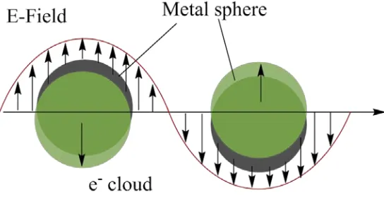
2.1.1 Noble metal NPs
Noble metal NPs such as gold NPs and silver NPs are an important class of nanomaterials
used in biomedicine due to their unique surface plasmon resonance (SPR) absorption.
The SPR is caused by the interaction between incident light and oscillation of electron
15
Figure 2.1 Schematic illustration of the SPR effect. The electromagnetic field of the light
induces a coherent dipole oscillation of the metal conduction electrons across the
nanoparticle (Adapted from Ref 2).
The response of noble NPs to oscillating electric field can be described by Mie theory, if
the diameter of a spherical NPs is much smaller than the wavelength of the incident light
(λ).3, 4
As shown in equation 2.1, the extinction cross section (Cext) of a particle (radius of
R), which defines the energy loss in the direction of incident light due to both absorption
and scattering, is described in term of dielectric function of the metal (ε = ε’+ iε”) and
dielectric constant of the medium εm.3 For small size of Au NPs (<60 nm), the absorption
cross section dominates in the Cext.5
Cext = 24π 2R3ε
m 3/2 λ
ε" �ε′+2ε
m�2+ε"2
Equation (2.1)
where ε = ε’ + iε” is the wavelength-dependent, complex dielectric function of the NPs material and εm is the dielectric constant of the surrounding/embedding medium.
The frequency of SPR is thus dependent on the size and composition of NPs, as well as
the dielectric constant of medium. The change in SPR frequency yields the change of
NPs color that can even be observed through bare eye. The SPR frequency is strongly
sensitive to the dielectric constant (ε) of the surrounding media, such as surface ligand
changes and inter-particle aggregation. This great sensitivity makes Au NPs and Ag NPs
well suited for bioassay applications. For example, analytes such as DNA, metal ions and
16
aggregation.6-8 Other examples are reviewed elsewhere including utilizing
surface-enhanced Raman scattering (SERS) for sensing.9, 10 In addition, the maximum
wavelength of SPR absorption for Au NPs fall into the visible range (520 nm to 600 nm),
which makes Au NPs as an excellent quencher to some common fluorophores for
bioimaging and biosensing application via resonance energy transfer process.11
Au NPs also find their applications in assisting drug delivery and therapy, due to the high
absorption cross-section. For instant, Halas et. al., reported near-infrared light triggered
gold nanoshells loaded hydrogels for deliverying a soluble drug.12 The mechanism is that
heat generated from the absorption of light by Au NPs triggered the hydrogels to collapse
and subsequently caused the release of the drug. Based on the similar concept, recently,
functionalized Au NPs or Au-hybrid NPs have been shown as a versatile tool for
photothermal cancer therapy 13, 14 and thermal ablation of pathogens.15 It is a great
advantage for such applications operating at NIR region, a transparent window for blood
and other types of biological samples.
2.1.2 Semiconductor quantum dots
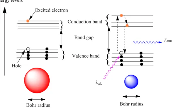
Quantum dots (QDs) are semiconductive and fluorescent crystalline particles that
typically have diameters ranging from 2 nm and 10 nm. Semiconductors have a valence
band filled with electrons and an empty conduction band separated by a band gap (Figure
2.2). An electron in the valence band can be excited into the highest level of the
conduction band by absorbing a photon with energy higher than the band gap energy,
leaving a hole of opposite charge in the valence band. An electron and its hole are
attracted towards each other by Coulomb force, and together form an exciton. The
distance between the excited electron and its hole is called the Bohr radius. QDs
fluorescence occurs when the excited electron reverts to its hole in the lowest level of the
valence band and emits a photon with equivalent to the band gap energy (lower than the
17
Figure 2.2 Size dependent photophysical properties of semiconductor quantum dots.
As the diameter of QDs is in the same order as its exciton Bohr radius, the excitons are
squeezed, leading to quantum confinement effect. The energy required to excite the
electron can be estimated in equation 2.2,16, 17
∆𝐸 =ħ2𝑅2𝜋22� 1 𝑚𝑒+
1 𝑚ℎ� −
1.786𝑒2
𝜀𝑅 −0.248𝐸𝑅𝑦∗ (Equation 2.2)
where me is the free electron mass, mh is the hole mass, and ε is the size-dependent
dielectric constant, R is the particle radius, ħ is the reduced plank constant and E*Ry is the
effective Rydberg energy that is usually small. The first term is band gap energy obtained
by “particle in a box” model. The second and third term indicate Coulomb force and
spatial correlation effect, respectively. The implications of the equation are clear that the
energy of the QDsare dependent on their size due to the quantum confinement effects.
Typically, smaller particles exhibit higher band gap energy and blue-shift emission.
The size-dependent fluorescent properties of QDs are superior to that of organic dyes in
biomedical applications. Firstly, QDs possess broad absorption spectra while maintaining
the same emission spectra. In addition, the emission profile is narrow and tunable,
allowing for multiplexing labeling. Furthermore, the bright fluorescence and significant
resistant to photobleaching, ensure a high ratio of signal to noise while applying them in
18
The pioneer work for utilizing QDs in biomedicine were reported by two groups in 1998,
demonstrating water-soluble bioconjugated QDs as an excellent labeling agent for cell
imaging.18, 19 Since then QDs have been tested in various biological applications due to
its bright fluorescence, including DNA array technology, immunofluorescence assays,
labeling in cell and animal biology.20 One benefit of using QDs as labeling agents is the
ability to excite and detect several species simultaneously using one single light source.21,
22
Besides severing as staining agents for bioimaging, QDs with bright fluorescence can
be also used to study the dynamic process inside living cells at molecule level .23 Apart
from directly labelling, the narrow emission profile makes QDs as excellent donors for
FRET based sensing and imaging (see also in Chapter 1). In addition to imaging, QDs
were also used as photosensitizing agents for photodynamic therapy (PDT).24
2.1.3 Magnetic Nanoparticles
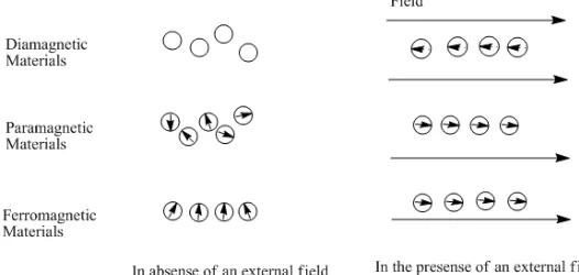
Magnetic nanoparticles (MNPs) are a class of NPs, which commonly consist of magnetic
elements such iron, nickel and cobalt or their chemical compounds. The magnetic
properties in matter are generally the consequence of the response of electrons magnetic
moments (due to their rotation around nucleus and spinning up and down) to an external
magnetic field. As shown in Figure 2.3, the materials can be classified into three
categories including diamagnetic (zero net moment, repelled by external field),
paramagnetic (zero net moment, attracted by external field), or ferromagnetic (exist net
moment, attracted by external field), depending on their response to an external magnetic
field.25
19
MNPs exploited in biomedical fields are commonly made of ferromagnetic materials. In
such materials, the unpaired electron spins of an atom are interacting, leading to a parallel
oriented magnetic moment and maintain a low energy state. The regions where the
parallel orientation occurs are called magnetic domains. For energetic reasons, the size of
the magnetic domain is usually smaller than the grain size. If the size of the magnetic
materials is reduced when only one domain is reached (ranging from ~20 nm to several
hundred nm), as in case of magnetic NPs, the magnetic behaviors are different from their
bulk materials.
In single domain particles, the electron spins rotate in unison, flipping the entire magnetic
moment of particles coherently and leaving a net magnetization and preferred aligned
direction. As the size of particle ( diameter d for spherical particles) is reduced, the
coercivity (Hc), which is the intensity of the applied field to overcome this magnetization,
drops to zero at the superparamagnetic limit dsp (typically less than 40 nm). In such a
case, particles that were originally aligned will have random directions at the
measurement time without external field, due to thermal fluctuations of energy kBT (kB is
Boltzman constant, T is measure temperature). For the same particles, their magnetic
behavior can be affected by temperature. If the superparamagnetic particles are cooled, at
certain temperature, the measurement time will be insufficient for complete magnetic
relaxation, i.e. the particles will exhibit hysteresis (Hc >0). This temperature is known as
the blocking temperature (Tb). For example, TB of 26 nm Fe3O4 NPs is about 300 K
(room temperature). Therefore, particles large than ~26 nm are predicated to exhibit
ferromagnetism, while smaller ones should have superparamagnetism.26
Blocking temperature can be affected by particle size. The relationship between them can
be obtained from Equation 2.3,27
𝑇𝐵= 𝑘𝐵𝐸𝑎ln 𝑓𝑡 (Equation 2.3)
Where t is the experimental measuring time, Eais the anisotropy energy barrier that the
20
be treated as constant. Ea = KV, where K is the anisotropy energy density constant and V
is the volume of particles. Thus, blocking temperature is size dependent.
Superparamagnetic NPs, in particular iron oxide MNPs, are widely designed for
pre-concentration, separation, and identification of molecules and specific biological units
and are particularly suitable for integration in micro fluidic devices.28, 29 Additionally, the
advantages of a very large surface area and good biocompatibility make these NPs
suitable for integration with biological system.
The most important utilization of MNPs is separation and targeting of analytes.
Numerous reports took the advantage of immuno-separation by antibody functionalized
MNPs in detection of bacteria. In one example, Mujika and co-workers reported a
magneto-resistive immunosensor for the analysis of Escherichia coli O157:H7 in food
and clinical samples. This biosensor enabled to detect and quantify small magnetic field
variations caused by the presence of superparamagnetic particles bound to the antigens
previously immobilized on the sensor surface via an antibody–antigen reaction.30
However, different methods for detection of MNPs become more and more important
while incorporation with immuno-separation. Other common reported detection methods
are impedimetric measurements 31 and electrochemical magneto-genosensing.32 In the
latter case, Liebana and colleagues reported the integration of immunomagnetic
separation/double-tagging PCR/electrochemical magneto-genosensing to detect
Salmonella in skimmed-milk samples with a limit of detection (LOD) of 1 colony
forming-unit (cfu)/mL. By combining the magnetic separation and miniature technology,
individual cells can now be separated through a microfluidic device system and
visualized in a low power microscopy.33 Another important aspect for taking the
advantage of magnetic attraction property is the transport of drugs and gens by magnetic
NPs. MNP medicated drug delivery can be performed via passive, active or direct ways.34
Research work about the functionalization of MNP with emphasis on the active in vitro
or in vivo drug delivery and related recent clinic results are reviewed by Pankhurst et.
21
In addition to separation and identification of target from matrix, MNPs have recently
been used as labels in biosensing and bioimaging. For example, Koets and co-workers
reported using streptavidin-coated superparamagnetic particles as detection labels for E.
coli and Salmonella via a Giant Magneto Resistance (GMR) sensor.36 An important use
of MNPs now is for magnetic resonance imaging (MRI) application where the NPs are
introduced as contrast-providing agents which have been summarized elsewhere.37-40
Despite of few examples, MNPs can also be used as antiseptic agents. There are main
two methods to modify magnetic NPs to obtain the antimicrobial NPs. The first one is to
functionalize NPs with biomolecules of antimicrobial activity. For example, in the study
conducted by Kaittanis, C. et. al., Con A-conjugated polysaccharide magnetic NPs have
been shown with significant and fast inhibition of the growth of E. coli and S. marcescens
in blood culture.41 In another work, Chen et. al. reported that the immobilization of
antimicrobial peptide LL-37 on the surface of polyacrylic acid coated NPs can effectively
kill E. coli.42 The second method to enable magnetic particles with antimicrobial
properties is to coat them with inorganic/organic antibacterial layers. For instance, silver
NPs and TiO2 layer have been deposited onto the surface of magnetic particles to produce
antibacterial agents.43, 44
2.2 Synthesis of colloid NPs for biomedical use
Colloidal inorganic NPs can be synthesized by various physical and chemical methods,
with the particles differing in their elemental composition, shape, size and chemical or
physical properties.45 The physical methods in general involve vapor deposition
approaches which dependent on sub-dividing of bulk materials to smaller NPs. Typical
chemical ways involve the reduction of ions into atoms in the presence of stabilizing
agents, followed by controlled growth of atoms into NPs (so called “bottom-up”
process).46 In the case of biomedical applications, solution based chemical synthesis
methods have been proved preferable ways, as they are more effective to control the size
distribution and ready for further modification or conjugation with biological species. In
this way, as-synthesized NPs are dispersed in a solvent either water- based or an organic
solvent for hydrophilic or hydrophobic particles, respectively; meanwhile amphophilic
22
2.2.1 Synthesis of NPs in water
A variety of NPs including Au, 47, 48 Ag,49 Co,50 Fe3O4,35 Fe2O3,51 SiO2 52 and CdTe 53, 54
have been synthesized in aqueous solution. These methods produce water-dispersible
NPs, a necessity for the application in biological systems. One typical example is the
synthesis of noble metal NPs such as Au NPs by reduction of Au(III) salts using reducing
agents such as sodium citrate, citric acid, ascorbic acid or amines.55 In addition,
biomolecules (e.g. starch) are also used as reducing agents providing a green protocol. In
another way, aqueous co-precipitation process, which involves nucleation growth,
coarsening and /or agglomeration, has been widely used in preparation of metallic and
metal (hydr) oxides NPs. The composition and morphology of the NPs are controlled by
precisely adjusting the reaction parameters.56 Stöber method, an aqueous sol-gel process,
is established to synthesize SiO2 NPs.52 Success has also been achieved in synthesis of
water-soluble semiconductor CdSe and CdTe quantum dots.54 However, limits in
controlling a narrow size distribution and low ordered crystalline structure remain as
main challenges, which subsequently affect their physical properties and stabilities.
2.2.2 Synthesis of NPs in organic phase
NPs composed of noble metals, transition metals, oxides and semiconducting materials
have been synthesized in organic phase via a thermal decomposition process. The process
general involves high temperature thermolysis of metal-organic precursor in the presence
of a hydrophobic capping agent, as well as in a non-polar organic solvent. The resultant
NPs usually gain high quality of nanocrystals.57 The growth of NPs, the crystal structure
and the cessation of growth depend on the environment and are fundamentally regulated
by the hydrophobic ligand. These ligands are either surfactant species such as fatty acid
or alkane thiols. The obtained NPs are thus inevitably hydrophobic. The ligands in some
cases also serve as solvent. One example is using tri-n-octyl phosphine oxide (TOPO) for
capping the semiconductor quantum dots.58
Hydrophilic NPs can also be produced in one-pot synthesis in organic phase by using
dedicatedly selected stabilisation ligands. For instant, amphiphilic ligands such as
23
However, only few examples have been reported due to the limit of amphiphilic
materials.
2.3 Surface Functionalization of NPs
A large portion of NPs used in biomedicine is produced in organic phase through high
temperature process. High-temperature synthesis in organic phase offers a number of
advantages over aqueous synthesis. Firstly, high temperature allows the impurities of NPs
be annealed out to obtain good crystallite structure. Furthermore, long chain of organic
ligands enables steric stabilization of NPs and allows higher concentration of NPs to be
produced. Moreover, temperature can be used to manipulate the morphology and size of
the NPs through controlling the growth kinetics of crystals. However, the resultant NPs
with apolar ligand are only soluble in organic solvents, e.g., hexane, toluene or
chloroform. They usually cannot be used directly for most applications in biomedicine, as
most biological process occurs at aqueous environment. Therefore, it is necessary to
bring them with water solubility, prior to the use them in biomedicine. One method to
circumvent this problem is development of strategies for surface functionalization of
NPs. Such strategies should satisfy certain criteria: i) they can enable NPs with good
water solubility, ii) they should offer a reduction of toxicity of NPs in some instances, iii)
they can provide additional functionalities for further conjugation of biomolecules for
targeting applications, and iv) they can stabilize the physical properties of NPs. Here a
variety of strategies for surface functionalization of NPs are evaluated and compared to
their effect on parameters relevant to stability, nonspecific binding, and biocompatibility
of NPs. The discussion falls in three categories: ligand exchange, ligand modification and
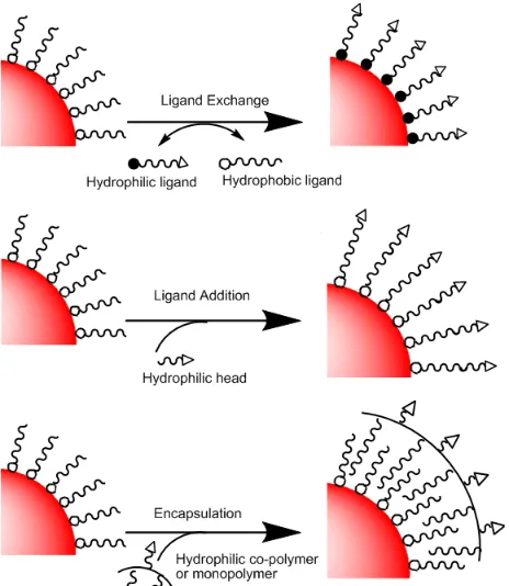
24
Figure 2.4 Schematic illustrations of common surface functionalization strategies.
2.3.1 Ligand exchange
For biomedical applications, ligand exchange of NPs usually involve a process that the
initial hydrophobic ligands are replaced by other more strongly bonding hydrophilic
ligands that allow the transferring of NPs from organic phase to aqueous solution. A
number of hydrophilic ligands have been reported to exchange the nature ligand on the
surface of NPs and bring them to aqueous solution, including small molecules with
functional headgroups (e.g. thiol, carboxyl, amine, phospine group, etc.), PEG derivatives
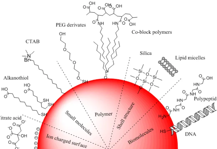
and biological molecules (Figure 2.5).
Small molecules. Small molecules with high affinity head functional group are primary
candidates for generating water-soluble particles, as they produce NPs with a smaller
hydrodynamic radius, which promotes in vivo trans membrane permeation and excretion
of NPs.60 Common examples of small molecules are alkylthiol terminated molecules that
can strongly bind to the inorganic surface of NPs, e.g. Au and Ag 61 or CdSe QDs,18, 62-65
by replacing weaker ligands. However, the colloid stability of the resultant NPs in buffer
25
NPs.64 To overcome this problem, bidentate ligands such as dihygrolipoic acid (DHLA)
and dithiocarbamate ligands are used to stabilize the NPs by increasing the number of
anchor points to particle surface.66-68 In addition to alkyl thiol terminated ligand
molecules, many molecules with other functional headgroups have also been developed
to transfer NPs from organic phase to aqueous solution. Excellent examples include
tetraalkylammonium salts such as tetraoctylammonium bromide (TOAB) for transfer of
AuNPs, and hexadecyltrimethylammonium bromide (CTAB) for iron oxide NPs;69, 70
oligomeric phosphine (oxide) ligands for transfer TOP/TOPO capped QDs;71 amphiphilic
species such as 2, 3-dimercaptosuccinic acid (DMSA) 72 and cyclodextrin 73 for
transfering of oleic acid capped NPs, respectively. However, one drawback is that the
small ligands rely on electrostatic interaction to stabilize NPs. Therefore, when the
solution condition such as pH and salt concentration changed, the NPs may be “salting
out” and forming aggregation.
26
PEG derivatives ligands. An alternative is using polymeric ligands as ligand exchange
agents to overcome the poor colloidal stability of NPs that capped by small molecules. A
number of polymers have been reported to offer NPs with good stability and water
solubility. Among them, poly (ethylene glycol) (PEG) is the most common reported
ligands for stabilizing NPs especially in biomedical applications. The ether group in the
backbone of PEG chains utilizes hydrogen bonding and steric stabilization for water
solubility, instead of electrostatic interactions. It is thus expected that PEG based NPs
would confer well stability over a wide pH range and even at high salt concentration.
Taking advantage of this, Mattoussi’s group developed a set of DHLA-PEG derivatives
for capping QDs through ligand exchange.74, 75 As a result, by combining the benefit of
strong bonding through dithiol moiety to particle surface, the PEG based QDs confer
water solubility over a wide pH range and high salt concentration. In addition to good
particle stability, PEG based NPs also were also found with low degree of nonspecific
binding to biological components, less cytotoxicity and longer circulation time in vivo.76,
77
Due to those advantages, it has been now increasing common to use PEG segments for
capping NPs to improve the biocompatibility.78 However, it should note that PEG itself is
simply a polyether, in general it should be modified with reactive terminal functional
group to serve as an anchor to the NPs.
Biomolecules. Other exchange ligands involved are biomolecules such as sugars, peptide,
nucleic acid and their derivatives. In one example, glucose and sucrose have been used in
reduction and post-synthesis stabilization of Ag and Au NPs in aqueous salts.79 Small
peptides are also used as QDs surface ligands. Pinaud and co-workers developed
phytochelatin-like peptides replacing TOPO for coating CdSe/ZnS QDs.80 The advantage
to use peptides as surface ligands is they offer an all-in-one solution for particles
solubilisation and biofunctionalization. Several other examples will be discussed in the
biofunctionalization part.
2.3.2 Ligand modification
A different strategy from ligand exchange is in situ modification of ligand for phase
transfer, which can prevent NPs aggregations due to irreversible desorption of replaced