
DOI: 10.7324/JAPS.2018.8820 ISSN 2231-3354
© 2018 Sudip Majumder et al. This is an open access article distributed under the terms of the Creative Commons Attribution License -NonCommercial- ShareAlikeUnported License (http://creativecommons.org/licenses/by-nc-sa/3.0/).
*Corresponding Author
Dr. Debasree Ghosh, Department of Chemistry, Amity School of Applied Sciences, Centre for Nano Science and Technology, Amity University Haryana, Amity Education Valley, Gurgaon, Haryana 122413, India. E-mail: ddebasreeghosh @ gmail.com
Dr. Tapasya Srivastava, Department of Genetics, University of Delhi South Campus Benito Juarez Road New Delhi-110021, India E-mail: tapasya @ south.du.ac.in
Synthesis, Characterization of Novel PLGA Encapsulated Indole
Nanoparticles and Study of its cytotoxic potential against A549 lung
cancer cell line
Sudip Majumder1,2, Neha Sharma1, Subhra Das3, Namita Pandey4, Tapasya Srivastava4*, Debasree Ghosha1,2*
1Department of Chemistry, Amity School of Applied Sciences, Amity University, Haryana, India. 2Centre for Nano Science and Technology, Amity University, Haryana, India.
3Department of Renewable Energy, Amity School of Applied Sciences, Amity University Haryana, India. 4Department of Genetics, University of Delhi, South Campus, New Delhi, India.
ARTICLE INFO ABSTRACT
Article history: Received on: 19/05/2018 Accepted on: 04/07/2018 Available online: 31/08/2018
Objectives: Indole and its derivatives are gaining importance because of their anti-cancer activity. Here, we have reported the synthesis and characterization of novel polymeric poly D, L-lactide-co-glycolide (PLGA) indole nanoparticles, and investigated their cytotoxic potential against A549 lung cancer cells. Materials and methods: Nanoparticles were synthesized by solvent emulsion-diffusion-evaporation method. Size determination was done
by Transmission Electron Microscopy (TEM), encapsulation efficiency using UV-Vis spectra, release kinetics using
dialysis, measurement of drug-polymer interaction by Fourier Transform Infra Red Spectroscopy (FTIR) and surface charge by zeta potential. Cell viability of lung cancer cells (A549) was determined by (3-(4,5-dimethylthiazol-2-yl)-2,5-diphenyltetrazolium bromide (MTT) assay and morphological analysis. Results: Nanoparticles were spherical
in shape with an average diameter of 65 nm, encapsulation efficiency was found to be about 78% and zeta potential was −15.2mV. Drug-loaded nanoparticles showed sustained release kinetics fitting well in exponential Higuchi and Zero order Model. FTIR studies showed a broadening of the peak of PLGA indole nanoparticles at 2100-3400 cm-1 indicating the formation of drug-loaded nanoparticles. These nanoparticles showed about 95% cytotoxicity against
A549 lung cancer cell lines. Results were supported by visible morphological changes in cells. Conclusion: PLGA encapsulated Indole nanoparticles were stable, having sustained release and good cytotoxic potential.
Key words:
Nanoparticles; biomaterials; polymers; sustained release; cytotoxicity.
INTRODUCTION
Cancer has emerged as a debilitating complex disease over the last many decades. Although advances in research in some cancers have led to the successful prognosis of the disease, in most cases especially in advanced stages, it continues to remain incurable. Development of new generation of drugs with
minimal side effects continues to be the mainstay of all research
work undertaken to combat this deadly disease. The advent of
nanotechnology has brought new hope in cancer treatment by targeted delivery, increased half-life, better stability and sustained release of the drug and hence helped in mitigating the problem of side effects (Hariharan et al., 2006). There are reports of different classes of nanoparticles that might serve as potential anti-cancer agents themselves. The class of nanoparticles ranges from transition metal oxides (Pandey et al., 2016; Tarnuzzer et al., 2005;
Sankar et al., 2014), chitosan derivatives (El-Sayed et al., 2017) to ceramics (which includes hydroxyapatite nanoparticles) (Kundu
et al., 2013). However, an alternate and popular approach in the
field of nanomedicine is by either encapsulating the anti-cancer
should be biocompatible in nature, and therefore, polymers have gained much importance. PLGA (Poly D, L-lactide-co-glycolide) is one such polymer widely used in drug delivery. These have been reported to have biocompatible nature, sustained release and non-immunogenic properties. Alternatively, other agents have
also been experimentally proven to have efficient anti-cancer
activities, however, the hydrophobic nature of the majority of
such compounds restricts their bioavailability and lack of proper targeting reduces their efficacy and brings about undesirable side
effects.
Natural products have served over the ages as potential
anti-cancer agents like curcumin, quercetin, Vitex agnus-castus
extract, etc. (Ghosh et al., 2012, Ghosh et al., 2010, Ibrahim
et al., 2017) in lieu of their potent antioxidant capabilities.
Nitrogen, containing aromatic heterocycles, makes a major class
of natural compounds that have medicinal applications. Among the aromatic nitrogenous heterocycles that serve as potential scaffolds for experimental anti-cancerous drug design, indole and its derivatives constitute a very important clan (Patel et al., 2012; Chu et al., 2011). Indole and its derivatives have shown promising anti-cancer activities rendering them of high interest to researchers (Patel et al., 2012). Indole conjugated Gold nanoparticles have been reported to function as anticancer agents when used individually and in combination with therapeutic doses of ionizing radiation (Jain et al., 2012). Indole has been found
to regulate several microbial processes like motility, biofilm
formation, quorum sensing, among others (Li and Young 2013;
Lee et al., 2009; Kim et al., 2010).
Till now, experimental work has been primarily done with indole derivatives like acetic acid (IAA),
Indole-3-carbinol for their putative anti-cancer properties. Indole and its derivatives are potent inhibitors of tubulin polymerization as well as DNA topoisomerases, leading to their screening as anti-cancer drugs. Indole-3-carbinol has been found to be effective against MCF-7 cell line, both in vitro and in vivo (Wang et al., 2013). These derivatives have shown better results and fewer side effects compared to conventional chemotherapeutic drugs against various cancer cell lines (Wang et al., 2013; Kim and Milner, 2005; Luo
et al., 2013).
However, reports suggest that Indole-3-carbinol have a tendency to oligomerize in acidic and even neutral conditions. This oligomerization reduces its bioavailability following oral
administration, and finally in the gastric conditions of the stomach
(Kim and Milner, 2005). To overcome this problem, chitosan nanoparticles of indole-3-carbinol have been prepared to improve
the stability of the compound for better efficacy (Luo et al., 2013). PLGA nanoparticles of Indole-3-Acetic acid have been reported to be effective against breast, colon, and prostate cancer cell lines
(Shaikh et al., 2009). Since indole derivatives have been shown
to be effective against human cancers; indole also seems to be very promising for being an effective anti-cancer agent. However,
keeping in mind the instability of indole and its derivatives, encapsulating indole in nanoparticles should work better in in-vitro and in vivo conditions.
Till now, there is no report available on the preparation of nanoparticles of indole itself. Here, we report an emulsion-diffusion-evaporation method for the preparation of PLGA encapsulated indole nanoparticles with controlled size and
sustained release properties fitting well with exponential Higuchi model and zero order model of drug release kinetics (Gouda et al., 2007; Thakkar et al., 2009). These nanoparticles have shown
efficient anti-cancer activities by increasing cell cytotoxicity
against A549 lung cancer cell line.
MATERIALS AND METHODS
Materials
Polylactide-co-glycolide (PLGA) (Resomer RG 85:15H),
Polyvinyl alcohol (PVA, MW:30000-70000), Indole were purchased from (Merck, India). A549 cell line used in the study was obtained from NCCS, Pune Dulbecco’s modified Eagle’s medium (DMEM), (GIBCO, USA) supplemented with 10% fetal bovine serum and 5 mg/ ml ciprofloxacin (Sigma, USA) was used for cell culture. Cells were cultured in a humidified incubator at standard condition of 18% O2 and 5% CO2 at 37οC for an appropriate period of time using similar
procedure (Pandey et al., 2016).
Preparation of PLGA encapsulated indole nanoparticle A modified emulsion-diffusion-evaporation method was used to make the nanoparticles using the similar procedure as
described by Ghosh et al. (2010). In brief, 50 mg of PLGA was dissolved in 5ml of ethyl acetate at room temperature. Indole (5 mg) was dissolved in 2 ml of ethyl acetate. The organic solution
of PLGA and drug in ethyl acetate was then emulsified with 10 ml of an aqueous phase containing 2% polyvinyl alcohol (PVA).
The resulting o/w emulsion was stirred at room temperature for three hours before homogenizing at 15,000 rpm for 5 min with a high-speed homogenizer. The organic solvent was removed by constant stirring on a water bath set at room temperature. The suspension was centrifuged at 105,000 g in Sorval RC 5B Plus using the rotor sorval T-865 for one hour. Nano pellet was washed and resuspended in PBS buffer.
Physicochemical characterization of nanoparticles
Encapsulation efficiency
To estimate the encapsulated drug in the nanoparticles,
the pellets were dissolved in ethyl acetate and kept for three days at
4οC. The O.D was measured at λ
max (Indole) 545 nm using a unicam UV-Vis spectrophotometer. The percentage of encapsulation was
calculated from the absorbance value.
Particle size analysis
The nanoparticle suspension was sonicated properly, and then a drop was added to a carboncoated copper grid, dried and observed under Transmission Electron Microscopy (TEM) for particle size analysis. TEM was performed at an accelerating
voltage of 300 kV. Photomicrographs were obtained using TEM
(Tecnai G2 30, FEI, Netherlands) using the same procedure as described by Ghosh et al. (2010).
Release kinetics
This was carried out in a phosphate buffer saline (PBS) medium, according to the method of Shaikh et al. (2009). A small
moderate shaking. At different time periods, a known amount of the sink solution was withdrawn and the absorbance was taken at 280 nm using a UV-Vis spectrophotometer. The amount withdrawn
was replaced by fresh medium. This experiment was performed in
triplicate and the mean value was taken.
Mathematical model fitting of the release kinetics data
Different mathematical models were utilized to understand drug release kinetics of indole from PLGA nanoparticle. Here, the data was fitted to Zero order, First order and Higuchi model (both in linear and exponential mode) respectively (Dash et al., 2010). For Zero order model, the working equation taken was Ct= C0+ K0t (Dash et al., 2010)
Ct= amount of drug released at time t, C0=initial concentration of drug at time t = 0, K0 = zero-order rate constant.
For the First order model, the equation used was DC/dt = −K1C.
K1 is the first order rate constant, expressed in time-1or per hour (Dash et al. 2010).
However, one of the most prominent models used in
current day drug release kinetics is the Higuchi model which is
based on both diffusion and dissolution of the drug (Subal, Patil
et al. 2007). The classical basic Higuchi equation is represented by Q = (Dδ/τ(2C − δ.Cs)Cs.t)1/2
Where, Q = cumulative amount of drug released in time t/ unit area, CO = initial drug concentration, CS =drug solubility in the matrix and D = diffusion coefficient of the drug molecule in the matrix. In all of these aforementioned models, suitable parameters were plotted and from the value of R2, the fitting of the data to a particular model was analyzed.
Fourier transform infrared spectroscopy (FTIR) analysis
Fourier transform infrared spectroscopy (FTIR) was used for detecting any possible drug-polymer interaction. For the FTIR analysis, the nanoparticle samples were dried and grounded with KBr pellets and analyzed on a Nicolet IR 200 (Thermo Electron Corp, US).
Surface charge
The nanoparticles were sonicated properly and their surface charges were measured using a zeta sizer (ZetasizerNanoZS; Malvern Instruments, Malvern, UK).
Cytotoxicity analysis
The cytotoxic potential of the PLGA-Indole nanoparticles was assessed using MTT assay (3-(4,5-dimethylthiazol-2-yl)-2,5-diphenyltetrazolium bromide) as described previously by (Pandey
et al., 2016). Wells containing complete medium, nanoparticles
and MTT reagent without cells were used as blanks. Cell morphology
About 3 × 10⁴ cells were seeded in four-well chamber
slide, and, after overnight culture, treated with PLGA indole
nanoparticles (10 μg/ml, 30 μg/ml, 50 μg/ml) for 6 days at 37οC.
Cells were then quickly washed with 1 ml PBS and visualized by a Zeiss Axiovert (Germany) 40 C microscope at a magnification
of 40X.
Statistical analysis
The mean and standard error was calculated for
cytotoxicity analysis and release kinetics data. Student’s ‘t-test’
was used for the statistical validation of cytotoxicity data. P < 0.05
was taken as the minimum level of significance. RESULTS AND DISCUSSION
Physicochemical characterization
Transmission electron microscopy (TEM) analysis
The PLGA-indole nanoparticles formed were spherical in shape with an average diameter of 65 nm (Figure 1a). The nanoparticles formed did not exhibit any agglomeration. The small size of nanoparticles is crucial for increased cellular activity
as the cellular uptake and biological functions of colloidal systems
are largely controlled by particle size where a reduced size results
in a significantly better uptake of these nanoparticles by the tumor
cells and increased half-life in plasma (Murugan et al., 2015).
Encapsulation efficiency
High encapsulation efficiency of the drug encapsulated nanoparticles is preferable for efficient use of the drugs used to
prepare the nanoparticles. This is also important since it requires a lesser amount of nanoparticles to achieve a particular effective dose, thus, reducing the chances of any possible toxicity that might result from the agent used to encapsulate the nanoparticles.
The encapsulation efficiency of the nanoparticles was found to be 78%, indicating better encapsulation of indole in PLGA as
compared to the other reported nanoparticles of indole derivatives till date (Luo et al. 2013).
Release kinetics
Release kinetics is an important parameter of nanomedicine as it indicates its efficacy and stability. Effective
anti-cancer treatment demands sustained release of the drugs. An early burst release is sometimes preferable, followed by a sustained release. The initial burst release gives maximum relief immediately and the following sustained release is desirable to avoid repeated administration (Preetha et al., 2015). However, generally, the most preferable release pattern for a drug encapsulated in a nanoparticle is sustained release over a
prolonged period of time. Here, in this work release pattern of
the PLGA encapsulated indole nanoparticles showed a sustained release over a period of 20 days (Figure 1b). After 20 days, the
cumulative release was 50%. The sustained release pattern is
desirable for nanoparticles in treating cancer cells as the drug
works much better than the nanoparticles showing only burst release kinetics. The chitosan nanoparticles of indole-3-carbinol
& 3,3´-diindolemethane synthesized by Luo et al. (2013) showed a burst release within the first half-an-hour, followed by a sustained release, about 80% over a period of only seven
hours. Indole nanoparticles showed a sustained release pattern persisting over a period of 20 days and without any burst release,
confirming the much better stability of the nanoparticles as
compared to previous reports till date (Luo et al., 2013).
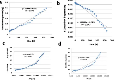
Mathematical model fitting of the release kinetics data
order model, First order model, Higuchi model (linear and exponential mode) respectively (Figure 2). R2 values of all them
were analyzed, and it was found that the exponential Higuchi
model and Zero order model show the best fit for the release kinetic data [R2 = 0.9853 and R2 = 0.972].
Fig. 1: Physicochemical Characterization of PLGA encapsulated indole nanoparticles showing Transmission Electron Micrographs (TEM) (a) release kinetics in in vitro conditions (PBS at 37οC), results are replicated in triplicates (b) and FTIR-spectrum (c) a) PLGA-Indole nanoparticles, b) Indole, c) PLGA.
Fig. 3: (a) In vitro cell cytotoxicity (MTT Assay) exhibited by PLGA-Indole nanoparticles on A549 cells, after 2 days (a1) and 6 days (a2) incubation. (* = P < 0.05 vs. DMSO control). [Each bar represents a minimum of 18 data points from three biological replicates and six technical replicates analyzed of each biological replicate.
The standard deviation is negligible in most cases.] (b) Representative microphotograph of DMSO treated control cells and PLGA-Indole nanoparticle-treated cells on
A549 cells after 6 days of incubation at 60X magnification.
Fourier transform infrared spectroscopy (FTIR) analysis
FTIR analysis is an important technique in determining any drug-polymer interaction. It is also helpful in judging the effective formation of drug encapsulated nanoparticles. A comparison of FTIR spectra of the free drug, polymer and the polymer encapsulated drug generally presents a good assessment of
the extent of encapsulation through the peak shifting of functional groups. Here in this work, the FTIR analysis (Figure 1c) revealed
no major shift as well as no loss of functional peaks of the spectra
of drug-loaded PLGA nanoparticles compared to that of indole and native PLGA, indicating no major drug-polymer interactions that can change the effective nature of any functional group. A similar observation was reported by Ghosh et al. (2012) for PLGA
encapsulated curcumin nanoparticles. The broadening of the peak
of PLGA encapsulated indole nanoparticles at the region 2100-3400 cm-1 is indicative of interaction of secondary N-H group of
indole with encapsulating PLGA moiety, suggesting the successful formation of PLGA encapsulated indole nanoparticles.
Surface charge
Zeta potential of the nanoparticles was found to be −15.2 mV (Figure 1d). Previous reports on A549 cells showed that these
cells indicated mean ZP of −10.2 mV with a deviation of ±19.7 mV (Honary et al., 2007). This suggests that these negatively
charged nanoparticles bind to some specific cationic sites of the
cell membrane in a cluster-wise manner (Patila et al., 2007). High
cellular uptake of negatively charged nanoparticles, therefore, seems to be a combination of non-specific adsorption and cluster
formation of nanoparticle on cell membrane surface.
Cytotoxicity analysis
The cytotoxic potential of PLGA indole nanoparticles against non-small cell lung cancer cell lines was studied. The
nanoparticles significantly decreased the viability of A549 cell
lines in a dose-dependent manner (Figure 3) from 85% to 10% in two days and from 75% to 15% in 6 days. The IC50 value for
PLGA indole nanoparticles in A549 was found to be ~50 µg/ml and ~30 µg/ml respectively for 48 hours (two days) and 144 hours (six days) treatment (Figure 3a). The prolonged incubation of A549 cells with 50 µg/ml of indole PLGA nanoparticles, showed
an increase in cellular cytotoxicity from 50% to 95% suggesting
a better effect of nanoparticle due to sustained release (Figure 3a1
and Figure 3a2). It was observed that indole PLGA nanoparticles
caused appreciably higher cytotoxicity to A549 cells at all doses as compared to DMSO control, the results were statistically
significant at P < 0.05.
Cell morphology
changes in a cellular structure like reduced cell size, pyknosis, karyorrhexis, indicating the cytotoxic effect of nanoparticles.
The decrease in a number of viable A549 cells with increasing
concentrations of PLGA indole nanoparticles (10 μg/ml, 30 μg/ ml, 50 μg/ml) as compared to untreated cells too suggested potent
cytotoxicity as shown by the nanoparticles.
CONCLUSIONS
Stable and biocompatible PLGA encapsulated indole nanoparticles were successfully synthesized. The nanoparticles showed considerably small particle size with good encapsulation
efficiency and sustained release properties fitting very well in
exponential Higuchi model and Zero order model. TEM and FTIR
analysis confirmed the formation of the drug-loaded nanoparticles. These nanoparticles showed about 95% cytotoxicity to A549
cancer cells at 50 µg/ml concentration after six days. Use of high
concentration of anticancer drugs is seen to have significant
side-effects in patients. Hence, there is a need to develop effective drug delivery systems containing low amounts of the anticancer drug with sustained release properties. The data obtained in our experiments suggest that effective cytotoxicity is achieved with a reduced concentration of the anti-cancer drug in the PLGA nanoparticles on increasing the duration of incubation of these nanoparticles with the cancer cells. This is a promising therapeutic intervention achieved with our formulation.
These highly stable PLGA encapsulated indole nanoparticles appear to be potentially promising anti-cancerous agents. Further in vitro and in vivo studies need to be performed to assess and characterize its function in vitro and in vivo.
ABBREVIATIONS
PLGA:poly D, L-lactide-co-glycolide; TEM: Transmission Electron Microscopy; FTIR: Fourier Transform Infra Red Spectroscopy; MTT: (3-(4,5-dimethylthiazol-2-yl)-2,5-diphenyltetra-zolium bromide.
CONFLICT OF INTEREST
The authors do not have any financial, personal or another conflict of interest.
ACKNOWLEDGMENTS
We thank Dr. Biswanath Kundu of Central Glass and Ceramic Research Institute, Kolkata, for his help in TEM data collection. TS is thankful to UGC-SAP (BRS-III), DU-DST
PURSE (Phase II) and DST-FIST (Level II). DG and SM
acknowledge Amity University Haryana for their support.
CONTRIBUTION FROM AUTHORS
Sudip Majumder has done the synthesis of the
drug-loaded nanoparticle and all the characterization like TEM, FTIR,
and Zeta potential and analyzed the data and also contributed in writing the manuscript, Neha Sharma has performed the release
kinetics and encapsulation efficiency and also contributed to the synthesis, Subhra Das has done the mathematical model fitting of the release kinetics data, Namita Pandey and Tapasya Srivastava
have done the cytotoxicity analysis and cell morphology study. Debasree Ghosha has conceptualized the entire idea, optimized
the formulation, written the manuscript and overall supervised the
work.
REFERENCES
Chu W, Zere TR, Weber MM, Wood TK, Whiteley M, Romano
E, Valenzuela BH, Mc Lean RJC. Microfluidic platform for rapid antibiotic
susceptibility testing of polymicrobial communities. Appl and Environ Microbiol, 2011; 78:411-419.
Dash S, Murthy PN, Nath L, Chowdhury P. Kinetic modeling on drug release from controlled drug delivery systems. Acta Pol Pharm, 2010; 67:217-223.
El-Sayed ST, Omar NI, El-Sayed MES, Shousha WG. Evaluation Antioxidant and cytotoxic activities of novel chitooligosaccharides prepared
from chitosan via enzymatic hydrolysis and ultrafiltration. Journal of App
Pharm Sci, 2017; 7(11):050-055
Ghosh D, Choudhury ST, Ghosh S, Mandal AK, Sarkar S, Ghosh
A, Saha KD, Das N. Nanocapsulated curcumin: Oral chemopreventive formulation against diethylnitrosamine induced hepatocellular carcinoma in rat. Chem Biol Interact, 2012; 195:206–214.
Ghosh, D, Ghosh S, Sarkar S, Ghosh A, Das N, Saha KD, Mandal
AK. Quercetin in vesicular delivery systems: Evaluation in combating arsenic-induced acute liver toxicity associated gene expression in rat model. Chem Biol Interact, 2010; 186:61–71.
Gouda R, Vaishya H, Qing Z. Application of mathematical models in drug release kinetics of carbidopa and levodopa ER tablets. J Dev
Drugs, 2007; 6(2):1-8.
Hariharan S, Bhardwaj V, Bala I, Sitterberg J, Bakowsky U, Ravi Kumar MNV. Design of Estradiol loaded PLGA Nanoparticulate
formulations: a potent oral delivery system for Hormone therapy. Pharm. Res, 2006; 23(1):184–195.
Honary S and Zahir F. Effect of zeta potential on the properties of Nano-Drug Delivery Systems – A review (Part 1). Trop J Pharm Res, 2013; 12(2):255–264.
Ibrahim AY, El-Newary SA, Youness ER, Ibrahim AMM, El
Kashak WA. Protective and therapeutic effect of Vitex agnus-castus against prostate cancer in rat. Journal of App Pharm Sci, 2017; 7(12):133-143.
Jain S, Hirst DG, Sullivan JMO. Gold nanoparticles as novel agents for cancer therapy. Br. J. Radiol, 2012; 85(1010):101-113.
Kim SY, Ryu JS, Li H, Park WJ, Yun HY, Baek KJ, Kwon NS,
Sohn UD, Kim DS. UVB-activated indole-3-acetic acid induces apoptosis
of PC-3 prostate cancer cells. Anticancer Res, 2010; 30 (11):4607-4612. Kim YS, Milner JA. Indole-3-carbinol inhibits cell proliferation and induces apoptosis in Hep-2 laryngeal cancer cells. J Nutr Biochem, 2005; 16:65-73.
Kundu B, Ghosh D, Sinha MK, Sen PS, Balla, VK., Das N, Basu
D. Doxorubicin-intercalated nano-hydroxyapatite drug-delivery system for liver cancer: An animal model. Ceramics International, 2013; 39(8):9557-9566.
Lee J, Maeda T, Hoong SH, Wood TK. Reconfiguring the quorum-sensing regulator SdiA of Escherichia coli to control biofilm
formation via indole and N-acylhomoserine lactones. Appl Environ Microbiol, 2009; 75:1703-1716.
Li G, Young KD. Indole production by the tryptphanase Tna Ain Escherischia coli is determined by the amount of exogenous tryprophan. Microbiology, 2013; 159:402-410.
Luo Y, Wang TT, Teng Z, Chen P, Sun J, Wang Q. Encapsulation of indole-3-carbinol and 3,3’-diindolylmethane in zein/carboxymethyl chitosan nanoparticles with controlled release property and improved stability. Food Chem, 2013; 201:224-230.
Mao CG, Tao ZZ, Chen Z, Chen C, Chen SM, Wan LJ. Expression of TWIST an inducer of epithelial-mesenchymal transition,
in nasopharyngeal carcinoma and its clinical significance. Exp Ther Med,
2014; 8(1):207-212.
Murugan K, Choonara YE, Kumar P, Bijukumar D, Toit LC, Pillay V. parameters and characteristics governing cellular internalization and trans-barrier trafficking of nanostructures. Int. J. Nanomedicine, 2015;
10:2191-2206.
Pandey N, Dhiman S, Srivastava, T, Majumder S. Transition metal oxide nanoparticles are effective in inhibiting lung cancer cell survival in the hypoxic tumor microenvironment. Chem Biol Interact, 2016; 254(4):221-230.
Patel H, Darji N, Pillai J, Patel B. Recent advance in anti-cancer activity of indole derivatives. Int. J. Drug Res. Tech, 2012; 2(3):225-230.
Patila S, Sandberg A, Heckert E, Self W, Seal S. Protein adsorption and cellular uptake of cerium oxide nanoparticles as a function
of zeta potential. Biomaterials, 2007; 28(31):4600-4607.
Preetha P, Srinivasa R, Pushpalatha P, Biphasic Drug Delivery in controlled release formulations-A review. Int. J Pharmacy Technology, 2015; 6(4)3046-3060.
Sankar R., Maheswari R., Karthik S, Shivashangari KS, Ravikumar V. Anticancer activity of Ficusreligiosa engineered copper oxide
nanoparticles. Mater. Sci. Eng. C, 2014; 44:234-239.
Shaikh J, Ankhola DD, Beniwal V, Singh D, Ravi Kumar MNV.
Nanoparticle encapsulation improves oral bioavailability of curcumin by at least 9-fold when compared to curcumin administered with piperine as
absorption enhancer. Eur. J. Pharm. Sci, 2009; 37(3-4):223–230.
Subal CB. 2006. Modelling of Drug release: The Higuchi equation and its application. Pharmabiz.com.
Tarnuzzer RW, Colon J, Patil S, Seal S. Vacancy engineered
ceria nanostructures for protection from radiation-induced cellular damage. Nano Lett, 2005; 5:2573-2577.
Thakkar VT, Shah PA, Soni TG, Parmar MY, Gohel MC et al. Goodness-of-fit model-dependent approach for release kinetics of levofloxacin hemihydrates floating tablet. Dissolution Technologies, 2009;
16:35-39.
Wang YQ, Chen C, Chen Z, Xu Y, Wang Y, Xiao BK, Chen SM, Tao ZZ. Indole-3-carbinol inhibits cell proliferation and induces apoptosis in Hep-2 laryngeal cancer cells. Oncol. Rep, 2013; 30(1):227-233.
How to cite this article:

![Poly[sodium(I) μ3 bis(ethylenedioxy)borato]](data:image/gif;base64,R0lGODlhAQABAIAAAP///wAAACH5BAEAAAAALAAAAAABAAEAAAICRAEAOw==)