Integrin expression and function
in osteoblasts
Sarah Moffatt Eastman Dental Institute University College London
ProQuest Number: U641867
All rights reserved
INFORMATION TO ALL USERS
The quality of this reproduction is dependent upon the quality of the copy submitted.
In the unlikely event that the author did not send a complete manuscript and there are missing pages, these will be noted. Also, if material had to be removed,
a note will indicate the deletion.
uest.
ProQuest U641867
Published by ProQuest LLC(2015). Copyright of the Dissertation is held by the Author.
All rights reserved.
This work is protected against unauthorized copying under Title 17, United States Code. Microform Edition © ProQuest LLC.
ProQuest LLC
789 East Eisenhower Parkway P.O. Box 1346
Abstract
List of abbreviations Abbreviation ANOVA AP a-mem BMP BSA BSP CAM cAMP Cbfa CCD CFU-F CFU-S CNS Col I/Col Col II DGEA DMEM Dmp-1 DMSO EOF ELISA EM ERK FACS FAK FCFC PCS FGF FITC GER GTP HBDCs HB-GAM HOBs ICAM used Definition
Analysis of variance Alkaline phosphatase
a-minimum essential medium Bone morphogenetic protein Bovine serum albumin Bone sialoprotein Cell adhesion molecule
Cyclic adenosine monophosphate Core binding factor-a
Cleidocranial Dysplasia
Colony forming unit fibroblastic Colony forming unit-spleen Central nervous system Type I collagen
Type II collagen Asp-Gly-Glu-Ala
Dulbecco’s modified Eagle’s medium Dentin matrix protein-1
Dimethylsulphoxide Epidermal growth factor
Enzyme linked immunosorbent assay Electron microscopy
Extracellular regulated kinase Fluorescence activated cell sorting Focal adhesion kinase
Fibroblast colony forming cell Foetal calf serum
Fibroblast growth factor
Anti-Fluorescein isothiocyanate Gly-Glu-Arg
Guanosine 5’ triphosphate Human bone derived cells
Heparin-binding growth associated molecule Primary human osteoblasts
Abbreviation Definition
IGF Insulin-like growth factor
ihh Indian hedgehog
ILK Integrin linked kinase
INK Janus kinase
MAPK Mitogen activated protein kinase
MAPKK Mitogen activated protein kinase kinase
MEK MAPK/ extracellular kinase 1
MEM Minimum essential medium
MG63s MG63 cells
mRNA Messenger ribonucleic acid
MTS [3-(4,5-dimethyl-2-yl)-5-(3-carboxymethoxyphenyl)-2-(4-sulfophenyl)-2H-tetrazolium
NaOH Sodium hydroxide
NCAM Neural cell adhesion molecule
OPG Osteoprotegrin
OPGL Osteoprotegrin ligand
PBS Phosphate buffered saline
PDGF Platelet derived growth factor
PI3K Phosphatidyl inositol 3 kinase
pfh Plasma fibronectin
PTE Parathyroid extract
PTH Parathyroid hormone
PTHrP Parathyroid hormone related protein
RGD Arg-Gly-Asp
RT-PCR Reverse transcription ploymerase chain reaction
SD Standard deviation
SEM Scanning electron microscopy
TIMP Tissue inhibitor of metalloproteinase
TNF Tumour necrosis factor
TRANCE TNF-related activation induced cytokine
une Uncoated
Contents Page number
Title page 1
Abstract 2
List of abbreviations used 3
Contents 5
List of tables and graphs 7
Thesis overview 9
1 Introduction 10
1.1 Introduction 10
1.2 Bone cells 11
1.3 Integrins 29
1.4 The extracellular matrix and integrin interaction 35
1.5 Cell adhesion molecules and bone cells 39
1.6 Bone cells, integrins and disease 47
1.7 Conclusion 48
2 Materials and methods 49
2.1 Methods - Cell culture 49
2.2 Methods - Characterisation of osteoblasts 53
2.3 Methods - Characterisation of integrin expression in osteoblasts 56 2.4 Methods - Characterisation of integrin function in osteoblasts 61
2.5 Statistical analysis 68
2.6 Scanning electron microscopy 68
3 Results o f characterisation studies 70
3.1 Aims of characterisation studies 70
3.2 Introduction 70
3.3 Results - Characterisation of osteoblasts 73 3.4 Results - Characterisation of integrin expression in MG63s and HOBS 80
3.5 Discussion 86
3.6 Summary of results 89
4 Integrins and ECM interaction - an important relationship in
osteoblast behaviour 90
4.1 Aims of adhesion assays 90
4.2 Introduction to integrins and adhesion in osteoblasts 90 4.3 Results - Adhesion assays on ECM substrates 92 4.4 Results - Adhesion assays with addition of RGD blocking peptide 95 4.5 Results - Adhesion assays with addition of integrin blocking antibodies 99 4.6 Results - Immunofluorescent staining of HOB adhesion assays 103 4.7 Results - EM of HOBs on coated coverslips 105
4.8 Discussion 108
5 Integrins and ECM regulate osteoblast migration 114
5.1 Aims of migration assays 114
5.2 Introduction to cell migration and migration assays 114
5.3 Results - Preliminary migration assays 116
5.4 Results - Migration assays with addition of integrin blocking antibodiesl21 5.5 Results - Migration and integrin signalling pathways 129
5.6 Results - Migration assay staining 131
5.7 Discussion 133
5.8 Summary of migration assay results 136
6 A link between integrins and the Cbfal transcription factor 138
6.1 Cbfal - introduction 138
6.2 Cbfal and osteoblast differentiation 138
6.3 Results to date - Characterisation of C2C12s - expression of osteoblastic
characteristics in vitro 140
6.4 Results to date - Characterisation of integrin expression in vitro 144
6.5 Discussion 146
7 Discussion and future work 147
7.1 Aims of the present study 147
7.2 Summary of results 147
7.3 Cell adhesion molecules and osteoblasts - the story so far 148
7.4 Conclusion 160
References 163
List of tables, graphs and photographs 1 Introduction
1(a) The pattern of gene and protein expression during the process of
osteoblast differentiation markers (adapted from Ducy, 2002) 14
1 (b) Cells of the marrow stromal lineage 21
1 (c) Summary of osteoblast characteristics of MG63s and HOBs 26 1(d) Schematic diagram showing the key features of integrin structure 30
1(e) Integrin mediated signalling 32
1(f) Fibronectin and proteolytic fragments 38
1(g) Results of studies into the integrin profile of osteoblasts 41
2 Materials and methods
2(a) Cell counting using a haemocytometer 51
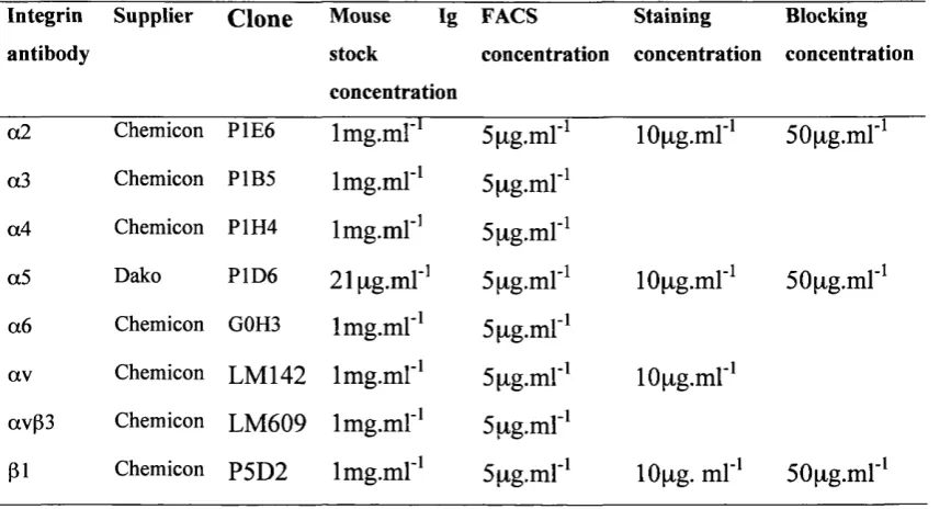
2(b) Details of antibodies used during the study 59
2(c) Cell dilutions 64
2(d) Boyden chamber migration insert 67
3 Results o f characterisation studies
3(a) Alkaline phosphatase activity in MG63s in response to
1,25-dihydroxy vitamin Da treatment. 75
3(b) Alkaline phosphatase activity in HOBs in response to
1,25-dihydroxyvitamin Da treatment. 76
3(c) Osteocalcin levels in MG63s in response to 1,25 dihydroxyvitamin Da
treatment 78
3(d) Osteocalcin levels in HOBs in response to 1,25 dihydroxyvitamin Da
treatment 79
3(e) FACS analysis of integrin expression on MG63s 81 3(f) FACS analysis of integrin expression on HOBs 82 3(g) Geometric means (GM) with standard deviations (SD) from the results
of FACS analysis on MG63s and HOBs using integrin antibodies 83
3(h-k) Integrin expression in HOBs 85
4 Integrins and ECM interaction - an important relationship in osteoblast behaviour
4(a) MG63 cell adhesion on ECM substrates 93
4(b) HOB adhesion on ECM substrates 94
4(c) MG63 cell adhesion on plasma fibronectin with RGD blocking peptide 96 4(d) MG63 cell adhesion on type I collagen with the RGD blocking peptide 97 4(e) HOB adhesion on plasma fibronectin with the RDG blocking peptide 98 4(f) MG63 cell adhesion on plasma fibronectin with addition of integrin
blocking antibodies 100
4(g) MG63 cell adhesion on type I collagen with the addition of integrin
4(h) HOB adhesion on plasma fibronectin and type I collagen with the
addition of integrin blocking antibodies 102
4(i-n) Immunofluorescent staining of pi integrin in HOBs 104 4(o-s) Immunofluorescent staining of a2 and a5 integrin in HOBs 106 4(t-y) Scanning electron micrographs of HOBs on type 1 collagen and plasma
fibronectin 107
5 Integrins and ECM regulate osteoblast migration
5(a) MG63 cell migration towards plasma fibronectin over a 4 hour time
course 117
5(b) HOB migation towards plasma fibronectin over a 4 hour time course 118 5(c) MG63 cell and HOB migration towards ECM substrates 120 5(d) MG63 cell migration towards plasma fibronectin with the addition of
integrin blocking antibodies 123
5(e) MG63 cell migration towards type 1 collagen with the addition of
integrin blocking antibodies 124
5(f) MG63 cell migration in response to the 120kDa plasma fibronectin
fragment with the addition of integrin blocking antibodies 126 5(g) HOB migration in response to plasma fibronectin and type 1 collagen
with the addition of integrin blocking antibodies 127 5(h) Representative images of transwell membranes during migration assays
with HOBs 128
5(i) MG63 cell and HOB Migration with UO126 (MAPKK inhibitor) 130 5(j-m) Integrin staining of HOB migration assays 132 5(n) Summary of migration and adhesion assay results 137
6 A link between integrins and the Cbfal transcription factor
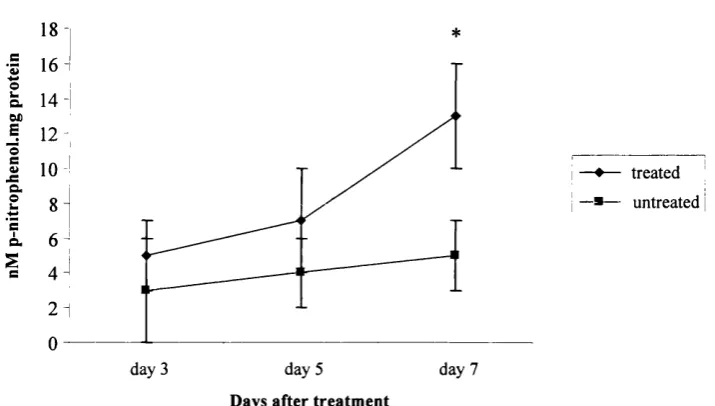
6(a) Alkaline phosphatase activity in C2C12 cells 141
6(b) Osteocalcin production in C2C12 cells 142
6(c-e) C2C12 cells stained for alkaline phosphatase after 7 days in culture 143
6(f-h) Immunofluorescent staining of integrins on C2C12 cells. 145
7 Discussion and future work
7(a) Integrin expression in cells of the osteoblast lineage (diagram taken fi-om
Bennett et al, 200la) 149
Thesis overview Chapter 1 - Introduction
The information available to date on osteoblast biology, integrins and integrin expression and function to date is explored in this introductory chapter.
Chapter 2 - Materials and methods
The materials and methods used in this thesis are detailed.
Chapter 3 - Results of characterisation studies
The results of studies into the integrin profile of primary human osteoblasts and MG63s are described in chapter 3. In addition, this chapter details the results of studies into the osteoblastic nature of primary human osteoblasts and MG63 cells.
Chapter 4 - Integrins and ECM interaction - an important relationship in osteoblast behaviour
This chapter contains the results of further studies into the role of specific integrins in osteoblast and ECM interaction.
Chapter 5 - Integrins and ECM regulate osteoblast migration
This chapter contains the results of studies into the role of specific integrins in osteoblast migration in response to ECM.
Chapter 6 - A link between integrins and the Cbfal transcription factor
This chapter contains preliminary results of studies into the potential link between integrins, the Cbfal transcription factor and osteoblast differentiation.
Chapter 7 - Discussion and future work
1 Chapter 1 - Introduction
The aims of this thesis were to:
• Ascertain the integrin profile of primary human osteoblasts and MG63 cells expressed in vitro; and
• Determine the role of specific integrins, in osteoblast behaviour in vitro.
1.1 Introduction
Bone is a dynamic tissue, constantly being broken down and replaced in the adult during the process of remodelling. This cycle of bone turnover occurs in response to stimuli from both within the bone and from external triggers. The two main cell types involved are the osteoblast, the principal bone producing cell, and the osteoclast,, which is associated with osteolysis. The continual breakdown and renewal of bone is dependent upon a level of communication between cells themselves and between cells and the surrounding extracellular matrix (ECM). Bone is an outstanding system in which to study these interactions as it has an extensive matrix secreted and mineralised by the osteoblast. As is the case with other cells types, cell adhesion molecules form a bridge between bone cells and their environment, playing an important role in the communication that occurs between the cells and their surroundings.
Integrins are known to act as members of cell signalling pathways, conveying information from the outside of the cell inwards. These cell adhesion molecules also have the capacity to transmit signals from inside the cell outwards. In addition, the presence of other cell adhesion molecules and cooperation between different receptors, growth factors and activation of multiple signalling pathways leads to a vast array of biological responses.
In this introduction, the current evidence regarding cell adhesion molecule expression in bone cells will be reviewed. Emphasis will be placed on the expression and function of integrins in osteoblasts. The areas of bone cell biology and cell adhesion molecules will also be reviewed.
1.2 Bone cells
There are three principal cell types present in bone: the osteoblast, the osteoclast and the osteocyte. In the following sections the characteristics of osteoblasts in vitro will be discussed in detail. Osteoblasts can be characterised ultra-structurally, biochemically and by molecular analysis and these will be discussed in the following sections. The key features of osteoclast biology will also be introduced.
1.2.1 Osteoblast ultra structure
Cells of the osteoblast lineage are responsible for the formation of bone, both during embryogenesis and throughout adult life. Located directly on the endosteal and periosteal surfaces of bone, these cells actively secrete a collagen rich matrix, osteoid, that is subsequently mineralised. Histologically, osteoblasts have a round nucleus at the base of the cell with well developed Golgi apparatus and endoplasmic reticulum. They can be observed as a layer of cuboidal cells on the bone matrix. Behind the layer of mature osteoblasts there also tends to be a layer of mesenchymal precursor cells and pre-osteoblasts (Puzas, 1996).
1.2.2 Osteoblast differentiation and biochemical analysis
In order to form a mature matrix-secreting cell, the osteoblast follows a pathway of differentiation that can be divided into several steps, based on both phenotypic observations and by analysis of gene expression in vitro (summarised in figure 1(a)). In the pre-osteoblast, genes universally associated with proliferation are expressed, for example c-myc and c-fos (Lian and Stein, 1993). In addition, genes associated with an osteoblast phenotype, matrix mineralisation and production are expressed, such as those encoding alkaline phosphatase, type 1 collagen (col 1), bone sialoprotein (BSP) and osteonectin. As the cell begins to mature, osteocalcin is also detected (Aubin et al, 1995). Recent studies have elucidated further factors associated with osteoblast differentiation such as Cbfal (Ducy, 2000) and Osx (Osterix) (Nakashima et al,
2002) and these will be discussed in 1.2.4. In vitro, osteoblast cultures may exhibit formation of nodules that resemble a mineralised matrix. This has been observed in rat calvarial cultures (reviewed in Aubin et al, 1993), adult rat stromal cell cultures (Malaval et al, 1994) and human trabecular explant cultures (Beresford et al, 1983).
Figure 1(a) - The pattern of gene and protein expression during the proeess of
osteoblast differentiation (adapted from Ducy, 2002)
Cblll Progaiitor ceU
Coil
AP
Osteoblast
pmgenitor
Cbfal CSx
Cbll
BSP ON AP
Cbfal OSx
Coll
BSP ON AP
OC
Cbfal Ckx
O steoblasts undergo a period o f differentiation from a progenitor cell to a mature osteoblast. This process can be divided into a series o f stages based on gen e and protein exp ression . Progenitor cells
are identified by the presence o f type II collagen (col 11), the cell then differentiates into an osteoblast
progenitor, sig n ified by type 1 collagen (col I), alkaline phosphatase (A P ), C b fa l and O sx. T hese gen es
1.2.3 Endocrine, autocrine and paracrine regulation of osteoblast
differentiation and function
There are many factors known to regulate osteoblast differentiation and function, a number of which will be discussed here. Several endocrine factors exert effects on osteoblasts, such as Parathyroid hormone (PTH), growth hormone, glucocorticoid hormones, oestrogen and progesterone. The biologically active form of vitamin D, 1,25-dihydroxyvitamin D3, also exerts endocrine effects on osteoblasts and bone. In vitro, the response of cells to PTH and 1,25-dihydroxyvitamin D3 have been used as markers of osteoblast differentiation. PTH is a polypeptide hormone involved in the maintenance of calcium homeostasis (Strewler et al, 1987). PTH stimulates cell signalling cascades that involve an increase cyclic adenosine monophosphate (cAMP) and results in an increase in Ca^^ levels in the osteoblast (reviewed in Swarthout et al, 2002). Overall, parathyroid hormone regulates many genes and proteins associated with osteoblast differentiation and function and also bone resorption. Prolonged parathyroid hormone treatment decreases collagen synthesis (Kream et al,
1986), osteocalcin levels (Beresford al, 1983; Lajeunesse et al, 1991), alkaline phosphatase activity (Luben g/ al, 1976), osteonectin (Termine et al, 1981), osteopontin (Noda and Rodan, 1989) and genes associated with DNA synthesis (Reid
et al, 1988). Evidence also suggests that PTH treatment could lead to activation of Cbfa-1 (core-binding factor-a) (Selvamuragan et al, 2000; reviewed in Swarthout et al 2002) and an increase in several further factors including, collagenase-3 (Meikle et al, 1992; Partridge et al, 1987), tissue inhibitors of metalloproteinases (TIMPS) (Partridge et al, 1987; Cook et al, 1994) and insulin-like growth factor (IGF-1) (McCarthy et al, 1989). PTH can also lead to the indirect activation of osteoclasts and factors associated with bone resorption (Teitelbaum, 2000). For further details see section 1.2.8. 1,25-dihydroxyvitamin D3 stimulates alkaline phosphatase (Fritsch
will promote the differentiation of pluripotent mesenchymal precursors into osteoblasts (Wang et al, 1993). BMP-2 treatment blocks myogenic differentiation and induces osteoblastic differentiation in myogenic cells (Katagiri et al, 1994). TGF-P treatment appears to inhibit osteoblastic differentiation (Spinella-Jaegle et al,
2001). Both TGF-P and BMPs are also secreted by osteoblasts themselves and can act in an autocrine manner.
Indian hedgehog (ihh) is a growth factor known to be important for osteoblast differentiation in vivo. The ihh gene is expressed in chondrocytes during development and controls the expression of parathyroid related peptide (PTHrP) (Vortkamp, 1996). Ihh knockout mice also show failure of osteoblast development in endochondral bones (St-Jacques et al, 1999).
1.2.4 Molecular control of osteoblast differentiation
Over recent years, advances in molecular biology have led to the characterisation of osteoblast differentiation at the molecular level, with increased knowledge on well- established markers such as alkaline phosphatase and osteocalcin and also the elucidation of several new factors associated with osteoblast differentiation and bone formation. In the following section molecular control of osteoblast differentiation and bone formation will be discussed.
1.2.4.1 The involvement of Cbfal in osteoblast differentiation
working in very different areas (Otto et al, 1997, Mundlos et al, 1997; Ducy et al, 1997). The Cbfal gene belongs to the runt family of genes and their protein products (Westendorf and Hiebert, 1999). For a review of Cbfal see Ducy, (2000).
1.2.4.2 Expression of Cbfal in bone cells
The expression profile of Cbfal is by no means straightforward, perhaps due to the plasticity of cells of stromal origin. During embryogenesis, skeletal development begins to take place with the condensation of undifferentiated cells (Hall and Miyake, 1992). Cbfal expression can be detected as early as 10.5 dpc in mice. Expression is seen in anlage destined to become either osteoblasts or chondrocytes (Ducy et al, 1997). Cbfal deficient mice have a complete loss of osteoblast differentiation and as a consequence the skeleton is composed of cartilage. Osteoclast formation also fails to occur (Ducy et al, 1997). In addition, Ducy et al, (1999) carried out further investigations using to determine the expression and role of Cbfal in postnatal mice. Transgenic mice expressing a truncated form of Cbfal driven by an osteocalcin promoter began to lose bone with no observed effect on osteoclasts. Mice transgenic for Cbfal show a decrease in the expression of important osteoblast markers, including type I collagen, osteocalcin and bone sialoprotein (Ducy et al,
1999).
In culture, Cbfal expression has been induced in cells other than osteoblasts. For example NIH3T3 fibroblasts, C3H10T1/2 fibroblasts, MC3T3 mouse pre-osteoblasts (Xiao et al, 1999) and the C2C12 mouse myoblast cell-line (Tsuji et al, 1998 and Lee et al, 1999). Cbfal not only appears to be important in the regulation of bone formation during development but also post-natally (Ducy et a l, 1997).
1.2.4.3 Role of Cbfal in the regulation of osteoblast specific genes
cultures with Cbfal specific anti-sense can lead to a decrease in the expression of these genes (Banerjee et al, 1997; Ducy et al., 1997).
Cbfal has also been shown to control the expression of dentin matrix protein 1 (dmp- 1) (Feng et al, 2002). Dmp-1 is a phosphoprotein (George et al, 1993) associated with mineralisation of both dentin and bone (Hirst et al, 1997; D’Souza et al, 1997) and has also been shown to be a marker of osteoblastic differentiation (Feng et al,
2002).
1.2.4.4 Control of Cbfal expression
(Gori et al, 1999), C2C12 cells (Nishimura et al, 1998; Lee et al, 1999) and 2T3 cells (Chen et al, 1998). The reader is referred to a comprehensive review on the regulation of osteoblast differentiation that details the effects of BMPs on Cbfal (Yamaguchi et al, 2000).
1.2.4.5 Use of Cbfal as an osteoblastic marker in vitro
Cbfal could be used as a marker of osteoblast differentiation in vitro and indeed several studies to date have done so. The most informative use of Cbfal as a marker of osteoblast differentiation would be in combination with other markers, for example alkaline phosphatase and osteocalcin, as this would provide an indication of differentiation status of cells.
1.2.4.6 Homeobox genes and osteoblast differentiation
There are several homeobox genes known to be involved in the control of osteoblast differentiation such as Dlx5 and Dlx6. These two genes are homologues of the drosophila distalless genes and are expressed in the cells of skeletal condensations during bone development (Simeone et al., 1994). Dlx5 knockout mice show delayed ossification of the membranous bones and a slight delay in the formation of long bones (Acampora et al, 1999). The gene Msx2 is also known to be involved in osteoblast differentiation (Davidson, 1995). Msx2 is the mammalian homologue of the Drosophila muscle segment gene. Knockout mice exhibit a delay in ossification of the skull and a decrease in overall bone volume (Satokata et al, 2000).
1.2.5 Osx - a further osteoblast transcription factor
entirely from cartilage. In addition, there was no expression of bone sialoprotein, osteonectin and osteocalcin. Further studies showed that Osx protein expression is absent in Cfbal-deficient mice but Cbfal expression is not absent in Osx-deficient mice, indicating the Osx acts downstream of Cbfal. Osx differs from Cbfal in that it does not result in cartilage defects whereas Cbfal does (Nakashima et al, 2002, Ducy
et al, 1997). Osx null cells in intramembranous bone appear to undergo chondrocyte differentiation when osteoblast differentiation is not possible (Nakashima et al, 2002)
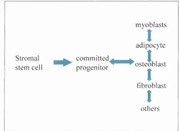
1.2.6 Osteoblasts and the marrow stromal cell lineage
Osteoblasts belong to a lineage of cells that arise from a bone marrow stromal precursor (see figure 1(b)). Evidence indicates that this cell is a common progenitor, not only for osteoblasts, but also osteoblastic, fibroblastic, adipocytic and reticular cells (Friedenstein et al, 1987; Bennett et al, 1991; Beresford et al, 1992). Recent data indicates that muscle cells (Ferrari et al, 1998; Gussoni et al, 1999) and neural tissues also arise from the same marrow stromal precursor cell (Kopen et al, 1999). Characterisation of marrow stromal cell cultures using both in vivo and in vitro
Figure 1(b) - Cells of the marrow stromal lineage
myoblasts
adipocyte
Stromal c o m m itte d ^ g ^
J
stem cell progenitor ^ ^ o s te o b la s t
t
fibroblast
t
& others
Research in this area began when studies into the haematopoietic system revealed the existence of a stem cell in marrow tissue capable of giving rise to bone (Friedenstein and Shapiro Piaetzky, 1966). Studies were carried out using a suspension of marrow stromal cells implanted within diffusion chambers in vivo. It was found that this suspension of marrow stromal cells gave rise to osteogenic tissue (Friedenstein and Shapiro Piaetzky, 1966). In vivo work was also carried out using transplantation of intact bone marrow either under the renal capsule or subcutaneously. Following transplantation, tissue formed that was analogous to bone and bone marrow (Tavassoli and Crosby, 1968; Friedenstein, 1976). In addition, in vivo work revealed the existence of a cell within the bone marrow stroma that was a precursor for colonies of fibroblastic cells. A suspension of marrow stromal cells were placed in diffusion chambers or transplanted under the renal capsule. In this system, a single cell termed fibroblastic colony forming cell (FCFC) or colony-forming unit fibroblastic (CFU-F) was found to give rise to a colony of fibroblastic cells. The term CFU-F was derived from the term colony unit forming spleen (CFU-S) (Friedenstein et al, 1974). Success of CFU-F in vitro culture is reportedly dependent on a number of culture conditions, including the presence of serum, hydrocortisone and EOF (reviewed by Owen, 1988; Bianco et al, 2001).
It has also been shown that bone and marrow stroma are of donor origin, whilst the haematopoietic tissue is of host origin (Friedenstein et al, 1978). Friedenstein et al
(1982) carried out further transplantation studies with suspensions of marrow cells and fibroblastic cells placed in porous sponges, grafted under the kidney capsule. This also gave rise to bone and marrow tissue (Friedenstein et al, 1982).
osteogenic tissue with corresponding presence of cells with an osteoblastic appearance and genetic characteristics. Three weeks after implantation, both cartilage and bone are present in the diffusion chambers (Ashton et al, 1980; Bab et al,
1984).
The multipotentiality of stromal stem cells has been the focus of several later studies, the results of which have perhaps proved rather surprising. Not only does it appear that the osteoblasts, fibroblasts, adipocytes and so-called reticular cells arise from a common precursor but that myogenic and neural cells may also arise from the same stromal origin. Ferrari et al (1998) demonstrated that bone marrow contained cells capable of myogenic tissue production after transplantation. In this study, marrow- derived cells were directly injected into damaged muscle and these cells underwent differentiation into skeletal muscle myocytes after 2-5 weeks. In addition, the study showed that in skeletal muscle injury, caused by whole bone marrow transplantation, donor marrow cells were found to contribute to the muscle fibres formed during healing. The clinical implications of these observations were explored by Gussoni et al (1999). Using a mouse model of muscular dystrophy, affected animals were given a bone marrow transplant from healthy, wild type mice. It was observed that marrow derived skeletal muscle cells developed and engrafted after transplantation and these cells expressed normal dystrophin in a small population of muscle fibrils after 12
weeks. This provides encouragement that, given optimum conditions, genetic therapy could be developed for inherited muscle defects. Bone marrow-derived stem cells have also been shown to differentiate into cardiac myocytes, following myocardial infarction in mice and rats (Kocher et al, 2001; Orlic et al, 2001). It has also been shown that bone marrow stem cells have the capacity to form neurogenic cells in vivo
1.2.7 In vitroosteoblast models
1.2.7.1 Osteoblasts derived from explant culture
A number of in vitro systems for studying osteoblasts are widely used. Cell-line cultures have been frequently used as a source of cells that are easily obtainable and straightforward to maintain in culture. Several attempts have been made to develop in vitro cultures of osteoblastic cells derived from bone (reviewed in Gallagher et al,
1996). Beresford et al (1983) successfully established a system for isolating cells exhibiting osteoblastic characteristics from explants of human trabecular bone. Cells isolated from explants of small trabecular bone chips in culture were shown to have many characteristics of osteoblasts. When cultured in medium containing glucocorticoid and ascorbate these cells were observed to produce an extensive matrix with mineralisation in vitro (Gundle and Beresford, 1995). Ascorbate-2-phosphate, a more stable derivative of ascorbic acid, is important for the expression of the procollagen gene and the biosynthesis of type I collagen (Tajima and Pinnell, 1982; Lyons and Schwartz, 1984). When used alone, glucocorticoids will inhibit collagen synthesis in vitro (Dietrich et al, 1979; Canalis, 1983). In contrast, the use of ascorbate with glucocorticoids will promote the secretion of a dense extracellullar matrix with the addition of |3-glycerophosphate as a source of phosphate (reviewed Gundle et al, 1998). In this thesis cells derived from trabecular explant cultures are termed HOBs.
1.2.7.2 Human osteoblast cell-lines
Table 1(c) - Summary of osteoblast characteristics of MG63 cells and HOBs Osteoblast characteristics Cells Alkaline phospha tase (AP) Osteo calcin (OC)
Effect of 1,25
(0H2)D3
Collagen type I
Effect of PTH
Fibro-nectin MG63s a,b,c a,b,c,d Elevates
AP a,b,c Elevates OC a,b,c,d a Elevates
cAMP and OC
e
b,c
HOBs a,g g Elevates
AP f,gj
Elevates OCf,h,j
Increase in cAMP activity and cell
proliferation i
Key:
a Clover and Gowen, (1994) f Beresford et al (1983) b Franceschi et al (1985) g Beresford gr a/. (1984) c Franceschi, (1988) h Beresford gr a/. (1986) d Lajeunesse et al (1990) i Macdonald, (1986) e Lajeunesse et al (1991) j Thavarajah et al (1993)
MG63 cells were shown to secrete alkaline phosphatase and osteocalcin and activity of alkaline phosphatase and levels of secreted osteocalcin were increased in response to 1,25-dihydroxyvitamin D3 treatment (Franceschi et al, 1985; Franceschi and Young, 1990; Clover and Gowen, 1994). Lajeunesse et al (1990) and (1991) also reported the secretion of osteocalcin and the elevation of this in response to 1,25- dihydroxyvitamin D3 treatment. Subsequent studies showed that osteocalcin secretion was regulated by PTH (parathyroid hormone) and PGE2 (prostaglandin E2) treatment in a cyclic adenosine monophosphatase (cAMP) dependent manner (Lajeunesse et al, 1991). MG63 cells have also been used for several studies investigating the effects of 1,25-dihydroxyvitamin D3 (Mahonen et al, 1990; Maenpaa et al, 1991; Pirskanen et al, 1991; Inaba et al, 1995). Due to the expression of osteoblast characteristics, summarised in figure 1 (c), MG63 cells are a valuable in vitro model.
1.2.7.3 Animal osteoblast cell-lines
Primary osteoblasts are frequently used that are isolated from rat calvaria. These cells are known to respond to parathyroid hormone and express alkaline phosphatase, type I collagen, osteopontin and bone sialoprotein. Under appropriate culture conditions rat calvarial osteoblasts will form nodules of mineralised tissue (Rodan and Noda, 1991; Lian and Stein, 1993). Several animal cell-lines are routinely used as in vitro osteoblast models, for example rat ROS 17/2.8 and UMR 106 (Rodan and Noda,
1991).
1.2.8 Osteoclasts and bone
(Udagawa et al, 1990; Simonet et al, 1997). The RANK system provided evidence that osteoclastogenesis required osteoclast contact with surrounding cells. RANKL, also known as OPGL (osteoprotegrin ligand), is expressed on osteoblasts/stromal cells and the RANK receptor is expressed on haematopoietic osteoclast precursor cells (Suda et al, 1992; 1997; Reddy and Roodman, 1998). The interaction of RANK and RANKL initiates a signalling and gene expression cascade that results in differentiation and maturation of osteoclast precursor cells to active osteoclasts, capable of resorbing bone (Simonet et al, 1997; Yasuda et al, 1998a; 1998b; Lacey
et al, 1998). A receptor termed OPG is also known to bind RANKL and binding to OPG, rather than RANK, leads to inhibition of osteoclastogensis (reviewed in Hofbauer et al, 2000; Teitelbaum, 2000). Many factors known to act on bone cells are known to stimulate the production of RANKL and inhibit the production of OPG, including 1,25-dixydroxyvitamin PTH and others. Oestrogen is thought to inhibit the production of RANKL and osteoclastogenesis. For further detail see Aubin and Bonnelye, (2000). Factors secreted by osteoblasts are also required for osteclastogenesis, such as tumour necrosis factor (TNF), 1,25-dihydroxyvitamin D], interleukins 1, 6 and 11, TGF-P and glucocorticoids (Suda et al, 1992; 1997; Reddy and Roodman, 1998).
The functional osteoclast migrates to areas of bone resorption where it attaches to the bone surface. The plasma membrane forms the tight sealing zone, also known as the clear zone, that is in close contact with the bone surface and is rich in F-actin filaments (Holtrop and King, 1977). Within the tight sealing zone is the ruffled border, the site of matrix degradation. For a comprehensive review see Vanananen and Horton, (1995). Integrin involvement in attachment of osteoclasts to the bone surface and the involvement of these cell adhesion molecules in the resorption cycle has been an area of intensive research and will be reviewed in section 1.5.8.
1.2.9 Bone formation during development
intramembranous ossification. The former requires the formation of a cartilage template that is then replaced by bone (Thompson et al, 1989; Hall and Miyake, 1992; Dunlop and Hall, 1995). Intramembranous ossification results in the formation of bone cells directly from mesenchymal precursors, without the need for a cartilage anlage. Recent studies suggest that there might be some overlap between the two processes of bone development (Nah et al, 2000).
1.3 Integrins
1.3.1 Integrin structure and function
Integrins are non-covalent heterodimers with an a and (3 chain. Figure 1(d) shows a summary diagram of an integrin. Each sub-unit has a large N terminal domain, a transmembrane spanning domain and a short cytoplasmic tail (Hynes, 1992; Sastry and Horwitz, 1993). The a integrin sub-unit varies in size from 120-180kDa. All contain seven tandem repeats of approximately 60 amino acids. It is thought that some of these repeats could contain cation-binding sites, such as those seen in calmodulin. The integrins LFA-1, a l and a l all contain an I domain between the second and third repeats. This is thought to take part in ligand binding. The p integrin sub-units are all 90-110kDa (with the exception of p4 that is 210kDa). At present, there are at least 24 heterodimers that are made up of combinations of 18
Figure 1(d) - Schematic diagram showing the key features of integrin structure
m atrix binding
(1 c hain d iv alen t
cystoino rich d o m a in s (I chain
p la sm a m e m b ra n e
COOH HOOC
talin a n d a-actinii> b in ding
cy to so l
F ro m T he A r t o f t-IBoC^ © 1 9 9 5 G a r la n d P u b l is h i n g , Inc.
Integrins are heterodim eric receptors com p osed o f an a chain (sh ow n in green) and a P chain (show n in
blue). Each chain has a large extracellular dom ain, transm em brane spanning region and an
intracellular dom ain. Cation binding sites are present on the extracellular portion o f the a chain.
1.3.2 Integrin signalling
Figure 1(e) - Integrin mediated signalling
Integrins ^--- ► Growth Factors
1
FAK --- ► Ras
PI3K
Rac
PI3K ► Raf
i
MAPK
Gcnc expression
!
Cell cycle progression, growth, cell survival adhesion and migration
1.3.2.1 Integrin affinity modulation
Binding of integrins to intracellular partners can also lead to a change in the affinity of integrins for external ligands and to the clustering or a more diffuse pattern of integrin expression on the cell surface, termed ‘inside out’ signalling (Hynes 1992; Schwartz et al, 1995). Several structural studies have been carried out to investigate the nature of integrin affinity modulation. It appears that structural changes occur upon integrins binding to ECM ligand and also in the reverse, changing the affinity of integrins for ECM ligands (Lee et al, 1995; Emsley et al, 2000; Takagi, 2001).
1.3.3 The role of integrins in cell adhesion
Cells adhere to underlying substratum via integrins. In this situation, the integrins tend to be located in small adhesive sites, called focal adhesions (Burridge et al, 1988). Within the sites of adhesion between the cell and substratum are both integrins and associated cytoskeletal proteins, for example talin, vinculin, a-actinin and FAK (Petit and Thiery, 2000; Liu et al, 2000). Cell adhesion via integrins not only plays a structural role but leads to the activation of signalling pathways involved in processes such as cell proliferation, differentiation and migration.
1.3.4 Integrins and cell migration
al, 1999). In addition, the ‘tail’ of the cell retracts and this is thought to involve decreased integrin ligation and integrin mediated contractility. One of the processes by which this is thought to occur is via the inhibition of myosin light chain kinase via cdc42 and rac. This leads to lowered myosin light chain phosphorylation and a decrease in stress fibre formation (Sanders et al, 1999; Manser et al, 1997).
1.3.5 Osteoblast migration
It has been proposed that osteoblast migration could be important for the recruitment of these cells to sites of bone formation, both during development and in the adult during the cycle of bone remodelling. It is likely that mature osteoblasts move to sites of bone formation during bone turnover in the adult (Bonewald, 1996). The mechanism of cell migration in osteoblasts is largely unknown although a variety of factors are known to stimulate motile behaviour in these cells. Migration of osteoblasts was first observed in the 1970s in studies using rat osteoblasts (Jones and Boyde, 1977). Using scanning electron microscopy (SEM), pieces of endocranial parietal bones of rats were cultured for up to 24 hours. Osteoblasts were seen to traverse the matrix surface of the bone and also migrated out of vascular channels. Glass spicules were placed on the bone surfaces and osteoblasts were seen to move over these. In the presence of PTE, the osteoblasts were elongated and aligned parallel to each other. In controls on glass, cells were less elongated and aligned. Isolated migrating cells in glass controls were seen to have membrane ruffles, whereas on the matrix they did not (Jones and Boyde, 1977). In retrospect, this study provided evidence for the cell spreading that takes place during the cell migration cycle that is now known to involve integrin mediated adhesion. Of course this work was carried out long before integrins were discovered.
that factors released during bone resorption acted as ehemotactic stimulants to osteoblasts. These findings were of particular interest as they provided a link (coupling) between bone resorption by the osteoclast and the subsequent production of new bone by the osteoblast. Imsiiet al (1998) showed that osteoblasts and osteoblast preeursors express the cell-surface receptor, N-syndecan. When placed in migration assays with the N-syndecan ligand, HB-GAM, these cells migrated rapidly. HB-GAM, also known as pleiotrophin, is an ECM-assoeiated protein rich in lysine and cysteine residues. Mice, transgenic for HB-GAM, exhibited increased bone thickness. The authors suggested that this could have been due to increased recruitment of osteoblasts to sites of HB-GAM expression, via the syndecan reeeptor
(\mdiietal, 1998).
1.4 The extracellular matrix and integrin interaction
An abundant ECM surrounds bone cells. Osteoblasts, chondrocytes and perhaps osteocytes are responsible for the synthesis of this matrix. Mature bone is known to contain abundant type I and type III collagen, osteopontin, bone sialoprotein and osteocalcin. Components of the ECM tend to have the ability to bind several integrins, for example fibronectin binds «5(31 and seven other integrins. There is little data available on the amount of fibronectin present in mature human bone.
1.4.1 Collagen
sites identified in type I collagen including several sites in the a 1 (1) chain such as the putative Asp-Glu-Gly-Ala (DGEA) motif. This sequence has been implicated in a2 p l binding to type I collagen (Staatz et al, 1990; 1991). The involvement of this sequence in a2 p l binding is controversial. Studies by Knight et al (1998) reported that peptide fragments containing the DGEA motif did not bind to the «2pi integrin. This study found that the Gly-Glu-Arg (GER) triplicate, present in type I collagen, was required for the binding of the a2 p l integrin. The GER sequence is similar to the binding motif for a i p i in collagen type IV (Eble et al, 1993). The crystal structure of a complex between the I domain of integrin a2 p i and a triple helical collagen peptide containing a critical GFOGER motif has been shown (Emsley et al, 2000).
The collagen binding integrins and the LFA-1 leukocyte integrin all contain a common feature, an A domain at the N terminus of the a sub-unit (Michishita et al,
1993; Tuckwell et al, 1995; Nolte et al, 1999). Binding at this site is cation dependent (Mn^^ or Mg^^ not Ca^^) (Tuckwell et al, 1995). Although both a i p i and a2 p i bind to collagen types I and IV, their relative affinities for the two types of collagen differ. The a l pi integrin binds to collagen type IV with higher affinity than to type I, whereas a2 p l binds to type I collagen with higher affinity (Kern et al,
1993). It has been suggested that the a i p i and a2pl collagen integrin receptors could have distinct signalling pathways. In a review by Heino, (2000) it was proposed that a i p i integrin-mediated signalling could lead to cell proliferation and a reduction in collagen synthesis, whereas a2pl signalling could lead to collagen production and also collagenase gene expression. It is also suggested that in 3D collagen gel cultures interaction of a2pl with collagen leads to activation of the p38/MAPK signalling pathway. It is likely that the cell signalling pathways linked to collagen binding are a great deal more complicated but the observations made to date in other systems may be applicable to osteoblasts and bone.
repeating amino acids termed type I, type II and type III repeats (Peterson et al, 1983; Ruoslahti, 1988). Fibronectin has a number of receptor binding sites, including several for integrins and also binding sites for other extracellular proteins. Knockout mice for fibronectin or the a5 integrin receptor resulted in the death of embryos early in development (Hynes et al, 1992) and studies have reported the importance of fibronectin and integrin interaction in this process (reviewed in Miyamoto et al,
1998).
Integrins are thought to bind to specific sequences in fibronectin. The most widely researched of these sequences has been the Arg-Gly-Asp (RGD) motif, located in a type III repeat (Ruoslahti, 1988; 1996). Many other components of the ECM contain this motif, for example fibronectin, vitronectin, osteopontin, collagens, thrombospondin, fibrinogen, and von Willebrand factor. Some integrins bind to the RGD sequence of a single adhesion protein only, whereas others recognise groups of them (Ruoslahti and Pierschbacher, 1987). In addition, fibronectin contains the ‘synergy’ site 1 that is also required for aSpi binding (Obara et al, 1988) (shown in figure 1(f)). Studies suggest that the p sub-unit binds the RGD sequence and the a sub-unit binds the synergy sequence (Obara et al, 1988; Kimizuka et al, 1991; Aota
Figure 1(f) - Fibronectin and proteolytic fragments
FIBRONECTIN
S aureus
T 3Π3-D -D 43-G U D -C JC 3-C 3-A A A A A A A A A A A A A A A A O A - -ET
Heparin
I
NH COOH
m û7
ÔT CQ
3 3
RGD
synergy
FIBRONECTIN: PROTEOLYTIC FRAGMENTS
es
30 KDa 45 KDa
• 120 KDa
Fibronectin is an ECM glycop rotein with a modular structure. It contains several binding sites for
1.5 Cell adhesion molecules and bone cells
1.5.1 Integrins and osteoblasts
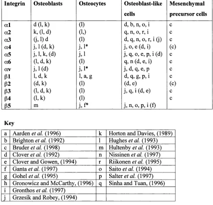
Several studies have sought to establish the profile and function of integrins present in osteoblasts and bone forming culture. The |31 family of integrins appears to be predominantly expressed, with recent studies indicating that a2pl and aSpi interaction with ECM may be of particular importance. The successful production of a mouse pi knockout has provided further evidence for the importance of this integrin family in osteoblast biology (Zimmerman et al, 2000). Many of the studies carried out to date have used cells from varying sources and with differing methods. This has made it difficult to draw firm conclusions as to the profile of specific integrins expressed in osteoblasts. In the following section the information currently available about integrin expression and function in osteoblastic systems will be reviewed.
1.5.2 The integrin profile of osteoblasts
Table 1(g) -Results of studies into the integrin profile of osteoblasts
Integrin Osteoblasts Osteocytes Osteoblast-like
cells
Mesenchymal
precursor cells
a l d (l,k ) (1) d, b, n, 0, i c
a2 k, (1, d) (1.) q, n, 0, r, i c
a3 a ,i)d (1) d, q, n, o, r, i (j) c
a4 j,i* j, 0, e (d, i) (c)
a5 j,l,k ,(d ) j , i j, q, 0, e, p, i (d) c
a6 (1, d, k) (1) q, n (d, e, i) c
av j. 1 (d) j.i* j, d, q, e, p c
p i l,d ,k 1, a,g d, q, g, p, i c
P2 (d,k) (1) (d, e) (c)
p3 (l,d,k) (1) j, q, i (d, e) c
(54 (l,k) (1) c
65 m j,f* j,n , 0, p, i(f)
Key
a Aarden et al. (1996) k Horton and Davies, (1989) b Brighton a/. (1992) 1 Hughes et al. (1993) c Bruder et al. (1998) m Hultenby et al. (1993) d Clover (1992) n Nissinen et al. (1997) e Clover and Gowen, (1994) r Riikonen fl/. (1995) f Ganta et al. (1997) 0 Saito et al. (1994) g Gohel et al. (1995) P Salter gr a/. (1997) h Gronowicz and McCarthy, (1996)
q
Sinha and Tuan, (1996) i Gronthos et al. (1997)j Grzesik and Robey, (1994)
1.5.3 The role of integrins in osteoblasts
In order to investigate the role of integrins in the osteoblast interaction with ECM, adhesion assays have been widely used. These have shown that osteoblasts will adhere at high levels to several matrix components, including plasma fibronectin and type I collagen. Addition of specific integrin blocking antibodies has shown that the pi integrins are of particular importance for osteoblast adhesion on the underlying ECM (Clover et al, 1992; Grzesik and Robey, 1994; Pistone et al, 1996; Gronthos et al, 1997).
Over the past few years studies have started to investigate the role of integrins in osteoblast behaviour further. Evidence has been provided for the presence, and importance, of the aSpi fibronectin binding integrin. Using osteoblasts derived from a rodent calvarial model, it was shown that fibronectin was required for the formation of mineralised nodules in culture. When anti-fibronectin antibodies were added into the culture system the nodular structures normally observed were absent. The authors suggested that the a5 integrin was involved (Moursi et al, 1996; 1997). Later work showed that mature rat osteoblasts were dependent upon fibronectin for survival (Globus et al, 1998). Studies using UMR-106-01 cell-lines showed that addition of «5, a 2 p i, pi and avp3 blocking antibodies significantly reduced mineralisation of these cells in culture (Schneider et al, 2001).
Several studies have sought to identify a role for collagen binding integrins in osteoblastic systems. As mentioned previously, type 1 collagen is the most abundant ECM component and has the potential to play an important role in osteoblast behaviour. Xiao et al (1998) showed that disruption of (%2-type 1 collagen binding, with either blocking antibodies or the DGEA peptide, prevents expression of alkaline phosphatase.
integrin-al and a2 antibodies to cultures significantly reduced the expression of early osteoblastic markers and mineralisation. Cells transfected with a constitutively active BMP-2 receptor were also sensitive to the effects of these integrin-blocking antibodies. As nientioned previously, Schneider et al. (2001) showed that addition of a2|31 integrin blocking antibodies to a UMR cell-line reduced mineralisation of the cells in culture.
1.5.4 pi knockout mice
The production of a pi knockout mouse model provided an insight into just how important pi integrins are in osteoblast biology. Zimmerman et al (2000) produced a mouse model with the pi gene deleted and targeted to mature osteoblasts with the aid of an osteocalcin promoter that is only functional in mature osteoblasts. The pi construct, driven by an osteocalcin promoter, circumvented the problem of pre-natal mortality. Transgenic mice resulting from these experiments had a greatly reduced bone mass. Previously, transgenic studies targeting specific integrin sub-units have proved a stumbling block in bone research, as exemplified by pi, a3, and a5 knockouts that have all been lethal at an antenatal stage of development (Faessler and Meyer, 1995 and Faessler et al, 1996).
Integrins, other than the pi binding partners, may also be expressed in osteoblast systems. Recent work has shown that the avp3 integrin may be important in osteoblast differentiation and bone mineralisation (Cheng et al, 2001). The murine MC3T3-E1 cell-line was transfected with av and p3 in separate vectors. Transfected cells appeared to exhibit increased proliferation but decreased mineralisation. Alkaline phosphatase activity was decreased, as was the expression of osteocalcin, type I collagen and bone sialoprotein. Osteopontin expression was increased. The avp3 integrin has also been shown to redistribute on the surface of primary human osteoblasts, in response to mechanical strain (Wozniak et al, 2000). Cheng et al
In summary, integrins play the following roles in osteoblasts: • Adhesion and migration of cells on ECM substrates;
• Differentiation of mesenchymal precursors and progression to mature osteoblast; and
• Cell survival.
1.5.5 Cadherins and osteoblasts
The cadherin superfamily of cell adhesion molecules consists of the classical cadherins and many cadherin-related molecules. Cadherins are important in many cell biology processes, such as cell polarity, cell proliferation, differentiation and, in turn, many whole tissue phenomena such as morphogenesis. (Takeichi, 1995; Gumbiner, 1996). Studies in osteoblastic systems have revealed expression of the classical cadherins (Cheng et al, 1998; Tsutsumimoto et al, 1999). Classical cadherins are calcium dependent with transmembrane spanning, intracellular and extracellular domains. The extracellular domain of these molecules contains five calcium binding repeats, termed EC1-EC5. The ECl domain is responsible for binding to cadherins on neighbouring cells and contains the His-Ala-Val (HAV) motif that mediates interaction between cadherin molecules on adjacent cells (Overduin et al, 1995; Shapiro et al, 1995). Classical cadherins tend to be located in adherens junctions that form between cells (Angst, 2001). The intracellular domains of these adhesion molecules interact with cytoplasmic proteins such as p-catenin and plakoglobin (PG) that form a link with a-catenin and consequently the actin cytoskeleton (Yap gf a/. 1997).
cadherin expression is important for the differentiation of the osteoblasts (Cheng et al, 1998; 2000; Ferrari et al, 2000).
Several studies have reported the expression of N-cadherin and cadherin-11. Tsutsumimoto, (1999) showed the MC3T3 cells expressed both functional N-cadherin and cadherin-11 (also termed OB cadherin). Treatment with TNF-a was shown to suppress the expression of N-cadherin. Cheng et al (1998) also observed cadherin-11 and N-cadherin in human trabecular derived osteoblasts, osteoprogenitor marrow stromal cells and the cell lines SaOS-2 and MG63. A low level of cadherin-4 mRNA was reported in the trabecular-derived osteoblasts and bone marrow stromal cells
{Cheng et al, 1998).
1.5.6 Role of cadherins in osteoblasts
The results of several studies indicate that cadherins play a role in osteoblast differentiation. A decapeptide containing the HAV motif of human N-cad partially inhibited Ca^^-dependent cell-cell adhesion and completely prevented BMP-2 induced stimulation of alkaline phosphatase activity in bone marrow stromal cells (Cheng et al, 1998). A later study using MC3T3 osteoblastic cells with a truncated dominant negative N-cadherin showed calcium dependent adhesion was decreased significantly in stably transfected clones. Expression of BSP, osteocalcin, type I collagen and alkaline phosphatase activity was also reduced (Cheng et al, 2000). Bone nodule formation in primary cultures of foetal rat calvaria and cell-cell contact in rat TRAB- 11 cells was inhibited by the HAV adhesion motif of N-cadherin (Ferrari et al,
1.5.7 Further non-integrin cell adhesion molecules and osteoblasts
There is little data on the expression and function of cell adhesion molecules, other than integrins and cadherins, in osteoblasts. The cell adhesion molecules intercellular adhesion molecule (ICAM-1) (Tanaka et al, 2000), VC AM (Tanaka et al, 1995) and NCAM (Lee and Chuong, 1992) are reported as being present. In addition, the glycoprotein CD44 has been shown in rat calvarial cultures (Jamal and Aubin, 1996). Expression of the cell-surface heparan sulfate, syndecan has been shown in the human osteosarcoma cell lines MG-63, TE-85, SaOS-2, and U20S, human osteoblast-like cells, rat calvarial osteoblasts and in human bone. Syndecan has been shown to play a role in cell adhesion and migration, and binding of growth factors (Birch and Skerry,
1999). A novel cell adhesion molecule has been reported, POEM. This molecule was cloned from a MC3T3 cDNA library. In situ hybridisation showed expression in bone, kidney, muscles and endocrine organs. It has been suggested that this molecule acts as a ligand for the aSpi integrin (Morimura et al, 2001).
1.5.8 Integrins and osteoclasts
Osteoclasts are known to express at least three integrin receptors, the avp3 vitronectin binding receptor, the «2^1 collagen binding reeeptor and av p l, also a vitronectin receptor. Here the main points to date will be covered; for further details the reader is referred to a several reviews published on the subject (Horton and Davies, 1989; Horton, 1995; Horton et al, 2002).
Although it is known that integrins are involved in bone resorption, the exact mechanism has not yet been elucidated. Some studies have suggested that the tight sealing zone is enriched with certain integrins that could mediate the bone resorbing function (Reinholt et al, 1990, Hultenby et al, 1993; Nakamura et al, 1996). For a comprehensive review of the evidence to date see Horton et al (2002).
Agents that target the av|33 integrin or the RGD sequence block bone resorption. It has been observed that the RGD containing snake venom proteins, for example echistatin (Sato et al, 1990), linear and cyclic RGD peptides and their analogues (Engleman et al, 1997) and antisense oligonucleotides (Villanova et al, 1999), will block bone resorption in vitro. Blocking the a2pi integrin with antibodies will also inhibit bone resorption in vitro. Knockout studies have been carried out to examine the role of integrins in osteoclasts. Bader et al (1998) produced an av knockout mouse. After birth, skeletal development appeared to be normal but the mice then died due to abnormalities of the vascular system. The p3 knockout mouse exhibited platelet defects, associated with human Glanzmann Thrombasthenia, the skeletal defects were surprisingly mild with bone sclerosis becoming evident in later life (McHugh et al, 2000).
1.6 Bone cells, integrins and disease
rheumatoid arthritis is likely to arise from a change in the growth factor and cytokine control of chondrocytes and ECM interaction, (reviewed in Horton et al, 2002).
1.7 Conclusion
2 Chapter 2 - Materials and methods
2.1 Methods - Cell culture
2.1.1 Introduction
The cell lines used were the MG63 human osteosarcoma cells and the C2C12 mouse myoblast cell line. The MG63 cell line was derived from a human osteosarcoma and has been shown to exhibit many osteoblast characteristics in vitro, for example the expression of alkaline phosphatase, osteocalcin and type I collagen (Franceschi et al,
1985 and Clover et al, 1992). C2C12 cells are a stromal cell line that, when grown at high serum levels (20%) in vitro, will differentiate into myotubes. The treatment of these cells with transforming growth factor-p (TGF-p) results in a prolonged period of proliferation and failure to fuse into myotubes. Bone morphogenetic protein-2 (BMP-2) treatment will promote the expression of osteoblastic characteristics by this cell-line (Katagiri et al, 1994). Maintenance of this cell-line at a low concentration of serum, 5%, will prolong proliferation of these cells.
2.1.2 Materials
All tissue culture plastic was obtained from Nunc (Biosciences Ltd, Dublin, Ireland). Culture medium, phosphate buffered saline (PBS) and trypsin-EDTA were obtained from Gihco (Paisley, Scotland). PBS was used at a Ix concentration, as supplied by the manufacturer. Routine cell-culture supplements and extracellular matrix (ECM) components were obtained from Sigma-Aldrich Company Ltd (Poole, Dorset, UK) unless otherwise stated. For culture of C2C12 cells, TGF-p and BMP-2 were obtained from R and D Systems UK Ltd (Abingdon, Oxon, UK). Each cell-line was grown both routinely and during experimental periods in a 37°C incubator with 5% CO2 and 100% humidity.
2.1.3 Routine culture of cells
(25U.mr^), streptomycin (lO^g.ml'^) and fungizone (250ng.ml'^). C2C12 cells were routinely cultured in DMEM with 20% PCS, benzyl penicillin (25U.ml' ^), streptomycin (lOfxg.mr^) and fungizone (250ng.ml'^). Once cells had reached confluence, they were passaged by removal of medium, washed with PBS and treated with 1ml trypsin with a five to ten minute incubation. The cells were then sub cultured by dilution in routine culture medium and placed in tissue culture flasks for growth in an incubator.
2.1.4 Cell counting
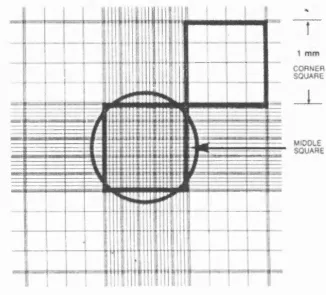
Cells were viewed by light microscopy and counted to obtain the correct cell number required for characterisation studies and functional assays. The following methodology was used:
After trypsinisation, cells were resuspended in routine culture medium and centrifuged at 10 GOOrpm for five minutes. The cells were then resuspended in 5mls of culture medium. lOpl of cell suspension was then placed on a haemotocytometer slide that has a pattern of grids (figure 2(a). The total number of cells in the four comer squares was counted and the average calculated. To get an accurate reading the average number of cells should have been between 40 and 70, if not, the cell suspension was diluted or concentrated as necessary. To further dilute the cell suspension l-5ml of routine culture medium was added to bring the cell count to between 40 and 70. To concentrate the cell suspension cells were centrifuged at
Figure 2(a) - Cell counting using a haemocytometer
CORNER SQUARE (ENLARGEMENT)
Count cells on and left
2.1.5 Preparation of cells for liquid nitrogen storage
Cell-lines were stored long-term in liquid nitrogen. After trypsinisation, cells were resuspended in 5mls of routine culture medium per flask and placed in a 15ml Falcon tube for centrifugation, with one flask of cells per Falcon tube. The cells were centrifuged for five minutes at 10 000 rpm to allow a pellet of viable cells to form at the bottom of the tube. The supernatant was removed, the cells were resuspended in 1ml of routine culture medium with 10% dimethylsulphoxide (DMSO), placed in a cryovial in a foam box for freezing overnight at -80°C and then transferred to liquid nitrogen for long-term storage.
2.1.6 Preparation of primary explant cultures
In addition to the cell-line cultures, primary human osteoblasts (HOBs) were used. These were obtained from trabecular explant cultures. The following methodology, which has been adapted from Beresford et al. (1983) and Gundle and Beresford, (1995), was used:
Trabecular bone, otherwise discarded from routine surgery, was obtained from the Eastman Dental Institute, Great Ormond Street Children’s Hospital and University College London Hospital and transported in sterile a-MEM culture medium. The bone was washed in sterile PBS supplemented with gentomycin (lOpg.mF^), until all non-bone tissue and blood was removed. The bone biopsy was then cut into small pieces of approximately 0.5cm^. The bone chips were then cultured in a-m em supplemented with 10%FCS, gentomycin (lOfxg.mf^) and fungizone (250ng.mF^), in either a 25cm^ (T25) or 80cm^ (T80) tissue culture flask, depending on the number of chips. The bone explant cultures were then placed in an incubator for growth.
2.1.7 Routine maintenance of primary explant cultures (HOBs)
characterisation studies and integrin function studies at sub-confluency. A proportion of cells were also stored in liquid nitrogen at this stage to build up a bank of HOBs for later studies.
2.1.8 Culture of C2C12 ceils for Cbfa-1 studies
The C2C12 cells were trypsinised, counted and diluted to obtain 2x10^ cells per ml of DMEM supplemented with 5% PCS, benzyl penicillin (25U.mT^), streptomycin (10|uig.ml'^) and fungizone (250ng.mP^). Cells were plated out in three 24 well plates with nine wells used per plate. Cells were then grown overnight in an incubator. On the next day (dayl) medium was removed, and the cells were cultured under three different conditions:
• DMEM with 5% PCS, supplemented as above;
• DMEM with 5% PCS, with the addition of 300ng/ml of BMP-2, supplemented as above; and
• DMEM with 5% PCS with the addition of TGP-pi at 5ng/ml of medium, supplemented as above.
C2C12 cells were plated out under each condition in triplicate for three, five and seven days (one 24 well plate for each day). At each of these time points the cells were tested for osteoblastic characteristics using the tests outlined in the following section.
2.2 Methods - Characterisation of osteoblasts
2.2.1 Materials