R E S E A R C H
Open Access
Identification of co-expression modules and
pathways correlated with osteosarcoma and
its metastasis
Jian-sheng Wang
1†, Yun-guo Wang
2†, Yong-sheng Zhong
3, Xue-dong Li
4, Shi-xin Du
4, Peng Xie
4,
Gui-zhou Zheng
4and Jing-ming Han
1*Abstract
Background:Osteosarcoma is the most common bone tumor that occurs in children.
Methods:To identify co-expression modules and pathways correlated with osteosarcoma and its clinical characteristics, we performed weighted gene co-expression network analysis (WGCNA) on RNA-seq data of osteosarcoma with 52 samples. Then we performed pathway enrichment analysis on genes from significant modules.
Results:A total of 5471 genes were included in WGCNA, and 16 modules were identified. Module-trait analysis identified that a module involved in microtubule bundle formation, drug metabolism-cytochrome P450, and IL-17 signaling pathway was negatively correlated with osteosarcoma and positively correlated with metastasis; a module involved in DNA replication was positively correlated with osteosarcoma; a module involved in cell junction was positively correlated with metastasis; and a module involved in heparin binding negatively correlated with osteosarcoma. Moreover, expression levels in four of the top ten differentially expressed genes were validated in another independent dataset.
Conclusions:Our analysis might provide insight for molecular mechanisms of osteosarcoma.
Keywords:Osteosarcoma, Metastasis, Co-expression modules, Pathways
Background
Osteosarcoma is an aggressive cancer in the skeletal system that most commonly occurs in children [1]. The proportion of patients who receive a complete clinical response after standard chemotherapy and multimodal treatment of sur-gery is about 70–75% [2]. For patients diagnosed with me-tastases, the overall 5-year survival rate was approximately 30%, and for patients with relapsed osteosarcoma, the rate decreased to 15% [3]. Since the development of chemother-apy, survival in localized high-grade osteosarcoma has improved considerably. However, there is still no worldwide consensus on a standard chemotherapy approach [3]. Therefore, exploring the molecular mechanism involved in disease progression and metastasis and its relationship with
drug response is critical for identifying effective drugs that overcome drug resistance of tumor.
During these decades, with the dramatic improvement on sequencing technology, the increasing accumulation of sequencing data provides us a data resource to investigate molecular mechanisms of diseases including critical genes, pathways, and networks based on an analysis of omics data. Weighted gene co-expression network analysis (WGCNA) is a method that is widely used in exploration of co-expression modules correlated with traits or phenotypes based on expression data (e.g., expression microarray, tran-scriptome data) [4]. And the R software package for WGCNA was developed to perform weighted correlation network analysis, including procedures of network construction, module identification, gene selection, calcula-tions of topological properties, data simulation, and visualization [4].
In this study, we performed weighted gene co-expression network analysis based on data from RNA-seq of osteosar-coma to identify critical co-expression modules and
* Correspondence:[email protected]
†Jian-sheng Wang and Yun-guo Wang contributed equally to this work.
1Department of Orthopedics Ward II, Shenzhen Children’s Hospital, Shenzhen
518000, China
Full list of author information is available at the end of the article
pathways correlated with osteosarcoma and its clinical characteristics, which might provide new insights for exploring the underlying molecular mechanisms of osteosarcoma.
Materials and methods
Expression profiles of osteosarcoma
Expression profiles of osteosarcoma (GSE87624) were obtained from the Gene Expression Omnibus (GEO) (https://www.ncbi.nlm.nih.gov/geo/). This data included RNA-seq profiles of 52 samples including 44 osteosar-coma tissues, 3 normal bone tissues, 1 osteoblast, and 4 osteosarcoma cell lines based on Illumina HiSeq 2000. After quality control, read mapping, and normalization of read counts of the RNA-seq data, the values of fragments per kilobase million (FPKM) in each gene were calculated.
Co-expression module analysis of osteosarcoma
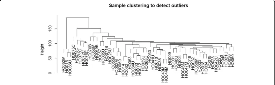
Weighted gene co-expression network analysis (WGCNA) was used to investigate co-expression mod-ules related with osteosarcoma and its clinical character-istics. Genes with the top 25% variance of expression values among samples were included in WGCNA. Then sample clustering was performed to detect whether there were outliers in these samples.
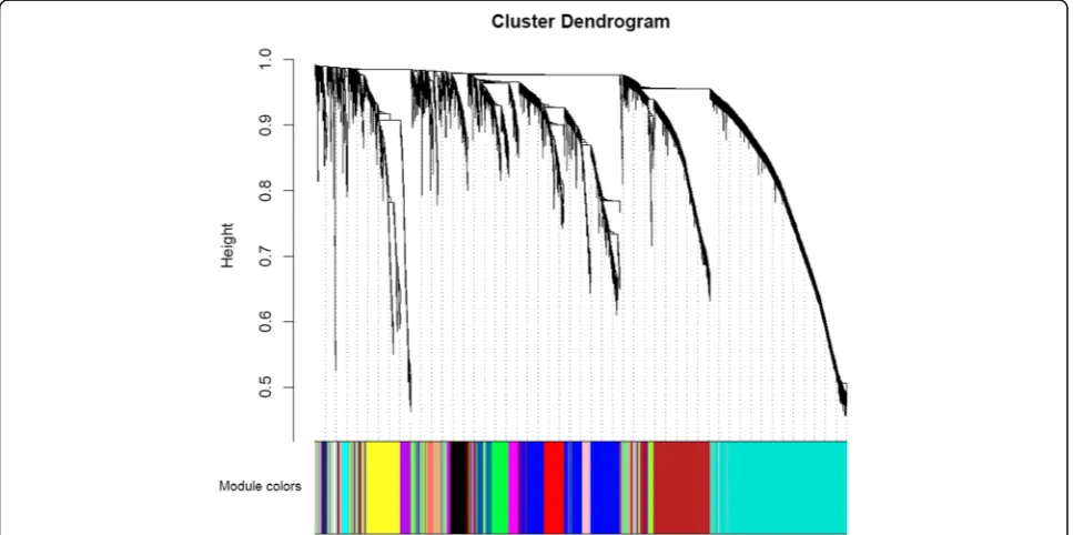
After sample clustering, scale independence and mean connectivity analysis of modules with different power values were performed to determine the soft threshold of module analysis. The power value was set from 1 to 20, and then the values of scale independence and mean connectivity were generated according to these power values. The power value was determined when the scale independence value was 0.9. Then co-expression matrix was calculated under the determined power value, with the minimal module size of 30 and the merge cut height of 0.25. A cluster dendrogram among modules and an eigengene adjacency heatmap between modules were generated.
Module-trait analysis based on clinical characteristics of osteosarcoma
Information on the clinical characteristics of samples with osteosarcoma, including sample type (osteosar-coma/normal), tissue type (osteosarcoma/normal), cell-line type (osteosarcoma/normal), and osteosar-coma type (metastasis/primary/unknown), was col-lected to identify significant co-expression modules related with the clinical characteristics (as trait). Module-trait relationships were calculated according to the correlation between modules and traits; modules that were significantly correlated with individual traits (P value < 0.05, module size < 500) were identified; and
genes in significant modules were then exported for further analysis.
Pathway enrichment analysis of significant co-expression modules
To identify the biological pathways and functions of sig-nificant modules, pathway enrichment analysis was con-ducted by using the R package“clusterProfiler v3.4.4” [5, 6] with genes in significant co-expression modules related with osteosarcoma. Pathways were annotated by informa-tion from the Kyoto Encyclopedia of Genes and Genomes (KEGG) database [7] and Gene Ontology (GO) terms [8], and theP value was adjusted by the Benjamin-Hochberg methods [9]. Pathways with aPvalue < 0.05 were consid-ered as significant pathways.
Validation of differentially expressed genes in independent dataset
To validate whether differentially expressed genes iden-tified in GSE87624 were also differentially expressed in other expression datasets, we investigated the expres-sion levels of the top ten differentially expressed genes in another expression dataset (GSE12865), which included 14 samples (12 osteosarcoma tumor samples and 2 normal human osteoblasts as control). Differen-tially expressed genes of osteosarcoma were analyzed by limma package in R.
Results
Expression profiles of osteosarcoma
For GSE87624, FPKM values of 21,884 genes from 52 samples were obtained. Then log2-transformed FPKM values were used for further analysis. Information of clin-ical characteristics of samples included the sample type (osteosarcoma/normal), tissue type (osteosarcoma/normal), cell-line type (osteosarcoma/normal), and osteosarcoma type (metastasis/primary/unknown). Clinical information for osteosarcoma patients is shown in Table1.
Co-expression module analysis of osteosarcoma
By selecting genes with the threshold of the top 25% variance of expression values, a total of 5471 genes were included in WGCNA. As shown in Fig. 1, by sample clustering, no outliers were observed in 52 samples, thus all samples were included in the analysis.
different colors, including 198 genes in a black module, 837 genes in a blue module, 689 genes in a brown mod-ule, 60 genes in a cyan modmod-ule, 408 genes in a green module, 95 genes in a green-yellow module, 31 in a light cyan module, 131 genes in a magenta module, 50 genes in a midnight blue module, 171 genes in a pink module, 127 genes in a purple module, 302 genes in a red module, 63 genes in a salmon module, 88 genes in a tan module, 1413 genes in a turquoise module, and 428 genes in a yellow module.
Module-trait analysis based on clinical characteristics of osteosarcoma
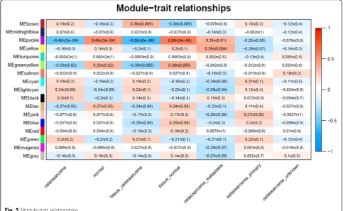
Clinical characteristics of samples with osteosarcoma were collected as a trait, and module-trait relation-ships were calculated according to the correlation be-tween modules and traits. As shown in Fig. 5, for the sample type of osteosarcoma, the most significant modules with negative correlation were purple and green-yellow; for the tissue type of osteosarcoma, the most significant modules with negative correlation
were purple and greenyellow and the positively corre-lated module was brown; and for the osteosarcoma type of metastasis, the most significant modules with positive correlation were purple and yellow.
Pathway enrichment analysis
By pathway enrichment analysis, with the threshold of Benjamin-adjustedP value < 0.05, we obtained significant GO terms and KEGG pathways enriched in significant modules (see Additional file 1). For the purple module (Additional file 1: Table S1), we identified 22 GO terms and 6 KEGG pathways and microtubule bundle formation, IL-17 signaling pathway, and drug metabolism-cyto-chrome P450 were identified; for the brown module (Add-itional file1: Table S2), we identified 58 GO terms and 3 KEGG pathways, which were mainly related with DNA replication and mitotic nuclear division; for the yellow module (Additional file1: Table S3), we identified 26 GO terms, which were mainly related with the anchored com-ponent of membrane and cell junction; and for the green-yellow module (Additional file 1: Table S4), we identified 3 GO terms and 2 KEGG pathways, which were mainly related with regulation of lipolysis in adipocytes and heparin binding.
Validation of differentially expressed genes in independent datasets
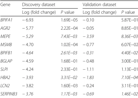
With the threshold of Benjamin-adjusted P value < 0.05 and |log2(fold change)| > 1, we obtained a total of 369 dif-ferentially expressed genes of osteosarcoma in GSE87624. The top ten differentially expressed genes were BPIFA1,
AGR2, MEPE, MSMB, BPIFB1, BGLAP, SLPI, HBA2,
LCN2, and SERPINB3. In an independent dataset
GSE12865, we observed that all ten genes had the same trend of expression level as that in GSE87624, in which four genes, MEPE, BPIFB1, HBA2, and Table 1Sample information in expression profiling
Type Number (%)
Osteosarcoma
Tissues 44 (83.0%)
Cell line 4 (7.5%)
Normal bone
Tissues 3 (5.7%)
Cell line 1 (1.9%)
Tumor type
Metastasis 9 (17.0%)
Primary 20 (37.8%)
Unknown 15 (28.3%)
SERPINB3, were also identified to have significantly differential expression levels (shown in Table 2.
Discussion
In this study, we performed a weighted gene co-expression network analysis (WGCNA) to investi-gate co-expression modules related with osteosarcoma and its clinical characteristics. Significant modules were identified to be correlated with osteosarcoma. For the purple module, which was mainly related with microtubule bundle formation, drug metabolism-cyto-chrome P450, and IL-17 signaling pathway and was identified to be negatively correlated with the trait of osteosarcoma, while being positively correlated with the trait of metastasis in osteosarcoma, previous
studies have reported that increase of microtubule destabilization was related with G1/G2 phase cell cycle arrest and apoptosis, and microtubule inhibitors could trigger autophagy and cell death in osteosar-coma cell line [10]. Besides, IL-17A/IL-17RA interaction promoted metastasis of osteosarcoma cells [11]. Moreover, the resistance of osteosarcoma to chemotherapy was re-lated to cytochrome P450 3A4 [12]. Our results might provide supporting evidence for these previous findings.
For the brown module, which was mainly involved in DNA replication and mitotic nuclear division and was observed to be positively correlated with the trait of osteosarcoma tissue, previous studies have revealed that genes involved with DNA replication and DNA damage were associated with radiosensitivity of osteosarcoma Fig. 2Scale independence and mean connectivity analysis
[13], as well as drug sensitivity [14]. Our results indi-cated that genes in the brown module might be related with carcinogenesis of osteosarcoma and might provide insights for exploring drug targets for the treatment of osteosarcoma.
For yellow module, which was mainly related with the anchored component of membrane and cell junction and was observed to be positively correlated with the trait of metastasis in osteosarcoma, it has been reported that pathways related with cell junction were involved in Fig. 4Eigengene adjacency heatmap between modules
metastasis in various tumors, e.g., lung cancer [15], osteosarcoma [16], and pancreatic cancer [17], which might be due to the molecules in cell junction, and the anchored component of membrane felicitated the migra-tion and invasion tumor cells into other tissues and organs [18]. Our results suggest that genes in the an-chored component of membrane and cell junction might also play important roles in the metastasis of osteosar-coma and were worthy of further investigation.
For the green-yellow module, which was mainly re-lated with regulation of lipolysis in adipocytes and hep-arin binding, it was observed to be negatively correlated with the trait of osteosarcoma. In accordance with our results, there were studies showing that heparin could reduce the osteosarcoma proliferation and growth [19, 20], and heparin binding sites might be potential thera-peutic targets for osteosarcoma [21].
In the validation section, among the top ten differen-tially expressed genes in GSE87624, we validated four genes, MEPE, BPIFB1, HBA2, and SERPINB3, also had significantly differential expression levels in another ex-pression dataset (GSE12865), demonstrating the results identified from the first dataset can be supported by an in-dependent dataset. For MEPE, there were researches re-vealing its involvement with osteosarcoma [22–24]; for BPIFB1, it has been reported that it could inhibit radiore-sistance in nasopharyngeal carcinoma [25, 26]; forHBA2, previous studies have identified different expression levels in the bone marrow of prostate cancer patients [27]; and for SERPINB3, it has been identified as mediators of Ras-driven inflammation and oncogenesis [28]. The effect of these genes on osteosarcoma was worthy of further investigation.
Conclusions
By WGCNA methods on expression data, we identified significant co-expression modules and pathways correlated
with osteosarcoma, as well as metastasis of osteosarcoma, and our analysis might provide insights for the molecular mechanisms of osteosarcoma.
Additional file
Additional file 1:Table S1.Significant pathways of the purple module.
Table S2.Significant pathways of the brown module.Table S3.
Significant pathways of the yellow module.Table S4.Significant pathways of the green-yellow module. (DOCX 27 kb)
Abbreviations
GEO:Gene Expression Omnibus; GO: Gene Ontology; KEGG: Kyoto Encyclopedia of Genes and Genomes; WGCNA: Weighted gene co-expression network analysis
Acknowledgements
We thank authors providing expression data of clubfoot.
Funding
The present research was supported by the Foundation of Shenzhen Health and Family Planning Commission (grant no. SZFZ 2017081), the National Natural Science Foundation of China (grant no. 81702110) and the Foundation of the Second Hospital of Tianjin Medical University (grant no. 2016ydey05).
Availability of data and materials
All data and material were available in the GEO database.
Authors’contributions
JMH contributed to the study concepts; JSW contributed to the study design and data analysis/interpretation; JSW and YGW contributed to the data acquisition, statistical analysis, manuscript preparation, and manuscript definition of intellectual content; YSZ and XDL contributed to the manuscript editing; and SXD, PX, GZZ, and JMH contributed to the manuscript revision/ review. All authors approved the final version of the manuscript.
Ethics approval and consent to participate
Not applicable.
Consent for publication
Not applicable.
Competing interests
The authors declare that they have no competing interests.
Publisher’s Note
Springer Nature remains neutral with regard to jurisdictional claims in published maps and institutional affiliations.
Author details
1
Department of Orthopedics Ward II, Shenzhen Children’s Hospital, Shenzhen 518000, China.2Department of Orthopedics, The Second Hospital of Tianjin
Medical University, Tianjin 300211, China.3Department of Neurosurgery, The First Affiliated Hospital of Shantou University Medical College, Shenzhen 518000, China.4Department of Orthopedics, The Third Affiliated Hospital of Shenzhen University Health Science Center, Shenzhen 518000, China.
Received: 7 October 2018 Accepted: 20 February 2019
References
1. Luetke A, Meyers PA, Lewis I, Juergens H. Osteosarcoma treatment - where do we stand? A state of the art review. Cancer Treat Rev. 2014;40:523–32. 2. Stiller CA. International patterns of cancer incidence in adolescents. Cancer
Treat Rev. 2007;33:631–45.
3. Anninga JK, Gelderblom H, Fiocco M, Kroep JR, Taminiau AH, Hogendoorn PC, Egeler RM. Chemotherapeutic adjuvant treatment for osteosarcoma: where do we stand? Eur J Cancer. 2011;47:2431–45.
Table 2Expression levels of the top ten genes in GSE87624 and GSE12865
Gene Discovery dataset Validation dataset
Log (fold change) Pvalue Log (fold change) Pvalue
4. Langfelder P, Horvath S. WGCNA: an R package for weighted correlation network analysis. BMC Bioinformatics. 2008;9:559.
5. Yu G, Wang LG, Han Y, He QY. clusterProfiler: an R package for comparing biological themes among gene clusters. Omics. 2012;16:284–7.
6. Yu G, Wang LG, Yan GR, He QY. DOSE: an R/Bioconductor package for disease ontology semantic and enrichment analysis. Bioinformatics. 2015;31:608–9. 7. Ogata H, Goto S, Sato K, Fujibuchi W, Bono H, Kanehisa M. KEGG: Kyoto
Encyclopedia of Genes and Genomes. Nucleic Acids Res. 1999;27:29–34. 8. Gene Ontology Consortium. Going forward. Nucleic Acids Res. 2015;43:D1049–56. 9. Huang d W, Sherman BT, Lempicki RA. Systematic and integrative analysis of
large gene lists using DAVID bioinformatics resources. Nat Protoc. 2009;4:44–57. 10. Vetter NS, Kolb EA, Mills CC, Sampson VB. The microtubule network and cell
death are regulated by an miR-34a/Stathmin 1/betaIII-tubulin axis. Mol Cancer Res. 2017;15:953–64.
11. Wang M, Wang L, Ren T, Xu L, Wen Z. IL-17A/IL-17RA interaction promoted metastasis of osteosarcoma cells. Cancer Biol Ther. 2012;14:155–63. 12. Mensah-Osman EJ, Thomas DG, Tabb MM, Larios JM, Hughes DP, Giordano TJ,
Lizyness ML, Rae JM, Blumberg B, Hollenberg PF, Baker LH. Expression levels and activation of a PXR variant are directly related to drug resistance in osteosarcoma cell lines. Cancer. 2007;109:957–65.
13. Dai N, Qing Y, Cun Y, Zhong Z, Li C, Zhang S, Shan J, Yang X, Dai X, Cheng Y, et al. miR-513a-5p regulates radiosensitivity of osteosarcoma by targeting human apurinic/apyrimidinic endonuclease. Oncotarget. 2018;9:25414–26. 14. Li S, Cui Z, Meng X. Knockdown of PARP-1 inhibits proliferation and ERK signals, increasing drug sensitivity in osteosarcoma U2OS cells. Oncol Res. 2016;24:279–86. 15. Liu MX, Zhou KC, Cao Y. MCRS1 overexpression, which is specifically
inhibited by miR-129*, promotes the epithelial-mesenchymal transition and metastasis in non-small cell lung cancer. Mol Cancer. 2014;13:245. 16. Aceto N, Bardia A, Miyamoto DT, Donaldson MC, Wittner BS, Spencer JA, Yu
M, Pely A, Engstrom A, Zhu H, et al. Circulating tumor cell clusters are oligoclonal precursors of breast cancer metastasis. Cell. 2014;158:1110–22. 17. Tan X, Tamori Y, Egami H, Ishikawa S, Kurizaki T, Takai E, Hirota M, Ogawa M. Analysis of invasion-metastasis mechanism in pancreatic cancer: involvement of tight junction transmembrane protein occludin and MEK/ERK signal transduction pathway in cancer cell dissociation. Oncol Rep. 2004;11:993–8. 18. Barbieri I, Pensa S, Pannellini T, Quaglino E, Maritano D, Demaria M, Voster A, Turkson J, Cavallo F, Watson CJ, et al. Constitutively active Stat3 enhances neu-mediated migration and metastasis in mammary tumors via upregulation of Cten. Cancer Res. 2010;70:2558–67.
19. Ichikawa J, Cole HA, Magnussen RA, Mignemi NA, Butler M, Holt GE, O'Rear L, Yuasa M, Pabla B, Haro H, et al. Thrombin induces osteosarcoma growth, a function inhibited by low molecular weight heparin in vitro and in vivo: procoagulant nature of osteosarcoma. Cancer. 2012;118:2494–506. 20. Saito M, Ichikawa J, Ando T, Schoenecker JG, Ohba T, Koyama K,
Suzuki-Inoue K, Haro H. Platelet-derived TGF-beta induces tissue factor expression via the Smad3 pathway in osteosarcoma cells. J Bone Miner Res. 2018. 21. Lamoureux F, Picarda G, Garrigue-Antar L, Baud'huin M, Trichet V, Vidal A,
Miot-Noirault E, Pitard B, Heymann D, Redini F. Glycosaminoglycans as potential regulators of osteoprotegerin therapeutic activity in osteosarcoma. Cancer Res. 2009;69:526–36.
22. Siggelkow H, Schmidt E, Hennies B, Hufner M. Evidence of downregulation of matrix extracellular phosphoglycoprotein during terminal differentiation in human osteoblasts. Bone. 2004;35:570–6.
23. Orimo H, Shimada T. Effects of phosphates on the expression of tissue-nonspecific alkaline phosphatase gene and phosphate-regulating genes in short-term cultures of human osteosarcoma cell lines. Mol Cell Biochem. 2006;282:101–8.
24. Prideaux M, Wijenayaka AR, Kumarasinghe DD, Ormsby RT, Evdokiou A, Findlay DM, Atkins GJ. SaOS2 osteosarcoma cells as an in vitro model for studying the transition of human osteoblasts to osteocytes. Calcif Tissue Int. 2014;95:183–93. 25. Wei F, Tang L, He Y, Wu Y, Shi L, Xiong F, Gong Z, Guo C, Li X, Liao Q, et al.
BPIFB1 (LPLUNC1) inhibits radioresistance in nasopharyngeal carcinoma by inhibiting VTN expression. Cell Death Dis. 2018;9:432.
26. Wei F, Wu Y, Tang L, He Y, Shi L, Xiong F, Gong Z, Guo C, Li X, Liao Q, et al. BPIFB1 (LPLUNC1) inhibits migration and invasion of nasopharyngeal carcinoma by interacting with VTN and VIM. Br J Cancer. 2018;118:233–47. 27. Guzvic M, Braun B, Ganzer R, Burger M, Nerlich M, Winkler S, Werner-Klein M,
Czyz ZT, Polzer B, Klein CA. Combined genome and transcriptome analysis of single disseminated cancer cells from bone marrow of prostate cancer patients reveals unexpected transcriptomes. Cancer Res. 2014;74:7383–94.