Journal of Experimental Pharmacology
Ouabain inhibits monocyte activation in vitro:
prevention of the proinflammatory mCD14
+
/
CD16
+
subset appearance and cell-size
progression
Raphael C Valente1 Elizabeth G Araujo2 Vivian M Rumjanek1 1Instituto de Bioquímica Médica,
Universidade Federal do Rio de Janeiro, Rio de Janeiro, Brazil;
2Departamento de Neurobiologia,
Universidade Federal Fluminense, Rio de Janeiro, Brazil
Correspondence: Vivian M Rumjanek Instituto de Bioquímica Médica, Universidade Federal do Rio de Janeiro, Av Carlos Chagas Filho 373, Centro de Ciências da Saúde, Sala H2-003, Cidade Universitária – Ilha do Fundão, CEP 21.941-902, Rio de Janeiro, Brazil Tel +55 21 2562 6780
Fax +55 21 2270 8647 Email [email protected]
Abstract: Classically described as a potent inhibitor of the sodium-potassium adenosine
triphosphatase enzyme, ouabain has been further shown to act as an effective immunomodulator in mammals. Recently, our group showed that this hormone downregulates membrane CD14 (mCD14) in human monocytes, though it is not known whether monocyte activation status could modify ouabain influence. Hence, we aimed to investigate ouabain effect during monocyte activation in vitro, analyzing mCD14, CD16 and CD69 expression in total monocytes after two periods of adhesion (2 hours and 24 hours) or in small and large monocyte subpopulations separately. Ouabain (100 nM) inhibited monocyte-size increase, characteristic of activation, only when added to cells immediately after 2 hours’ adhesion. Moreover, downregulation of both mCD14 and CD16 expression by ouabain was more effective in small monocytes and in cells after 2 hours’ adhesion. Since monocytes after 24 hours’ adhesion showed no lack of ouabain binding and no CD69 upregulation, it seems that ouabain is somehow incapable of triggering an appropriate cell-signaling induction once monocytes become activated. Furthermore, though p38 MAPK activation was crucial for the impairment in cell-size progression induced by ouabain, its inhibition did not alter ouabain-induced CD69 upregulation, suggesting that other molecules may participate in the response to this hormone by monocytes. Our data suggest that ouabain inhibits monocyte activation in vitro, preventing both cell-size increase and the appearance of the proinflammatory mCD14+/CD16+ subpopulation. Thus, the findings
suggest that individuals suffering from disorders commonly associated with high ouabain plasma levels, like hypertension, may present defective monocyte activation under inflamma-tion or infecinflamma-tion.
Keywords: ouabain, human monocytes, p38 MAPK, mCD14, CD16, CD69
Introduction
Inflammation is a protective mechanism that defends the host from distinct sources of harmful stimuli, such as pathogens or damaged cells, in order to perform the clearance of the hazard signal and start the healing process. In general, the first line of defense of the host during inflammation involves the action of pattern-recognition molecules present in immune cells, which are responsible for the detection of antigens commonly
present in pathogens.1
In this manner, it is known that both infection and sterile conditions are able to promote strong immune responses. Such ability is possible because the immune system does not simply recognize pathogen-associated molecular patterns but also
Dove
press
O R i g i n A l R E S E A R C H open access to scientific and medical research
Open Access Full Text Article
Journal of Experimental Pharmacology downloaded from https://www.dovepress.com/ by 118.70.13.36 on 24-Aug-2020
For personal use only.
Number of times this article has been viewed
This article was published in the following Dove Press journal: Journal of Experimental Pharmacology
endogenous molecules induced under stress or injury conditions, named damage-associated molecular patterns
(DAMPs).2
In general terms, DAMPs are the endogenous equiva-lents of pathogen-associated molecular patterns and include intracellular molecules released from necrotic cells,
heat-shock proteins, uric acid, defensins, and interleukin (IL)-1α,
amongst others. However, in both cases, inflammation can be triggered, for instance, via activation of Toll-like recep-tors (TLRs), leading to the secretion of proinflammatory cytokines and chemokines, thus demonstrating a funda-mental relationship between tissue damage, infection, and inflammation.2,3
Amongst the cells involved in this process, the monocyte/ macrophage plays a crucial role, performing a broad spectrum of functions. Monocytes are mononuclear cells with a very short life span in the circulation, from approximately 1 to
3 days.4 Once activated, they migrate into target tissues or
organs and undergo activation and differentiation, normally into macrophages. They play a key role in the onset, amplifi-cation, and termination of the immune response, participating in several features of immune regulation, such as cytokine production and recognition of pathogen-associated molecular
patterns,5 the capture, processing, and antigen presentation
to T-lymphocytes,6 the clearance of apoptotic cells,7 and also
phagocytosis of either bacteria or fungi.8,9
However, peripheral monocytes are not a homogeneous population. About 20 years ago, Ziegler-Heitbrock and col-leagues described the existence of two subsets of monocytes
in human blood.10 Before that, monocytes were only classified
using the expression of mCD14 on their plasma membrane, but since then monocytes have been able to be further catego-rized based on the expression of mCD14 and CD16, namely the lipopolysaccharide receptor membrane CD14 and the
immunoglobulin Fcγ receptor type III, respectively.
The mCD14high/CD16– monocytes were designated
clas-sical monocytes, due to their abundance in peripheral blood, accounting for 90%–95% of the total count of monocytes
in healthy individuals. The other subpopulation, mCD14+/
CD16+, comprises around 5%–10% of the total monocytes
in blood and is commonly referred to as “nonclassical” or “proinflammatory,” as a result of the observation that these
monocytes secrete high amounts of tumor necrosis factor-α
(TNFα) in response to stimulation with TLR2 or TLR4
agonists.11 The subdivision of these two subpopulations is not
merely owing to their phenotypes but also to functional
dif-ferences, as mCD14+/CD16+ monocytes are increased both in
acute and chronic infection12 and in inflammatory diseases or
sepsis.13,14 Furthermore, proinflammatory monocytes present
higher phagocytic and antigen-presenting activities than the
classical subpopulation as well.12
Monocyte differentiation is a complex matter and may vary depending on the composition of the surrounding envi-ronment. In this manner, monocytes can give rise to M1 or M2 macrophages. Monocyte differentiation into M1 cells is induced by IFNγ but inhibited by IL-10. On the other hand, differentiation into M2 macrophages is induced by IL-10. These macrophages are distinct both phenotypically and functionally, since M1 macrophages are the typical antigen-presenting cells, expressing both MHC class II and B7 molecules for the stimulation of T cells, and M2 mac-rophages lack those receptors but express CD16, allowing
them to destroy antibody-coated target cells or pathogens.15
Besides the regulation of monocyte and macrophage function by immune mediators, these cells are also strongly regulated by hormones. Amongst them, glucocorticoids are usually employed in clinics to treat inflammatory and autoimmune diseases, based on their ability to shut down the inflammatory response. However, the prolonged use of these hormones is associated with several side effects, including
osteoporosis and metabolic disease.16
Nevertheless, another hormone, namely ouabain, was also shown to influence the immune system. Ouabain was first described as a cardiac glycoside extracted from plants with the ability of inhibiting the sodium-potassium adenosine
triphosphatase enzyme (Na+/K+-ATPase), thus promoting a
positive inotropic effect in the cardiac muscle.17 However,
20 years ago, an endogenous analogue of this compound was firstly described in humans18 and later in other mammals,19–21
whose secretion is performed by adrenal glands and the
hypothalamus.19,22 Interestingly, several actions of this
hormone are not related to the inhibition of Na+/K+-ATPase
activity, since the activation of cell-signaling pathways in different cell types, like kidney cells, cardiomyocytes, and retinal ganglion cells, amongst others, has been described. As expected for a multifaceted hormone, the outcomes induced by ouabain are diverse and may include cell proliferation, survival, or even cell death, depending on the concentration
and period of administration.23–26
In the immune system, several actions elicited by ouabain
have been described,27 like the inhibition of mitogen-induced
lymphocyte proliferation and upregulation of the
proinflam-matory cytokines IL-1 and TNFα in vivo.28,29 Conversely, it
has been reported that repeated in vivo treatment with ouabain reduces the inflammation induced by several inflammatory
substances.30 Additionally, our group reported that ouabain
Dovepress
Valente et al
Journal of Experimental Pharmacology downloaded from https://www.dovepress.com/ by 118.70.13.36 on 24-Aug-2020
also induces mitogen-activated cell death in lymphocytes31,32
and calcium mobilization at nanomolar concentrations in thymocytes, a feature that correlated with increased
expres-sion of CD69.33,34 CD69 is a transmembrane glycoprotein
expressed in several cell types, classically described as the earliest antigen cell-surface molecule upregulated in lymphocytes following activation. Although its ligands remain unknown in monocytes and macrophages, CD69 acts as a potent trigger of monocyte activation, being rapidly
upregulated after stimulation with γ-interferon, TNFα, and
lipopolysaccharide.35
Recently, we demonstrated that ouabain induces mCD14 downregulation on human monocytes, a facet that relies on the transactivation of the epidermal growth factor receptor and activation of p38 MAPK.36 However, it is not known
whether the degree of cell activation could be important for the onset of its effects. That observation is critical, as monocytes undergo activation in culture over time, becoming morphologically indistinguishable from tissue
macrophages.37,38
Hence, the aim of the present work was to study whether ouabain may influence in vitro activation/differentiation of human monocytes and if, in turn, monocyte activation sta-tus could modulate ouabain effects. For that, we evaluated activation through phenotypical analysis of cell size and the expression of mCD14 and CD16, receptors that confer critical functional skills to monocytes in the recognition, phagocyto-sis, and pathogen killing.5,7–9,39 We also studied the cellular
mechanisms involved in ouabain actions, particularly the binding of this hormone to the cell surface and the induction of cell-signaling pathways, namely p38 MAPK activation and the induction of CD69.
Materials and methods
Obtainment of blood samples
and monocyte separation
The ethics committee of the Hospital Universitário Clem-entino Fraga Filho-UFRJ agreed with the study protocol, which is registered under the approval number 148/09. Blood samples were obtained from buffy coats via a partnership with the Hemotherapy Unit of the Hospital Universitário Clementino Fraga Filho-UFRJ or from peripheral blood samples collected from healthy volunteers using sodium heparin (Roche, Rio de Janeiro, Brazil) as anticoagulant. Peripheral blood mononuclear cells were separated by density gradient, through centrifugation for 30 minutes at
400 g using a Ficoll-Histopaque reagent (GE Healthcare
Bio-Sciences, Piscataway, NJ). After separation, mononuclear
cells were removed and cells were plated in 24-well plates (Techno Plastic Products, Trasadingen, Switzerland) with culture medium Roswell Park Memorial Institute (RPMI) 1640 (Sigma, St Louis, MO), supplemented with 10% fetal
bovine serum (FBS), at a concentration of 2 × 10⁶ cells/mL
for 2 hours in a humidified chamber with 5% CO2 atmosphere
at 37°C. This procedure was performed in order to allow monocyte adhesion and further separation from lympho-cytes, which remained in suspension. To evaluate possible alterations owing to a higher activation state, a prolonged period of 24-hour adhesion was also performed. After those periods, lymphocytes were removed by washing three times with phosphate-buffered saline (PBS) and attached mono-cytes were incubated again in RPMI medium supplemented with 10% FBS for the analysis in the presence or absence of ouabain.
incubation with ouabain and p38 MAPK
inhibitor
Following adhesion, cells were once more incubated in cul-ture medium with FBS in the presence or absence of 100 nM ouabain, a concentration that was shown to induce a
sig-nificant downregulation in mCD14 expression.36 Moreover,
to verify a broader role of p38 MAPK in the modulation induced by ouabain, we made use of the p38 MAPK
inhibi-tor SB202190 (used at 20 µM; Sigma). The experiments
were performed with incubation in the presence or absence of ouabain for a period of 24 hours, since short incubations
were shown to be ineffective.36
Measurement of mCD14, CD16,
and CD69 expression
To assure that only monocytes would be evaluated, we employed a gate within the monocyte population, using cell size and complexity or granularity as parameters, as described
before.40 For CD16 analysis, only monocytes were evaluated
(mCD14-positive cells), as natural killer lymphocytes, a cell type that also expresses CD16 in the plasma membrane,
exhibit cell size comparable with that of monocytes.41
Briefly, after incubation, monocytes were removed using a cell scraper and labeled with human fluorescein isothiocyanate–conjugated anti-CD14 (1:20 dilution factor; eBioscience, San Diego, CA), with human phycoerythrin– conjugated anti-CD16 (1:15 dilution factor) or human phycoerythrin–conjugated anti-CD69 (1:15 dilution factor), both from BD Pharmingen (San Diego, CA), for 30 minutes at 4°C. Following antibody incubation, cells were washed
twice with PBS + FBS 5% solution, resuspended in the same
Dovepress Ouabain modulates mainly nonactivated monocytes
Journal of Experimental Pharmacology downloaded from https://www.dovepress.com/ by 118.70.13.36 on 24-Aug-2020
solution and kept in ice until measurement of fluorescence in a flow cytometry apparatus (FACScan, Becton Dickinson, San Diego, CA), equipped with an air-cooled Argon Laser tuned to emit 15 mW at 488 nm.
Determination of large-monocyte
subpopulation
Monocytes are known to undergo activation in culture over time, reflecting in an increment in their cell size.37,38 To verify
whether in our experimental conditions these cells were indeed undergoing activation induced by adhesion to the sub-strate, we analyzed the percentage of large monocytes after distinct periods in culture. For that, we made use of the flow cytometry profiles of forward scatter versus side scatter after 2-hour adhesion (always using a gate with mCD14-positive monocytes). Using this moment as a parameter, regions of small and large monocytes were set, and the percentages of large monocytes were evaluated immediately after 2-hour and 24-hour adhesion periods or after 2-hour adhesion followed by maintenance in culture up to 48 hours.
To investigate the effect of ouabain during monocyte activation, the control in the absence of ouabain was used to set the gates of small and large subpopulations. Then, the percentages of large monocytes were determined after the incubation for 24 hours with several ouabain concentrations (1 pM, 1 nM, 10 nM, and 100 nM), comparing the effects in monocytes following 2-hour or 24-hour adhesion.
Measurement of ouabain binding
Flow cytometry was employed to quantify the binding of the fluorescent analogue ouabain-bodipy (Molecular Probes; Life Technologies, Carlsbad, CA). Monocytes were plated
on 24-well plates at a concentration of 2 × 10⁶ cells/mL, and
the experiments were performed 2 or 24 hours after seeding. Then, the medium containing FBS was removed and a fresh RPMI medium in the absence of FBS was added.
Ouabain-bodipy was added in a concentration of 1 µM for 30 minutes,
as described elsewhere.42 After that, cells were gently washed
with PBS and harvested in PBS solution using cell scraper with the plate on ice. Samples were then kept on ice, and fluorescence was measured by flow cytometry in the FACScan device through a 530-nm-long pass filter. Given that ouabain-bodipy concentration employed was high, we also performed a pretreatment with 100 nM unlabeled ouabain for 30 minutes or 24 hours to verify modulation of the staining induced by Na+/K+-ATPase endocytosis.43
Statistical analysis
Each experiment was repeated at least six times, using dif-ferent individuals, except for ouabain-bodipy binding, where the experiments were repeated with at least three donors. Flow cytometry analyses were performed using the software Summit 4.3 (Dako, Fort Collins, CO). Data are expressed
as means ± standard error of the mean or median and were
analyzed using Prism 5.0 software (GraphPad Software,
La Jolla, CA) performing Mann–Whitney or paired t-tests
for comparison of the differences. Values of P, 0.05 were
considered statistically significant.
Results
Monocytes are known to undergo activation and
differentia-tion in culture over time in distinct culture condidifferentia-tions.37,38
However, it was important to analyze if the same results would be achieved under our experimental protocol. So we studied monocyte activation by measuring cell size as a
Large monocytes (%)
24 h adhesion 2 h adhesion
0 20
10 30 40 50
70 *
60
2 h ad + 48 h 2 h ad + 24 h
a,b
a
2 h adhesion 0
20 40
Large monocytes (%)
60 80 100
24 h adhesion
2 h adhesion
Cell granularity
0
0 64 128 192 256
64 128 256 192
0
0 64 128 192 256
64 128 256 192
Cell size
Small Large
Small Large
A B
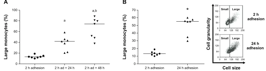
Figure 1 (A and B) Percentage of large-monocyte increases in culture. Using 2 hours’ adhesion as a parameter, gates delimiting small- and large-monocyte subpopulations were determined (B, inset), using data obtained from flow cytometric analysis of cell size versus cell granularity, as described in Materials and methods. (A) Comparison of large-cell numbers among monocytes evaluated immediately after 2-hour adhesion and monocytes after 2-hour adhesion, followed by removal of lymphocytes and further maintenance in culture for 24 hours and 48 hours. Values represent the medians of large monocytes in each condition. (B) Comparison of large-cell amount between monocytes after 2-hour and 24-hour adhesion in the presence of lymphocytes. Values represent the medians of large monocytes. Both experiments were performed using seven individuals.
Notes:a,bStatistical difference from 2-hour adhesion or 2-hour adhesion plus 24 hours in culture, respectively (aP, 0.001; bP, 0.05, Mann–Whitney test); *statistical
difference from 2-hour adhesion (P, 0.001, Mann–Whitney test).
Dovepress
Valente et al
Journal of Experimental Pharmacology downloaded from https://www.dovepress.com/ by 118.70.13.36 on 24-Aug-2020
function of plating time, using two distinct conditions: after lymphocyte removal following 2-hour adhesion and further maintenance in culture for 24 or 48 hours (Figure 1A); or in the presence of lymphocytes during entire adhesion time, comparing monocytes attached for 2 hours to monocytes after 24-hour adhesion (Figure 1B). Figure 1A shows that after 2-hour adhesion, large monocytes comprised just 15% of the total population. Moreover, after 24 and 48 hours in culture in the absence of lymphocytes, the large-monocyte percentage increased to 40% and 75%, respectively, of the total monocyte population. The increase in monocyte size as a function of time in culture was also observed even in the presence of lymphocytes for 24 hours (Figure 1B), show-ing that size increase induced by adhesion to the substrate
relies on the incubation time, independently of the action of surrounding cells, corroborating the data obtained by other authors.37,38
Our next goal was to investigate if ouabain alters mono-cyte activation. Using the standard protocol of adhesion (2 hours), the influence of physiological (1 nM and 10 nM) and pharmacological (100 nM) concentrations of ouabain on the number of large monocytes after incubation for 24 hours was evaluated. Only 100-nM ouabain promoted a significant difference, almost a 60% decrease in the median of large-monocyte percentage, when compared to the con-trol (Figure 2A). Moreover, in some experiments we also employed a concentration of 1 pM ouabain, but no alteration was detected (data not shown).
Large monocytes (%)
100 nM oua Ctrl
0 20 10 30 40 50 100
*
*
60 70 80 90
10 nM oua 100 nM oua
1 nM oua Ctrl
0 10
Large monocytes (%
)
20 30 40
Cell granularity
2 h adhesion 24 h adhesion
A
B
Ctrl 1 nM oua
10 nM oua 100 nM oua
0
0 64 128 192 256
64 128 256
192 0
0 64 128 192 256
64 128 256
192
0
0 64 128 192 256
64 128 256
192 0
0 64 128 192 256
64 128 256
192
Cell size
Small Large Small Large
Small Large Small Large
Large monocytes (%)
100 nM oua Ctrl
0 10
5 15 20 25 50
30 35 40 45
Figure 2 (A and B) Ouabain (Oua) inhibits the appearance of large monocytes in culture. (A) After 2-hour adhesion, cultures containing peripheral blood mononuclear cells were washed for lymphocyte removal, and monocytes were incubated in the presence or absence of several Oua concentrations for a further 24 hours. Next, the number of large monocytes was determined in each condition, using the control (Ctrl) as a parameter to create gates delimiting small- and large-monocyte subpopulations. inset portrays a representative experiment showing the dot plot of cell size versus cell granularity obtained by flow cytometry. (B) Monocytes were allowed to attach to the substrate for either 2 hours or 24 hours. Then, cultures were washed for lymphocyte removal, and monocytes were incubated for a further 24 hours in the presence or absence of 100 nM Oua. Analysis of large-monocyte percentage was similar to that of Figure 2A.
Notes: Values in A and B refer to the medians of large monocytes; *statistical difference from the respective Ctrl (P, 0.01, Mann–Whitney test).
Dovepress Ouabain modulates mainly nonactivated monocytes
Journal of Experimental Pharmacology downloaded from https://www.dovepress.com/ by 118.70.13.36 on 24-Aug-2020
Considering that monocytes after 2-hour and 24-hour adhesion present a remarkable difference in the amount of large monocytes, thus denoting alterations in the cellular activation status, the question of whether ouabain could produce the same effects on cell-size increment in both condi-tions was addressed. For that, experiments were performed to compare the percentage of large monocytes in cultures after two periods of adhesion: 2 and 24 hours. Then, following adhesion, lymphocytes were removed, and monocytes were incubated for further 24 hours in the presence or absence of 100 nM ouabain. As shown in Figure 2B (left panel), oua-bain promoted a significant decrease in the number of large monocytes only in cultures following 2-hour adhesion, as the median of large-monocyte percentage was nearly 45% lower than that seen in control. On the other hand, no significant changes in the median of large monocytes were observed when ouabain was given to monocytes after 24-hour adhesion (Figure 2B, right panel). This finding suggests that monocytes with a higher degree of activation are less susceptible to ouabain influence. Moreover, as cultures immediately evalu-ated after 2-hour adhesion presented nearly 15% of large
monocytes (Figure 1), the decreased amount of large cells seen after incubation with ouabain may be interpreted as an impairment of the normal monocyte-activation process.
Besides phenotypical peculiarities, monocyte subpopula-tions can also be distinguished by means of expression of
functional surface receptors, especially mCD14 and CD16.11
These receptors are important for proper monocyte response and activation, since they are involved in the recognition and response against distinct pathogens, including bacteria, fungi, and antibody-coated particles, culminating in pathogen phagocytosis and killing.8,9,39
To test whether these molecules could be differentially modulated by ouabain, depending on the degree of monocyte activation, we also performed a comparison between mono-cytes attached to the substrate for 2 and 24 hours, followed by incubation with 100 nM ouabain for a further 24 hours, given that only this concentration induced a significant inhibition in the appearance of large monocytes in vitro.
The evaluation of mCD14 expression was performed via
measurement of mCD14high cells, denoting cells with high
expression of this protein, separated by gates delimiting
mCD14 expression (% of mCD14
high
cells)
100 nM oua 100 nM oua
Ctrl Ctrl
0 20 10 30 40 50 100
60 70 80 90
Number of events
2 h adhesion 24 h adhesion
Ctrl
100 nM oua
CD14high cells
CD14high cells
0
100 101 102 103 104 100 101 102 103 104
62 125 250
187 0 62 125 250
187
Fluorescence intensity A
C
MFI variation of CD14 expression compared with control (ctrl = 1) 2 h adhesion 24 h adhesion + 100 nM oua 24 h Ctrl
0.00 0.25 0.50 1.25
*
*
0.75 1.00
B
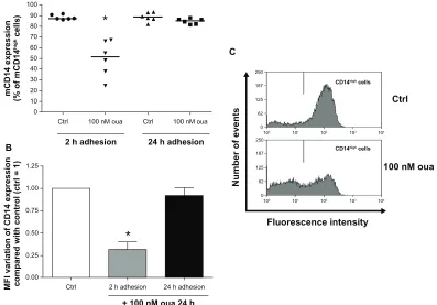
Figure 3 (A–C) Ouabain (Oua) modulates mCD14 expression only in monocytes after 2-hour adhesion. Monocytes were allowed to attach to the substrate for either 2 hours or 24 hours. After that, cultures were washed for lymphocyte removal, and monocytes were then incubated with 100 nM Oua for a further 24 hours or left untreated. Next, monocytes were incubated with antihuman CD14, as described in Materials and methods. (A) Medians of mCD14high cell percentages, denoting cells with a high expression of
mCD14 in their surface; *difference from the respective control (Ctrl) (P, 0.01, Mann–Whitney test). (B) Variation of the means of fluorescence intensities (MFi) of mCD14 expression from monocytes incubated with Oua after 2-hour or 24-hour adhesion, compared with the Ctrl (set as 1); *statistically different from Ctrl (P, 0.0005, paired
t-test). (C) Representative experiment showing the decrease in mCD14high cells (gate located in the right) after treatment with Oua. These experiments were performed
using six individuals.
Dovepress
Valente et al
Journal of Experimental Pharmacology downloaded from https://www.dovepress.com/ by 118.70.13.36 on 24-Aug-2020
the peak of flow cytometry histograms obtained in control monocytes (Figure 3C, upper panel). Figure 3A shows that after 2-hour or 24-hour adhesion, the vast majority
of monocytes (nearly 90%) were mCD14high. However, it
can be seen that treatment with 100 nM ouabain induced a remarkable decline in mCD14 expression in monocytes attached for only 2 hours prior to ouabain treatment. This
effect was verified both in the percentage of mCD14high cells
(Figure 3A and C), where a 40% decrease was detected, and also in the mean of fluorescence intensity (MFI) of mCD14 expression, whose decline in the presence of ouabain was around 70% (Figure 3B). Although mCD14 downregula-tion was also observed in a previous work from our group
using the same experimental protocol,36 the observation that
cells allowed to attach to the substrate for 24 hours do not present any alteration in mCD14 levels induced by ouabain was surprising. Noteworthy was the fact that both protocol conditions differed only in the period of attachment to the
substrate (2 or 24 hours) prior to the incubation with ouabain. So such disparity might be explained by the fact that mono-cytes after 24-hour adhesion display morphological changes indicating a higher degree of activation, when compared to cells attached for only 2 hours.
Next, the same comparison was performed to analyze the modulation of CD16 expression by ouabain, particularly in
the population mCD14+, in order to ensure that only
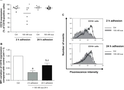
mono-cytes would be evaluated. Similarly to that seen for mCD14 analysis, CD16 expression was more efficiently modulated by ouabain in monocytes attached for only 2 hours to the substrate (Figure 4A). In this case, the median of CD16+ monocytes was about 13% lower in cells treated with ouabain when compared to the control. However, no significant dif-ference was detected in monocytes after 24-hour adhesion (Figure 4A and C). Conversely, it was observed that ouabain induced a decrease in the MFI of CD16 expression even in monocytes treated with ouabain after the prolonged adhesion,
CD16 expressio
n
(% of CD16-positive cells)
100 nM oua 100 nM oua
Ctrl Ctrl
40 50 100
60 70 80 90
Number of events
2 h adhesion 24 h adhesion
2 h adhesion
24 h adhesion
Ctrl 100 nM oua
Ctrl 100 nM oua
CD16+ cells CD16+ cells
0
100 101 102 103 104
100 101 102 103 104
45 91 183
137 0 45 91 183
137
Fluorescence intensity A
C
MFI variation of CD16 expressio
n
compared with control (ctrl = 1) 2 h adhesion 24 h adhesion
+ 100 nM oua 24 h Ctrl
0.00 0.25 0.50 1.25
a
b,c *
0.75 1.00
B
Figure 4 (A–C) Ouabain (Oua) modulates CD16+ cell percentage only in monocytes after 2-hour adhesion. Monocytes were allowed to attach to the substrate either
for 2 hours or 24 hours. After that, cultures were washed for lymphocyte removal, and monocytes were then incubated with 100 nM Oua for a further 24 hours or left untreated. After that, monocytes were incubated with antihuman CD16, as described in Materials and methods. (A) Medians of CD16+ cell percentages; *statistical difference
from the control (Ctrl) (P, 0.05, Mann–Whitney test). (B) Variation in the means of fluorescence intensities (MFi) of CD16 expression from monocytes incubated with Oua after 2-hour or 24-hour adhesion, compared with the control (set as 1); a,bdifferences from the Ctrl (P, 0.001 and P, 0.01, respectively, paired t-test); cstatistical difference
from 2-hour adhesion (P, 0.05, paired t-test). (C) Representative experiment showing CD16 fluorescence histograms obtained by flow cytometry. Black curves indicate control staining, and gray-filled histograms designate monocytes treated with 100 nM Oua for 24 hours after 2-hour adhesion (upper panel) or 24-hour adhesion (lower panel). These experiments were performed using six individuals.
Dovepress Ouabain modulates mainly nonactivated monocytes
Journal of Experimental Pharmacology downloaded from https://www.dovepress.com/ by 118.70.13.36 on 24-Aug-2020
approximately 40% lower than the control. Nevertheless, cells attached for only 2 hours exhibited a much higher varia-tion, with a 67% decrease in the MFI values (Figure 4B).
As mentioned before, the results shown in Figure 1 dem-onstrate that in our experimental protocol, human monocytes become larger after 24-hour adhesion, indicating that after this period monocytes are indeed more activated. Therefore, it was important to assess whether small- and large-monocyte subpopulations display different degrees of susceptibility to this glycoside. For that, the expression of both mCD14 and CD16 was analyzed in monocytes after 2-hour adhesion, evaluating these two monocyte subpopulations separately.
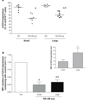
Examining firstly the mCD14high cell percentage, it can be
seen that small and large monocytes both showed a decrease
in the number of mCD14high cells when exposed to ouabain
(Figure 5A). However, the mCD14high cell median decrease
was slightly but significantly minor in large monocytes (approximately 10%, when compared to monocytes after 2-hour adhesion). This feature might possibly occur owing to the elevated mCD14 expression levels observed in these cells under control conditions, roughly twofold higher than seen in small monocytes (Figure 5B, inset). In addition to
mCD14high cell percentage, small and large subpopulations
also presented a similar pattern of modulation in mCD14 MFI induced by ouabain treatment (Figure 5B).
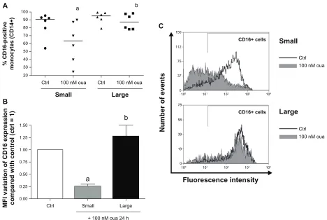
Evaluating CD16 expression after ouabain treatment in these two groups, it is apparent that small monocytes are highly more susceptible than the large subpopulation. The
median of CD16+ monocyte percentage in small cells is about
30% lower in the presence of ouabain, but no significant
mCD14 expression (% of mCD1
4
high
cells)
100 nM oua 100 nM oua
Ctrl Ctrl
0 10 100
20 30 40 50 60 70 80 90
Small Large
A
MFI variation of CD14 expressio
n
comparing with control (ctrl = 1)
Small Large
100 nM oua
Ctrl 0.00
0.25 0.50 1.25
a a,b
a a,b
0.75 1.00
B
MFI of CD14 expression (au)
Small
0 25 50 150
75 100 125
Large *
Figure 5 (A and B) Ouabain (Oua) modulation of mCD14 expression in small and large monocytes. Monocytes were maintained for adhesion in culture for 2 hours, and mCD14 expression was evaluated as described in Figure 3, analyzing small and large subpopulations separately. (A) Medians of mCD14high cell percentages in small and
large monocytes; astatistical difference from control (Ctrl) (P, 0.001, paired t-test); bstatistical difference from small monocytes treated with Oua (P, 0.01, paired t-test).
(B) Variation in the means of fluorescence intensities (MFi) of mCD14 expression from small and large monocytes incubated with Oua, compared with the Ctrl (set as 1);
astatistical difference from Ctrl (P, 0.001, paired t-test); bstatistical difference from small monocytes treated with Oua (P, 0.05, paired t-test). inset: MFi values of mCD14
expression in small and large monocytes; *statistically different (P, 0.05, paired t-test). These experiments were performed using seven individuals.
Dovepress
Valente et al
Journal of Experimental Pharmacology downloaded from https://www.dovepress.com/ by 118.70.13.36 on 24-Aug-2020
alteration was observed in the large-size subpopulation (Figure 6A and C). This scenario was similarly seen when evaluating the MFI of CD16 expression in monocytes treated with this hormone (Figure 6B), where small monocytes showed a loss in CD16 expression of approximately 75%, but the expression of this receptor in large cells was not statistically different from the control.
The data obtained so far suggest that activated monocytes present some degree of resistance to ouabain effects, at least in terms of mCD14 and CD16 modulation. Supporting this idea, it is known that increased CD16 expression is associ-ated with a higher degree of monocyte response, either in
phagocytosis or antigen presentation.12 Interestingly, in our
experimental protocol, monocytes evaluated immediately after 2-hour adhesion presented CD16 levels 47 times lower than monocytes after 24-hour adhesion. Moreover, only 45%
of monocytes were CD16+ promptly after 2-hour adhesion,
contrasting with a vast proportion seen after 24-hour adhesion (90% of monocytes; data not shown).
Thus, two major hypotheses could explain this decreased response in either large monocytes and/or monocytes attached to the substrate for 24 hours: (1) acti-vated monocytes might exhibit a decrease in ouabain-bind-ing sites on their surface or (2) ouabain is unable to trigger cell-signaling events once monocytes become activated.
To test the first conjecture, the fluorescent analogue ouabain-bodipy was used. This probe is useful to measure
ouabain binding to its receptor, which is the Na+/K+-ATPase
alpha subunit.42 If the first assumption were correct, activated
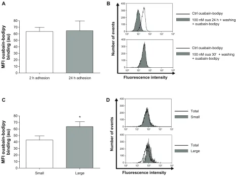
monocytes would display minor ouabain-bodipy fluores-cence in their plasma membrane, compared to nonactivated cells. However, monocytes after 2-hour or 24-hour adhesion presented the same fluorescence levels for this compound (Figure 7A). Furthermore, evaluating small and large monocytes after 2-hour adhesion, it is apparent that large monocytes present even higher levels of ouabain-bodipy binding (Figure 7C and D), refuting the idea that these cells
% CD16-positive
monocytes (CD14+)
100 nM oua 100 nM oua
Ctrl Ctrl
20 30 100
40 50 60 80 70 90
Number of events
Small Large
Small
Large
Ctrl 100 nM oua
Ctrl 100 nM oua
CD16+ cells CD16+ cells
0
100 101 102 103 104
100 101 102 103 104
19 39 78
58 0 37 75 150
112
A
C
MFI variation of CD16 expression compared with control (ctrl = 1) Small Large
+ 100 nM oua 24 h Ctrl
0.00 0.25 0.50 1.50
a
b
a b
0.75 1.00 1.25
B
Fluorescence intensity
Figure 6 (A–C) Ouabain (Oua) differentially modulates CD16 expression in small and large monocytes. Monocytes were maintained for adhesion in culture for 2 hours, and CD16 expression was evaluated as described in Figure 4, analyzing small and large subpopulations separately. (A) Medians of CD16-positive cell percentages in small and large monocytes; astatistical difference from control (Ctrl) (P, 0.05, paired t-test); bstatistical difference from small monocytes treated with Oua (P, 0.05, paired t-test).
(B) Variation in the means of fluorescence intensities (MFi) of CD16 expression from small and large monocytes incubated with Oua, compared with the Ctrl (set as 1);
astatistical difference from Ctrl (P, 0.05, paired t-test); bstatistical difference from small monocytes treated with Oua (P, 0.01, paired t-test). (C) Representative experiment
showing CD16 fluorescence histograms obtained by flow cytometry. Black curves indicate control staining, and gray-filled histograms designate treatment with 100 nM Oua for 24 hours in small monocytes (upper panel) and large monocytes (lower panel). These experiments were performed using six individuals.
Dovepress Ouabain modulates mainly nonactivated monocytes
Journal of Experimental Pharmacology downloaded from https://www.dovepress.com/ by 118.70.13.36 on 24-Aug-2020
80
40 50 60 70
30 20 10 0
2 h adhesion
MFI ouabain-bodipy
binding (au)
24 h adhesion Fluorescence intensity
Number of events
Ctrl ouabain-bodipy 100 nM oua 24 h + washing + ouabain-bodipy
Ctrl ouabain-bodipy 100 nM oua 30' + washing + ouabain-bodipy
100 101 102 103 104
0 100 200 400
300
100 101 102 103 104
0 100 200 400
300
Fluorescence intensity
Number of events
Total Small
Total Large
100 101 102 103 104
0 100 200 400
300
100 101 102 103 104
0 100 200 400
300
A B
80
40 50 60 70
30 20 10 0
Small
MFI ouabain-bodipy
binding (au)
Large
*
C D
Figure 7 (A–D)Large monocytes do not lack ouabain (Oua)-binding sites. Monocytes were incubated with 1 µM Oua-bodipy for 30 minutes in Roswell Park Memorial institute medium, as described in Materials and methods. Values refer to the means of fluorescence intensities of Oua-bodipy labeling in (A) monocytes after 2-hour and 24-hour adhesion, and (C) small and large monocytes after 2-hour adhesion; *statistical difference (P, 0.05, paired t-test). (B and D) Representative experiments showing Oua-bodipy fluorescence histograms obtained by flow cytometry. (B) Black curves indicate control (Ctrl) Oua-bodipy staining in total monocyte population, and gray-filled histograms indicate monocytes pretreated with 100 nM unlabeled Oua for 24 hours or 30 minutes, followed by washing and further staining with Oua-bodipy. (D) Black curves indicate total population binding levels, and gray-filled histograms designate fluorescence levels in small monocytes (upper panel) and large monocytes (lower panel). These experiments were performed at least three times.
do not respond properly to ouabain owing to a decrease in
their Na+/K+-ATPase content.
Though the concentration of ouabain-bodipy used in
the assay was high (1 µM) and could possibly provoke
unspecific binding, lower concentrations did not allow fluorescence measurement. Nevertheless, in order to verify the specificity of the method, we performed experiments pretreating monocytes with 100 nM unlabeled ouabain for 24 hours or 30 minutes prior to ouabain-bodipy staining. Only pretreatment with ouabain for 24 hours induced a sig-nificant decline in ouabain-bodipy fluorescence in human monocytes, when compared to control cells (Figure 7B). Such a result is not surprising, as prolonged incubation with
ouabain is known to induce Na+/K+-ATPase endocytosis,43
but that is an important control to assure that the staining is not related to nonspecific ouabain-bodipy binding to the cell surface.
Our group has previously shown that ouabain increases CD69 expression both in thymocytes and in peripheral blood
lymphocytes.33,44 This protein seems to be the earliest
induc-ible cell-surface glycoprotein acquired during lymphoid activation and functions as a signal-transmitting receptor
in lymphocytes.45 Although CD69 expression is induced in
vitro in cells of most hematopoietic lineages, its constitutive
expression has been described in monocytes.46
To check the second hypothesis, ie, whether ouabain is unable to trigger cell-signaling responses in large monocytes, the expression of CD69 was assessed in human monocytes, comparing cells after 2-hour and 24-hour adhesion as well as small and large monocytes after 2-hour adhesion. As depicted in Figure 8A, incubation with ouabain induced a
twofold increase in the percentage of CD69+ monocytes after
2-hour adhesion. Interestingly, this effect was observed even when analyzing small- and large-monocyte subpopulations
Dovepress
Valente et al
Journal of Experimental Pharmacology downloaded from https://www.dovepress.com/ by 118.70.13.36 on 24-Aug-2020
separately. The medians of CD69+ monocytes incubated with ouabain were approximately 70% and 60% higher, for small and large monocytes, respectively (Figure 8B). However, ouabain did not exert any effect on CD69 expression in monocytes after 24-hour adhesion (Figure 8C and D).
Despite the fact that CD69 is well known as a receptor engaged in lymphocyte activation,45 the data obtained in
Figure 8 indicate that the number of monocytes expressing this receptor declines about 50% in monocytes after 24-hour
adhesion + 24 hours in culture when compared to monocytes
after 2-hour adhesion + 24 hours in culture. Thus, this result
suggests that CD69 might be transiently expressed in human monocytes in vitro.
In a previous report, our group revealed that p38 MAPK had a central role in the modulation of mCD14 expression
induced by ouabain.36 After the observation in the present
work that ouabain induces a broader effect in monocytes, preventing the activation induced by adhesion, it was impor-tant to verify whether this kinase could play a role in this ouabain facet.
As seen in Figure 9, though 100 nM ouabain signifi-cantly reduced the median of large-monocyte percentage in culture (from 21.71% in the control to 10.78% in the pres-ence of ouabain), incubation with the p38 MAPK inhibitor
SB202190 significantly reverted this effect (medians:
SB202190 = 24.03 and SB202190 + ouabain = 19.15).
Thus, our findings suggest that p38 MAPK might have a central role in the effects triggered by ouabain in monocytes, also controlling the effects of this hormone in cell-size increase. In order to confirm this hypothesis, we tested whether p38 MAPK function could also be important for the ouabain-induced CD69 upregulation in human mono-cytes, given that the pattern of modulation observed for this receptor is the opposite of that observed for both mCD14 and CD16. Therefore, monocytes after 2-hour adhesion were incubated with 100 nM ouabain in the presence or absence of the p38 MAPK inhibitor, and the expression of CD69 was subsequently assessed. As described in Table 1, p38 MAPK inhibition did not alter CD69 induction triggered by ouabain in monocytes, refuting the theory that p38 MAPK could act in regulating all effects triggered by ouabain in human monocytes.
Discussion
The modulation of human monocytes by ouabain is a com-plex matter and involves the activation of several signaling pathways, varying with each cell type. In our experimental protocol, the concentration of 100 nM ouabain was relatively 50
40
30
20
10
0 Ctrl
CD69-positive cells (%
)
100 nM oua
Fluorescence intensity
Number of events
15
12
9
6
3
0 Ctrl
CD69-positive cells (%
)
100 nM oua
CD69-positive cells (%)
Oua large Oua small
Ctrl small Ctrl large
0 10 70
20 30 40 50 60 Ctrl
100 nM oua CD69+ cells
100 101 102 103 104
0 75 150 300 225
Fluorescence intensity
Number of events
Ctrl 100 nM oua CD69+ cells
100 101 102 103 104
0 75 150 300 225
B
a
b
CD69-positive cells (%)
Oua large Oua small
Ctrl small Ctrl large
0 5 30
10 15 20 25 D A
C
*
CD69+ cells
Figure 8 (A–D)Ouabain fails to induce CD69 expression in monocytes after 24-hour adhesion. Monocytes were maintained for adhesion in culture for 2 hours (A and B) or 24 hours (C and D), and then incubated with 100 nM Oua for 24 hours or left untreated. After that, monocytes were incubated with antihuman CD69, as described in Materials and methods.
Notes: (A and C) Means of CD69-positive cells in total population; *statistical difference from control (Ctrl) (P, 0.001, Mann–Whitney test). (B and D) Medians of CD69-positive cells in small and large monocytes; a,bstatistical difference from small and large monocyte Ctrl, respectively (aP, 0.01 and bP, 0.05, Mann–Whitney test). These
experiments were performed using six individuals.
Dovepress Ouabain modulates mainly nonactivated monocytes
Journal of Experimental Pharmacology downloaded from https://www.dovepress.com/ by 118.70.13.36 on 24-Aug-2020
higher than the physiological range for this hormone in human plasma, from approximately 100 pM to nanomolar concentrations, depending on the experimental conditions and possibly on the individuals tested.18,47 However,
con-trasting with several observations from other authors, which made use of excessively high quantities of this hormone when studying the regulation of cardiac hypertrophy and renal cell
toxicity induced by ouabain,23–25 the concentration used in
the present study was observed in the plasma of individuals suffering from cardiac dysfunction treated with ouabain or even under acute stress conditions.47,48 Moreover, taking
into account that adrenocorticotropic hormone stimulation induces the secretion of both corticosterone/cortisol and oua-bain by adrenal glands,49,50 it has been suggested that ouabain
also acts as a stress hormone,51 whose secretion is likely to be
above its physiological range in any type of situation where plasma cortisol levels are elevated.
Though monocytes seemed to be robustly modulated
by ouabain in terms of mCD14 expression,36 the present
data support the idea that ouabain influence may fluctuate according to the extent of cellular activation, as cells after 2-hour and 24-hour adhesion are distinctly regulated. Interestingly, both mCD14 and CD16 downregulation induced by ouabain were significantly more pronounced in cells after short periods of adhesion (2 hours). At this time, monocytes were clearly less activated, reflected in the low number of large monocytes, when compared to cells after the prolonged adhesion of 24 hours.
Furthermore, our data showed that CD16 modulation induced by ouabain was considerably higher in the small-monocyte subpopulation. Taking into account that the amount
of CD16+ monocytes increases in culture, our results
sug-gest that ouabain prevents the appearance of the mCD14+/
CD16+ subpopulation in vitro. However, it is not known so
far whether in vivo administration of ouabain could induce
a similar effect, preventing the increase of mCD14+/CD16+
monocytes in either infection or inflammatory diseases. That is an important issue, as the rise of the mCD14+/
CD16+ subpopulation is related to the aggravation of several
immune disorders, such as inflammatory arthritis or sepsis,
by means of the secretion of proinflammatory cytokines.12–14
Nevertheless, there is growing evidence in the literature suggesting that ouabain is able to inhibit the secretion of the
proinflammatory cytokines TNFα, IL-1β, and IL-6 induced
by lipopolysaccharide in vitro and even to protect mice against lipopolysaccharide-induced lethal endotoxemia
Large monocytes (%
)
Oua SB202190
SB202190
oua + SB202190
CD14
100 nM oua
100 nM oua + SB202190
Ctrl
0 20
10 30 40 50 60
a,b
Cell granularity
0
0 64 128 192 256 64
128 256 192
0
0 64 128 192 256 64
128 256 192
Cell size
Small Large
Small Large
0
0 64 128 192 256 64
128 256 192
0
0 64 128 192 256 64
128 256 192
Small Large
Small Large
Figure 9 Role of p38MAPK inhibition in the modulation of monocyte cell-size progression induced by ouabain (Oua).
Notes: Monocytes were maintained for adhesion in culture for 2 hours and then incubated with 100 nM Oua for 24 hours, in the presence or absence of the p38MAPK inhibitor SB202190 (used at 20 µM). Values refer to the medians of large monocytes in each condition (evaluating only CD14+ cells); astatistical difference from control
(P, 0.01, paired t-test); bstatistical difference from Oua + SB202190 (P, 0.05, paired t-test). Inset: Representative experiment showing monocyte size and
cell-granularity profiles obtained by flow cytometry, where gates delimiting small and large subpopulations are depicted, as well as both Oua and SB202190 effects on large-monocyte subpopulation number. These experiments were performed using six individuals.
Table 1 Effect of the p38 MAPK inhibitor SB202190 on
ouabain-induced CD69 upregulation in human monocytes
Treatment CD69+ monocytes (means ± SEM)
Control 17.51 ± 3.04
Ouabain 100 nM 41.19 ± 7.58*
SB202190 23.19 ± 7.40
SB202190 + ouabain 100 nM 38.80 ± 7.79*
Notes: Monocytes were maintained for adhesion in culture for 2 hours and then incubated with 100 nM ouabain for 24 hours, in the presence or absence of the p38 MAPK inhibitor SB202190 (used at 20 µM). Values refer to the percentage of large monocytes in each condition ± standard error of mean (SEM); *statistical difference from the respective controls (P, 0.05, paired t-test). These experiments were performed using six individuals.
Dovepress
Valente et al
Journal of Experimental Pharmacology downloaded from https://www.dovepress.com/ by 118.70.13.36 on 24-Aug-2020
and inflammation induced by zymosan,29,30 thus supporting
the hypothesis that the mCD14+/CD16+ subpopulation may
be modulated by ouabain even in vivo. Additionally, it is not known whether ouabain treatment in vivo could evoke preferential differentiation towards M1-type macrophages, which lack CD16 in their surface and play an important role in antigen presentation and facilitating the onset of Th1 cell response.
Additionally, we found that ouabain action was not merely restricted to the modulation of cell-surface molecules. Analyzing the percentage of large monocytes after 24-hour incubation with 100 nM ouabain (Figure 2A and B), it was clear that this subpopulation is almost 50% lower in the presence of ouabain than that observed in the control. This finding raised the possibility that this hormone could
modu-late monocyte size due to impairment of Na+/K+-ATPase
ion transport, which could result in cell shrinking, as seen
in other experimental models.52,53
Nevertheless, some important observations reject this proposition. First, the studies observing cell shrinkage
induced by Na+/K+-ATPase inhibition were performed with
ouabain concentrations far higher than that employed in
the present work, ranging from 50 to 100 micromolars.52,53
Moreover, prolonged stimulation with high ouabain con-centrations was shown to be associated with cell death, but viability assay using annexin V and propidium iodide, which stain cells undergoing apoptosis or necrosis, respectively, indicated the opposite in our experimental protocol. In this manner, incubation with 100 nM ouabain led to only a slight increase in the amount of cells stained with annexin V (nearly 7%) in monocytes after 2-hour and 24-hour adhesion (data
not shown), corroborating previous results.36
Second, the number of large monocytes after incubation with ouabain for 24 hours (Figure 2) is almost the same as that seen in monocytes immediately observed after 2-hour adhesion (Figure 1), indicating that this hormone is indeed preventing cell-size progression stimulated by adhesion to the substrate, instead of inducing cell shrinkage. Furthermore, flow cytometry analyses of size versus granularity patterns did not reveal any significant alteration in the size of small monocytes treated with ouabain (Figure 2A, inset). Finally, the observation in Figure 9 that the p38 MAPK inhibitor practically reverted the ouabain effect on cell size reinforces the idea that this facet has no association with ion-transport inhibition.
The association between monocyte size and activation/ differentiation process in vitro has long been known. Even under distinct experimental conditions, monocytes may
undergo activation by adhesion to the substrate and
differenti-ate into macrophages.37,38 In this fashion, the more monocytes
are able to attach to a certain surface, the faster the activation/ differentiation process is. To exemplify this relationship, Hsu et al54 investigated the ability of human monocytes
to attach to distinct polyester-type surfaces covered with different amounts of gold nanoparticles or left untreated. They observed that the presence of activated monocytes dis-playing large size and spread morphology was significantly higher in the untreated polyester-type surface after 96 hours in culture. Moreover, the subcutaneous implantation of this material in rats also induced a higher formation of a fibrous capsule after 19 days, when compared to the gold-covered surfaces. Thus, the authors confirmed that the size of the fibrous capsule in vivo was consistent with the degree of monocyte activation/differentiation in vitro, demonstrating unequivocally the close relationship between monocyte size
and activation induced by adhesion.54
The observation that p38 MAPK inhibition influences
both ouabain-induced downregulation of mCD1436 and
ouabain-induced cell size–progression restraint in culture, seen in the present work, raised the idea that p38 MAPK might play a central role in the cell-signaling events trig-gered by this hormone in monocytes. However, as described in Table 1, p38 MAPK inhibition showed no effect on the regulation of CD69 expression by ouabain, refuting the notion that all ouabain effects in monocytes may be a result of p38 MAPK function.
These data reinforce the idea that the response of human monocytes to ouabain relies on the activation of several signal-ing pathways, which may act together to evoke a broad modi-fication in the phenotype, morphology, and possibly function of monocytes. Corroborating this assumption, several authors have postulated that other important signaling events are also induced by ouabain, like ERK1/2 activation and reactive
oxy-gen species oxy-generation,24 tyrosine-kinase receptors and PKC
activation,26 cytosolic calcium mobilization,33,34 production
and secretion of the proinflammatory cytokines TNFα and
IL-1β,29 amongst others. Hence, it is likely that one or more
of those molecules might be involved in CD69 upregulation induced by ouabain.
Aiming to understand why activated cells failed to respond to ouabain, we firstly analyzed whether those cells presented fewer binding sites for this hormone, namely the
Na+/K+-ATPase enzyme. However, the binding of the
fluo-rescent analogue ouabain-bodipy was similar in monocytes after either 2-hour or 24-hour adhesion. Additionally, large cells after 2-hour adhesion exhibited a slight increase in
Dovepress Ouabain modulates mainly nonactivated monocytes
Journal of Experimental Pharmacology downloaded from https://www.dovepress.com/ by 118.70.13.36 on 24-Aug-2020
ouabain binding compared to small monocytes, demonstrat-ing that activated monocytes are not less responsive due to a decline in ouabain-binding sites. The reason for the rise in ouabain-binding sites observed in large monocytes is not known at the moment. However, it is possible that large
cells may require more copies of Na+/K+-ATPase in order to
maintain a proper ion homeostasis or that, in turn, activated
cells might express another Na+/K+-ATPase alpha-subunit
presenting higher affinity for ouabain.
The observation that altered Na+/K+-ATPase activity and/
or content in the plasma membrane is/are not the cause for the lack of response to ouabain seen in activated cells are not surprising, because it is well known that several actions of this hormone are intrinsically related to the activation of
cell-signaling molecules.23–26 Corroborating this assumption,
CD69 expression was only induced by ouabain in monocytes after 2-hour adhesion, although this result was obtained both in small and large monocytes. So these data suggest that monocytes after 24-hour adhesion fail to respond to ouabain owing to an impediment on the activation of one or more cell-signaling pathways triggered by this hormone.
Our data show, for the first time, that similarly to that observed in lymphocytes, ouabain also induces CD69 upregulation in human monocytes. Though CD69 function is still unknown in monocytes, this molecule acts as a signal-transmitting receptor in lymphocytes, being the earliest
cel-lular glycoprotein acquired under activation.45 However, given
that control monocytes after 24-hour adhesion plus 24 hours in culture displayed half the percentage of CD69+ cells of that seen in control monocytes after 2-hour adhesion plus 24 hours in culture, it seems that CD69 expression in monocytes is transitory in vitro. Therefore, CD69 expression levels could not be effectively related to monocyte-activation status.
The results obtained in the present work show that cell size is not the only attribute for the evaluation of whether monocytes are ouabain-sensitive or not. It is clear that the period of adhesion to the substrate is crucial, probably requir-ing alterations in gene-expression patterns. Though altera-tions in total protein expression by ouabain were not assessed in the present study, the differential expression induced by ouabain of CD16 and CD69 in monocytes after 2-hour and 24-hour adhesion confirms this assumption. Moreover, CD69 upregulation induced by ouabain clearly indicates that monocytes chronically treated with this hormone do not become unresponsive, but probably perform different physiological roles.
In conclusion, adhesion of monocytes to endothelial cells and their subsequent migration into tissues is one of the
earliest changes detectable during immune responses.55 So
as ouabain preferentially modulates monocytes displaying a phenotype comparable to that found in circulation, our results suggest that this hormone could act in preventing the exac-erbation of inflammatory responses either in inflammatory diseases or infection. Alternatively, it is likely that individuals presenting one of the pathophysiological conditions where ouabain plasma levels are constantly elevated, like chronic stress or essential hypertension, may possibly exhibit defi-cient monocyte activation.
Acknowledgments
This work was supported by grants from Coordenação de Aperfeiçoamento de Pessoal de Nível Superior (CAPES), Fundação de Amparo à Pesquisa do Estado do Rio de Janeiro (FAPERJ), and Conselho Nacional de Desenvolvimento Científico e Tecnológico (CNPq). The authors are indebted to the Hemotherapy Unit from Hospital Universitário Clementino Fraga Filho-UFRJ for the kind donation of buffy coats used in the present study.
Disclosure
The authors report no conflicts of interest in this work.
References
1. Ferrero-Miliani L, Nielsen OH, Andersen PS, Girardin SE. Chronic inflammation: importance of NOD2 and NALP3 in interleukin-1beta generation. Clin Exp Immunol. 2007;147(2):227–235.
2. Piccinini AM, Midwood KS. DAMPening inflammation by modulating TLR signalling. Mediators Inflamm. 2010;2010:672395.
3. Bianchi ME. DAMPs, PAMPs and alarmins: all we need to know about danger. J Leukoc Biol. 2007;81(1):1–5.
4. Heidenreich S. Monocyte CD14: a multifunctional receptor engaged in apoptosis from both sides. J Leukoc Biol. 1999;65(6):737–743. 5. Akira S, Hemmi H. Recognition of pathogen-associated molecular
patterns by TLR family. Immunol Lett. 2003;85(2):85–95.
6. Tacke F, Ginhoux F, Jakubzick C, van Rooijen N, Merad M, Randolph GJ. Immature monocytes acquire antigens from other cells in the bone marrow and present them to T cells after maturing in the periphery. J Exp Med. 2006;203(3):583–597.
7. Gregory CD. CD14-dependent clearance of apoptotic cells: relevance to the immune system. Curr Opin Immunol. 2000;12(1):27–34. 8. Schiff DE, Kline L, Soldau K, et al. Phagocytosis of gram-negative
bacteria by a unique CD14-dependent mechanism. J Leukoc Biol. 1997; 62(6):786–794.
9. Netea MG, Gijzen K, Coolen N, et al. Human dendritic cells are less potent at killing Candida albicans than both monocytes and macrophages. Microbes Infect. 2004;6(11):985–989.
10. Ziegler-Heitbrock HW, Passlick B, Flieger D. The monoclonal antimono-cyte antibody My4 stains B lymphoantimono-cytes and two distinct monoantimono-cyte subsets in human peripheral blood. Hybridoma. 1988;7(6): 521–527. 11. Strauss-Ayali D, Conrad SM, Mosser DM. Monocyte subpopulations
and their differentiation patterns during infection. J Leukoc Biol. 2007;82(2):244–252.
12. Nockher WA, Scherberich JE. Expanded CD14+ CD16+ monocyte subpopulation in patients with acute and chronic infections undergoing hemodialysis. Infect Immun. 1998;66(6):2782–2790.
Dovepress
Valente et al
Journal of Experimental Pharmacology downloaded from https://www.dovepress.com/ by 118.70.13.36 on 24-Aug-2020