T E C H N I C A L A D V A N C E
Open Access
Development and validation of a high
throughput, closed tube method for the
determination of haemoglobin alpha gene
(
HBA1
and
HBA2
) numbers by gene ratio
assay copy enumeration-PCR (GRACE-PCR)
Andrew Turner
1*, Jurgen Sasse
1and Aniko Varadi
2Abstract
Background:Deletions of theα-globin genes are the most common genetic abnormalities in the world. Currently multiplex Gap-PCRs are frequently used to identify specific sets of common deletions. However, these assays require significant post-amplification hands on time and cannot be used to identify novel or unexpected deletions. The aim of the current study was to develop a rapid screening test for the detection of all deletions of theα-globin genes that can be integrated into a high volume clinical laboratory workflow.
Methods:A gene ratio assay copy enumeration (GRACE) PCR method was developed by simultaneous amplification of targets in theα-globin genes (HBA1andHBA2) and the chloride channel voltage sensitive 7 (CLCN7) reference gene. A novel application of High Resolution Melting (HRM) analysis then allowed rapid determination ofα-globin gene copy numbers. The assay was validated using 105 samples with previously determined and 62 samples with unknownα-globin genotypes.
Results:The GRACE-PCR assay detected abnormalα-globin gene copy numbers in 108 of the 167 samples evaluated. The results were consistent with those from a commercial reverse hybridization assay and no allele drop out was observed.
Conclusions:We have successfully developed and validated a GRACE-PCR screening test for the detection of deletions and duplications of theα-globin genes. The assay is based on copy number determination and has the ability to detect both known and novel deletions of theα-globin genes. It is a closed tube technique; consequently the risk of amplicon contamination is negligible. Amplification, detection and analysis can be completed within one hour, making it faster, cheaper and simpler than other existing tests and thus well suited as a rapid first step in a clinical laboratory workflow.
Keywords:Alpha thalassaemia, Copy number determination, Gene quantification, qPCR, GRACE-PCR
* Correspondence:[email protected]
1Department of Pathology and Laboratory Medicine, Sheikh Khalifa Medical
City, Abu Dhabi, United Arab Emirates
Full list of author information is available at the end of the article
Background
Deletions of the α-globin genes are the most common genetic disorders in the world. The World Health Organization (WHO) estimates that 20 % of the world’s population may have a deletion in one or moreα-globin genes [1]. The majority of these cases are asymptomatic carriers, where the deletion of one or twoα-globin genes may provide some protection against malaria [1, 2]. However, deletion of three genes causes a form of thalas-saemia intermedia known as Hb H disease and often re-sults in significant anaemia. Deletion of all fourα-globin genes leads to Hb Barts hydrops foetalis syndrome and normally results in foetal death; such pregnancies are also associated with a significant risk of maternal mor-bidity or mortality [3]. It has been estimated that around 13,000 pregnancies are affected annually by severe forms ofα-thalassaemia [1].
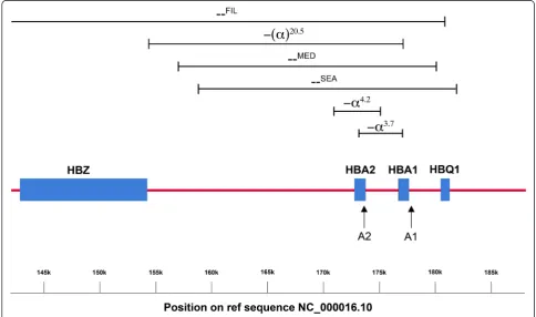
Normal haemoglobin is a tetramer composed of two α-globin and two β-globin chains. There are two func-tional α-globin genes, designated Haemoglobin Alpha 1 (HBA1) and Haemoglobin Alpha 2 (HBA2). These genes are 97 % homologous and are located 2941 bp apart on chromosome 16 in a region known as theα-globin locus (Fig. 1) [4]. In contrast toβ-thalassaemia, where deletions are relatively rare, the majority of α-thalassaemia cases
arise from gene deletions [2]. The designation α+is used to describe a chromosome with reduced α-globin gene expression, while αo describes a chromosome with no gene expression [4]. Typically, α+ thalassaemia alleles re-sult from a small deletion that leaves just one functional α-globin gene on the chromosome, although there are also non-deletional forms that arise as the result of a point mutation. αo Thalassaemia alleles are normally caused by larger deletions affecting both theHBA1 and HBA2genes [2].
Identification of α-globin gene deletions is often per-formed in a clinical laboratory setting using either Gap-PCR or reverse hybridization. A number of multiplexed assays based on these techniques have been described for the most common deletions [5–9]. These techniques are simple and fairly robust, but the use of either agarose gel electrophoresis or hybridization for identification makes them relatively time consuming and labour inten-sive. Thus, these technologies are not particularly con-venient for high throughput testing in a routine clinical laboratory. An additional limitation of these techniques is that they only identify specific targeted deletions and rare or novel deletions may be missed.
Due to these limitations, methods for the detectionα -globin gene deletions based on a number of alternative
[image:2.595.55.540.400.686.2]techniques have been developed. These include Multi-plex Ligation-dependent Probe Amplification (MLPA) [10, 11], Denaturing High Performance Liquid Chroma-tography (DHPLC) [12, 13], Quantitative PCR (qPCR) [14–18] and melting curve analysis [19–21]. Melting curve analysis methods are particularly attractive, since they do not require post amplification processing by electrophoresis or chromatography. A novel technique based on co-amplification of a target and a reference gene, followed by melting analysis has been described for the Adenomatous Polyposis Coli (APC) gene [22]. More recently this technique has also been applied to the Ataxia telangiectasia mutated serine/threonine kin-ase (ATM) and the Phosphate and tensin homolog (PTEN) genes [23]. Here we describe the use of a similar approach to develop a novel gene ratio assay copy enu-meration (GRACE) screening test for the simultaneous copy number determination of the HBA1 and HBA2 genes relative to the reference gene chloride channel voltage sensitive 7 (CLCN7) [14]. This GRACE-PCR screening test is a single tube assay that can detect any rearrangements of the α-globin genes that affect the 3′ ends of the HBA1 or HBA2 genes. In contrast to most previously described assays for deletion or duplication of the α-globin genes, the GRACE-PCR assay is a simple closed tube technique, which requires no further hands on time after the initial PCR setup. Data analysis is per-formed by a novel application of the standard features of the HRM analysis software, making it high throughput and well suited for performing rapid initial screening of samples in the clinical laboratory.
Methods
Samples
This study was conducted using anonymous, archived material from blood specimens submitted to the Sheikh Khalifa Medical City (SKMC) laboratory, Abu Dhabi, United Arab Emirates forα-thalassaemia screening. Eth-ical clearance was obtained from the SKMC Institutional Research and Ethics Committee to use this material for the current study. In total 167 samples were used.
The performance characteristics of the GRACE-PCR assay were originally established using 105 samples with known genotype. These samples were selected to repre-sent as many different α-globin genotypes as possible. Samples with point mutations of the HBA2 gene in the stop codon or untranscribed region (UTR) were also in-cluded to assess their impact on assay performance. The assay was further validated using an additional 62 sam-ples of unknown genotype.
DNA Extraction
Genomic DNA was extracted from whole blood using the QIAamp DNA Blood Mini kit (Qiagen, Germany) in
accordance with the Manufacturer’s instructions. The concentration of the extracted DNA was measured using a NanoDrop 2000 spectrophotometer (Thermo Scien-tific, USA) and adjusted to 10 ng/μl with 10 mM Tris, 0.5 mM EDTA (pH 9.0).
Genotyping
All samples included in the study were genotyped using the CE-IVD (Conformité Européene - In Vitro Diagnos-tics) marked α-Globin StripAssay (ViennaLab Diagnos-tics, Austria) in accordance with the manufacturer’s instructions. The StripAssay is able to detect theαααanti3.7 duplication and the following deletions: -α3.7, -α4.2, -(α)20.5,
–SEA
, –Med, –Fil, –Thai in addition to a number of point mutations including αConstant Springα, αIcariaα,αpolyA-1α and αpolyA-2α
.
Primer design and synthesis
To detect deletions in HBA1 and HBA2simultaneously three primer pairs targeting the HBA1, HBA2 and CLCN7 genes were used in the GRACE-PCR assay (Table 1). All primers were designed using Oligo Primer Analysis Software version 7.56 (Molecular Insights, USA). The approximate location of the amplification sites for the HBA1 and HBA2 genes along with the locations of the deletions identified in the current study are shown in Fig. 1. Primers were specifically designed to work at the same annealing temperatures, but to generate products with melting temperatures approximately 3 °C apart. Alternative primer pairs were also developed to amplify HBA2 and CLCN7 genes by GRACE-PCR to investigate anomalous re-sults from the original GRACE-PCR (Additional file 1: Table S1).
High Performance Liquid Chromatograph (HPLC) purified primers were commercially synthesized (Meta-bion, Germany).
GRACE-PCR and data analysis
Each 12.5 μL GRACE-PCR reaction contained 20 ng of genomic DNA, 0.25 units of Kappa HiFi HotStart poly-merase (Kappa Biosystems, Boston, USA), 0.25 units of Platinum Taq polymerase (Invitrogen, Carlsbad, USA), 0.5 μL of LightCycler 480 ResoLight dye (Roche Diag-nostics, Mannheim, Germany), 1x Kappa GC buffer (Kappa Biosystems), 0.3 mM of each dNTP (Kappa Bio-systems), 0.45 μM of each primer for the HBA1 and HBA2genes and 0.16μM of each primer for theCLCN7 gene.
at 72 °C. Melting was performed at a rate of 0.2 °C/s over the temperature range 77 °C to 87 °C, with data ac-quisition on the HRM channel. PCR conditions for the alternative primers are given in the Additional file 1: Method section.
Data analysis was performed using the HRM module Rotor-Gene Q series software version 1.7.
Results
GRACE-PCR design and optimisation
[image:4.595.56.539.99.205.2]Initially, we used Platinum Taq polymerase in combin-ation with 1.2 M betaine during the development of the GRACE-PCR assay. This strongly favoured the amplifica-tion ofHBA2overHBA1, which made the quantification of the latter difficult. This issue could not be resolved by a
Table 1Primers used for the GRACE-PCR alpha globin copy number assay
Primer Direction Primer Sequence 5′-3′ Target Gene symbol
Primer Concentration (μM)
Product size (bp)
Product Tm (°C)
Position on Ref Sequence NC_000016.10
Forward CACCCGGCCTCATGGAT CLCN7 0.16 155 79.4 1,462,101 to 1,462,255
Reverse AAGAGAACTACAGACCAACACCC
Forward CCATCTTTACGTTTCTGGGCACTC HBA1 0.45 131 82.2 177,800 to 177,930
Reverse GCCATGCTGGAGTGGGACTTC
Forward CCGTTAAGCTGGAGCCTCGGT*A HBA2 0.45 171 85.2 173,594 to 173,764
Reverse ACACCTCCATTGTTGGCACAT
*indicates the use of phosphorothioate (PTO) to block 3′exonuclease activity
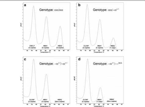
[image:4.595.61.537.342.692.2]simple redesign of the primers. Consequently, various al-ternative enzymes and enhancers were tried and with the use of an enzyme cocktail containing Platinum Taq and Kappa HiFi HotStart polymerases simultaneous and com-parable amplification of HBA1 and HBA2 was achieved (Fig. 2). The Kappa HiFi HotStart polymerase has 3′to 5′ exonuclease proofreading activity, thus to avoid misprim-ing of the HBA2forward primer toHBA1 a phosphoro-thioate (PTO) block was incorporated in the primer sequence (Table 1) [24]. Amplification of theCLCN7 con-trol gene [14, 25] was more efficient than that of the target α-globin genes, which was corrected by reducing the con-centration of the CLCN7primers (Table 1). Limiting the number of PCR cycles resulted in termination of amplifi-cation during the exponential phase of the PCR reaction. This ensured that the relative amount of each PCR prod-uct remained proportional to the initial number of copies of the gene present.
Data analysis
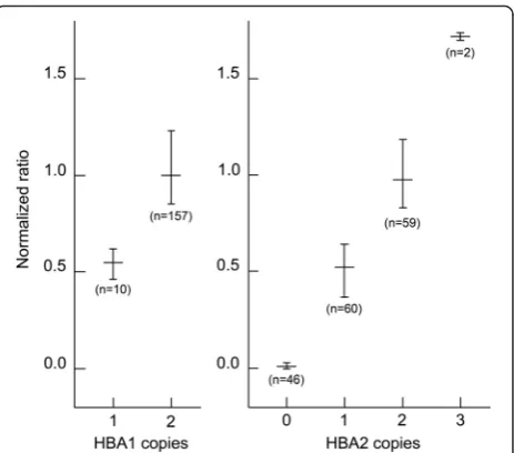
The amount of PCR product from each of the three genes targeted in the GRACE-PCR assay is proportional to the height of the peak on the -dF/dT versus temperature plots (Fig. 2). Initially we determined the gene copy numbers by measuring peak heights on the -dF/dT plots (Fig. 2) and then normalized the data to a wild type control sample included in the same run using the following calculation: Normalized ratio = (RWT Con-trol/TWT Control)/(RUnknown/TUnknown); where R and T are
the peak heights for the reference (CLCN7) and the tar-get (HBA1 or HBA2) genes, respectively. The subscript
‘WT Control’ indicates the wild type control sample. This process worked well and allowed the correct copy number for bothα-globin genes to be determined for all samples tested (Fig. 3). However, this method of data analysis was complex and time consuming. Conse-quently, we developed a simplified visual method for data analysis, which utilized features available in the HRM module Rotor-Gene Q series software (Fig. 4) and eliminated the need for manual calculations.
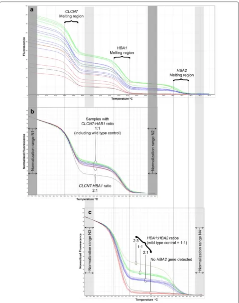
As expected in the GRACE-PCR assay three distinct melt transition steps could be observed in the melting curves corresponding to the melting temperatures of the PCR products initially designed to be 3°C apart (Fig. 4a, Table 1). Therefore, the steps could be assigned to the melting of the products from CLCN7, HBA1 and HBA2 genes as indicated in Fig. 4a. Normalization of the raw HRM data was performed in two stages using the Rotor-Gene Q series software. First the data was normalized by setting normalization ranges before the melting of the CLCN7product and after the melting of theHBA1 prod-uct (normalization ranges N1 and N2 indicated by the dark grey bars in Fig. 4). This resulted in a plot in which the height of the curve between the CLCN7 and HBA1
melt regions is dependent on the gene copy number ra-tio of CLCN7:HBA1. Inclusion of a known wild type control sample in each run allowed rapid visual deter-mination of any samples with abnormal copy numbers for HBA1 (Fig. 4b). Since the number of copies of the CLCN7 reference gene is assumed to be two, this ratio can be used to determine the copy number for the HBA1 gene. In the second stage of the analysis normalization ranges were set before the melting of the HBA1product and after the melting of theHBA2 prod-uct (using the normalization ranges N3 and N4 indi-cated by the light grey bars in Fig. 4c). This resulted in a plot allowing the gene copy number ratio of HBA1:HBA2 to be determined (Fig. 4c). Since the copy number for the HBA1 gene was already determined in the first stage of the analysis this ratio allowed the copy number for HBA2 to be easily derived. This alternative visual method of analysis led to identical results as the more complex calculation method for all 167 samples included in the study, but proved far quicker and sim-pler to perform.
Assessment and validation of the GRACE-PCR assay
An initial assessment of the assay was carried out using 105 samples that had been previously genotyped with the commercial α-Globin StripAssay (ViennaLab Diagnostics, Austria). These 105 samples included 70 samples that had deletions or duplications of one or more of the α-globin genes. Samples with mutations of
Fig. 3Normalized ratios of -dF/dT peak heights for different copy numbers of theHBA1andHBA2genes. The horizontal bars indicate the mean ratio and the vertical bars indicate the range from the minimum to maximum ratio observed for each gene copy number. The data was normalized with the equation: Normalized ratio = (RWT
Control/TWT Control)/(RSample/TSample), where R and T is reference gene
[image:5.595.307.539.86.290.2]the HBA2 stop codon and UTR were also included to ensure that they did not interfere with the assay performance.
GRACE-PCR correctly identified the 70 samples shown by the α-globin StripAssay to have deletions or duplications of the α-globin genes (Table 2). Mutations of the stop codon or UTR did not affect the copy num-ber result, however, the -(α)20.5deletion (Fig. 1) was de-tected as a deletion of the HBA2 gene only. This was because the -(α)20.5 deletion removes all of the HBA2 gene and the 5′end of theHBA1gene, but does not ex-tend to the primer target at the 3′ end of the HBA1 gene. The performance of the GRACE-PCR assay was further validated by the analysis of an additional 62
samples prior to genotyping by the α-globin StripAssay. GRACE-PCR detected α-globin gene deletions in 38 of these samples, all of which were subsequently confirmed by genotyping (Table 2). Additionally, 143 samples were analysed using alternative primers (Additional file 1: Table S2) and no allele drop out was observed.
In total 108 of the samples were identified by the α -globin StripAssay as having a deletion or duplication of the α-globin genes. GRACE-PCR returned abnormalα -globin gene copy numbers for all 108 positive samples and did not generate any false positives (Table 2). This equates to a sensitivity of 100.0 % (95 % confidence interval 96.6-100.0 %) and a specificity of 100.0 % (95 % confidence interval 93.9-100.0 %).
(See figure on previous page.)
[image:7.595.57.540.392.672.2]Fig. 4Normalization of GRACE-PCR assay to determine gene copy numbers. Raw melting curves show three distinct steps corresponding to the melting of theCLCN7, HBA1andHBA2gene products generated during the GRACE-PCR Screening Test (a). Setting the normalization regions N1 and N2 (dark grey bars) in the HRM analysis software allowed the gene copy number ratio forCLCN7:HBA1to be determined (b). Re-setting the normalization regions to N3 and N4 (light grey bars) allowed the determination of the gene copy number ratioHBA1:HBA2(c). The copy number forCLCN7is assumed to be two, which allows the number ofHBA1copies to be calculated, which in turn allows the determination of the number ofHBA2copies
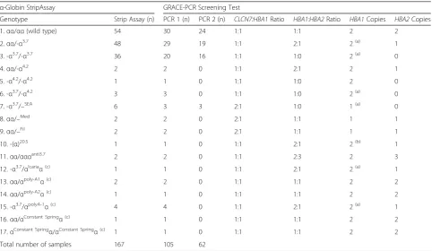
Table 2Genotypes of samples used in the development and validation of the alpha globin gene ratio analysis copy enumeration PCR assay (GRACE-PCR)
α-Globin StripAssay GRACE-PCR Screening Test
Genotype Strip Assay (n) PCR 1 (n) PCR 2 (n) CLCN7:HBA1Ratio HBA1:HBA2Ratio HBA1Copies HBA2Copies
1.αα/αα(wild type) 54 30 24 1:1 1:1 2 2
2.αα/-α3.7 48 29 19 1:1 2:1 2(a) 1
3. -α3.7/-α3.7 36 20 16 1:1 1:0 2(a) 0
4.αα/-α4.2 2 2 0 1:1 2:1 2 1
5. -α4.2/-α4.2 1 1 0 1:1 1:0 2 0
6. -α3.7/-α4.2 3 3 0 1:1 1:0 2(a) 0
7. -α3.7/–SEA 6 3 3 2:1 1:0 1(a) 0
8.αα/–Med 2 2 0 2:1 1:1 1 1
9.αα/–Fil 2 2 0 2:1 1:1 1 1
10. -(α)20.5 1 1 0 1:1 2:1 2(b) 1
11.αα/αααanti3.7 2 2 0 1:1 2:3 2 3
12. -α3.7/αIcariaα(c) 1 1 0 1:1 2:1 2(a) 1
13.αα/αpoly-A1α(c) 2 2 0 1:1 1:1 2 2
14.αα/αpoly-A2α(c) 1 1 0 1:1 1:1 2 2
15. -α3.7/αpolyA-1α(c) 4 4 0 1:1 2:1 2(a) 1
16.αα/αConstant Springα(c) 1 1 0 1:1 1:1 2 2
17.αConstant Springα/αConstant Springα(c) 1 1 0 1:1 1:1 2 2
Total number of samples 167 105 62
The GRACE-PCR assay was assessed using samples with previously determined (PCR1) and unknown (PCR 2) genotypes. GRACE-PCR was able to detect all 108 samples identified by the commercialα-globin StripAssay as havingα-globin gene rearrangements (genotypes 2 to12)
(a) Due to the positioning of primers at the 3′ends of theα-globin genes, the hybrid -α3.7gene is counted as anHBA1gene in this assay (b) The -(α)20.5
deletion does not extend to the 3′end on theHBA1gene targeted by the GRACE-PCR screening test primers, consequently only the deletion of the
HBA2gene is detected
Discussion
Conventional Gap-PCR and reverse hybridization assays are commonly used methods to detectα-globin gene de-letions [5–9]. These techniques are reliable and allow identification of the specific targeted gene rearrange-ments. However, Gap-PCR is not ideal for high volume testing in a clinical laboratory, since it is relatively time consuming and labour intensive. PCR protocols for Gap-PCR require a long extension step in order to generate fairly large amplicons; consequently cycling is slow and typically takes approximately 3 h. The subsequent detec-tion by agarose gel electrophoresis involves considerable hands on time, adding around another 2 h to the ana-lysis. In common with Gap-PCR, reverse hybridization assays are open tube techniques that take several hours to complete. Therefore, a stepwise clinical laboratory in-vestigation incorporating a rapid screening step of α -globin gene deletions has been suggested to be a more efficient approach [26]. In our proposed workflow here, all samples are first screened by the rapid and inexpen-sive GRACE-PCR assay, and then all those identified with a variant copy number are analysed further with a different method that can identify the specific genotype. The initial step in our workflow has the additional ad-vantage that it can detect novel rearrangements not identified by other assays, for example Gap-PCR.
Fragment analysis by capillary electrophoresis have also been used to determine α-globin gene copy num-bers [26–29]. These assays make use of the eight base length differences between the HBA1 and HBA2 genes. One limitation of these assays is that due to rearrange-ments of the α-globin genes, such as the African α2 Polymorphism or theα12 allele, this difference in length between theHBA1 andHBA2 genes can be absent [30– 32]. Indeed, these polymorphisms are reported to be very common, with the Africanα2 Polymorphism reach-ing a frequency of up to 12 % in some African popula-tions [30]. In contrast, GRACE-PCR is not affected by these gene rearrangements and it does not require the use of a capillary electrophoresis based genetic analyser making it a more universally applicable and faster screening method.
In common with most PCR based techniques, GRACE-PCR could potentially yield incorrect results due to a SNP within a priming site inhibiting primer an-nealing and therefore causing allele dropout. The 5′-end of the reverse primer for HBA2 coincided with the stop codon and consequently, there was a risk that mutations of this codon might result in allele dropout. This was tested by including samples from individuals carrying a mutation in the stop codon, Hb Constant Spring (HBA2:c.427T > C) and Hb Icaria (HBA2:c.427T > A), in the assay validation. Our results showed that the assay design was robust and the presence of the mutations of
theHBA2stop codon did not affect the results (Table 2, Genotypes 12, 16 and 17).
Some other low frequency SNPs have also been de-scribed within the GRACE-PCR priming sites, the most frequent of which (rs2261869) has a minor allele fre-quency (MAF) of T = 0.0016 [33]. Thus, the possibility of allele dropout needs be considered if an abnormal GRACE-PCR result is obtained that cannot be con-firmed by other methods. One possibility for checking the original GRACE-PCR screening result is the use of alternative primers for HBA2 and CLCN7 (Additional file 1: Table S1) before proceeding to more complex tests such as MLPA. In the current study all results of the
‘original’ and ‘alternate’ GRACE-PCRs were consistent with the genotype from the StripAssay indicating that no allele dropout had occured, suggesting that such events should be rare.
We also considered the impact that point mutations might have on the performance of GRACE-PCR. Point mutations may alter the melt curve shapes, but would have a negligible impact on total fluorescence, which is dependent on the amplicon amount produced. Conse-quently, point mutations are not expected to have a sig-nificant impact on copy number determination, since GRACE-PCR interpretation is dependent on the fall in fluorescence as each amplicon melts. Indeed, inclusion of samples with theαPoly A1(HBA2:c.*92A > G) andαPoly A2 (HBA2:c.*94A > G) mutations in the method validation confirmed that point mutations within the amplicons did not affect the result interpretation (Table 2, Genotypes 13 to 15).
Other melt curve based methods have been previously described for the detection of α-globin gene deletions. Most of these are modified Gap-PCR assays that use melting curve analysis as means of detection, thus eliminating the need for agarose gel electrophoresis [13, 18, 19, 34]. This makes them more convenient than conventional Gap-PCR, but they have only been developed for a limited set of deletions such as –SEA and –THAI [19]. A melt curve method that compares ratios of HBA1 and HBA2 gene PCR products has also been described, but this method did not include a reference gene and thus, required additional tubes to detect certain genotypes [20].
In common with GRACE-PCR, both fragment analysis by capillary electrophoresis and qPCR quantify the α -globin genes relative to a reference gene that is assumed to have a copy number of two. The current GRACE-PCR assay used the well characterizedCLCN7 gene for refer-ence [14, 25, 35]. This gene is well suited for this applica-tion because it has no pseudo-genes [14] and mutaapplica-tions lead to autosomal dominant osteopetrosis, an uncommon sclerosing bone disorder [35]. Consequently, individuals without obvious bone abnormalities are expected to have two intact copies of the gene. There is a common SNP (rs2744995, MAF G = 0.3808) at the ultimate 5′ base of the Screening TestCLCN7gene reverse primer. Due to its location at the 5′end of the primer this SNP would not be expected to affect the assay results. Indeed, CLCN7 copy numbers were identical when alternative primers were used (Additional file 1: Table S2). No information was available on bone abnormalities for the archived ma-terial used in the current study, but the genotyping data indicated that all subjects included in the study had two copies of theCLCN7gene and neither rs2744995 nor any other SNPs that may have been present affected the re-sults (Table 2). Additionally, in most cases copy number changes or mispriming of the CLCN7 reference gene would result in an improbable HBA1 copy number highlighting samples for further investigation.
A limitation of methods that detect gene deletions or duplications by copy number determination is that the presence of deletions or duplications is inferred from the total number of genes, rather than detected directly. When an individual co-inherits both a deletion and du-plication of a gene, the total number of genes does not change and consequently such cases would appear nor-mal. For example, the genotype -α3.7/αααanti3.7 has the same number ofα-globin genes asαα/ααand thus these genotypes cannot be distinguished by copy number. This limitation applies equally to GRACE-PCR, qPCR, capil-lary electrophoresis fragment analysis and to MLPA. Such combinations would be rare and would not affect the phenotype of the individual carrying the mutations, but they may be of clinical interest in some situations, for example pre-marital genetic counselling.
Copy number based assays do not provide the identity of the specific deletions/duplications detected. However, the GRACE-PCR assay does differentiate between cases with two alleles carryingα+deletions (-α/-α) from cases with a single αo deletion (αα/–). This clinically import-ant distinction can be made because the test uses targets at the 3′ end of each α-globin gene. The advantage of this approach is that the -α3.7 hybrid allele types as an HBA2gene deletion, thus allowing compound heterozy-gotes -α3.7/-α4.2 to be distinguished from a single large αo
deletion (αα/–). Assays that utilize targets 5′ of the α-globin genes cannot make this distinction.
Finally, assays based on DHPLC have been described to detect the common -α3.7, -α4.2and–SEAdeletions [12, 13]. DHPLC requires a specialized chromatography system for detection and the post amplification processing takes around 10 min for each sample. The GRACE-PCR assay has a detection step that is far more rapid, taking around 10 min for a batch of 72 samples. Furthermore, with the use of a plate based PCR system GRACE-PCR batch sizes could be increased up to 384 samples without increasing the post amplification processing time.
Conclusion
The GRACE-PCR test described here is a simple and ro-bust method for simultaneous copy number assignment of both theHBA1 andHBA2genes. This method is well suited for rapid initial screening of a large number of samples since: (1) unlike Gap-PCR, DHPLC and capil-lary electrophoresis based methods it does not require extensive post-amplification processing of samples (i.e. electrophoresis or chromatography); (2) in contrast to fragment analysis by capillary electrophoresis-based as-says it does not require the use of a capillary electro-phoresis based genetic analyser and is not affected by the common Africanα2 polymorphism; (3) in this closed tube assay the risk of amplicon contamination is negli-gible; and (4) the data analysis is very simple and allows instant visual identification of any samples in a batch with abnormalα-globin gene copy numbers.
Additional file
Additional file 1:Alternative GRACE-PCR primers.(DOCX 918 kb)
Abbreviations
CLCN7:Chloride channel voltage sensitive 7 gene; DHPLC: Denaturing high performance liquid chromatography; EDTA: Ethylenediaminetetraaceitc acid; GRACE: Gene ratio assay copy enumeration;HBA1: Haemoglobin alpha 1 gene;HBA2: Haemoglobin alpha 2 gene; HPLC: High performance liquid chromatography; HRM: High resolution melting; MLPA: Multiplex ligation-dependent probe amplification; PCR: Polymerase chain reaction; PTO: Phosphorothioate; qPCR: Quantitative polymerase chain reaction; WHO: World Health Organization.
Competing interests
The authors declare that they have no competing interests.
Authors’contributions
AT conducted the experimental work and wrote the manuscript. JS and AV were both significantly involved in the study design, writing and critical revision of the manuscript. All authors read and approved the final manuscript.
Author details
1Department of Pathology and Laboratory Medicine, Sheikh Khalifa Medical
City, Abu Dhabi, United Arab Emirates.2Department of Biological, Biomedical
and Analytical Sciences, Faculty of Health and Applied Sciences, University of the West of England, Bristol, UK.
References
1. Modell B. Global epidemiology of haemoglobin disorders and derived service indicators. Bull World Health Organ. 2008;86:480–7.
2. Harteveld CL, Higgs DR.α-thalassaemia. Orphanet J Rare Dis. 2010;5:13. 3. Leung WC, Leung KY, Lau ET, Tang MHY, Chan V. Alpha-thalassaemia. Semin
Fetal Neonatal Med. 2008;13:215–22.
4. Higgs DR, Weatherall DJ. The Alpha Thalassaemias. Cell Mol Life Sci. 2009;66:1154–62.
5. Chong SS, Boehm CD, Higgs DR, Cutting GR. Single-tube multiplex-PCR screen for common deletional determinants ofα-thalassemia. Blood. 2000;95:360–2.
6. Tan ASC, Quah TC, Low PS, Chong SS. A rapid and reliable 7-deletion multiplex polymerase chain reaction assay forα-thalassemia. Blood. 2001;96:250–1. 7. de Mare A, Groeneger AH-O, Schuurman S, van den Bergh FATJM, Slomp J.
A Rapid Single-Tube Multiplex Polymerase Chain Reaction Assay for the Seven Most PrevalentαThalassemia Deletions andααα-anti 3.7αGlobin Gene Triplication. Hemoglobin. 2010;34:184–90.
8. Liu Y, Old J, Miles K, Fisher C, Weatherall D, Clegg J. Rapid detection of α-thalassaemia deletions andα-globin gene triplication by multiplex polymerase chain reactions. British J Haematol. 2000;108:1–5. 9. Puehringer H, Najmabadi H, Law HY, Krugluger W, Viprakasit V, Pissard S,
et al. Validation of a reverse-hybridization StripAssay for the simultaneous analysis of commonα-thalassemia point mutations and deletions. Clin Chem Lab Med. 2007;45:605–10.
10. Harteveld CL, Refaldi C, Cassinerio E, Cappellini MD, Giordano P. C. Segmental duplications involving theα-globin gene cluster are causingβ -thalassemia intermedia phenotypes inβ-thalassemia heterozygous patients. Blood Cells Mol Dis. 2008;40:312–6.
11. Colosimo A, Gatta V, Guida V, Leodori E, Foglietta E, Rinaldi S, et al. Application of MLPA assay to characterize unsolvedα-globin gene rearrangements. Blood Cells Mol Dis. 2011;46:139–44.
12. Hung C-C, Lee C-N, Chen C-P, Jong Y-J, Hsieh W-S, Lin W-L, et al. Molecular assay of -α3.7 and -α4.2 deletions causingα-thalassemia by denaturing high-performance liquid chromatography. Clin Biochem. 2007;40:817–21. 13. Liu J, Jia X, Tang N, Zhang X, Wu X, Cai R, et al. Novel technique for rapid
detection ofα-globin gene mutations and deletions. Trans Res. 2010;155: 148–155.
14. Fallah M-S, Mahdian R, Aleyasin S-A, Jamali S, Hayat-Nosaeid M, Karimipour M, et al. Development of a quantitative real-time PCR assay for detection of unknownα-globin gene deletions. Blood Cells Mol Dis. 2010;45:58–64. 15. Grimholt R, Urdal P, Klingenberg O, Piehler A. Rapid and relaible detection
ofα-globin copy number variations by quantitative real-time PCR. BMC Hematol. 2014;14:1–8.
16. Zhou W, Wang G, Zhao X, Xiong F, Zhou S, Peng J, et al. A Multiplex qPCR Gene dosage assay for rapid genotyping and large-scale population screening for deletionalα-Thalassemia. J Mol Diag. 2013;15:642–51. 17. Sangkitporn SW, Wangkahat K, Sangnoi A, Songkharm B, Charoenporn P,
Sangkitporn S. Rapid diagnosis ofαo-thalassemia using relative quantitative PCR and dissociation curve analysis. Clin Lab Haematol. 2003;25:359–65. 18. Pornprasert S, Sukunthamala K, Sacome J, Phusua A, Saetung R.
Sanguansermsri et al. Analysis of Real-Time SYBR-Polymerase Chain Reaction Cycle Threshold for Diagnosis of theαThalassemia-1 Southeast Asian Type Deletion: Application to Carrier Screening and Prenatal Diagnosis of Hb Bart’s Hydrops Fetalis. Hemoglobin. 2008;32:393–402.
19. Munkongdee T, Vattanaviboon P, Thummarati P, Sewamart P, Winichagoon P, Fucharoen S, et al. Rapid diagnosis ofα-Thalassemia by melting curve analysis. J Mol Diag. 2010;12:354–8.
20. Jia X, Liu J, Wang L, Yao L, Tang N, Cai R, et al. A rapid detection forα -thalassemia by PCR combined with dissociation curve analysis. Exp Mol Pathol. 2011;91:626–30.
21. Pornprasert S, Phusua A, Suanta S, Saetung R, Sanguansermsri T. Detection of alpha-thalassemia-1 Southeast Asian type using real-time gap-PCR with SYBR Green1 and high resolution melting analysis. Eur J Haematol. 2008;80:510–4. 22. Torrezan GT, da Silva FCC, Krepischi ACV, dos Santos RMM, Rossi BM, Carraro
DM. A novel SYBR-based duplex qPCR for the detection of gene dosage: detection of an APC large deletion in a familial adenomatous polyposis patient with an unusual phenotype. BMC Med Genet. 2012;13:1–1. 23. Koboldt DC, Fulton RS, McLellan MD, Schmidt H, Kalicki-Veizer J, McMichael
JF, et al. Hereditary breast and ovarian cancer: assessmentof point mutations and copy number variations in Brazilian patients. BMC Med Genet. 2014;490:61–70.
24. Skerra A. Phosphorothioate primers improve the amplification of DNA sequences by DNA polymerases with proofreading activity. Nucleic Acids Res. 1992;20:3551–4.
25. Babashah S, Jamali S, Mahdian R, Nosaeid MH, Karimipoor M,
Alimohammadi R, et al. Detection of unknown deletions inβ-globin gene cluster using relative quantitative PCR methods. Eur J Haematol. 2009;83:261–9.
26. Alkindi SS, AlZadjali S, Daar S, Sindhuvi E, Wali Y, Pathare AV, et al. A stepwiseα-thalassemia screening strategy in high-prevalence areas. Eur J Haematol. 2013;91:164–9.
27. Liao Y-M, Lin S-K, Liu T-C, Chiou S-S, Lu H-C, Kao C-F, et al. Rapid identification of the copy number ofα-globin genes by capillary electrophoresis analysis. Clin Biochem. 2012;45:798–805.
28. Mo Z-P, Yu C-S, Li Y-J, Cao W-X, Zeng Z-Y, Zhan Y-X, et al. Detection ofα -globin gene deletion and duplication using quantitative multiplex PCR of short fluorescent fragments. Clin Chem Lab Med. 2012;50:649–54. 29. Turner A, Sasse J, Varadi A. Hb Fontainebleau (HBA2: c.64G > C) in the
United Arab Emirates. Hemoglobin. 2014;38:216–20.
30. Segbena A, Prehu C, Wajcman H, Bardakdjian F, Messie K, Feteke L, et al. Hemoglobins in Togolese Newborns: Hb S, Hb C, Hb Bart’s, andα-Globin Gene Status. Am J Hematol. 1998;59:208–13.
31. Law H, Luo H, Wang W, Ho J, Najmabadi H, Ng I, et al. Determining the cause of patchwork HBA1 and HBA2 genes: recurrent gene conversion or crossing over fixation events. Haematologica. 2006;91:297–302.
32. Borgio JF, AbdulAzeez S, Al-Nafie AN, Naserullah ZA, Al-Jarrash S, Al-Madan MS, et al. A novel HBA2 gene conversion in cis or trans:“α12 allele”in a Saudi population. Blood Cells Mol Dis. 2014;53:199–203.
33. Sherry ST, Ward M-H, Kholodov M, Baker J, Phan L, Smigielski EM, et al. dbSNP: the NCBI database of genetic variation. Nucleic Acids Res. 2001;29:3008–311.
34. Seeratanachot T, Sanguansermsri T, Shimbhu D. Detection of Hb H disease genotypes common in northern thailand by quantitative real-time polymerase chain reaction and high resolution melting analyses. Hemoglobin. 2013;37:574–83.
35. Waguespack SG, Hui SL, DiMeglio LA, Econs MJ. Autosomal Dominant Osteopetrosis: Clinical Severity and Natural History of 94 Subjects with a Chloride Channel 7 Gene Mutation. J Clin Endocr Metab. 2007;92:771–8.
• We accept pre-submission inquiries
• Our selector tool helps you to find the most relevant journal • We provide round the clock customer support
• Convenient online submission • Thorough peer review
• Inclusion in PubMed and all major indexing services • Maximum visibility for your research
Submit your manuscript at www.biomedcentral.com/submit